Preterm Birth, Short Cervix, and Transvaginal Ultrasound: A New ...
Preterm Birth, Short Cervix, and Transvaginal Ultrasound: A New ...
Preterm Birth, Short Cervix, and Transvaginal Ultrasound: A New ...

You also want an ePaper? Increase the reach of your titles
YUMPU automatically turns print PDFs into web optimized ePapers that Google loves.
A SUPPLEMENT TO<br />
October 2009<br />
CONTENTS<br />
Introduction<br />
Emily A. DeFranco, DO<br />
<strong>Preterm</strong> <strong>Birth</strong>:<br />
Clinical Considerations<br />
Emily A. DeFranco, DO<br />
Screening With<br />
<strong>Transvaginal</strong> <strong>Ultrasound</strong><br />
for <strong>Short</strong> <strong>Cervix</strong>: The<br />
Solution to <strong>Preterm</strong> <strong>Birth</strong>?<br />
Rachel M. Gutkin, MD<br />
Lawrence D. Platt, MD<br />
Disclosures<br />
Emily A. DeFranco, DO,<br />
has no financial arrangements<br />
or affiliations with<br />
commercial or equipment<br />
companies during the past<br />
3 years.<br />
Rachel M. Gutkin, MD,<br />
has no financial arrangements<br />
or affiliations with<br />
commercial or equipment<br />
companies during the past<br />
3 years.<br />
Lawrence D. Platt, MD,<br />
is a consultant to <strong>and</strong><br />
receives research support<br />
from GE Medical Systems<br />
<strong>and</strong> Philips Medical Systems<br />
<strong>and</strong> is an invited<br />
speaker for Columbia<br />
Laboratories, Inc.<br />
<strong>Preterm</strong> <strong>Birth</strong>, <strong>Short</strong> <strong>Cervix</strong>,<br />
<strong>and</strong> <strong>Transvaginal</strong> <strong>Ultrasound</strong>:<br />
A <strong>New</strong> Urgency<br />
<strong>Preterm</strong> birth (PTB) is<br />
a critical public health<br />
burden. With a continuous<br />
rise in the prevalence of<br />
the condition <strong>and</strong> increasing<br />
survival rates for extremely<br />
preterm <strong>and</strong> low birthweight<br />
neonates, PTB has become one<br />
of the most vexing complex diseases<br />
encountered in contemporary<br />
medicine. The current<br />
rate of PTB (defined as delivery<br />
prior to 37 weeks of gestation)<br />
is 12.8%, the highest reported<br />
rate ever in the United States.<br />
This represents a 21% increase<br />
since 1990. 1<br />
Emily A. DeFranco, DO, is Assistant<br />
Professor, Maternal-Fetal Medicine,<br />
Department of Obstetrics <strong>and</strong> Gynecology,<br />
University of Cincinnati College of<br />
Medicine, OH.<br />
Introduction<br />
Emily A. DeFranco, DO<br />
The current rate<br />
of PTB (defined as<br />
delivery prior to 37 weeks<br />
of gestation) is 12.8%,<br />
the highest reported rate<br />
ever in the United States.<br />
This represents a 21%<br />
increase since 1990.<br />
Despite its prevalence, PTB<br />
remains a relatively underappreciated<br />
condition compared with<br />
other serious <strong>and</strong> life-threatening<br />
disorders. The 2 leading<br />
causes of death in the United<br />
States, heart disease <strong>and</strong> cancer,<br />
each take the lives of more<br />
than 500,000 individuals annu-<br />
Supported by an educational grant from Columbia Laboratories, Inc.
Introduction<br />
ally. 2 Approximately 1 million<br />
myocardial infarctions <strong>and</strong> 1.4<br />
million new cases of cancer<br />
occur each year. 3,4 Comparatively,<br />
PTB is also a highly<br />
prevalent condition. More<br />
than 500,000 babies are born<br />
prematurely every year. 5 The<br />
economic burden related to<br />
PTB has been estimated at<br />
more than $26 billion annually,<br />
which is a reflection of<br />
the high cost associated with<br />
providing care to child <strong>and</strong><br />
adult survivors of this potentially devastating disease.<br />
6 Depending on the degree of prematurity<br />
<strong>and</strong> breadth of complications encountered in the<br />
neonatal period, survivors are at risk of requiring<br />
lifelong medical assistance. Without question,<br />
the financial <strong>and</strong> emotional ramifications of PTB<br />
to an unsuspecting family are immeasurable.<br />
In an effort to address the “seemingly intractable<br />
problem of preterm birth,” <strong>and</strong> in response<br />
to the 2006 Prematurity Research Expansion <strong>and</strong><br />
Education for Mothers who deliver Infants Early<br />
(PREEMIE) Act (PL 109-450), the Office of the<br />
Surgeon General convened a multidisciplinary<br />
conference to focus on awareness, research, <strong>and</strong><br />
future directions to mitigate the disease. 7 The<br />
recommendations from this conference specifically<br />
addressed the importance of making the<br />
prevention of preterm delivery a coordinated<br />
national health priority. Experts participating in<br />
the conference specifically outlined the need to<br />
identify improved biomarkers to predict PTB <strong>and</strong><br />
broaden our underst<strong>and</strong>ing of the mechanisms of<br />
prophylactic progestins, a promising intervention<br />
for the prevention of prematurity.<br />
Employing cervical length screening as a<br />
method of identifying a larger number of women<br />
who may benefit from PTB intervention could<br />
yield a significant impact on the overall reduction<br />
Employing cervical<br />
length screening as a<br />
method of identifying a<br />
larger number of women<br />
who may benefit from<br />
PTB intervention could<br />
yield a significant impact<br />
on the overall reduction<br />
in the rate of PTB.<br />
in the rate of PTB. As universal<br />
cervical length screening<br />
is not a routinely employed<br />
test, the estimate of its effect<br />
is difficult to ascertain. The<br />
cost-effectiveness of using<br />
this screening tool is currently<br />
unknown. But if routine<br />
screening of cervical length<br />
has the potential to identify a<br />
large number of women destined<br />
to deliver early, with a<br />
cost lower than that incurred<br />
by the potentially preventable<br />
cases of PTB, a change in our current practice<br />
of prenatal care may be in our near future.<br />
Whether to incorporate routine cervical length<br />
screening by transvaginal ultrasonography in<br />
the midtrimester of pregnancy into the routine<br />
prenatal care of all women is a dilemma frequently<br />
debated. This question is soon likely to<br />
be answered as new studies further evaluating<br />
both the efficacy of treatment interventions for a<br />
short cervix <strong>and</strong> the cost-effectiveness of cervical<br />
length screening become available. <br />
References<br />
1. Hamilton BE, Martin JA, Ventura SJ. <strong>Birth</strong>s: preliminary data<br />
for 2005. Natl Vital Stat Rep. 2006;55(11):1-18.<br />
2. Heron M. Deaths: leading causes for 2004. Natl Vital Stat Rep.<br />
2007;56(5):1-95.<br />
3. American Heart Association. Heart attack <strong>and</strong> angina statistics<br />
(2005). Available at: www.americanheart.org/presenter.jhtml?<br />
identifier=4591. Accessed September 3, 2009.<br />
4. American Cancer Society, Surveillance <strong>and</strong> Health Policy<br />
Research. Estimated <strong>New</strong> Cancer Cases <strong>and</strong> Deaths by Sex, US,<br />
2009. Available at: www.cancer.org/downloads/stt/CFF2009_<br />
EstCD_3.pdf. Accessed September 3, 2009.<br />
5. March of Dimes. PeriStats. March of Dimes Perinatal Data<br />
Center. Available at: www.marchofdimes.com/peristats.<br />
Accessed September 3, 2009.<br />
6. Behrman R, Butler A. <strong>Preterm</strong> birth: causes, consequences, <strong>and</strong><br />
prevention. Institute of Medicine (US) Committee on Underst<strong>and</strong>ing<br />
Premature <strong>Birth</strong> <strong>and</strong> Assuring Healthy Outcomes.<br />
Washington, DC: National Academies Press; 2007.<br />
7. Ashton DM, Lawrence HC 3rd, Adams NL 3rd, Fleischman AR.<br />
Surgeon General’s Conference on the Prevention of <strong>Preterm</strong><br />
<strong>Birth</strong>. Obstet Gynecol. 2009;113(4):925-930.<br />
The Female Patient Supplement OCTOBER 2009
<strong>Preterm</strong> <strong>Birth</strong>: Clinical Considerations<br />
Emily A. DeFranco, DO<br />
<strong>Preterm</strong> birth (PTB) is a<br />
complex disorder with<br />
multifactorial etiologies<br />
that remains poorly understood.<br />
Because of the complexity<br />
of the condition, the<br />
identification of an effective<br />
intervention to prevent PTB<br />
remains elusive. The pathophysiologic<br />
mechanisms leading<br />
to PTB may differ from<br />
those that occur at term, <strong>and</strong><br />
it is known that if one or more<br />
risk factors for PTB exist, the likelihood of PTB<br />
increases. Numerous risk factor scoring systems<br />
have been proposed for the prediction of<br />
PTB. Unfortunately, these scoring systems have<br />
not been proved to reliably predict spontaneous<br />
PTB, <strong>and</strong> most lack clinically important reference<br />
st<strong>and</strong>ards. 1<br />
Risk Factors<br />
The known risk factors for PTB have differing<br />
relative risks <strong>and</strong> vary based upon the<br />
population in which the risk factors have been<br />
assessed. Nonetheless, the identification of<br />
risk factors for PTB can in some instances<br />
allow treatments to be provided that may be<br />
helpful to reduce the risk, such as with bacterial<br />
vaginosis in women with a prior PTB.<br />
Also, identification of some risk factors such<br />
as prior PTB may lead to increased surveillance<br />
by screening for other risks, such as<br />
infections or shortened cervical length (the<br />
best predictor of PTB among all risk factors).<br />
For these reasons, risk factor identification<br />
remains an important part of routine prenatal<br />
care for all pregnancies.<br />
Emily A. DeFranco, DO, is Assistant Professor, Maternal-<br />
Fetal Medicine, Department of Obstetrics <strong>and</strong> Gynecology,<br />
University of Cincinnati College of Medicine, OH.<br />
A short cervical<br />
length, measured by<br />
transvaginal ultrasound<br />
in the midtrimester of<br />
pregnancy, has proved<br />
to be the most useful<br />
predictor of PTB in all<br />
populations studied so far.<br />
Prior history of PTB: Previous<br />
PTB has long been<br />
regarded as one of the leading<br />
risk factors for PTB. The<br />
rate of recurrence of PTB has<br />
been estimated to range from<br />
15% with a single prior PTB<br />
to more than 30% with 2<br />
prior PTBs. 2-4 Other studies<br />
have demonstrated a recurrence<br />
rate higher than 50%. 5<br />
The risk of recurrent PTB<br />
increases with an increasing<br />
number of prior PTBs as well as very early gestational<br />
age of prior birth. 2,5-8<br />
<strong>Short</strong> cervical length: A short cervical length,<br />
measured by transvaginal ultrasound in the<br />
midtrimester of pregnancy, has proved to be the<br />
most useful predictor of PTB in all populations<br />
studied so far. These populations include both<br />
singleton <strong>and</strong> twin gestations, women with a prior<br />
PTB, asymptomatic low-risk women, asymptomatic<br />
high-risk women, <strong>and</strong> women presenting with<br />
symptoms of preterm labor, among others. 9 The<br />
risk of PTB, <strong>and</strong> thus the positive predictive value<br />
of shortened cervix, increases as the cervical length<br />
measurement decreases. Likewise, the same is true<br />
when shortened cervix is identified at earlier gestational<br />
ages. 10-12 Some studies have indicated that<br />
the combination of obstetric history with cervical<br />
length has a greater than 80% sensitivity to predict<br />
PTB with a 10% false-positive rate. 13<br />
Even when screening a low-risk population<br />
with an overall low prevalence of shortened cervix,<br />
it has been reported that the risk of an early<br />
PTB with a significantly shortened cervix (
<strong>Preterm</strong> <strong>Birth</strong>: Clinical Considerations<br />
less of historical factors, may be a useful tool<br />
to identify women at the highest risk for PTB.<br />
In turn, identifying the highest risk population<br />
may enable the implementation of focused interventions<br />
for those most likely to demonstrate<br />
a benefit, avoid treatment side effects in those<br />
least likely to experience PTB, <strong>and</strong> ultimately<br />
identify an optimal population on which to<br />
focus future studies.<br />
Other known risk factors for<br />
PTB include the following, as<br />
well as others 2,15,16 :<br />
Race: Black race increases the<br />
risk of PTB, especially with a<br />
history of prior PTB. 6<br />
Age: Extremes of maternal<br />
age, ie, younger than 18 <strong>and</strong><br />
older than 40, increase risk.<br />
Infection <strong>and</strong> inflammation:<br />
The link between infection/<br />
inflammation <strong>and</strong> PTB has<br />
been accepted for many years.<br />
<strong>New</strong>er evidence demonstrates that when vaginal<br />
infections (sexually transmitted <strong>and</strong> other) are<br />
treated, the rate of PTB is decreased. 2,17,18<br />
Placental implantation abnormalities: Abnormalities<br />
such as placenta previa are also known<br />
to increase the likelihood of PTB, as is vaginal<br />
bleeding, which commonly occurs with placental<br />
abnormalities.<br />
Genetics: Many risk factors for PTB implicate<br />
an underlying genetic basis for abnormalities of<br />
birth timing. Studies have demonstrated PTB<br />
clustering in twins, nontwin siblings, <strong>and</strong> children<br />
of those affected by PTB. Likewise, studies<br />
revealing a close association in the recurrence of<br />
birth timing among women giving birth to more<br />
than one infant support a genetic contribution to<br />
PTB. More recently, specific genes <strong>and</strong> potential<br />
patterns of inheritance associated with PTB have<br />
been suggested. 2,19,20<br />
Environmental factors: Exposure to tobacco<br />
<strong>and</strong> illicit drugs has been linked to PTB. So have<br />
other factors related to the social <strong>and</strong> behavioral<br />
environment, such as lack of prenatal care, short<br />
Some studies have<br />
indicated that the<br />
combination of obstetric<br />
history with cervical<br />
length has a greater<br />
than 80% sensitivity to<br />
predict PTB with a 10%<br />
false-positive rate.<br />
intervals between pregnancies, <strong>and</strong> low socioeconomic<br />
status. 5,21,22<br />
Infertility treatment: The increasing utilization<br />
of infertility treatments has contributed to a<br />
rise in PTB, both due to an association between<br />
infertility <strong>and</strong> PTB <strong>and</strong> to the increase in the<br />
frequency of multifetal gestations resulting from<br />
ovarian hyperstimulation. 23<br />
The PTB Patient History<br />
<strong>and</strong> Examination<br />
A routine part of prenatal care<br />
should be early consideration<br />
of preexisting risk factors for<br />
adverse pregnancy outcomes.<br />
Subsequent integration of<br />
screening tests <strong>and</strong> treatments<br />
as necessary is commonplace<br />
in obstetric care to<br />
optimize perinatal outcomes.<br />
Examples of beneficial prenatal<br />
screening <strong>and</strong> treatment<br />
routines in pregnancy include first-trimester testing<br />
for sexually transmitted infections, bacteriuria,<br />
anemia, <strong>and</strong> many other conditions. Similarly,<br />
ultrasound screening in pregnancy for aneuploidy<br />
<strong>and</strong> congenital malformations is widely accepted as<br />
routine obstetric care, despite the fact that many<br />
identified abnormalities are not treatable during<br />
the antepartum period.<br />
With a growing underst<strong>and</strong>ing of the relative<br />
importance of preexisting historical risk<br />
factors for PTB, such as a previous PTB, it has<br />
become part of routine prenatal screening to<br />
query whether such a history exists. Now, with a<br />
breadth of data supporting the predictive value of<br />
cervical length measures for PTB, some have suggested<br />
that routine screening of cervical length<br />
be considered as a method to assess PTB risk. As<br />
the cost-effectiveness of this approach remains<br />
uncertain, others have suggested an approach<br />
incorporating maternal history (ie, prior PTB or<br />
uterine anomalies) <strong>and</strong> cervical length assessment<br />
as a way to optimize the predictive value when<br />
choosing a PTB screening approach. 12,13<br />
The Female Patient Supplement OCTOBER 2009
DeFranco<br />
Cervical Surveillance<br />
The optimal gestational ages for screening <strong>and</strong><br />
follow-up intervals for patients chosen for cervical<br />
length screening have not been well defined. For<br />
a low-risk patient in which screening with a single<br />
measure of cervical length may be desirable, a<br />
reasonable approach would be to assess the cervix<br />
at the time of the midtrimester comprehensive<br />
fetal anatomic survey (18 to 22 weeks) <strong>and</strong> then to<br />
schedule follow-up if the cervical<br />
length is abnormally short,<br />
ie, less than 25 mm. Some may<br />
suggest an approach of routine<br />
transabdominal assessment of<br />
the lower uterine segment followed<br />
by targeted transvaginal<br />
cervical length assessment only<br />
if the cervix appears subjectively short or evidence<br />
of funneling exists. 24 The disadvantage of this<br />
approach is the potential for false-negatives with<br />
the transabdominal approach, since some studies<br />
have demonstrated a weak correlation of transabdominal<br />
<strong>and</strong> transvaginal measures of cervical<br />
length. 25 The optimal approach for cervical surveillance<br />
in this population remains uncertain.<br />
In women with a significant historical risk factor<br />
for PTB, such as a prior PTB or uterine anomaly,<br />
planned cervical surveillance with transvaginal<br />
ultrasound measurement in the midtrimester<br />
may more selectively identify those at the highest<br />
risk <strong>and</strong> subsequently those most likely to benefit<br />
from therapeutic interventions. 12,13,24 In this<br />
population, planned cervical length assessments<br />
could be initiated early in the second trimester,<br />
at approximately 16 weeks, with routine followup<br />
thereafter. 24 If the cervix becomes abnormally<br />
short, weekly follow-up cervical length assessments<br />
may be prudent. If the patient remains<br />
asymptomatic <strong>and</strong> the initial cervical length is<br />
normal, a follow-up interval of every 2 weeks<br />
until 24 to 28 weeks could be considered.<br />
Fetal Fibronectin Screening<br />
The presence of fetal fibronectin in the cervicovaginal<br />
fluids has been shown to be one of the<br />
If the cervix becomes<br />
abnormally short, weekly<br />
follow-up cervical length<br />
assessments may be prudent.<br />
best predictors of PTB in many populations,<br />
including both low- <strong>and</strong> high-risk asymptomatic<br />
women, those with preterm labor, <strong>and</strong> twins. 26,27<br />
Knowledge of the results of fetal fibronectin testing<br />
has also been associated with a reduced rate<br />
of PTB before 37 weeks, further demonstrating<br />
its utility not only as a screening tool but also a<br />
method of evaluation of women most likely to<br />
benefit from an intervention. 28<br />
The positive predictive value<br />
(PPV) of fetal fibronectin for<br />
PTB varies widely based on<br />
the population studied, gestational<br />
ages, <strong>and</strong> whether a<br />
single screen versus sequential<br />
screening is employed. Overall,<br />
the PPV of fetal fibronectin<br />
ranges from 9% to 46%. 27 Some studies have<br />
applied a strategy of fetal fibronectin <strong>and</strong> cervical<br />
length as a combined screening approach to<br />
maximize the PPV for PTB, but due to variations<br />
of patient populations studied (asymptomatic vs<br />
with symptoms of preterm labor), cutoff values<br />
considered for an abnormal screen, gestational<br />
ages at screening, <strong>and</strong> differing outcome measures,<br />
the true utility of combining the 2 tools<br />
to improve the prediction of PTB is still incompletely<br />
understood. <br />
References<br />
1. Honest H, Bachmann LM, Sundaram R, Gupta JK, Kleijnen J,<br />
Khan KS. The accuracy of risk scores in predicting PTB: a<br />
systematic review. J Obstet Gynaecol. 2004;24(4):343-359.<br />
2. ACOG. ACOG practice bulletin: Cervical insufficiency. Obstet<br />
Gynecol. 2003;102(5 Pt 1):1091-1099.<br />
3. Petrini JR, Callaghan WM, Klebanoff M, et al. Estimated<br />
effect of 17 alpha-hydroxyprogesterone caproate on preterm<br />
birth in the United States. Obstet Gynecol. 2005;105(2):267-672.<br />
4. Carr-Hill RA, Hall MH. The repetition of spontaneous preterm<br />
labour. Br J Obstetrics Gynaecol. 1985;92(9):921-928.<br />
5. Meis PJ, Klebanoff M, Thom E, et al. Prevention of recurrent<br />
preterm delivery by 17 alpha-hydroxyprogesterone caproate.<br />
N Engl J Med. 2003;348(24):2379-2385.<br />
6. Kistka ZA, Palomar L, Lee KA, et al. Racial disparity in the<br />
frequency of recurrence of preterm birth. Am J Obstet Gynecol.<br />
2007;196(2):131.e1-e6.<br />
7. da Fonseca EB, Bittar RE, Carvalho MH, Zugaib M. Prophylactic<br />
administration of progesterone by vaginal suppository to<br />
reduce the incidence of spontaneous preterm birth in women at<br />
increased risk: a r<strong>and</strong>omized placebo-controlled double-blind<br />
study. Am J Obstet Gynecol. 2003;188(2):419-424.<br />
The The Female Patient SUPPLEMENT Supplement November OCTOBER 2007 2009
<strong>Preterm</strong> <strong>Birth</strong>: Clinical Considerations<br />
8. O’Brien JM, Adair CD, Lewis DF, et al. Progesterone vaginal<br />
gel for the reduction of recurrent preterm birth: primary results<br />
from a r<strong>and</strong>omized, double-blind, placebo-controlled trial.<br />
<strong>Ultrasound</strong> Obstet Gynecol. 2007;30(5):687-696.<br />
9. Berghella V, Baxter JK, Hendrix NW. Cervical assessment by<br />
ultrasound for preventing preterm delivery. Cochrane Database<br />
Syst Rev. 2009;(3):CD007235.<br />
10. Berghella V, Roman A, Daskalakis C, Ness A, Baxter JK. Gestational<br />
age at cervical length measurement <strong>and</strong> incidence of<br />
preterm birth. Obstet Gynecol. 2007;110(2 Pt 1):311-317.<br />
11. Grimes-Dennis J, Berghella V. Cervical length <strong>and</strong> prediction of<br />
preterm delivery. Curr Opin Obstet Gynecol. 2007;19(2):191-195.<br />
12. Crane JM, Hutchens D. <strong>Transvaginal</strong> sonographic measurement<br />
of cervical length to predict preterm birth in asymptomatic<br />
women at increased risk: a systematic review. <strong>Ultrasound</strong><br />
Obstet Gynecol. 2008;31(5):579-587.<br />
13. Celik E, To M, Gajewska K, Smith GC, Nicolaides KH.<br />
Cervical length <strong>and</strong> obstetric history predict spontaneous<br />
preterm birth: development <strong>and</strong> validation of a model to provide<br />
individualized risk assessment. <strong>Ultrasound</strong> Obstet Gynecol.<br />
2008;31(5):549-554.<br />
14. Fonseca EB, Celik E, Parra M, Singh M, Nicolaides KH. Progesterone<br />
<strong>and</strong> the risk of preterm birth among women with a<br />
short cervix. N Engl J Med. 2007;357(5):462-469.<br />
15. Pschirrer ER, Monga M. Risk factors for preterm labor. Clin<br />
Obstet Gynecol. 2000;43(4):727-734.<br />
16. Robinson JN, Regan JA, Norwitz ER. The epidemiology of<br />
preterm labor. Semin Perinatol. 2001;25(4):204-214.<br />
17. Swadpanich U, Lumbiganon P, Prasertcharoensook W, Laopaiboon<br />
M. Antenatal lower genital tract infection screening <strong>and</strong><br />
treatment programs for preventing preterm delivery. Cochrane<br />
Database Syst Rev. 2008;(2):CD006178.<br />
18. Thinkhamrop J, Hofmeyr GJ, Adetoro O, Lumbiganon P.<br />
Prophylactic antibiotic administration in pregnancy to prevent<br />
infectious morbidity <strong>and</strong> mortality. Cochrane Database Syst Rev.<br />
2002;(4):CD002250.<br />
19. Chaudhari BP, Plunkett J, Ratajczak CK, Shen TT,<br />
DeFranco EA, Muglia LJ. The genetics of birth timing:<br />
insights into a fundamental component of human development.<br />
Clin Genet. 2008;74(6):493-501.<br />
20. Plunkett J, Feitosa MF, Trusgnich M, et al. Mother’s genome<br />
or maternally-inherited genes acting in the fetus influence<br />
gestational age in familial preterm birth. Hum Hered.<br />
2009;68(3):209-219.<br />
21. DeFranco EA, Lian M, Muglia LA, Schootman M. Area-level<br />
poverty <strong>and</strong> preterm birth risk: a population-based multilevel<br />
analysis. BMC Public Health. 2008;8:316.<br />
22. DeFranco EA, Stamilio DM, Boslaugh SE, Gross GA, Muglia LJ.<br />
A short interpregnancy interval is a risk factor for preterm<br />
birth <strong>and</strong> its recurrence. Am J Obstet Gynecol. 2007;197(3):<br />
264.e1-e6.<br />
23. Reddy UM, Wapner RJ, Rebar RW, Tasca RJ. Infertility,<br />
assisted reproductive technology, <strong>and</strong> adverse pregnancy outcomes:<br />
executive summary of a National Institute of Child<br />
Health <strong>and</strong> Human Development workshop. Obstet Gynecol.<br />
2007;109(4):967-977.<br />
24. ACOG. ACOG Practice Bulletin No 101: Ultrasonography in<br />
pregnancy. Obstet Gynecol. 2009;113(2 Pt 1):451-461.<br />
25. To MS, Skentou C, Cicero S, Nicolaides KH. Cervical assessment<br />
at the routine 23-weeks’ scan: problems with transabdominal<br />
sonography. <strong>Ultrasound</strong> Obstet Gynecol. 2000;15(4):<br />
292-296.<br />
26. Ashton DM, Lawrence HC 3rd, Adams NL 3rd, Fleischman AR.<br />
Surgeon General’s Conference on the Prevention of <strong>Preterm</strong><br />
<strong>Birth</strong>. Obstet Gynecol. 2009;113(4):925-930.<br />
27. Leitich H, Egarter C, Kaider A, Hohlagschw<strong>and</strong>tner M,<br />
Berghammer P, Husslein P. Cervicovaginal fetal fibronectin<br />
as a marker for preterm delivery: a meta-analysis. Am J Obstet<br />
Gynecol. 1999;180(5):1169-1176.<br />
28. Berghella V, Hayes E, Visintine J, Baxter JK. Fetal fibronectin<br />
testing for reducing the risk of preterm birth. Cochrane Database<br />
Syst Rev. 2008;(4):CD006843.<br />
The Female Patient Supplement OCTOBER 2009
Screening With <strong>Transvaginal</strong> <strong>Ultrasound</strong> for<br />
<strong>Short</strong> <strong>Cervix</strong>: The Solution to <strong>Preterm</strong> <strong>Birth</strong>?<br />
Rachel M. Gutkin, MD; Lawrence D. Platt, MD<br />
Screening for short cervix<br />
with transvaginal<br />
ultrasound (TVU) is a<br />
challenging conundrum: The<br />
technique has been shown<br />
to accurately predict preterm<br />
birth (PTB), but the options<br />
for utilization are still unclear.<br />
Who should be screened, <strong>and</strong> what action, if<br />
any, should be taken based on the results?<br />
It is well established that the relative risk of<br />
PTB increases with decreasing cervical length<br />
combined with increasing gestational age. 1,2<br />
It is also well established that a short cervix is<br />
present in all preterm births. 3 It makes sense,<br />
then, that predicting which women have a<br />
risk of short cervix <strong>and</strong> identifying gravid<br />
women who have short cervix will improve<br />
the ability to prevent PTB. While TVU is<br />
a screening test, not a diagnostic test, it is a<br />
perfect tool with which to accomplish these<br />
important goals.<br />
The accuracy of TVU over transabdominal<br />
ultrasound has been established. 4 It has been<br />
clearly proved to be reproducible when proper<br />
technique is applied. 3 The body of evidence supporting<br />
the use of TVU for identifying the short<br />
cervix is abundant. TVU has been established as<br />
very safe, without statistically significant bacterial<br />
inoculation effect. 5 It has also been used in<br />
the scenario of preterm premature rupture of<br />
membranes. 6 However, in these cases it should be<br />
used with caution. The use of TVU has been well<br />
received by patients. 7<br />
Who should be<br />
screened, <strong>and</strong> what action,<br />
if any, should be taken<br />
based on the results?<br />
It is surprising, therefore,<br />
that the routine use of TVU<br />
is more limited than would<br />
be expected. As an effective,<br />
reproducible technique, TVU,<br />
if put to optimal use in clinical<br />
practice, has the potential to<br />
identify a pregnancy at risk for<br />
PTB much earlier. Once identified, the potential<br />
to take preventive action exists. Whether that<br />
action is to simply put the patient on bed rest,<br />
place a cerclage, insert a pessary, or treat with<br />
progesterone remains in question. 8,9 This article<br />
will briefly explore the current clinical guidelines,<br />
data on TVU screening, <strong>and</strong> issues with clinical<br />
management, as well as provide an overview of<br />
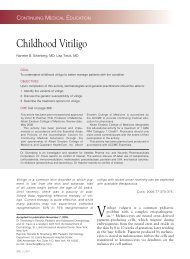
TVU screening technique (Sidebar on pages 10<br />
<strong>and</strong> 11 <strong>and</strong> Figure 1).<br />
Current Clinical Guidelines:<br />
Who Should Be Screened?<br />
As is often the case, current clinical guidelines on<br />
screening for short cervix with TVU have not yet<br />
caught up with the evidence. 8,9,12,15 Although the<br />
ACOG 2008 Practice Bulletin number 98 found<br />
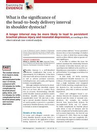
Internal os<br />
External os<br />
Rachel M. Gutkin, MD, is Maternal-Fetal Medicine<br />
Fellow, Department of Obstetrics <strong>and</strong> Gynecology, David<br />
Geffen School of Medicine, UCLA, Los Angeles, CA.<br />
Lawrence D. Platt, MD, is Director, Center for Fetal<br />
Medicine <strong>and</strong> Woman’s <strong>Ultrasound</strong>, Los Angeles; <strong>and</strong> Professor<br />
of Obstetrics <strong>and</strong> Gynecology, David Geffen School<br />
of Medicine, UCLA, Los Angeles, CA.<br />
Figure 1. Normal cervix visualized on transvaginal ultrasound.<br />
The Female Patient Supplement OCTOBER 2009
Screening With <strong>Transvaginal</strong> <strong>Ultrasound</strong> for <strong>Short</strong> <strong>Cervix</strong>: The Solution to <strong>Preterm</strong> <strong>Birth</strong>?<br />
limited evidence to routinely<br />
screen with TVU for cervical<br />
length, a number of studies<br />
have added to the body of<br />
evidence that supports TVU<br />
screening in a larger number of<br />
patients. 16 ACOG further states<br />
that an effective screening protocol<br />
for assessing risk factors of<br />
PTB has not been established.<br />
ACOG does support TVU<br />
screening in patients with a<br />
history of one or more pregnancy<br />
losses in the second or<br />
early third trimesters. Research<br />
is ongoing regarding the effectiveness<br />
of TVU screening in<br />
various populations.<br />
Studies Supporting Predictive Value<br />
of <strong>Short</strong> <strong>Cervix</strong> on TVU<br />
The optimal time frame for cervical measurement<br />
is between 14 <strong>and</strong> 30 weeks; before or after<br />
that time period, measurement of cervical length<br />
as a PTB predictor is not accurate or useful. 2,17<br />
Cervical length of less than 25 mm has been<br />
found to have the best predictive accuracy for<br />
increased risk of PTB. 1,17<br />
Some of the most important trials that have<br />
established the evidence for short cervix screening<br />
are summarized here.<br />
• <strong>Short</strong> <strong>Cervix</strong> on TVU Predicts PTB<br />
In an important study reported by Iams et al<br />
for the Maternal Fetal Medicine Network,<br />
the risk of PTB increases as the length of<br />
the cervix decreases as measured by TVU. 1<br />
While this was not the first trial showing<br />
this important relationship, it was the first<br />
to establish that the length of the cervix is<br />
directly correlated with the duration of pregnancy—the<br />
shorter the cervix, the higher the<br />
risk of PTB.<br />
• TVU Is Better Than Manual (Digital) Exam<br />
Cervical length measured by TVU has been<br />
shown to be a better predictor of PTB than<br />
The optimal time frame<br />
for cervical measurement is<br />
between 14 <strong>and</strong> 30 weeks;<br />
before or after that time<br />
period, measurement of<br />
cervical length as a PTB<br />
predictor is not accurate<br />
or useful. Cervical length<br />
of less than 25 mm has<br />
been found to have the<br />
best predictive accuracy for<br />
increased risk of PTB.<br />
cervical length measured<br />
by manual examination<br />
in a study of singleton<br />
pregnancies at high<br />
risk for PTB that were<br />
followed from 14 to 30<br />
weeks. 3 TVU was more<br />
predictive of PTB, most<br />
likely because manual<br />
examination is more subjective<br />
<strong>and</strong> has a high<br />
interobserver variability.<br />
• TVU Is Very Effective at<br />
Predicting PTB<br />
In a retrospective cohort<br />
study, a short cervix seen<br />
on a second-trimester<br />
sonogram was a strong<br />
predictor of PTB. Almost<br />
50% of patients with a cervical length of<br />
15 mm or less had an early spontaneous<br />
preterm delivery. 18<br />
• TVU Is Best in High-Risk Patients<br />
<strong>Short</strong> cervix on TVU is a better predictor of<br />
PTB in high-risk patients than in their lowrisk<br />
counterparts. The sensitivity is greater<br />
than 50% with a positive predictive value of<br />
45% between weeks 14 <strong>and</strong> 18. 3 The populations<br />
that will most benefit from screening<br />
are those with prior PTB, multiple gestations,<br />
uterine anomalies, prior LEEP or cold knife<br />
cone, multiple prior D&Es, or those with<br />
symptomatic preterm labor. 16<br />
• Benefit of Serial TVU Screenings<br />
High-risk women may benefit from several<br />
screenings, ie, one at 15 weeks <strong>and</strong> one at<br />
18 weeks, although the optimal interval for<br />
repeat TVU has not been established. 3<br />
• TVU in Special Situations<br />
In a recent trial, screening in asymptomatic<br />
high-risk women predicted spontaneous PTB<br />
at less than 35 weeks. 19 In another recent trial,<br />
it was shown that women with a history of<br />
PTB with preterm labor <strong>and</strong> intact membranes<br />
at onset of labor have shorter cervices<br />
The Female Patient Supplement OCTOBER 2009
Gutkin • Platt<br />
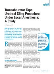
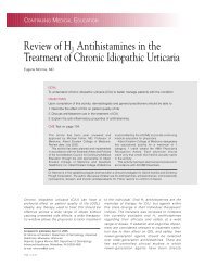
Funnel<br />
<strong>Short</strong><br />
cervix<br />
Cerclage<br />
stitches<br />
Figure 2. Funneling.<br />
Figure 3. Cerclage in cervix.<br />
than women with a history of PTB <strong>and</strong><br />
preterm premature rupture of membranes at<br />
the onset of labor. Both groups had shorter<br />
cervices than low-risk controls<br />
(Figure 2). 20<br />
Should Low-Risk Patients<br />
Be Excluded From TVU<br />
Screening?<br />
At the current time, low-risk<br />
populations (such as low-risk<br />
singleton pregnancies) are<br />
excluded from screening. The<br />
reasons often cited not to<br />
incorporate low-risk women<br />
are low positive predictive<br />
value, the lack of preventive<br />
therapy, <strong>and</strong> the cost of TVU.<br />
Arguments against using low predictive value as<br />
an exclusion criteria, however, include the fact<br />
that ultrasounds for genetic abnormalities are<br />
routinely performed with even lower sensitivity.<br />
Most low-risk women undergo genetic ultrasound<br />
examinations during 16 to 23 weeks. A<br />
TVU may be performed at time of these scans.<br />
This additional technique would add little<br />
time <strong>and</strong> may be of benefit in the reduction<br />
of prematurity. Perhaps the most persuasive<br />
argument that TVU should not be limited to<br />
Perhaps the most<br />
persuasive argument<br />
that TVU should not<br />
be limited to high-risk<br />
women, such as those<br />
with a previous preterm<br />
delivery, is that these women<br />
contribute only 30% of<br />
total spontaneous PTB.<br />
high-risk women, such as those with a previous<br />
preterm delivery, is that these women contribute<br />
only 30% of total spontaneous PTB. 21<br />
Clinical Management<br />
of At-Risk Patients<br />
The lack of patient management<br />
tools available to prevent<br />
PTB once a short cervix<br />
has been found on TVU is<br />
frustrating. Use of tocolytics<br />
is limited in the absence of<br />
uterine contractions. Home<br />
uterine activity monitoring<br />
<strong>and</strong> antibiotics are also<br />
employed sparingly. Bed rest<br />
now appears to be the main<br />
intervention utilized. Cerclage<br />
<strong>and</strong> pessaries are also used when deemed<br />
appropriate (Figure 3).<br />
Cerclage does not prevent PTB in all women<br />
with short cervical length on TVU. 22 A metaanalysis<br />
showed that some populations may benefit<br />
from cerclage (singletons with history of preterm<br />
delivery), but in others it may have no effect on<br />
outcome or even be detrimental (twin gestations).<br />
Overall, cerclage was not associated with any<br />
effect on PTB in patients with a short cervical<br />
length determined by TVU. 22 <strong>New</strong> evidence sug-<br />
The The Female Patient NEWSLETTER Supplement Supplement November OCTOBER 2008 2009
Screening With <strong>Transvaginal</strong> <strong>Ultrasound</strong> for <strong>Short</strong> <strong>Cervix</strong>: The Solution to <strong>Preterm</strong> <strong>Birth</strong>?<br />
Effective Technique for <strong>Ultrasound</strong> of the <strong>Cervix</strong><br />
A<br />
normal cervical length is considered to be between<br />
1 <strong>and</strong> 2 inches (25 to 50 mm). Thus, a short cervix<br />
is usually considered to be less than 25 mm.<br />
While transvaginal ultrasound (TVU) is now considered the<br />
gold st<strong>and</strong>ard for cervical measurement, other techniques<br />
are still currently in use. What are the drawbacks?<br />
Transabdominal <strong>Ultrasound</strong><br />
Transabdominal ultrasound (TAU) was the original method<br />
of visualizing the gravid cervix, but it has the limitation of<br />
being optimally utilized with a full bladder, which is necessary<br />
to eliminate fetal parts from obscuring the cervix. 10<br />
This can lead to artificial elongation of the cervix, masking<br />
of funneling, <strong>and</strong> the potential for a dilated internal<br />
os to appear closed. In addition, the external os can be<br />
difficult to identify with this method. Furthermore, manual<br />
pressure may be required, due to the distance of the<br />
cervix from the TAU probe, which can affect assessment.<br />
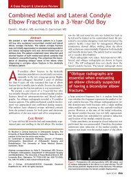
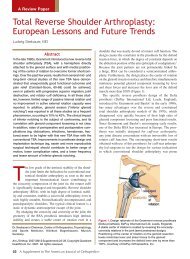
Because of these drawbacks, many cases of short cervix<br />
are missed with TAU (Figure 4). Thus, the role of TAU is<br />
limited, <strong>and</strong> experts agree that clinical decisions should<br />
not be made based on TAU alone.<br />
Despite these drawbacks, if TAU is the only option in a<br />
clinical situation, or if the patient declines TVU, screening<br />
by TAU is still preferable to manual examination<br />
alone. If the cervical length is less than 30<br />
mm or if the cervical length cannot be well visualized,<br />
then it is imperative to refer for TVU.<br />
when compared with the transvaginal approach. In cases<br />
where the cervix is successfully visualized, translabial<br />
cervical lengths have been shown to correlate well with<br />
those obtained transvaginally. 11 We personally find this<br />
technique more cumbersome <strong>and</strong> have reserved this<br />
mainly for patients with ruptured membranes. Overall,<br />
TVU is the superior method. The same caveats apply to<br />
TLU as to TAU regarding patient management.<br />
<strong>Transvaginal</strong> <strong>Ultrasound</strong><br />
Most clinical trials conducted over the past 10 years<br />
examining short cervix <strong>and</strong> preterm birth (PTB) have used<br />
TVU successfully, leading to the recognition of TVU as<br />
the gold st<strong>and</strong>ard for cervical length evaluation. TVU provides<br />
the best cervical visualization without obstruction,<br />
<strong>and</strong> bladder filling is not required. In fact, we prefer to<br />
have an empty bladder. TVU is safe, easy for the health<br />
care professional to perform, <strong>and</strong> the technique has high<br />
patient acceptability.<br />
Technique for Performing TVU 10,12<br />
Operators should have completed 50 or more procedures<br />
to become proficient. Less than accurate measurements<br />
may result if the bladder is partially full, if excessive probe<br />
Translabial <strong>Ultrasound</strong><br />
Translabial ultrasound (TLU) has been used<br />
because visualization is not obstructed by fetal<br />
parts, bladder filling is not required, <strong>and</strong> visualization<br />
is improved in comparison to TAU, due to<br />
the closer proximity of the probe to the cervix.<br />
However, even with these improvements, the full<br />
length of the cervix is visible in only 80% of cases<br />
Figure 4. Transabdominal vs transvaginal ultrasound scans.<br />
gests that therapeutic cerclage placement may be<br />
more efficacious in patients with shortened cervix<br />
who have a history of a prior term rather than a<br />
prior preterm delivery. 23<br />
With regard to pessary usage in the setting of<br />
shortened cervix, there are limited data in American<br />
publications. Several European studies have<br />
suggested a possible role for this noninvasive device.<br />
Additional studies must be performed to evaluate<br />
the efficacy of pessaries for this indication.<br />
ACOG advises that when a short cervix is present,<br />
assessment should be made for potential fetal<br />
anomalies, uterine activity should be evaluated, <strong>and</strong><br />
maternal chorioamnionitis should be excluded. 8 It<br />
is reasonable to suggest that performing TVU in<br />
isolation without performing a detailed fetal anat-<br />
10 The Female Patient Supplement OCTOBER 2009
Gutkin • Platt<br />
Figure 5. Effect of probe pressure.<br />
pressure is applied, if the cervix is curved, or if uterine<br />
contraction occurs during the examination (Figure 5).<br />
The patient should have an empty bladder. The<br />
probe should be covered with a single-use latex (or<br />
nonlatex if the patient indicates<br />
Funnel<br />
latex allergy) disposable sleeve.<br />
The probe is inserted (some<br />
patients prefer to insert it themselves),<br />
<strong>and</strong> a sagittal view of<br />
the cervix is visualized when the<br />
probe is in the anterior fornix.<br />
The probe is then withdrawn<br />
until the image is blurred. Light<br />
pressure is then applied, just<br />
enough to see the cervix clearly,<br />
at which point the image is<br />
enlarged to fill two-thirds or<br />
more of the screen. This is the<br />
stage at which the measurement<br />
is taken. Measurement from the external os to<br />
the internal os along the endocervical canal is taken.<br />
Several measurements are obtained, using the shortest<br />
as the best. Some operators obtain another set of<br />
measurements after applying transfundal pressure with<br />
the h<strong>and</strong> for 15 seconds. Little extra time is required for<br />
this entire process.<br />
Funneling <strong>and</strong> Other Anomalies<br />
The risk of PTB is also correlated to funneling<br />
(Figure 6). This is due to the fact that funneling,<br />
or dilation of the internal os, is part of the<br />
process of cervical shortening. However, due<br />
to its interobserver variability, funneling is less<br />
reproducible than cervical length. It has been well<br />
documented that funneling is not a predictor of an<br />
increased risk for PTB when it is associated with<br />
a long cervix. 13<br />
Lower uterine contraction can mimic funneling,<br />
but the echogenicity is different. The examination<br />
should continue to monitor the cervical appearance<br />
after the contraction disappears. If the<br />
contraction remains throughout the exam, the patient<br />
should be reevaluated in 1 to 2 days to monitor for<br />
resolution of the contraction.<br />
<strong>Short</strong><br />
cervix<br />
Figure 6. Funneling with shortened<br />
cervical length.<br />
Funnel<br />
Sludge<br />
<strong>Short</strong><br />
cervix<br />
Figure 7. Amniotic fluid sludge.<br />
Amniotic fluid sludge has also been reported as an<br />
independent risk factor for PTB. In association with shortened<br />
cervical length, studies have correlated it with a<br />
higher risk for PTB than the risk found with either variable<br />
alone. 14 Further research regarding the significance of<br />
sludge <strong>and</strong> its origin is needed to determine the significance<br />
of this finding on TVU (Figure 7).<br />
omy scan is insufficient. In addition, if not already<br />
made, lifestyle changes should be implemented<br />
that would affect PTB, including smoking cessation,<br />
moderation of activity, <strong>and</strong> discontinuation of<br />
coitus. ACOG further advises that because of the<br />
lack of evidence for the effectiveness of cerclage,<br />
elective cerclage should be reserved for patients<br />
with a history of 3 or more early pregnancy losses<br />
or PTBs. 8 Studies are ongoing to reevaluate the<br />
efficacy of cerclage, so a different approach cannot<br />
be recommended until trial results are available.<br />
While cerclage still has a place as an intervention,<br />
other possible interventions, such as treatment<br />
with progesterone, are on the horizon.<br />
While ACOG recommends that progesterone be<br />
offered to women with a prior singleton PTB, the<br />
The The Female Female Patient Patient NEWSLETTER Supplement Supplement OCTOBER 2009 11
Screening With <strong>Transvaginal</strong> <strong>Ultrasound</strong> for <strong>Short</strong> <strong>Cervix</strong>: The Solution to <strong>Preterm</strong> <strong>Birth</strong>?<br />
drug is not yet FDA-approved for this indication. 9<br />
Several current trials are investigating the efficacy<br />
of progesterone for the prevention of PTB.<br />
Conclusion<br />
<strong>Short</strong> cervix measured by TVU is the most accurate<br />
screening tool currently available to predict<br />
PTB. Its use is safe, effective, reproducible, <strong>and</strong><br />
acceptable in all studied populations. A cervical<br />
length of less than 25 mm when found on TVU<br />
between weeks 14 <strong>and</strong> 30 is highly predictive<br />
of PTB. It is our own practice to monitor with<br />
increased surveillance patients who have cervical<br />
length less than 30 mm during this time period.<br />
The need for effective interventions is critical.<br />
While bed rest <strong>and</strong> cerclage are currently the main<br />
interventions available, studies are in progress that<br />
may point to progesterone as an option. <br />
References<br />
1. Iams JD, Goldenberg RL, Meis PJ, et al. The length of the<br />
cervix <strong>and</strong> the risk of spontaneous premature delivery. N Engl J<br />
Med. 1996;334(9):567-572.<br />
2. Berghella V, Roman A, Daskalakis C, Ness A, Baxter JK. Gestational<br />
age at cervical length measurement <strong>and</strong> incidence of<br />
preterm birth. Obstet Gynecol. 2007;110(2 Pt 1):311-317.<br />
3. Berghella V, Tolosa JE, Kuhlman K, Weiner S, Bolognese RJ,<br />
Wapner RJ. Cervical ultrasonography compared with manual<br />
examination as a predictor of preterm delivery. Am J Obstet<br />
Gynecol. 1997;177(4):723-730.<br />
4. To MS, Skentou C, Cicero S, Nicolaides KH. Cervical assessment<br />
at the routine 23-weeks’ scan: problems with transabdominal<br />
sonography. <strong>Ultrasound</strong> Obstet Gynecol. 2000;15(4):292-296.<br />
5. Krebs-Jimenez J, Neubert AG. The microbiological effects<br />
of endovaginal sonographic assessment of cervical length.<br />
J <strong>Ultrasound</strong> Med. 2002;21(7):727-729.<br />
6. Carlan SJ, Richmond LB, O’Brien WF. R<strong>and</strong>omized trial of<br />
endovaginal ultrasound in preterm premature rupture of membranes.<br />
Obstet Gynecol. 1997;89(3):458-461.<br />
7. Clement S, C<strong>and</strong>y B, Heath V, To M, Nicolaides KH. <strong>Transvaginal</strong><br />
ultrasound in pregnancy: its acceptability to women <strong>and</strong><br />
maternal psychological morbidity. <strong>Ultrasound</strong> Obstet Gynecol.<br />
2003;22(5):508-514.<br />
8. ACOG. ACOG practice bulletin: Cervical insufficiency. Obstet<br />
Gynecol. 2003;102(5 Pt 1):1091-1099.<br />
9. Society for Maternal Fetal Medicine Publications Committee.<br />
ACOG Committee Opinion number 419 October 2008<br />
(replaces no. 291, November 2003): Use of progesterone to<br />
reduce preterm birth. Obstet Gynecol. 2008;112(4):963-965.<br />
10. American College of Radiology. ACR Appropriateness Criteria:<br />
assessment of gravid cervix. Available at: www.acr.org/<br />
Secondary MainMenuCategories/quality_safety/app_criteria/<br />
pdf/ExpertPanelonWomensImaging/PrematureCervical<br />
DilatationDoc8.aspx. Accessed August 7, 2009.<br />
11. Cicero S, Skentou C, Souka A, To MS, Nicolaides KH. Cervical<br />
length at 22-24 weeks of gestation: comparison of transvaginal<br />
<strong>and</strong> transperineal-translabial ultrasonography. <strong>Ultrasound</strong><br />
Obstet Gynecol. 2001;17(4):335-340.<br />
12. Abuhamad AZ; ACOG Committee on Practice Bulletins–Obstetrics.<br />
ACOG Practice Bulletin, clinical management guidelines for<br />
obstetrician-gynecologists number 98, October 2008 (replaces<br />
Practice Bulletin number 58, December 2004): Ultrasonography<br />
in pregnancy. Obstet Gynecol. 2008;112(4):951-961.<br />
13. To MS, Skentou C, Liao AW, Cacho A, Nicolaides KH. Cervical<br />
length <strong>and</strong> funneling at 23 weeks of gestation in the prediction<br />
of spontaneous early preterm delivery. <strong>Ultrasound</strong> Obstet<br />
Gynecol. 2001;18(3):200-203.<br />
14. Kusanovic JP, Espinoza J, Romero R, et al. Clinical significance<br />
of the presence of amniotic fluid ‘sludge’ in asymptomatic<br />
patients at high risk for spontaneous preterm delivery. <strong>Ultrasound</strong><br />
Obstet Gynecol. 2007;30(5):706-714.<br />
15. ACOG. ACOG Practice Bulletin: Assessment of risk factors for<br />
preterm birth: clinical management guidelines for obstetriciangynecologists.<br />
Number 31, October 2001 (Replaces Technical<br />
Bulletin number 206, June 1995; Committee Opinion number<br />
172, May 1996; Committee Opinion number 187, September<br />
1997; Committee Opinion number 198, February 1998; <strong>and</strong><br />
Committee Opinion number 251, January 2001). Obstet Gynecol.<br />
2001;98(4):709-716.<br />
16. Grimes-Dennis J, Berghella V. Cervical length <strong>and</strong> prediction<br />
of preterm delivery. Curr Opin Obstet Gynecol. 2007;19(2):<br />
191-195.<br />
17. Berghella V, Talucci M, Desai A. Does transvaginal sonographic<br />
measurement of cervical length before 14 weeks predict<br />
preterm delivery in high-risk pregnancies? <strong>Ultrasound</strong> Obstet<br />
Gynecol. 2003;21(2):140-144.<br />
18. Hassan SS, Romero R, Berry SM, et al. Patients with an ultrasonographic<br />
cervical length ≤15 mm have nearly a 50% risk<br />
of early spontaneous preterm delivery. Am J Obstet Gynecol.<br />
2000;182(6):1458-1467.<br />
19. Crane JM, Hutchens D. <strong>Transvaginal</strong> sonographic measurement<br />
of cervical length to predict preterm birth in asymptomatic<br />
women at increased risk: a systematic review. <strong>Ultrasound</strong><br />
Obstet Gynecol. 2008;31(5):579-587.<br />
20. Crane JM, Hutchens D. Use of transvaginal ultrasonography to<br />
predict preterm birth in women with a history of preterm birth.<br />
<strong>Ultrasound</strong> Obstet Gynecol. 2008;32(5):640-645.<br />
21. To MS, Skentou CA, Royston P, Yu CK, Nicolaides KH. Prediction<br />
of patient-specific risk of early preterm delivery using<br />
maternal history <strong>and</strong> sonographic measurement of cervical<br />
length: a population-based prospective study. <strong>Ultrasound</strong> Obstet<br />
Gynecol. 2006;27(4):362-367.<br />
22. Berghella V, Odibo AO, To MS, Rust OA, Althuisius SM.<br />
Cerclage for short cervix on ultrasonography: meta-analysis<br />
of trials using individual patient-level data. Obstet Gynecol.<br />
2005;106(1):181-189.<br />
23. Poggi SH, Vyas N, Pezzullo JC, L<strong>and</strong>y HJ, Ghidini A. Therapeutic<br />
cerclage may be more efficacious in women who develop<br />
cervical insufficiency after a term delivery. Am J Obstet Gynecol.<br />
2009;200(1):68.e1-e3.<br />
12 The Female Patient Supplement OCTOBER 2009