Madelung Deformity With Prior Distal Radius Fracture: A Case ...
Madelung Deformity With Prior Distal Radius Fracture: A Case ...
Madelung Deformity With Prior Distal Radius Fracture: A Case ...

You also want an ePaper? Increase the reach of your titles
YUMPU automatically turns print PDFs into web optimized ePapers that Google loves.
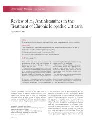
A <strong>Case</strong> Report <strong>With</strong> Literature Review<br />
<strong>Madelung</strong> <strong>Deformity</strong> <strong>With</strong> <strong>Prior</strong> <strong>Distal</strong><br />
<strong>Radius</strong> <strong>Fracture</strong>: A <strong>Case</strong> Report<br />
Justin S. Field, MD, and Marco Rizzo, MD<br />
<strong>Madelung</strong> 1 first described the wrist deformity bearing<br />
his name in 1878. Although previous authors<br />
had described lesions that we might now term<br />
<strong>Madelung</strong> deformity, <strong>Madelung</strong> was the first to<br />
accurately describe it clinically and to propose both an etiology<br />
and a treatment. The deformity results from epiphyseal<br />
arrest on the ulnar and volar half of the distal radius, which<br />
causes the articular surface to be directed ulnarly and volarly.<br />
In 1992, Vickers and Nielsen 2 further described the abnormal<br />
physeal and ligamentous anatomy on the volar aspect<br />
of the radiocarpal joint. They identified a thick fibrous band<br />
spanning the radial metaphysis to the proximal carpal row<br />
(Vickers ligament) seen in congenital <strong>Madelung</strong> deformity.<br />
The etiologies of <strong>Madelung</strong> deformity have been thoroughly<br />
described in the literature. Although confusion<br />
continues as to the exact etiology of this condition, most<br />
reports show the deformity to be a condition inherited by<br />
an autosomal-dominant trait with variable expressivity.<br />
Common to congenital <strong>Madelung</strong> deformity is the presence<br />
of Vickers ligament, which tethers the volar-ulnar<br />
distal radial physis. Other reports have described an<br />
acquired or pseudo-<strong>Madelung</strong> deformity wherein injury to<br />
the immature physis produces a bony bridge with progression<br />
to a deformity that may mimic that of a true congenital<br />
<strong>Madelung</strong> deformity. In this report, we present the case of<br />
an acquired <strong>Madelung</strong> deformity secondary to trauma with<br />
the presence of a Vickers ligament and absence of a bony<br />
bridge affecting the volar-ulnar distal radial physis. In the<br />
literature, presence of Vickers ligament associated with<br />
posttraumatic <strong>Madelung</strong> deformity is unique; it has been<br />
described only in the congenital variant.<br />
<strong>Case</strong> Report<br />
At age 7, a right-hand–dominant girl fell, landed on<br />
her hyperflexed left hand, and sustained a nondisplaced<br />
extra-articular left distal radius fracture that was treated<br />
Dr. Field is Orthopaedic Surgery Resident, Division of<br />
Orthopaedic Surgery, Duke University Medical Center, Durham,<br />
North Carolina.<br />
Dr. Rizzo is Attending Orthopaedic Surgeon, Department of<br />
Orthopaedic Surgery, Mayo Clinic, Rochester, Minnesota.<br />
Requests for reprints: Marco Rizzo, MD, The Mayo Clinic,<br />
Department of Orthopedic Surgery, 200 First Street, SW, Rochester,<br />
MN 55905 (tel, 507-284-3689; fax, 507-284-5539; e-mail, rizzo.<br />
marco@mayo.edu).<br />
Am J Orthop. 2007;36(6):E91-E93. Copyright 2007, Quadrant<br />
HealthCom Inc.<br />
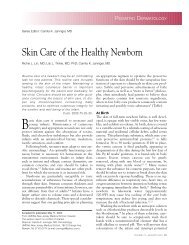
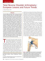
Figure1. (A, B) Left nondisplaced<br />
extra-articular distal radius fracture<br />
in 7-year-old girl.<br />
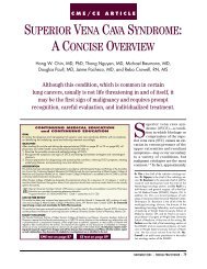
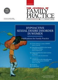
Figure 2. <strong>Distal</strong> radius deformity<br />
in same child 5 years after injury.<br />
with closed reduction and casting for 6 weeks (Figure<br />
1). She healed uneventfully and did well until age 12,<br />
when she had increased pain and swelling, particularly<br />
of the ulnar side of the left wrist, as well as decreased<br />
wrist range of motion.<br />
On presentation, this now 12.5-year-old patient reported<br />
no significant past medical, surgical, or family history<br />
suggesting bony or soft-tissue abnormalities. She had no<br />
allergies and was not taking any medications. She actively<br />
participated in activities, including gymnastics, soccer,<br />
and softball.<br />
The most recent preoperative plain films suggested a<br />
fused growth plate on the ulnar side of the distal radius<br />
(Figure 2) leading to a significant deformity, with the radial<br />
aspect of the radius continuing to grow with radial inclination<br />
increased well beyond normal, and also with the ulna<br />
continuing to grow and cause significant positive ulnar<br />
variance. The lateral plain film showed the ulna subluxating<br />
dorsally. Computed tomography with minor cuts did<br />
not reveal a significant growth plate anomaly, but a magnetic<br />
resonance (MR) image suggested a small physeal bar<br />
near the old fracture site.<br />
The patient was premenarchal and of normal stature for<br />
her age. Physical examination revealed limitation of motion<br />
June 2007<br />
E91
<strong>Madelung</strong> <strong>Deformity</strong> <strong>With</strong> <strong>Prior</strong> <strong>Distal</strong> <strong>Radius</strong> <strong>Fracture</strong><br />
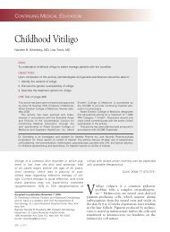
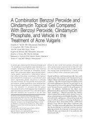
Figure 3. Vickers ligament.<br />
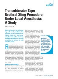
Figure 4. Two years postoperatively.<br />
Patient healed well, maintaining<br />
her position with normal resumption<br />
of radial physeal growth.<br />
with pronation of 20° on the left versus 80° on the right side.<br />
Supination was 80° bilaterally. Wrist flexion was limited as<br />
well, to 45° on the left versus 60° on the right and with left<br />
wrist extension of 35° versus 70° on the right. Radial deviation<br />
and grip strength were significantly diminished on the<br />
left as well. The patient also had a prominent ulnar head,<br />
with pain and tenderness to palpation. Her contralateral wrist<br />
had no physical or radiologic abnormalities.<br />
The surgical plan consisted of corrective osteotomy<br />
of the left distal radius with placement of structural<br />
allograft and plating secondary to continued symptoms.<br />
A modified Henry approach to the left distal radius<br />
was performed. Pronator quadratus was elevated off of<br />
the distal radius on the radial border, and Vickers ligament<br />
was identifiable in the ulnar aspect of the distal<br />
radius (Figure 3). This was an interesting finding, as<br />
the impression of this injury was that it was a malunited<br />
distal radius with a bony bridge. However, there was<br />
no evidence of a bony bridge on the ulnar aspect of<br />
the distal radius on direct visualization. These findings<br />
suggested that this was a unique posttraumatic<br />
<strong>Madelung</strong> deformity. Despite the past history of trauma,<br />
a Vickers ligament was present, not a bony bridge.<br />
Subsequently, the Vickers ligament was released off the<br />
distal radius, an osteotomy was made 3 cm proximal to<br />
the growth plate, allograft was inserted, deformity was<br />
corrected in all planes, and plate fixation was completed<br />
under fluoroscopy.<br />
Two years after surgery, the patient was well healed and<br />
maintaining her position with normal resumption of radial<br />
physeal growth (Figure 4). She had a painless arc of motion<br />
and no ulnar prominence or tenderness. She could flex her<br />
left wrist 65° and extend it 80°. Grip strength, pronation/<br />
supination, and deviation improved to normal as well.<br />
Discussion<br />
The clinical manifestations of <strong>Madelung</strong> deformity are<br />
characterized by the insidious onset of pain in one or both<br />
wrists and increasing prominence of the dorsal ulnar head<br />
and bowing of the distal radius. <strong>Deformity</strong> progresses<br />
until the growth plate of the distal radius closes. Wrist<br />
motion, particularly extension and supination, is limited.<br />
Radiographically, there is an increase in the normal palmar<br />
and ulnar inclination of the distal radius. The ulna is unaffected<br />
and remains in its usual dorsal position.<br />
Henry and Thorburn 3 classified <strong>Madelung</strong> deformity<br />
into 4 etiologies: dysplastic, chromosomal/genetic, idiopathic/primary,<br />
and posttraumatic.<br />
Bone dysplasias associated with <strong>Madelung</strong> deformity<br />
include multiple hereditary osteochondromatosis, Ollier<br />
disease, achondroplasia, multiple epiphyseal dysplasias,<br />
and mucopolysaccharidoses. However, the most important<br />
associated dysplasia is dyschondrosteosis, a type of mesomelic<br />
dwarfism that should be suspected if the condition<br />
is bilateral or if there is a positive family history, short<br />
stature, or mesomelia.<br />
One third of cases of <strong>Madelung</strong> deformity are transmitted<br />
in an autosomal-dominant fashion, with variable<br />
expression and 50% penetrance. The deformity is bilateral<br />
50% of the time, and females are affected 4 times as often<br />
as males. Molecular genetic studies have shown that an<br />
X-chromosomal translocation is associated with <strong>Madelung</strong><br />
deformity, Turner syndrome, and dyschondrosteosis. 4-6<br />
The posttraumatic deformity can occur from repetitive<br />
stress or from a single event that disrupts growth of the<br />
distal radial ulnar-volar physis. The literature includes<br />
many case reports of pseudo-<strong>Madelung</strong> deformity in<br />
gymnasts secondary to the sport’s repetitive stress to<br />
the wrists. Vender and Watson 7 reported a case of a<br />
high-level gymnast with bilateral closure of the ulnar<br />
side of the distal radius physeal plate, with clinical and<br />
radiographic changes like those found in a congenital<br />
<strong>Madelung</strong> deformity. They concluded that cumulative<br />
microtrauma to the ulnar side of the distal radius<br />
physeal plate may cause premature closure leading to a<br />
<strong>Madelung</strong>-like deformity.<br />
Conclusions<br />
Thorough history-taking and physical examination indicated<br />
that the etiology of our patient’s wrist deformity<br />
was unlikely to be genetics or a dysplastic disorder. The<br />
patient had an acquired <strong>Madelung</strong> deformity. Her diagnosis<br />
and etiology indicated that the initial injury to the physis<br />
caused the abnormal growth volarly and ulnarly at the distal<br />
radius. MI images suggested a physeal bridge. However,<br />
intraoperative findings included a Vickers ligament without<br />
evidence of a bony bridge. To our knowledge, there have<br />
been no other case reports citing a postfracture <strong>Madelung</strong><br />
deformity with a Vickers ligament and absent a bony<br />
bridge. Whether there is a link between the initial fracture<br />
and the subsequent <strong>Madelung</strong> deformity with Vickers<br />
ligament remains unclear. In patients with past history of<br />
E92 The American Journal of Orthopedics ®
J. S. Field and M. Rizzo<br />
fracture and continued wrist pain, an acquired <strong>Madelung</strong><br />
deformity secondary to a bony bridge or a Vickers ligament<br />
should be considered as part of the differential diagnosis.<br />
Author’s Disclosure Statement<br />
and Acknowledgement<br />
The authors report no actual or potential conflict of interest<br />
in relation to this article.<br />
References<br />
1. <strong>Madelung</strong> OW. Die spontane Subluxation der Hand nach vorne<br />
[in German]. Verh Dtsch Ges Chir. 1878;7:259.<br />
2. Vickers D, Nielsen G. <strong>Madelung</strong>’s deformity: surgical prophylaxis<br />
(physiolysis) during the late growth period by resection of the<br />
dyschondrosteosis lesion. J Hand Surg Br. 1992;17:401-407.<br />
3. Henry A, Thorburn MJ. <strong>Madelung</strong>’s deformity. A clinical and cytogenetic<br />
study. J Bone Joint Surg Br. 1967;49:66-73.<br />
4. Palka G, Stuppia L, Guanciali Franchi P. Short arm rearrangements<br />
of sex chromosomes with haploinsufficiency of the SHOX<br />
gene are associated with Leri-Weill dyschondrosteosis. Clin<br />
Genet. 2000;57:449-453.<br />
5. Schwartz RP, Sumner TE. <strong>Madelung</strong>’s deformity as a presenting<br />
sign of Turner’s syndrome. J Pediatr. 2000;136:563.<br />
6. Shears DJ, Vassal HJ, Goodman FR. Mutation and deletion of the<br />
pseudoautosomal gene SHOX cause Leri-Weill dyschondrosteosis.<br />
Nat Genet. 1998;19:70-73.<br />
7. Vender MI, Watson HK. Acquired <strong>Madelung</strong>-like deformity in a<br />
gymnast. J Hand Surg Am. 1988;13:19-21.<br />
June 2007<br />
E93