Total Internal Reflection Fluorescence (TIRF ... - TeachLine
Total Internal Reflection Fluorescence (TIRF ... - TeachLine
Total Internal Reflection Fluorescence (TIRF ... - TeachLine

You also want an ePaper? Increase the reach of your titles
YUMPU automatically turns print PDFs into web optimized ePapers that Google loves.
<strong>Total</strong> <strong>Internal</strong> <strong>Reflection</strong> <strong>Fluorescence</strong><br />
(<strong>TIRF</strong>) microscopy in cell biology.<br />
QuickTime and a<br />
Animation decompressor<br />
are needed to see this picture.<br />
Uri Ashery<br />
Department of Neurobiochemistry, Wise Faculty of life Sciences,<br />
Tel Aviv University, Tel Aviv,<br />
Israel<br />
Ofer Yizhar, Irit Gottfried, Orly Raviv, Barak Rotblat
<strong>Total</strong> <strong>Internal</strong> <strong>Reflection</strong> <strong>Fluorescence</strong><br />
(<strong>TIRF</strong>) microscopy in cell biology.<br />
• Principles of the technique<br />
• Configuration of the microscope<br />
• Examples:<br />
• Vesicle trafficking and fusion<br />
•Analysis<br />
• Structure of receptors<br />
• Endocytosis<br />
•Intracellular signaling<br />
• Advantages disadvantages
<strong>Total</strong> <strong>Internal</strong> <strong>Reflection</strong> <strong>Fluorescence</strong><br />
(<strong>TIRF</strong>) microscopy in cell biology.<br />
Optical technique that restricts the excitation and detection<br />
of a fluorophores to a thin region of the specimen.<br />
• Allows direct visualization and detection of sub-membrane<br />
events:<br />
• movement and fusion of single vesicles<br />
• conformational dynamics of single ion channels and receptors<br />
• translocation of proteins to and from the plasma membrane<br />
• protein-protein interaction at the plasma membrane<br />
• cell adhesion processes
Synaptic transmission is carried out by fusion<br />
of neurotransmitter-containing vesicles<br />
Molecular mechanisms of vesicle exocytosis and endocytosis:<br />
<strong>TIRF</strong> allows direct visualization of single vesicles<br />
(movement, transport, fusion and recycling in an intact cell)<br />
NT-containing<br />
vesicles<br />
Pre-synaptic<br />
Post-synaptic
The synaptic vesicle cycle<br />
Docking<br />
Priming<br />
Fusion-competent<br />
Ca 2+<br />
Ca 2+<br />
Plasma membrane
The synaptic vesicle cycle<br />
Endocytosis<br />
Plasma membrane
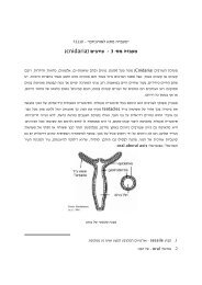
The chromaffin cell - a model system for neuroexocytosis:<br />
Chromaffin cells secrete adrenaline into the<br />
blood stream<br />
What’s inside<br />
Chromaffin cell<br />
1µm
<strong>Total</strong> <strong>Internal</strong> <strong>Reflection</strong> <strong>Fluorescence</strong> (<strong>TIRF</strong>) or<br />
Evanescent-Wave <strong>Fluorescence</strong> Microscopy<br />
Refracted<br />
laser beams<br />
A cell (specimen):<br />
low refraction index<br />
Glass coverslip:<br />
high refraction index<br />
Evanescent<br />
field<br />
200-300 nm.<br />
Reflected<br />
laser beam<br />
Incident<br />
angle<br />
Incident<br />
angle<br />
Laser beams<br />
(various angles)<br />
- - -<br />
Objective
QuickTime and a<br />
TIFF (Uncompressed) decompressor<br />
are needed to see this picture.<br />
.
<strong>Total</strong> <strong>Internal</strong> <strong>Reflection</strong> <strong>Fluorescence</strong> (<strong>TIRF</strong>) or<br />
Evanescent-Wave <strong>Fluorescence</strong> Microscopy<br />
A cell filled with fluorescently labeled vesicles<br />
Glass coverslip<br />
Evanescent<br />
field<br />
200-400 nm.<br />
fluorescence
Chromaffin cell<br />
Epi-fluorescence<br />
<strong>TIRF</strong>
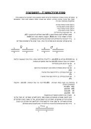
•Snell’s law: n1sinQ1=n2SinQ2<br />
•Critical angle Qc (Q1), Q2=90 o and (sin 90 o =1)<br />
•n1sinQc=n2<br />
•Qc=sin -1 (n2/n1)<br />
•Qc= sin -1 (1.5/1.36)<br />
•Qc=63.8 o<br />
•wide objectives<br />
n1 high refraction index<br />
n1=1.51 (Immersion Oil)<br />
Q2<br />
Q2=90<br />
n2 low refraction index; n2=1.36<br />
Q1 Qc<br />
n1>n2<br />
Evanescent<br />
field<br />
100-200 nm.<br />
Incident<br />
angle > Qc<br />
Immersion<br />
Oil<br />
Objective<br />
Qc: critical angle
Air n=1.00, water n=1.33, Cytosol n=1.36-1.38, Immersion oil<br />
n=1.51<br />
Thus Objective NA must be > 1.4; NA=1.45; one uses 10% of <strong>TIRF</strong><br />
The numerical aperture of a microscope objective is a measure<br />
of its ability to gather light and resolve fine specimen detail at a<br />
fixed object distance.<br />
Numerical Aperture (NA )= n(sin m)<br />
n is the refractive index of the imaging medium<br />
m=A/2<br />
http://www.micro.magnet.fsu.edu/primer/java/tirf/penetration/index.htmlhttp://www.micro.magnet.fsu.edu/primer/java/tirf/highnaobjective/index.htm
Decay of the evanescence field<br />
Evanescent<br />
field<br />
100-200 nm.<br />
1<br />
Excitation Relative Excitation energy Energy (relative)<br />
0.8<br />
0.6<br />
0.4<br />
0.2<br />
E = C×e − z τ<br />
C = Constant<br />
z = Distance from membrane<br />
τ = Extinction coefficient<br />
(penetration depth of the<br />
evanescence field<br />
0<br />
0 200 400 600 800 1000<br />
Distance from interface (nm)<br />
Chromaffin vesicle<br />
Distance from interface (nm)
Decay of the evanescence field<br />
1<br />
Excitation Relative Excitation energy Energy (relative)<br />
0.8<br />
0.6<br />
0.4<br />
0.2<br />
E = C × e − z τ<br />
C = Constant<br />
z = Distance from membrane<br />
τ = Extinction coefficient<br />
0<br />
0 200 400 600 800 1000<br />
Distance from interface (nm)<br />
Chromaffin vesicle<br />
Distance from interface (nm)<br />
http://www.micro.magnet.fsu.edu/primer/java/tirf/penetration/index.html
QuickTime and a TIFF (Uncompressed) decompressor are needed to see this picture.<br />
I(z) = I(o)e -z/d<br />
d = λ/4 p• (n(1) 2sin 2q(1) - n(2) 2)-1/2<br />
Have a look at this web address for more details<br />
http://www.micro.magnet.fsu.edu/primer
Types of <strong>TIRF</strong> microscopes
Objective type <strong>TIRF</strong>; Prism lesss (inverted microscope)<br />
Objectives:<br />
APO100XOHR<br />
(NA 1.65)<br />
PLAPO60XO<strong>TIRF</strong>M<br />
(NA1.45)
http://www.micro.magnet.fsu.edu/primer/java/tirf/tirfalign/index.html<br />
Laser<br />
CCD<br />
http://www.micro.magnet.fsu.edu/primer/java/tirf/tirfalign/index.html
Prism type <strong>TIRF</strong> (upright microscope)<br />
Benefit of the prism <strong>TIRF</strong>M<br />
By varying the angle of the laser beam, one can<br />
modulates the penetration depth.
•Examples:<br />
• Vesicle trafficking and fusion<br />
• Structure of receptors<br />
•Endocytosis<br />
•Cellular signaling
Evanescent-Wave <strong>Fluorescence</strong> Microscopy<br />
The synaptic vesicle cycle<br />
NT uptake<br />
Docking<br />
Priming<br />
Fusion-competent<br />
Ca 2+<br />
Ca 2+<br />
Plasma membrane<br />
Allows direct visualization of number of docked vesicles, fraction of<br />
fusion-competent vesicles and vesicles transport in an intact cell.
Staining chromaffin vesicles<br />
with fluorescent proteins<br />
• Acidic dyes (Acridine Orange, Lysotracker)<br />
• Vesicular proteins:<br />
– Membrane<br />
– Lumen<br />
Electroporation<br />
NPY-mRFP<br />
Endocytotic dyes
What’s it good for?<br />
Epi-fluorescence<br />
<strong>TIRF</strong><br />
QuickTime and a<br />
YUV420 codec decompressor<br />
are needed to see this picture.<br />
5Hz recording, 20Hz playback
Filtering images for better contrast<br />
C<br />
D<br />
3 steps filtering
Identification of individual vesicles<br />
Peak intensity<br />
Half-width
Identification of individual vesicles<br />
Y<br />
X<br />
Z
Information derived from image sequences:<br />
• X/Y position – By fitting the intensity distribution with a<br />
gaussian curve, finding the location of the peak<br />
• Z position – indicated by peak intensity
Tracking single vesicles:
Tracking vesicle motion<br />
Data from tracking single vesicle<br />
Peak intensity<br />
6<br />
4<br />
2<br />
0 5 10 15<br />
QuickTime and a<br />
YUV420 codec decompressor<br />
are needed to see this picture.<br />
X (µm)<br />
10.9<br />
10.7<br />
10.5<br />
0 5 10 15 20<br />
Y(µm)<br />
6.4<br />
6.2<br />
5Hz recording, 20Hz playback<br />
6<br />
0 5 10 15 20<br />
Time (s)
Tracking vesicle motion<br />
3-D trajectory of a single vesicle 3-D reconstruction of vesicle trajectories<br />
QuickTime and a<br />
YUV420 codec decompressor<br />
are needed to see this picture.
Synaptic transmission is carried out by fusion<br />
of neurotransmitter-containing vesicles<br />
Molecular mechanisms of vesicle exocytosis and endocytosis:<br />
NT-containing<br />
vesicles<br />
Pre-synaptic<br />
Post-synaptic
Tomosyn inhibits exocytosis<br />
Tomosyn doesn’t inhibit docking<br />
Control cell<br />
Tomosyn cell<br />
600<br />
exocytosis<br />
∆Cm (fF)<br />
400<br />
200<br />
Tomosyn inhibits vesicle priming<br />
control<br />
tomosyn<br />
0<br />
Control<br />
Tomosyn
EM Analysis of Vesicle Distribution With Tomosyn<br />
Overexpression<br />
Control cell<br />
Tomosyn cell<br />
Docked Primed Fusion-Competent<br />
Can we define physically primed vesicle?
Calculation of vesicle mobility<br />
MSD plot<br />
Diffusion constant<br />
-4 2<br />
D=1.65×10 (µm /sec)<br />
0.5<br />
-3 2<br />
D=1.39×10 (µm /sec)<br />
0.5<br />
Vesicle<br />
Diameter<br />
(~200nm)<br />
∆Y(µm)<br />
Y (µm)<br />
0<br />
∆Y(µm)<br />
Y (µm)<br />
0<br />
-0.5<br />
-0.5 0 0.5<br />
X (µm)<br />
∆X (µm)<br />
-0.5<br />
-0.5 0 0.5<br />
X (µm)<br />
∆X (µm)
In untreated cells, ~50% of membraneproximal<br />
vesicles are immobilized<br />
Distribution of vesicle diffusion coefficients in untreated cells:<br />
100<br />
% of Vesicles<br />
90<br />
80<br />
70<br />
60<br />
50<br />
40<br />
30<br />
Y (µm)<br />
0.5<br />
0<br />
Immobilized<br />
Pool<br />
-0.5<br />
-0.5 0 0.5<br />
X (µm)<br />
20<br />
10<br />
0<br />
5.0E-04<br />
1.0E-03<br />
1.5E-03<br />
2.0E-03<br />
2.5E-03<br />
3.0E-03<br />
3.5E-03<br />
4.0E-03<br />
4.5E-03<br />
5.0E-03<br />
5.5E-03<br />
6.0E-03<br />
2<br />
D (µm /sec)
Studying the roles of synaptic proteins<br />
through vesicle mobility<br />
Tomosyn reduces the population of immobilized vesicles<br />
Is this immobile pool represent ready-to-fuse vesicles?<br />
Yizhar et al.
Studying the roles of synaptic proteins<br />
through vesicle mobility; vesicle fusion<br />
QuickTime and a<br />
YUV420 codec decompressor<br />
are needed to see this picture.<br />
10Hz recording, 20Hz playback<br />
Do we measure fusion or undocking?
pH-sensitive green fluorescent protein<br />
(pHluorin) fused to Synatpobrevin<br />
(SynaptopHluorin)<br />
Exocytosis relieves the proton-dependent<br />
quenching of ecliptic-pHluorin<br />
fluorescence
Dual color <strong>TIRF</strong> imaging of NPY-mRFP and SynaptopHluorin
Diffusion constants of vesicles<br />
immediately before fusion<br />
100<br />
90<br />
80<br />
70<br />
60<br />
50<br />
40<br />
30<br />
20<br />
10<br />
0<br />
80% of pre-fusion vesicles are immobilized<br />
0.9<br />
0.8<br />
0.7<br />
0.6<br />
0.5<br />
0.4<br />
0.3<br />
0.5<br />
0<br />
-0.5<br />
-0.5 0 0.5<br />
X (µm)<br />
Immobilization<br />
Precede Vesicle Fusion<br />
and Might be Correlated with<br />
Priming<br />
0.2<br />
0.1<br />
0<br />
5.0E-04<br />
1.0E-03<br />
1.5E-03<br />
2.0E-03<br />
2.5E-03<br />
3.0E-03<br />
3.5E-03<br />
4.0E-03<br />
4.5E-03<br />
5.0E-03<br />
5.5E-03<br />
6.0E-03<br />
6.5E-03<br />
7.0E-03<br />
7.5E-03<br />
8.0E-03<br />
8.5E-03<br />
9.0E-03<br />
9.5E-03<br />
1.0E-02<br />
Y (µm)<br />
5.0E-04<br />
1.0E-03<br />
1.5E-03<br />
2.0E-03<br />
2.5E-03<br />
3.0E-03<br />
3.5E-03<br />
4.0E-03<br />
4.5E-03<br />
5.0E-03<br />
5.5E-03<br />
6.0E-03<br />
Vesicle D (µM 2 /s)<br />
Yizhar et al.
Exocytosis of single synaptic vesicles (diameter 50nm) in<br />
Retinal bipolar neurons<br />
QuickTime and a<br />
Animation decompressor<br />
are needed to see this picture.<br />
QuickTime and a<br />
Animation decompressor<br />
are needed to see this picture.<br />
Zenisek, Steyer, Almers 2000
Hot spots for exocytosis in Retinal bipolar neurons<br />
Zenisek, Steyer, Almers 2000
Exocytosis of different pools of vesicles in Retinal bipolar<br />
neurons<br />
Zenisek, Steyer, Almers 2000
Conformational rearrangements associated with the gating of G<br />
protein-coupled Potassium channel<br />
Spectroscopic Measurements of FRET under <strong>TIRF</strong><br />
Microscopy<br />
Inbal Riven , Eli Kalmanzon , Lior Segev , and Eitan Reuveny<br />
Neuron, Vol 38, 225-235, 24 April 2003
Spectroscopic Measurements of FRET under <strong>TIRF</strong> Microscopy<br />
FRET Efficiencies Change during Channel Gating<br />
Inbal Riven , Eli Kalmanzon , Lior Segev , and Eitan Reuveny<br />
Neuron, Vol 38, 225-235, 24 April 2003
A Model Representing the Possible Rearrangement of<br />
the N- and C-Terminal Cytosolic Domains during<br />
Channel Gating<br />
Since FRET efficiency depends heavily on the distance between<br />
the donor and the acceptor (R 6 th power dependence) one can<br />
estimate distances between subunits.<br />
fluorescence resonance energy transfer (FRET)<br />
Inbal Riven , Eli Kalmanzon , Lior Segev , and Eitan Reuven<br />
Neuron, Vol 38, 225-235, 24 April 2003
Clathrin mediated endocytosis<br />
Clathrin: coat assembly<br />
Dynamin: GTPase activity is needed for pinching off<br />
Slepnev and De Camilli 2000
Tracking endocytosis with <strong>TIRF</strong><br />
Merrifield et al, Almers 2002<br />
Santini and Keen 2002
Merrifield et al, Almers 2002<br />
Santini and Keen 2002
The role of Huntingtin interacting protein (HIP1) in<br />
clathrin mediated endocytosis<br />
PIP 2 Binding AP2 Binding Actin Binding<br />
218 604<br />
Slepnev and De Camilli 2000
The role of HIP1 in clathrin mediated endocytosis<br />
EpiFluorescense<br />
<strong>TIRF</strong>M<br />
Clathrin-EGFP<br />
HIP218-EGFP<br />
Gottfried et al.
Behavior of HIP1 and clathrin in live cos7 cells<br />
HIP218<br />
Clathrin<br />
QuickTime and a<br />
YUV420 codec decompressor<br />
are needed to see this picture.
Kinetic analysis of HIP and clathrin clusters<br />
60<br />
HIP(218-604)<br />
HIP 218-604<br />
60<br />
Clathrin<br />
clathrin<br />
Life Time<br />
No. of vesicle<br />
50<br />
40<br />
30<br />
20<br />
10<br />
0<br />
N=160<br />
10 30 50 70 90 110 130 150 170 190 210 230 250<br />
No. of Vesicle<br />
50<br />
40<br />
30<br />
20<br />
10<br />
0<br />
N=138<br />
10 30 50 70 90 110 130 150 170 190 210 230 250<br />
Life Tim e (se c)<br />
Life Time (sec)<br />
HIP1(218-604) Mobility<br />
Clathrin Mobility<br />
50<br />
50<br />
Mobility<br />
No. of Vesicles<br />
40<br />
30<br />
20<br />
10<br />
No. of Vesicles<br />
40<br />
30<br />
20<br />
10<br />
0<br />
10 20 30 40 50 60 70 80 90 100 110 120 130 More<br />
Averaged Point to Point Velocity (nm/sec)<br />
0<br />
10 20 30 40 50 60 70 80 90 100 110 120 130 More<br />
Averaged Point to Point Velocity (nm/sec)<br />
HIP avg 8-12 Sec.<br />
Clathrin avg 8-12 Sec.<br />
Intensity<br />
<strong>Fluorescence</strong> Intensity (A.U)<br />
14<br />
12<br />
10<br />
8<br />
6<br />
4<br />
2<br />
0<br />
N=32<br />
2 4 6 8 10 12<br />
<strong>Fluorescence</strong> Intensity (A.U)<br />
14<br />
12<br />
10<br />
8<br />
6<br />
4<br />
2<br />
0<br />
N=30<br />
2 4 6 8 10 12<br />
Time (sec)<br />
Time (sec)
Dual-View Imaging<br />
Clathrin-dsRed<br />
HIP218-EGFP<br />
Merge<br />
These quantitative and simultaneous measurements of<br />
clathrin and HIP1 will indicate if HIP1 precedes or<br />
follows clathrin recruitment to the CP or if it functions<br />
after the formation of coated vesicles.<br />
QuickTime and a<br />
decompressor<br />
are needed to see this picture.<br />
QuickTime and a<br />
decompressor<br />
are needed to see this picture.<br />
QuickTime and a<br />
decompressor<br />
are needed to see this picture.
Identification of a new cytoplasmic nanoparticle, the<br />
rasosome, as a carrier of multiple Ras molecules and its<br />
signals<br />
Rotblat et al. 2006
<strong>Total</strong> <strong>Internal</strong> <strong>Reflection</strong> <strong>Fluorescence</strong><br />
(<strong>TIRF</strong>) microscopy in cell biology.<br />
• Focus on an optical section at least 5 times thinner<br />
than confocal microscope, as fluorophores above<br />
100-200nm range will not be excited.<br />
• Z resolution on the nenometric scale by variation<br />
of the evanescent field depth.<br />
– Elimination of background fl. From outside of the focal<br />
plane--->improved signal to noise ratio<br />
– Improved spatial resolution
<strong>Total</strong> <strong>Internal</strong> <strong>Reflection</strong> <strong>Fluorescence</strong><br />
(<strong>TIRF</strong>) microscopy in cell biology.<br />
– Applications:<br />
– Imaging of minute structures or single molecules<br />
– Monitoring the interaction between intracellular protein<br />
and the substratum, focal adhesions<br />
– Vesicle translocation, docking, fusion and endocytosis<br />
– Monitoring protein-protein interaction by FRET<br />
– Characterization of force extracted on the substratum<br />
during cell motility<br />
•<strong>TIRF</strong> is fast non-invasive non destructive, sensitive and versatile technique<br />
•Can be added to conventional microscopes
<strong>Total</strong> <strong>Internal</strong> <strong>Reflection</strong> <strong>Fluorescence</strong><br />
(<strong>TIRF</strong>) microscopy in cell biology:<br />
• Restricted to one section<br />
• Problems:<br />
1. Variable vesicle fluorophore content<br />
2. Membrane adherence to glass<br />
Drawbacks<br />
3. Uncertainty of evanescent field within cytosol<br />
• Expression of fluorescent proteins or staining of<br />
vesicles in neurons/primary cultures (pSFV,<br />
elecroporation).<br />
• Analysis software/procedure for vesicle movements<br />
• CCD detectors: spatial and time resolution
Many thanks<br />
Orly Raviv<br />
Dragoslav Zikich<br />
Adam Grundland<br />
Ofer Yizhar<br />
Aviv Mezer<br />
Reut Frierdrich<br />
Irit Gottfried<br />
Rely Melamed<br />
Naama Zabari<br />
Uri Nili<br />
Noa Bilopolski<br />
Uri Ashery<br />
Keren Sirota