COUNTERCURRENT SYSTEM and the LOOP OF HENLE - TeachLine
COUNTERCURRENT SYSTEM and the LOOP OF HENLE - TeachLine
COUNTERCURRENT SYSTEM and the LOOP OF HENLE - TeachLine

Create successful ePaper yourself
Turn your PDF publications into a flip-book with our unique Google optimized e-Paper software.
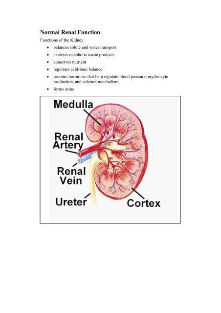
Normal Renal Function<br />
Functions of <strong>the</strong> Kidney:<br />
• balances solute <strong>and</strong> water transport<br />
• excretes metabolic waste products<br />
• conserves nutrient<br />
• regulates acid-base balance<br />
• secretes hormones that help regulate blood pressure, erythrocyte<br />
production, <strong>and</strong> calcium metabolism.<br />
• forms urine
Formation of Urine in <strong>the</strong> Kidney<br />
A kidney contains around one million nephrons. In general, <strong>the</strong> kidneys can<br />
adequately function with only one third of <strong>the</strong> normal number of nephrons.<br />
Less than that, <strong>the</strong> body will retain waste products, especially urea <strong>and</strong><br />
creatinine.<br />
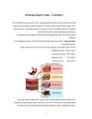
Nephrons process <strong>the</strong> blood to make urine. A tuft of capillaries called <strong>the</strong><br />
glomerulus is contained in each nephron. The glomerulus is surrounded by<br />
Bowman's capsule. The capillaries are extremely porous, allowing large<br />
amounts of solute-rich fluids to pass from <strong>the</strong> capillaries into <strong>the</strong> capsule. This<br />
fluid is <strong>the</strong> raw material of urine.
This fluid leaves <strong>the</strong> capsule <strong>and</strong> is channeled into <strong>the</strong> proximal convoluted<br />
tubule (PCT) of <strong>the</strong> nephron. This is where <strong>the</strong> primary active transport of <strong>the</strong><br />
sodium ion accounts for about 80% of sodium reabsorption. During <strong>the</strong><br />
primary active transport of sodium, chloride is simultaneously reabsorbed as<br />
are all amino acids <strong>and</strong> glucose. Primary active transport also occurs to some<br />
extent in <strong>the</strong> o<strong>the</strong>r tubules except in <strong>the</strong> descending loop of Henle.<br />
The secondary active secretion of H + during Na + reabsorption is called<br />
countertransport since <strong>the</strong> ions move in opposite directions. Normally, only<br />
20% of total Na + reabsorption occurs during active secretion of H + <strong>and</strong> K + .<br />
When H + ions are not available for exchange as in alkalemia, K + ions are<br />
secreted. This is why alkalemia may lead to hypokalemia. In H + <strong>and</strong> K +<br />
secretion, HCO 3 - ions are reabsorbed in place of Cl - . When chloride ions are in<br />
short supply, <strong>the</strong>re is an increased dem<strong>and</strong> for H + <strong>and</strong> K + secretion to reabsorb<br />
sodium.<br />
The proximal tubules are responsible for <strong>the</strong> iso-osmotic reabsorption of<br />
water, electrolytes, non-electolytes. As much as 80% of <strong>the</strong> filtrate is<br />
reabsorbed into <strong>the</strong> capillaries that line <strong>the</strong> tubules.<br />
1. All glucose <strong>and</strong> amino acids filtered are completely reabsorbed.<br />
2. Almost all potassium is reabsorbed.<br />
3. Almost all uric acid is reabsorbed.<br />
4. 90% of bicarbonate is reabsorbed.<br />
5. Two- thirds of filtered sodium is reabsorbed.<br />
6. H 2 O, chloride, <strong>and</strong> urea are reabsorbed by passive transport.<br />
7. Hydrogen ion is secreted, creatinine is secreted.<br />
8. Most of <strong>the</strong> calcium <strong>and</strong> phosphate is reabsorbed.
Past <strong>the</strong> PCT is <strong>the</strong> loop of Henle which consists of both ascending <strong>and</strong><br />
descending limbs. The descending limb is freely permeable to water, while <strong>the</strong><br />
ascending is less permeable. In <strong>the</strong> Loop of Henle <strong>the</strong>re is continued<br />
reabsorption of water, sodium, chloride.<br />
The distal convoluted tubule receives fluid from <strong>the</strong> loop of Henle. The distal<br />
tubules are important in <strong>the</strong> final regulation of water balance <strong>and</strong> acid-base<br />
balance since hydrogen ion is excreted with ammonia as ammonium <strong>and</strong> with<br />
phosphate buffers. Bicarbonate is regenerated in this process <strong>and</strong> retained in<br />
<strong>the</strong> body.<br />
The collecting tubule <strong>the</strong>n receives <strong>the</strong> newly formed urine from <strong>the</strong> nephrons.<br />
In <strong>the</strong> collecting duct water reabsorption is completed. The final concentration<br />
of urine takes place here under <strong>the</strong> control of anti-diuretic hormone (ADH). In<br />
<strong>the</strong> presence of ADH, more water is reabsorbed.<br />
The urine flows through <strong>the</strong> minor <strong>and</strong> major calyces of <strong>the</strong> renal pelvis into<br />
<strong>the</strong> ureter. From <strong>the</strong> ureter, urine makes its way to bladder.
<strong>COUNTERCURRENT</strong> <strong>SYSTEM</strong> <strong>and</strong> <strong>the</strong> <strong>LOOP</strong> <strong>OF</strong> <strong>HENLE</strong><br />
1. The Loop of Henle establishes medullary hyperosmolarity<br />
The ascending limb of <strong>the</strong> loop of Henle transports solutes (NaCl) out of <strong>the</strong> tubule<br />
lumen with little or no water, generating an hyperosmotic medullary interstitium <strong>and</strong><br />
delivering an hyposmotic tubule fluid to <strong>the</strong> distal tubule. This is called <strong>the</strong> "single<br />
effect".<br />
The osmolarity of <strong>the</strong> interstitium rises progressively from cortex to medulla <strong>and</strong><br />
papilla through multiplication of <strong>the</strong> "single effect" by countercurrent flow in <strong>the</strong><br />
branches of <strong>the</strong> loop: The single effect in fluid processed by loop segments located<br />
near <strong>the</strong> tip of <strong>the</strong> papilla occurs in fluid already subject to <strong>the</strong> single effect when <strong>the</strong><br />
fluid was in loop segments located closer to <strong>the</strong> cortex.<br />
Countercurrent exchange of solutes between ascending <strong>and</strong> descending vasa recta (<strong>the</strong><br />
renal medullary capillaries) minimizes solute washout from <strong>the</strong> medullary<br />
interstitium.<br />
2. The countercurrent system permits forming a concentrated urine<br />
In <strong>the</strong> presence of ADH, which increases water permeability, <strong>the</strong> hyposmotic fluid<br />
that enters <strong>the</strong> distal tubule (DT) from <strong>the</strong> thick ascending limb (TAL) looses most of<br />
its water by osmotic equilibration with <strong>the</strong> surrounding cortical interstitium along <strong>the</strong><br />
CNT <strong>and</strong> cortical collecting duct (CCD). It also continues loosing NaCl through<br />
reabsorptive transport along DT, CNT <strong>and</strong> CCD, until <strong>the</strong> tubule fluid becomes<br />
isoosmotic with plasma, by <strong>the</strong> end of <strong>the</strong> CCD.<br />
The relatively small amount of isoosmotic fluid that flows into <strong>the</strong> medullary<br />
collecting ducts losses progressively more <strong>and</strong> more water to <strong>the</strong> hyperosmotic<br />
medullary <strong>and</strong> papillary interstitia <strong>and</strong> is finally excreted as hyperosmotic, highly<br />
concentrated urine.<br />
3. The countercurrent system permits forming a dilute urine<br />
In <strong>the</strong> absence of ADH, <strong>the</strong> hyposmotic fluid that enters <strong>the</strong> DT from <strong>the</strong> loop of<br />
Henle, continues to be diluted by transport of NaCl via NaCl (thiazide sensitive)<br />
cotransporters into DT cells <strong>and</strong> via Na channels (amiloride sensitive) along <strong>the</strong> CD.<br />
Water reabsorption is limited so that <strong>the</strong> tubule fluid becomes more <strong>and</strong> more dilute<br />
along DT, CNT <strong>and</strong> collecting ducts (CCD, OMCD <strong>and</strong> IMCD), until it is excreted as<br />
a large volume of hyposmotic urine.<br />
4. Mechanism of hyperosmotic reabsorption in <strong>the</strong> TAL<br />
There is apical Na-K-2Cl reabsorptive cotransport with K recycling through apical K-<br />
channels, <strong>and</strong> basolateral transport of Na via <strong>the</strong> Na-K-ATPase <strong>and</strong> of Cl via Clchannels,<br />
in <strong>the</strong> water impermeable epi<strong>the</strong>lium of <strong>the</strong> TAL.
A lumen positive electrical potential difference is generated by <strong>the</strong> luminal Na-K-2Cl<br />
cotransporter operating in parallel with channels that allow K to recycle into <strong>the</strong><br />
lumen. The lumen positive potential drives passive paracellular reabsorption of more<br />
Na+ <strong>and</strong> of o<strong>the</strong>r cations (Mg++, Ca++)<br />
The higher <strong>the</strong> delivery of Cl (Km=50 mM), <strong>the</strong> higher <strong>the</strong> activity of <strong>the</strong> luminal Na-<br />
K-2Cl cotransporters <strong>and</strong> <strong>the</strong> higher <strong>the</strong> rate of hyperosmotic Na reabsorption at <strong>the</strong><br />
TAL.<br />
5. Mechanism for hyperosmotic reabsorption in <strong>the</strong> tAL (thin ascending limb)<br />
Water abstraction along <strong>the</strong> early part of <strong>the</strong> thin descending limb (tDL) is driven by<br />
<strong>the</strong> high osmolarity (at least half due to urea) present in <strong>the</strong> medullary interstitium. In<br />
<strong>the</strong> deep nephrons, water reabsorption increases <strong>the</strong> tubule fluid osmolarity (up to<br />
1200 mOsm/L) <strong>and</strong> <strong>the</strong> Na concentration (up to 300 mEq/L) by <strong>the</strong> bend of <strong>the</strong> loop.<br />
Along <strong>the</strong> water impermeable tAL, Na diffuses from <strong>the</strong> tubule lumen into <strong>the</strong><br />
medullary interstitium driven by its concentration gradient <strong>and</strong> some urea enters from<br />
<strong>the</strong> interstitium into <strong>the</strong> lumen; <strong>the</strong> osmolarity decreases as <strong>the</strong> fluid ascends along <strong>the</strong><br />
tAL.<br />
Operation of this passive mechanisms of Na reabsorption along <strong>the</strong> tAL is critically<br />
dependent on efficient medullary recirculation of urea from IMCD to interstitium, to<br />
tAL.<br />
5. O<strong>the</strong>r functions of <strong>the</strong> Loop of Henle<br />
Bicarbonate reabsorption through Na-H exchange<br />
Reabsorption of cations such as Ca 2+ <strong>and</strong> Mg 2+<br />
Generation of cortical to medullary gradients of gaseous NH 3 <strong>and</strong> O 2 <strong>and</strong> of<br />
medullary to cortical gradients of CO 2 <strong>and</strong> lactic acid<br />
Production of Tamm-Horsfall mucoprotein (casts)<br />
Cells survive in <strong>the</strong> hyperosmotic medullary environment through slow accumulation<br />
of osmolytes (75 mM sorbitol <strong>and</strong> 25 mM glycerophosphocholine (GPC) by<br />
syn<strong>the</strong>sis, <strong>and</strong> 25 mM betaine <strong>and</strong> 10 mM inositol by Na + driven cotransport), which<br />
can be rapidly released from <strong>the</strong> cells through channels that open when <strong>the</strong> osmolarity<br />
decreases.
The Loop of Henle: Concentration.<br />
The proximal tubule reabsorbs about 70 percent<br />
of <strong>the</strong> fluid filtered from <strong>the</strong> blood by <strong>the</strong><br />
glomerular capillaries. As <strong>the</strong>re is no such thing<br />
as active transport for water (no water pump), it<br />
accomplishes this by reabsorbing electrolytes by<br />
active transport <strong>and</strong> thus dragging water across<br />
from <strong>the</strong> urine <strong>and</strong> into <strong>the</strong> blood by osmosis.<br />
This is essentially <strong>the</strong> same mechanism used by<br />
water absorbing (gut) <strong>and</strong> water secreting<br />
(salivary gl<strong>and</strong>s) epi<strong>the</strong>lia throughout <strong>the</strong> body.<br />
The proximal tubule even gets a helping h<strong>and</strong> in<br />
this process because <strong>the</strong> blood that circulates in<br />
<strong>the</strong> capillaries near <strong>the</strong> proximal tubule is <strong>the</strong><br />
very same blood that was filtered in <strong>the</strong><br />
glomerular capillaries <strong>and</strong> is thus slightly<br />
hyperosmotic to begin with. Anyway, <strong>the</strong>re is a limitation to this mechanism for<br />
water reabsorption. Not volume, it is possible to reabsorb vast quantities of<br />
fluid using this sort of mechanism, but ra<strong>the</strong>r concentration. The problem with<br />
this sort of mechanism for fluid absorption is that it works on a very small<br />
osmotic gradient <strong>and</strong> consequently, water absorption is practically isotonic.<br />
This means that <strong>the</strong> urine will also be isotonic. It won't contain <strong>the</strong> same<br />
substances as plasma (e.g. no glucose or bicarbonate etc. <strong>and</strong> more PAH or<br />
DDT etc) but <strong>the</strong> overall osmolarity will be <strong>the</strong> same. Suppose <strong>the</strong> body has<br />
taken on an excess sodium chloride load <strong>and</strong> is desperate to get rid of it<br />
because it is disturbing <strong>the</strong> osmolarity of <strong>the</strong> body. If <strong>the</strong> urine is isotonic, <strong>the</strong><br />
salt can't be got rid of. Think about it. If <strong>the</strong> urine is always isotonic with <strong>the</strong><br />
plasma you can adjust <strong>the</strong> amount of salt <strong>and</strong> water in <strong>the</strong> body but not <strong>the</strong><br />
concentration. To get rid of excess salt, you would have to drink sufficient<br />
water to restore normal osmolarity <strong>and</strong> <strong>the</strong>n excrete <strong>the</strong> excess salt with <strong>the</strong><br />
excess water.<br />
To dispose of an excess salt load, you must be able to generate a<br />
concentrated urine. Enter <strong>the</strong> renal medulla <strong>and</strong> <strong>the</strong> loop of Henle. The<br />
medulla is very poorly drained (more on that later) <strong>and</strong> so it is possible to set<br />
up a large osmotic gradient in <strong>the</strong> medulla <strong>and</strong> keep it <strong>the</strong>re (anywhere else<br />
<strong>and</strong> <strong>the</strong> blood would wash <strong>the</strong> gradient away). The o<strong>the</strong>r part of <strong>the</strong> problem is<br />
how to create a large osmotic gradient. Active transport is <strong>the</strong> key, but even<br />
<strong>the</strong> mighty sodium pump can't shift enough sodium to create a worthwhile<br />
gradient by itself. The o<strong>the</strong>r part of <strong>the</strong> solution, <strong>and</strong> <strong>the</strong> reason that <strong>the</strong> loop<br />
of Henle is in fact a loop lies in <strong>the</strong> principle of countercurrent multiplication. In<br />
a countercurrent multiplier, <strong>the</strong> combined action of active pumping <strong>and</strong><br />
circulation <strong>and</strong> re-circulation of solutes around <strong>the</strong> loop of Henle create an<br />
osmotic gradient with <strong>the</strong> following properties.
1) A huge concentration gradient<br />
between <strong>the</strong> solution in <strong>the</strong> middle<br />
of <strong>the</strong> loop <strong>and</strong> that at <strong>the</strong><br />
beginning (or <strong>the</strong> end) of <strong>the</strong> loop<br />
2) Little concentration difference<br />
between solution entering <strong>the</strong> loop<br />
<strong>and</strong> that leaving <strong>the</strong> loop. In fact,<br />
<strong>the</strong> fluid leaving <strong>the</strong> loop may be<br />
hypotonic.<br />
3) Little concentration difference between any two adjacent segments in <strong>the</strong><br />
loop.<br />
Ok. Now we have a huge concentration gradient<br />
extending into <strong>the</strong> medulla, The osmolarity of <strong>the</strong><br />
medulla may be as high as 1400mOsm/l,<br />
compared to <strong>the</strong> normal plasma osmolarity of<br />
300 mOsm/l, but it isn't doing much good in<br />
concentrating <strong>the</strong> urine. True <strong>the</strong> urine gets very<br />
concentrated as it descends <strong>the</strong> loop of Henle<br />
into <strong>the</strong> medulla but it gets progressively weaker<br />
again as it ascends back out into <strong>the</strong> cortex. The<br />
latter part of <strong>the</strong> loop of Henle <strong>and</strong> <strong>the</strong> distal<br />
tubule are sometimes known as <strong>the</strong> 'diluting<br />
segment' of <strong>the</strong> nephron because <strong>the</strong> urine can<br />
in fact be more dilute as it enters <strong>the</strong> distal tubule<br />
than it was leaving <strong>the</strong> proximal tubule. This used<br />
to drive renal physiologists nuts. They knew that<br />
<strong>the</strong> loop of Henle was involved in concentrating <strong>the</strong> urine somehow but so far<br />
as <strong>the</strong>y could tell, all it did was dilute it. The answer is in <strong>the</strong> collecting ducts.<br />
After going through <strong>the</strong> distal tubule, <strong>the</strong> urine passes into <strong>the</strong> collecting<br />
ducts. The collecting ducts travel through <strong>the</strong> medulla on <strong>the</strong>ir way out of <strong>the</strong><br />
kidney. Right past <strong>the</strong> high osmolarity part of <strong>the</strong> medulla in fact. If <strong>the</strong><br />
collecting ducts are water permeable <strong>the</strong>n <strong>the</strong> huge concentration gradient<br />
between <strong>the</strong> medulla <strong>and</strong> <strong>the</strong> collecting ducts will drag water out of <strong>the</strong> urine<br />
into <strong>the</strong> medulla. Finally we have a concentrated urine. Next stop after <strong>the</strong><br />
collecting ducts is <strong>the</strong> bladder. The extent to which <strong>the</strong> urine is concentrated<br />
depends mainly on how water permeable <strong>the</strong> collecting ducts are. The water<br />
permeability of <strong>the</strong> collecting ducts is controlled by anti-diuretic hormone<br />
(ADH). In <strong>the</strong> absence of ADH, <strong>the</strong> collecting ducts are water impermeable,<br />
no water is reabsorbed <strong>and</strong> a large volume of dilute urine is produced. In <strong>the</strong><br />
presence of a high concentration of ADH <strong>the</strong> collecting ducts are highly water<br />
permeable, a lot of water is reabsorbed <strong>and</strong> a small volume of very<br />
concentrated urine is produced. ADH, working via cAMP as a second<br />
messenger stimulates insertion of water channels into <strong>the</strong> plasma membrane.
The V2 receptors for ADH is yet ano<strong>the</strong>r example of a 7-membrane-spanningdomain-G-protein-coupled<br />
receptor). All intermediate stages of water<br />
absorption are possible so that <strong>the</strong> volume <strong>and</strong> <strong>the</strong> concentration of urine can<br />
respond to <strong>the</strong> changing needs of <strong>the</strong> body in regulating fluid <strong>and</strong> electrolyte<br />
homeostasis.<br />
Before I forget. One of <strong>the</strong> requirements to build a countercurrent multiplier is<br />
to keep it away from <strong>the</strong> blood supply, o<strong>the</strong>rwise <strong>the</strong> blood will wash it away<br />
before it gets started. The capillaries that accompany <strong>the</strong> long loops of Henle<br />
on <strong>the</strong>ir trip to <strong>the</strong> medulla <strong>and</strong> back are called <strong>the</strong> vasa recta. These<br />
capillaries also form long loops <strong>and</strong> run in parallel to <strong>the</strong> loop of Henle, most<br />
importantly, <strong>the</strong> arterial <strong>and</strong> venous ends of <strong>the</strong> capillaries run in parallel with<br />
each o<strong>the</strong>r (in o<strong>the</strong>r words a hairpin loop). This allows a process called<br />
countercurrent exchange to occur. In short, countercurrent exchange allows<br />
exchange of solute between <strong>the</strong> ascending <strong>and</strong> descending limbs of <strong>the</strong><br />
capillaries <strong>and</strong> prevents <strong>the</strong>m from dissipating <strong>the</strong> medullary concentration<br />
gradient. Among <strong>the</strong> solutes exchanged in this process are oxygen <strong>and</strong><br />
carbon dioxide (e.g. oxygen moves from <strong>the</strong> descending limb of <strong>the</strong> vasa<br />
recta to <strong>the</strong> ascending limb without travelling around <strong>the</strong> loop, carbon dioxide<br />
moves in <strong>the</strong> opposite direction). All of which makes <strong>the</strong> vasa recta a terrible<br />
blood supply by all normal criteria. It has been suggested that <strong>the</strong> cells at <strong>the</strong><br />
base of <strong>the</strong> loop of Henle have make ATP anaerobically (by glycolysis) to<br />
survive.