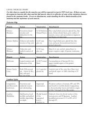
Atlas of the muscle motor points for the lower limb: implications for ...
Atlas of the muscle motor points for the lower limb: implications for ...
Atlas of the muscle motor points for the lower limb: implications for ...

You also want an ePaper? Increase the reach of your titles
YUMPU automatically turns print PDFs into web optimized ePapers that Google loves.
Eur J Appl Physiol (2011) 111:2461–2471 2469<br />
position <strong>of</strong> <strong>the</strong> two gastrocnemius <strong>motor</strong> <strong>points</strong> at *25%<br />
<strong>of</strong> respective reference lines, with poor (<strong>for</strong> medial gastrocnemius)<br />
or fair (<strong>for</strong> lateral gastrocnemius) uni<strong>for</strong>mity<br />
across subjects.<br />
The innervation <strong>of</strong> <strong>the</strong> tibialis anterior and peroneus<br />
longus <strong>muscle</strong>s is provided by <strong>the</strong> common peroneal nerve<br />
which first originates from <strong>the</strong> sciatic nerve in <strong>the</strong> popliteal<br />
fossa region and travels across <strong>the</strong> lateral head <strong>of</strong> <strong>the</strong><br />
gastrocnemius toward <strong>the</strong> fibular head, and <strong>the</strong>n separates<br />
into <strong>the</strong> superficial and deep peroneal nerves. The superficial<br />
peroneal nerve innervates <strong>the</strong> peroneus longus and<br />
brevis <strong>muscle</strong>s, while <strong>the</strong> deep peroneal nerve innervates<br />
<strong>the</strong> anterior leg <strong>muscle</strong>s. In agreement with our findings,<br />
Lee et al. (2011) recently demonstrated that <strong>the</strong> <strong>motor</strong> entry<br />
<strong>points</strong> <strong>of</strong> <strong>the</strong> superficial peroneal nerve supplying <strong>the</strong> peroneus<br />
longus <strong>muscle</strong> were located from 10 to 60% <strong>of</strong> <strong>the</strong><br />
distance between <strong>the</strong> apex <strong>of</strong> <strong>the</strong> fibular head and <strong>the</strong> apex<br />
<strong>of</strong> <strong>the</strong> lateral malleolus. To our knowledge, no previous<br />
study attempted to localize <strong>the</strong> <strong>motor</strong> <strong>points</strong> <strong>of</strong> <strong>the</strong> deep<br />
peroneal nerve to <strong>the</strong> tibialis anterior <strong>muscle</strong>. However, it<br />
has been shown that <strong>the</strong> <strong>muscle</strong>s <strong>of</strong> <strong>the</strong> deep posterior<br />
compartment <strong>of</strong> <strong>the</strong> leg (popliteus, flexor hallucis longus,<br />
tibialis posterior, flexor digitorum longus) are innervated<br />
by one or two primary <strong>motor</strong> branches arising from <strong>the</strong><br />
tibial nerve (Apaydin et al. 2008). Similarly, it is not surprising<br />
that <strong>the</strong> tibialis anterior <strong>muscle</strong> had more than one<br />
<strong>motor</strong> point in our study, which could correspond to <strong>the</strong><br />
entry <strong>points</strong> <strong>of</strong> different nerve branches supplying different<br />
portions <strong>of</strong> <strong>the</strong> <strong>muscle</strong>. In fact, <strong>the</strong> bipennate <strong>muscle</strong><br />
architecture (Maganaris and Baltzopoulos 1999) could<br />
imply that <strong>the</strong> superficial and deep unipennate parts have a<br />
distinct pattern <strong>of</strong> innervation: this hypo<strong>the</strong>sis is in agreement<br />
with our observation <strong>of</strong> a differential activation <strong>of</strong><br />
<strong>the</strong> superficial and deep portion <strong>of</strong> <strong>the</strong> <strong>muscle</strong> following<br />
stimulation <strong>of</strong> <strong>the</strong> proximal and distal <strong>motor</strong> point,<br />
respectively.<br />
Implications <strong>for</strong> electrical stimulation procedures<br />
and electrode positioning<br />
The demonstration that different <strong>motor</strong> <strong>points</strong> can be<br />
identified in <strong>the</strong> three superficial <strong>muscle</strong>s <strong>of</strong> <strong>the</strong> quadriceps,<br />
<strong>the</strong> most <strong>of</strong>ten stimulated <strong>muscle</strong> <strong>for</strong> NMES rehabilitation,<br />
‘‘prehabilitation’’, and training purposes (Bax et al. 2005;<br />
Maffiuletti 2010), may have relevant <strong>implications</strong> <strong>for</strong> <strong>the</strong><br />
placement <strong>of</strong> stimulation electrodes. It is well-known that<br />
<strong>motor</strong> unit recruitment during NMES is spatially fixed<br />
(Bigland-Ritchie et al. 1979; Gregory and Bickel 2005),<br />
thus implying that <strong>the</strong> same <strong>muscle</strong> units are repeatedly<br />
activated by <strong>the</strong> same amount <strong>of</strong> electrical current, which,<br />
in turn, hastens <strong>the</strong> onset <strong>of</strong> <strong>muscle</strong> fatigue (Bigland-<br />
Ritchie et al. 1979; Binder-Macleod and Snyder-Mackler<br />
1993). Such early occurrence <strong>of</strong> fatigue represents a major<br />
limitation <strong>of</strong> NMES. In order to maximize <strong>the</strong> spatial<br />
recruitment during NMES, thus minimizing <strong>the</strong> extent <strong>of</strong><br />
<strong>muscle</strong> fatigue, it has been recommended to adopt different<br />
subterfuges during a treatment session such as <strong>the</strong> progressive<br />
increase in current intensity, alteration in <strong>muscle</strong><br />
length, and displacement <strong>of</strong> active electrodes (Maffiuletti<br />
2010). The existence <strong>of</strong> different <strong>motor</strong> <strong>points</strong> within each<br />
<strong>of</strong> <strong>the</strong> three superficial heads <strong>of</strong> <strong>the</strong> quadriceps suggests<br />
that a change in <strong>the</strong> population <strong>of</strong> activated fibers could<br />
also be obtained through a multichannel stimulation technique<br />
that involves a non-synchronous activation <strong>of</strong> different<br />
<strong>muscle</strong> volumes. Consistently, Malesević et al.<br />
(2010) recently showed in paraplegic patients that NMES<br />
delivered to one quadriceps via multi-pad electrodes (one<br />
anode positioned at <strong>the</strong> distal part <strong>of</strong> <strong>the</strong> quadriceps and<br />
four cathodes distributed over <strong>the</strong> quadriceps <strong>muscle</strong>s)<br />
delayed <strong>the</strong> occurrence <strong>of</strong> fatigue with respect to a conventional<br />
stimulation (one electrode positioned over <strong>the</strong> top<br />
<strong>of</strong> <strong>the</strong> quadriceps and <strong>the</strong> o<strong>the</strong>r over <strong>the</strong> distal part <strong>of</strong> <strong>the</strong><br />
<strong>muscle</strong>).<br />
Besides neuromuscular training, NMES finds application<br />
in <strong>the</strong> in vivo assessment <strong>of</strong> <strong>muscle</strong> contractile properties,<br />
fatigue pr<strong>of</strong>ile, and level <strong>of</strong> voluntary activation<br />
(Maffiuletti 2010). For example, supramaximal stimulation<br />
<strong>of</strong> <strong>the</strong> femoral nerve during a maximal voluntary contraction<br />
is an established technique <strong>for</strong> <strong>the</strong> assessment <strong>of</strong><br />
quadriceps activation (Gandevia 2001). Place et al. (2010)<br />
recently showed that quadriceps <strong>muscle</strong> belly stimulation<br />
can be used to assess <strong>the</strong> level <strong>of</strong> voluntary activation as a<br />
valid alternative to <strong>the</strong> femoral nerve stimulation that may<br />
be associated with discom<strong>for</strong>t and/or stimulation electrode<br />
displacement during <strong>the</strong> voluntary ef<strong>for</strong>t. It may be<br />
hypo<strong>the</strong>sized that a multichannel NMES technique per<strong>for</strong>med<br />
with several electrodes placed over <strong>the</strong> different<br />
<strong>motor</strong> <strong>points</strong> <strong>of</strong> <strong>the</strong> superficial heads <strong>of</strong> quadriceps can<br />
increase <strong>the</strong> validity <strong>of</strong> <strong>the</strong> neuromuscular testing and<br />
minimize subjective discom<strong>for</strong>t.<br />
Electrical stimulation <strong>of</strong> <strong>the</strong> gastrocnemii is usually<br />
per<strong>for</strong>med with two large rectangular electrodes placed<br />
below <strong>the</strong> popliteal cavity and over <strong>the</strong> distal portion <strong>of</strong> <strong>the</strong><br />
two <strong>muscle</strong> heads, with <strong>the</strong> major side <strong>of</strong> <strong>the</strong> electrodes<br />
perpendicular to <strong>the</strong> longitudinal axis <strong>of</strong> <strong>the</strong> triceps surae<br />
(Bergquist et al. 2011). On <strong>the</strong> basis <strong>of</strong> <strong>the</strong> <strong>motor</strong> point<br />
distribution we found in <strong>the</strong> gastrocnemii, it may be proposed<br />
that an alternative placement <strong>of</strong> <strong>the</strong> electrodes (one<br />
over <strong>the</strong> lateral head and <strong>the</strong> o<strong>the</strong>r over <strong>the</strong> medial head),<br />
with <strong>the</strong>ir major side parallel to <strong>the</strong> <strong>muscle</strong> longitudinal<br />
axis, can be used as a valid and more effective alternative<br />
to <strong>the</strong> usual electrode placement.<br />
Finally, <strong>the</strong> existence <strong>of</strong> two distinct <strong>motor</strong> <strong>points</strong> could<br />
imply that <strong>the</strong> tibialis anterior has to be stimulated with <strong>the</strong><br />
two electrodes located above both <strong>motor</strong> <strong>points</strong> in case <strong>of</strong><br />
bipolar arrangement, or with <strong>the</strong> active electrode properly<br />
123