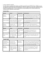
Atlas of the muscle motor points for the lower limb: implications for ...
Atlas of the muscle motor points for the lower limb: implications for ...
Atlas of the muscle motor points for the lower limb: implications for ...

You also want an ePaper? Increase the reach of your titles
YUMPU automatically turns print PDFs into web optimized ePapers that Google loves.
2468 Eur J Appl Physiol (2011) 111:2461–2471<br />
A B C<br />
1<br />
2<br />
3<br />
4<br />
7<br />
8<br />
5<br />
6<br />
1<br />
2<br />
3<br />
4<br />
1<br />
2<br />
(2010) showed a dominant innervation pattern consisting <strong>of</strong><br />
two primary <strong>motor</strong> branches supplying <strong>the</strong> upper and <strong>lower</strong><br />
part <strong>of</strong> <strong>the</strong> <strong>muscle</strong>, with a proximal and a distal entry point<br />
located, respectively, at *20 and *60% <strong>of</strong> <strong>the</strong> distance<br />
between ischial tuberosity and medial femoral epicondyle.<br />
They also indicated that <strong>the</strong> <strong>motor</strong> entry point <strong>of</strong> <strong>the</strong> <strong>lower</strong><br />
part <strong>of</strong> <strong>the</strong> semitendinosus and that <strong>of</strong> <strong>the</strong> semimembranosus<br />
were closely located to each o<strong>the</strong>r. Since <strong>the</strong><br />
semimembranosus can be found at a deeper level than <strong>the</strong><br />
semitendinosus, it may be hypo<strong>the</strong>sized that <strong>the</strong> stimulation<br />
level we adopted to localize <strong>the</strong> <strong>motor</strong> point <strong>of</strong> <strong>the</strong> semimembranosus<br />
also activated <strong>the</strong> <strong>lower</strong> part <strong>of</strong> <strong>the</strong> more<br />
superficial semitendinosus. The inspection and palpation<br />
method we adopted has <strong>the</strong> advantages <strong>of</strong> simplicity and<br />
quickness. However, visual inspection <strong>of</strong> <strong>muscle</strong> contraction<br />
and surface palpation <strong>of</strong> semitendinosus and semimembranosus<br />
and <strong>the</strong>ir distal tendons could not aid in<br />
distinguishing between <strong>the</strong> two <strong>muscle</strong>s. Accordingly, a<br />
localization <strong>of</strong> <strong>the</strong> distal <strong>motor</strong> point <strong>of</strong> <strong>the</strong> semitendinosus<br />
more precise than that obtained by <strong>the</strong> inspection and<br />
palpation method we adopted may be required.<br />
The course <strong>of</strong> <strong>the</strong> tibial nerve has been widely studied.<br />
However, only a few authors investigated <strong>the</strong> course <strong>of</strong> its<br />
Fig. 4 Schematic view <strong>of</strong> <strong>the</strong> <strong>motor</strong> branches <strong>of</strong> <strong>the</strong> femoral nerve<br />
supplying <strong>the</strong> quadriceps <strong>muscle</strong>. a 1 Femoral nerve; 2 inguinal<br />
ligament; 3 <strong>motor</strong> branch <strong>of</strong> <strong>the</strong> rectus femoris; 4 <strong>motor</strong> branch <strong>of</strong> <strong>the</strong><br />
vastus lateralis with its superior (5) and inferior (6) sub-branches; 7<br />
<strong>motor</strong> branch <strong>of</strong> <strong>the</strong> vastus intermedius; 8 <strong>motor</strong> branch <strong>of</strong> <strong>the</strong> vastus<br />
medialis (adapted from Sung et al. 2003). Dotted lines indicate <strong>the</strong><br />
deep course <strong>of</strong> <strong>the</strong> <strong>motor</strong> branch <strong>of</strong> <strong>the</strong> vastus lateralis and its subbranches<br />
(which run under <strong>the</strong> vastus medialis and rectus femoris<br />
<strong>muscle</strong>s). b Superficial proximal (1) and deep proximal (2) subbranches<br />
<strong>of</strong> <strong>the</strong> proximal primary branch <strong>of</strong> <strong>the</strong> vastus lateralis; middistal<br />
(3) and distal (4) sub-branches <strong>of</strong> <strong>the</strong> distal primary branch <strong>of</strong><br />
<strong>the</strong> vastus lateralis (adapted from Becker et al. 2010). c Short and<br />
lateral (1) and long and medial (2) sub-branches <strong>of</strong> <strong>the</strong> <strong>motor</strong> branch<br />
<strong>of</strong> <strong>the</strong> vastus medialis (adapted from Thiranagama 1990)<br />
<strong>motor</strong> branches and <strong>the</strong> <strong>motor</strong> point distribution <strong>of</strong> <strong>the</strong><br />
gastrocnemius <strong>muscle</strong>s. Yoo et al. (2002) per<strong>for</strong>med <strong>the</strong><br />
anatomic dissection <strong>of</strong> 40 cadaver knees and localized <strong>the</strong><br />
<strong>motor</strong> <strong>points</strong> <strong>of</strong> <strong>the</strong> medial and lateral gastrocnemius<br />
<strong>muscle</strong>s at an absolute distance <strong>of</strong> *4.0 and *3.5 cm,<br />
respectively, from <strong>the</strong> medial and lateral epicondyle <strong>of</strong> <strong>the</strong><br />
femur. In ano<strong>the</strong>r dissection study, Sook Kim et al. (2002)<br />
showed a dominant innervation pattern consisting <strong>of</strong> one<br />
<strong>motor</strong> branch supplying <strong>the</strong> lateral gastrocnemius and one<br />
<strong>motor</strong> branch supplying <strong>the</strong> medial gastrocnemius, with <strong>the</strong><br />
respective <strong>motor</strong> entry <strong>points</strong> located at *10% <strong>of</strong> <strong>the</strong><br />
distance between <strong>the</strong> intercondylar and <strong>the</strong> intermalleolar<br />
line. The same authors also localized, in healthy subjects,<br />
<strong>the</strong> <strong>motor</strong> <strong>points</strong> <strong>of</strong> <strong>the</strong> medial and lateral gastrocnemius at<br />
*12 and *10% <strong>of</strong> <strong>the</strong> same distance, respectively (Lee<br />
et al. 2009). Fur<strong>the</strong>r, Kim et al. (2005) showed that <strong>the</strong><br />
<strong>motor</strong> <strong>points</strong> <strong>of</strong> <strong>the</strong> medial and lateral gastrocnemius were<br />
diffusely distributed along <strong>the</strong> longitudinal bulk <strong>of</strong> <strong>the</strong> two<br />
<strong>muscle</strong> heads: <strong>the</strong> range <strong>of</strong> <strong>motor</strong> point locations was from<br />
*10 to *37% <strong>of</strong> <strong>the</strong> distance between <strong>the</strong> intercondylar<br />
and <strong>the</strong> intermalleolar line <strong>for</strong> <strong>the</strong> medial gastrocnemius,<br />
and from *12 to *38% <strong>of</strong> <strong>the</strong> same distance <strong>for</strong> <strong>the</strong> lateral<br />
gastrocnemius. Consistently, we found an average<br />
123