Atlas of the muscle motor points for the lower limb: implications for ...
Atlas of the muscle motor points for the lower limb: implications for ...
Atlas of the muscle motor points for the lower limb: implications for ...

You also want an ePaper? Increase the reach of your titles
YUMPU automatically turns print PDFs into web optimized ePapers that Google loves.
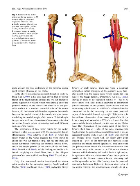
Eur J Appl Physiol (2011) 111:2461–2471 2467<br />
Fig. 3 Position <strong>of</strong> <strong>the</strong> <strong>motor</strong><br />
<strong>points</strong> <strong>for</strong> <strong>the</strong> leg <strong>muscle</strong>s in 53<br />
healthy subjects, along <strong>the</strong><br />
respective reference lines:<br />
a tibialis anterior (blue circles,<br />
proximal <strong>motor</strong> point; yellow<br />
circles, distal <strong>motor</strong> point);<br />
b peroneus longus; c medial<br />
(blue circles) and lateral (yellow<br />
circles) gastrocnemii. The<br />
arrows indicate <strong>the</strong> average<br />
positions <strong>of</strong> <strong>the</strong> <strong>motor</strong> <strong>points</strong><br />
along <strong>the</strong> respective reference<br />
lines<br />
could explain <strong>the</strong> poor uni<strong>for</strong>mity <strong>of</strong> <strong>the</strong> proximal <strong>motor</strong><br />
point position observed in this study.<br />
In <strong>the</strong> above-mentioned anatomical dissection study by<br />
Sung et al. (2003), it has also been shown that <strong>the</strong> <strong>motor</strong><br />
branch <strong>of</strong> <strong>the</strong> rectus femoris divides into two sub-branches:<br />
(a) <strong>the</strong> superior sub-branch, which runs laterally under <strong>the</strong><br />
posterior surface <strong>of</strong> <strong>the</strong> <strong>muscle</strong> and enters it on <strong>the</strong> posterior<br />
surface at a proximal one-third point <strong>of</strong> <strong>the</strong> rectus<br />
femoris; (b) <strong>the</strong> inferior sub-branch that pierces <strong>the</strong> <strong>muscle</strong><br />
fascia at <strong>the</strong> medial border <strong>of</strong> <strong>the</strong> <strong>muscle</strong> and runs downward<br />
along <strong>the</strong> medial margin <strong>of</strong> <strong>the</strong> <strong>muscle</strong>. This finding is<br />
in agreement with our observation <strong>of</strong> two <strong>motor</strong> <strong>points</strong> <strong>for</strong><br />
<strong>the</strong> rectus femoris whose stimulation activated different<br />
portions <strong>of</strong> <strong>the</strong> <strong>muscle</strong>.<br />
The observation <strong>of</strong> two <strong>motor</strong> <strong>points</strong> <strong>for</strong> <strong>the</strong> vastus<br />
medialis is also in agreement with two anatomical studies<br />
(Thiranagama 1990; Lefebvre et al. 2006) in which <strong>the</strong><br />
<strong>motor</strong> branch <strong>of</strong> <strong>the</strong> vastus medialis has been shown to<br />
divide into two sub-branches (Fig. 4c): (a) <strong>the</strong> short and<br />
lateral sub-branch supplying <strong>the</strong> proximal <strong>muscle</strong> fibers,<br />
that is <strong>the</strong> longus portion <strong>of</strong> <strong>the</strong> <strong>muscle</strong> (Lieb and Perry<br />
1968; Travnik et al. 1995), and (b) <strong>the</strong> long and medial subbranch<br />
supplying <strong>the</strong> distal fibers, that is <strong>the</strong> obliquus<br />
portion <strong>of</strong> <strong>the</strong> <strong>muscle</strong> (Lieb and Perry 1968; Travnik et al.<br />
1995).<br />
Only few anatomical studies investigated <strong>the</strong> <strong>motor</strong><br />
point location <strong>for</strong> <strong>the</strong> hamstring <strong>muscle</strong>s. Sunderland and<br />
Hughes (1946) and Seidel et al. (1996) studied <strong>the</strong> biceps<br />
femoris <strong>of</strong> adult cadaver <strong>limb</strong>s and found a dominant<br />
innervation pattern consisting <strong>of</strong> two primary <strong>motor</strong> branches<br />
issued from <strong>the</strong> sciatic nerve which supply <strong>the</strong> long<br />
head <strong>of</strong> <strong>the</strong> biceps femoris. Differently, An et al. (2010)<br />
showed in most <strong>of</strong> <strong>the</strong> examined <strong>muscle</strong>s (41 out <strong>of</strong> 50<br />
<strong>lower</strong> <strong>limb</strong>s from adult human cadavers) an innervation<br />
pattern consisting <strong>of</strong> one primary <strong>motor</strong> branch with <strong>the</strong><br />
<strong>motor</strong> entry point located at *40% <strong>of</strong> a reference line that<br />
connected <strong>the</strong> ischial tuberosity to <strong>the</strong> most proximal<br />
aspect <strong>of</strong> <strong>the</strong> medial femoral epicondyle. This result is in<br />
line with our observation <strong>of</strong> one <strong>motor</strong> point <strong>of</strong> <strong>the</strong> biceps<br />
femoris long head located at *33% <strong>of</strong> a reference line that<br />
connected <strong>the</strong> ischial tuberosity to <strong>the</strong> apex <strong>of</strong> <strong>the</strong> fibular<br />
head. Our observation <strong>of</strong> one <strong>motor</strong> point <strong>of</strong> <strong>the</strong> biceps<br />
femoris short head at *60% <strong>of</strong> <strong>the</strong> same reference line<br />
(starting from <strong>the</strong> proximal anatomical landmark) is also in<br />
agreement with <strong>the</strong> study <strong>of</strong> An et al. (2010) who observed<br />
one primary <strong>motor</strong> branch with <strong>the</strong> <strong>motor</strong> entry point<br />
located at *56% <strong>of</strong> <strong>the</strong> reference line between ischial<br />
tuberosity and medial femoral epicondyle. They also found<br />
one primary <strong>motor</strong> branch <strong>for</strong> <strong>the</strong> semimembranosus with<br />
<strong>the</strong> <strong>motor</strong> entry point located at *60% <strong>of</strong> <strong>the</strong> distance<br />
between ischial tuberosity and medial femoral epicondyle,<br />
in agreement with our observation <strong>of</strong> one <strong>motor</strong> point at<br />
*60% <strong>of</strong> <strong>the</strong> distance between ischial tuberosity and<br />
medial epicondyle <strong>of</strong> <strong>the</strong> tibia (starting from <strong>the</strong> proximal<br />
anatomical landmark). Differently from our observation <strong>of</strong><br />
one <strong>motor</strong> point <strong>for</strong> <strong>the</strong> semitendinosus <strong>muscle</strong>, An et al.<br />
123