Atlas of the muscle motor points for the lower limb: implications for ...
Atlas of the muscle motor points for the lower limb: implications for ...
Atlas of the muscle motor points for the lower limb: implications for ...

You also want an ePaper? Increase the reach of your titles
YUMPU automatically turns print PDFs into web optimized ePapers that Google loves.
2466 Eur J Appl Physiol (2011) 111:2461–2471<br />
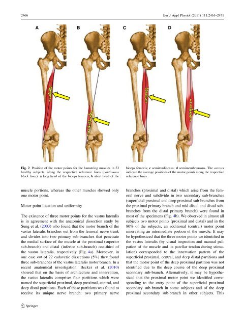
Fig. 2 Position <strong>of</strong> <strong>the</strong> <strong>motor</strong> <strong>points</strong> <strong>for</strong> <strong>the</strong> hamstring <strong>muscle</strong>s in 53<br />
healthy subjects, along <strong>the</strong> respective reference lines (continuous<br />
black lines): a long head <strong>of</strong> <strong>the</strong> biceps femoris; b short head <strong>of</strong> <strong>the</strong><br />
biceps femoris; c semitendinosus; d semimembranosus. The arrows<br />
indicate <strong>the</strong> average positions <strong>of</strong> <strong>the</strong> <strong>motor</strong> <strong>points</strong> along <strong>the</strong> respective<br />
reference lines<br />
<strong>muscle</strong> portions, whereas <strong>the</strong> o<strong>the</strong>r <strong>muscle</strong>s showed only<br />
one <strong>motor</strong> point.<br />
Motor point location and uni<strong>for</strong>mity<br />
The existence <strong>of</strong> three <strong>motor</strong> <strong>points</strong> <strong>for</strong> <strong>the</strong> vastus lateralis<br />
is in agreement with <strong>the</strong> anatomical dissection study by<br />
Sung et al. (2003) who found that <strong>the</strong> <strong>motor</strong> branch <strong>of</strong> <strong>the</strong><br />
vastus lateralis branches out from <strong>the</strong> femoral nerve trunk<br />
and divides into two primary sub-branches that penetrate<br />
<strong>the</strong> medial surface <strong>of</strong> <strong>the</strong> <strong>muscle</strong> at <strong>the</strong> proximal (superior<br />
sub-branch) and distal (inferior sub-branch) one-third <strong>of</strong><br />
<strong>the</strong> vastus lateralis, respectively (Fig. 4a). Moreover, in<br />
one case out <strong>of</strong> 22 cadaveric dissections (5%) <strong>the</strong>y found<br />
three sub-branches <strong>of</strong> <strong>the</strong> vastus lateralis <strong>motor</strong> branch. In a<br />
recent anatomical investigation, Becker et al. (2010)<br />
showed that on <strong>the</strong> basis <strong>of</strong> architecture and innervation,<br />
<strong>the</strong> vastus lateralis comprises four partitions which were<br />
named <strong>the</strong> superficial proximal, deep proximal, central, and<br />
deep distal partitions. Each <strong>of</strong> <strong>the</strong>se partitions was found to<br />
receive its unique nerve branch: two primary nerve<br />
branches (proximal and distal) which arise from <strong>the</strong> femoral<br />
nerve and subdivide in two secondary sub-branches<br />
(superficial proximal and deep proximal sub-branches from<br />
<strong>the</strong> proximal primary branch and mid-distal and distal subbranches<br />
from <strong>the</strong> distal primary branch) were found in<br />
most <strong>of</strong> <strong>the</strong> specimens (Fig. 4b). We observed in almost all<br />
subjects two <strong>motor</strong> <strong>points</strong> (proximal and distal) and in <strong>the</strong><br />
80% <strong>of</strong> <strong>the</strong> subjects, an additional (central) <strong>motor</strong> point<br />
innervating an intermediate portion <strong>of</strong> <strong>the</strong> <strong>muscle</strong>. It may<br />
be hypo<strong>the</strong>sized that <strong>the</strong> three <strong>motor</strong> <strong>points</strong> we identified in<br />
<strong>the</strong> vastus lateralis (by visual inspection and manual palpation<br />
<strong>of</strong> <strong>the</strong> <strong>muscle</strong> and its patellar tendon during stimulation)<br />
corresponded to <strong>the</strong> innervation pattern <strong>of</strong> <strong>the</strong><br />
superficial proximal, central, and deep distal partitions and<br />
that <strong>the</strong> <strong>motor</strong> point <strong>of</strong> <strong>the</strong> deep proximal partition was not<br />
identified due to <strong>the</strong> deep course <strong>of</strong> <strong>the</strong> deep proximal<br />
secondary sub-branch. Alternatively, it may be hypo<strong>the</strong>sized<br />
that <strong>the</strong> proximal <strong>motor</strong> point we identified corresponding<br />
to <strong>the</strong> entry point <strong>of</strong> <strong>the</strong> superficial proximal<br />
secondary sub-branch in some subjects and <strong>of</strong> <strong>the</strong> deep<br />
proximal secondary sub-branch in o<strong>the</strong>r subjects. This<br />
123