Atlas of the muscle motor points for the lower limb: implications for ...
Atlas of the muscle motor points for the lower limb: implications for ...
Atlas of the muscle motor points for the lower limb: implications for ...

You also want an ePaper? Increase the reach of your titles
YUMPU automatically turns print PDFs into web optimized ePapers that Google loves.
2462 Eur J Appl Physiol (2011) 111:2461–2471<br />
proximity <strong>of</strong> only one <strong>of</strong> <strong>the</strong> two electrodes and not at <strong>the</strong><br />
o<strong>the</strong>r (Merletti et al. 1992). The active stimulation electrode<br />
(usually called ‘‘negative’’ electrode) has small<br />
dimensions (usually a few square centimeters) and is<br />
located ei<strong>the</strong>r near a nerve (nerve stimulation) or above a<br />
<strong>muscle</strong> <strong>motor</strong> point (<strong>muscle</strong> stimulation). The second<br />
electrode (usually called ‘‘reference’’ or ‘‘dispersive’’ or<br />
‘‘positive’’ or ‘‘return’’ electrode) is larger than <strong>the</strong> active<br />
electrode (around tens <strong>of</strong> square centimeters) and is generally<br />
placed over <strong>the</strong> antagonist <strong>muscle</strong> or opposite to <strong>the</strong><br />
active electrode. With this electrode configuration, <strong>for</strong> a<br />
certain current level, <strong>the</strong> current density in <strong>the</strong> proximity <strong>of</strong><br />
<strong>the</strong> active electrode may exceed <strong>the</strong> excitation level <strong>of</strong> <strong>the</strong><br />
axons/axonal branches, whereas <strong>the</strong> large dimensions <strong>of</strong><br />
<strong>the</strong> reference electrode assure that in its proximity <strong>the</strong><br />
current density remains below <strong>the</strong>ir excitation threshold.<br />
There<strong>for</strong>e, this technique allows <strong>the</strong> stimulation <strong>of</strong> localized<br />
populations <strong>of</strong> superficial <strong>motor</strong> units. In <strong>the</strong> bipolar<br />
arrangement, two electrodes <strong>of</strong> similar dimensions are<br />
applied over <strong>the</strong> <strong>muscle</strong>. With respect to <strong>the</strong> monopolar<br />
stimulation, current distribution is more confined in space<br />
and current density is more uni<strong>for</strong>m along <strong>the</strong> current path.<br />
Ano<strong>the</strong>r difference between <strong>the</strong> two configurations is <strong>the</strong><br />
number <strong>of</strong> electrodes needed <strong>for</strong> a multichannel stimulation:<br />
in <strong>the</strong> monopolar arrangement <strong>the</strong>re is only one reference<br />
electrode shared by all <strong>the</strong> active ones, while in <strong>the</strong><br />
bipolar case each active electrode has its own reference<br />
electrode. In both monopolar and bipolar <strong>muscle</strong> stimulation,<br />
<strong>the</strong> position <strong>of</strong> <strong>the</strong> stimulation electrodes is a critical<br />
issue: a proper electrode positioning over <strong>the</strong> main <strong>muscle</strong><br />
<strong>motor</strong> <strong>points</strong> is necessary to optimize <strong>the</strong> stimulation paradigm,<br />
that is, to maximize <strong>muscle</strong> tension and <strong>the</strong>re<strong>for</strong>e<br />
<strong>for</strong>ce output (Gobbo et al. 2011) and avoid/minimize<br />
discom<strong>for</strong>t.<br />
Muscle <strong>motor</strong> point, also known as <strong>motor</strong> entry point,<br />
represents <strong>the</strong> location where <strong>the</strong> <strong>motor</strong> branch <strong>of</strong> a nerve<br />
enters <strong>the</strong> <strong>muscle</strong> belly. It can be non-invasively identified<br />
by NMES as <strong>the</strong> skin area above <strong>the</strong> <strong>muscle</strong> in which an<br />
electrical pulse evokes a visible <strong>muscle</strong> twitch with <strong>the</strong><br />
least injected current. Its precise localization is paramount<br />
not only <strong>for</strong> proper positioning <strong>of</strong> <strong>the</strong> stimulation electrodes,<br />
but also <strong>for</strong> improving <strong>the</strong>rapeutic effectiveness and<br />
minimizing complications <strong>of</strong> anes<strong>the</strong>tic or neurolytic <strong>motor</strong><br />
nerve blocks (Karaca et al. 2000). Despite <strong>the</strong> pervasive<br />
diffusion <strong>of</strong> anatomic <strong>motor</strong> point charts (Prentice 2005;<br />
Reid 1920), that are <strong>of</strong>ten provided with <strong>the</strong> user manuals<br />
<strong>of</strong> commercially available stimulators, <strong>the</strong> uni<strong>for</strong>mity <strong>of</strong><br />
<strong>the</strong> <strong>motor</strong> point position <strong>for</strong> <strong>lower</strong> <strong>limb</strong> <strong>muscle</strong>s has never<br />
been investigated in a large group <strong>of</strong> healthy subjects.<br />
There<strong>for</strong>e, <strong>the</strong> aim <strong>of</strong> this study was to assess <strong>the</strong> interindividual<br />
variability <strong>of</strong> <strong>muscle</strong> <strong>motor</strong> point positions.<br />
Some <strong>of</strong> <strong>the</strong>se data have been presented in abstract <strong>for</strong>m<br />
(Oprandi et al. 2010).<br />
Materials and methods<br />
Subjects<br />
Fifty-three healthy subjects <strong>of</strong> both genders (28 males, 25<br />
females; age range: 18–50 years; body mass, mean ± SD:<br />
64.4 ± 11.4 kg; stature: 1.69 ± 0.08 m; body mass index:<br />
22.5 ± 3.5 kg/m 2 ) volunteered to participate in <strong>the</strong> study.<br />
They were free from neuromuscular or skeletal impairments.<br />
Health status was assessed by medical history,<br />
physical exam, blood count and chemistry, urinalysis, and<br />
electrocardiogram. The subjects received a detailed<br />
explanation <strong>of</strong> <strong>the</strong> procedures and gave written in<strong>for</strong>med<br />
consent prior to participation. The study con<strong>for</strong>med with<br />
<strong>the</strong> guidelines in <strong>the</strong> Declaration <strong>of</strong> Helsinki and was<br />
approved by <strong>the</strong> local ethics committee.<br />
Motor point identification, stimulation technique,<br />
and ultrasound examination<br />
The <strong>muscle</strong> <strong>motor</strong> <strong>points</strong> were identified <strong>for</strong> <strong>the</strong> dominant<br />
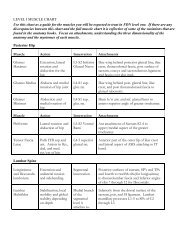
side <strong>of</strong> ten <strong>muscle</strong>s <strong>of</strong> <strong>the</strong> <strong>lower</strong> <strong>limb</strong> (see <strong>the</strong> list in<br />
Table 1). The <strong>motor</strong> <strong>points</strong> were identified while <strong>the</strong> subjects<br />
were positioned as follows: (a) seated, with <strong>the</strong> knee<br />
angle at 90°, <strong>for</strong> <strong>the</strong> investigation <strong>of</strong> <strong>the</strong> quadriceps <strong>muscle</strong>s;<br />
(b) prone, with <strong>the</strong> knee fully extended and <strong>the</strong> ankle<br />
at 150° (180° corresponded to full plantar flexion), <strong>for</strong> <strong>the</strong><br />
investigation <strong>of</strong> <strong>the</strong> hamstring <strong>muscle</strong>s and gastrocnemii;<br />
(c) supine, with <strong>the</strong> knee fully extended and <strong>the</strong> ankle at<br />
150° (180° corresponded to full plantar flexion), <strong>for</strong> <strong>the</strong><br />
investigation <strong>of</strong> <strong>the</strong> tibialis anterior and peroneus longus<br />
<strong>muscle</strong>s.<br />
For each <strong>muscle</strong>, <strong>the</strong> position <strong>of</strong> <strong>the</strong> identified <strong>motor</strong><br />
<strong>points</strong> was determined as absolute and relative distances<br />
along a reference line which was measured between a<br />
proximal and a distal anatomical landmark (see <strong>the</strong> list in<br />
Table 1).<br />
The <strong>muscle</strong> <strong>motor</strong> <strong>points</strong> corresponded to <strong>the</strong> locations<br />
<strong>of</strong> <strong>the</strong> skin area above <strong>the</strong> <strong>muscle</strong> in which an electrical<br />
pulse evoked a <strong>muscle</strong> twitch (as determined by visual<br />
inspection and manual palpation <strong>of</strong> <strong>the</strong> <strong>muscle</strong> and its<br />
proximal or distal tendon) with <strong>the</strong> least injected current.<br />
These locations were identified by scanning <strong>the</strong> skin surface<br />
with a stimulation pen electrode (small size cathode:<br />
1cm 2 surface; Globus Italia, Codognè, Italy) and with a<br />
large (50 9 80 mm) reference electrode placed over <strong>the</strong><br />
antagonist <strong>muscle</strong> to close <strong>the</strong> stimulation current loop<br />
(monopolar stimulation). The pen electrode was moved<br />
over <strong>the</strong> skin, while <strong>the</strong> stimulation current was slowly<br />
increased (starting from 1 to 2 mA) by <strong>the</strong> operator until a<br />
clear <strong>muscle</strong> twitch could be observed. Then, <strong>the</strong> stimulation<br />
current was decreased to a value that could still elicit a<br />
small mechanical response <strong>of</strong> <strong>the</strong> <strong>muscle</strong>. This <strong>motor</strong> point<br />
123