Organic & Biomolecular Chemistry
Organic & Biomolecular Chemistry
Organic & Biomolecular Chemistry

Create successful ePaper yourself
Turn your PDF publications into a flip-book with our unique Google optimized e-Paper software.
Downloaded by Dalian University of Technology on 04 December 2010<br />
Published on 06 May 2010 on http://pubs.rsc.org | doi:10.1039/C004148C<br />
View Online<br />
<strong>Organic</strong> &<br />
<strong>Biomolecular</strong><br />
<strong>Chemistry</strong><br />
www.rsc.org/obc Volume 8 | Number 13 | 28 June 2010 | Pages 2873–3084<br />
ISSN 1477-0520<br />
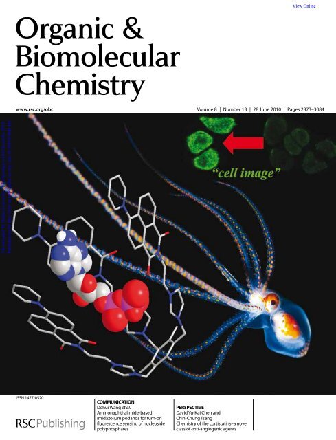
COMMUNICATION<br />
Dehui Wang et al.<br />
Aminonaphthalimide-based<br />
imidazolium podands for turn-on<br />
fluorescence sensing of nucleoside<br />
polyphosphates<br />
PERSPECTIVE<br />
David Yu-Kai Chen and<br />
Chih-Chung Tseng<br />
<strong>Chemistry</strong> of the cortistatins–a novel<br />
class of anti-angiogenic agents
View Online<br />
COMMUNICATION<br />
www.rsc.org/obc | <strong>Organic</strong> & <strong>Biomolecular</strong> <strong>Chemistry</strong><br />
Aminonaphthalimide-based imidazolium podands for turn-on fluorescence<br />
sensing of nucleoside polyphosphates†<br />
Dehui Wang, Xiaolin Zhang, Cheng He and Chunying Duan*<br />
Received 16th March 2010, Accepted 26th April 2010<br />
First published as an Advance Article on the web 6th May 2010<br />
DOI: 10.1039/c004148c<br />
Downloaded by Dalian University of Technology on 04 December 2010<br />
Published on 06 May 2010 on http://pubs.rsc.org | doi:10.1039/C004148C<br />
New aminonaphthalimide imidazolium podands, which<br />
worked as luminescence chemosensors for selectively sensing<br />
nucleoside polyphosphates through a “turn-on” manner,<br />
were prepared for fluorescent imaging of ADP and ATP in<br />
living cells.<br />
Recently, a great deal of interest has been focused on the<br />
molecular recognition of nucleotide polyphosphates because they<br />
play a major role in understanding and evaluating several key<br />
biological processes. 1 While considerable efforts have been devoted<br />
to developing fluorescent chemosensors for various nucleotides<br />
like ATP, 2 GTP, 3 UTP/UDP 4 in the past decades. It is still not<br />
easy to find an example in which all of the twelve ribonucleoside<br />
polyphosphates were examined to evaluate the selectivity. 5 The<br />
distinguishability of ATP from adenosine di-phosphate (ADP)<br />
and adenosine mono-phosphate (AMP) is also urgently desired<br />
for an improved sensor, since ATP is made from ADP and AMP,<br />
and converted back into these precursors in metabolism. 6 In<br />
particular, ATP and ADP are basic and important components<br />
in bioenergetic conversion processes of living organisms, with<br />
their polyphosphate chains being the center for chemical energy<br />
storage and transfer. 7 Thus a fluorescent sensor, which can signal<br />
the concentration of ADP/ATP from the hindrance of other<br />
nucleoside polyphosphates is needed.<br />
As a continuance of our research work on a tripodal receptor<br />
with arms comprising benzoimidazolium hydrogen bonding<br />
moieties, 8 herein, we report a new approach to the preparation of<br />
chemosensors that have the potential to distinguish ADP and ATP<br />
from other ribonucleoside polyphosphates, by incorporating 1,8-<br />
naphthalimide and imidazolium moieties into the preorganized<br />
tripodal receptors. Since amino-naphthalimide is a promising signaling<br />
subunit emitting in the green region (l ~ 540–550 nm) with<br />
high quantum yields (U f ), 9 the chemosensors are also successfully<br />
applied to cells imaging for ADP and ATP, respectively.<br />
Compound TIA1 was synthesized by reaction of N-(2-(1Himidazol-1-yl)ethyl)-4-piperidine-1,8-naphthalimide<br />
with 1,3,5-<br />
tris(bromomethyl)-2,4-dimethylbenzene in CHCl 3 and characterized<br />
by EA, 1 H NMR and MS (Scheme 1). TIA1 exhibited a<br />
1,8-naphthalimide characteristic absorption band at 425 nm (log<br />
e = 4.40) in acetonitrile solution. The addition of all the twelve<br />
ribonucleotides did not cause any significant spectra variation.<br />
Free receptor TIA1 exhibited a strong green emission at 548 nm<br />
(Fig. 1), assignable to the 1,8-naphthalimide (U f = 0.04) 10 upon<br />
excitation at 465 nm. The addition of ADP, caused fluorescence<br />
State Key Laboratory of Fine Chemicals, Dalian University of Technology,<br />
Dalian, 116012, China. E-mail: cyduan@dlut.edu.cn<br />
† Electronic supplementary information (ESI) available: Experimental<br />
details and additional spectroscopic data. See DOI: 10.1039/c004148c<br />
Scheme 1 The aminonaphthalimide-based imidazolium podands TIA1<br />
for fluorescent sensing of ADP. FE is the fluorescence enhancement.<br />
Fig. 1 Fluorescence responses of TIA1 (20 mM) in acetonitrile solution<br />
upon addition of ADP (0 to 0.4 mM). Inset: fluorescence responses for<br />
several ribonucleotide di- and triphosphates (0.4 mM). The bars represent<br />
the value of [(F - F 0 )/F 0 ], where F and F 0 are the emission intensities in<br />
the presence and absence of the ribonucleotides, respectively. Intensity was<br />
recorded at 548 nm, excitation at 465 nm.<br />
enhancement, and showed a steady and smooth increase until<br />
a plateau was reached (U f = 0.12). The Hill-plot profile of the<br />
titration curve suggested a 1 : 3 stoichiometry of the host–guest<br />
complexation species with the association constant (log K ass ) 11<br />
calculated as 10.44 (Fig. S5). Under the same conditions no significant<br />
fluorescence enhancements of TIA1 (20 mM) were observed in<br />
the presence of other ribonucleotide di- and triphosphates. These<br />
results suggest that TIA1 is a useful probe for the fluorescence<br />
detection of ADP with high selectivity and strong binding affinity.<br />
Since the aminonaphthalimide absorbance band did not change<br />
upon the addition of increasing amounts of ADP, the enhancement<br />
of the fluorescence was attributed to the PET motif. 12 It seemed<br />
that the enhancement of the fluorescence was due to a reduced<br />
electron-charge density at the imidazole site after the binding with<br />
diphosphate of the ADP, which reduced the “push–pull” nature<br />
of the ICT excited state of TIA1 (caused by the electron-donating<br />
amine and the electron-withdrawing imidazole). 13 The selectivity<br />
This journal is © The Royal Society of <strong>Chemistry</strong> 2010 Org. Biomol. Chem., 2010, 8, 2923–2925 | 2923
View Online<br />
Downloaded by Dalian University of Technology on 04 December 2010<br />
Published on 06 May 2010 on http://pubs.rsc.org | doi:10.1039/C004148C<br />
of the response with ADP over other adenosine polyphosphates<br />
was ascribed the suitable length of the linked group for the<br />
diphosphate group. The discrimination of ADP from other<br />
nucleotide diphosphates seems to come from the possible hydrogen<br />
bonding interactions between the adenosine and the receptor.<br />
1<br />
H NMR spectra of the receptor TIA1 (1 mM) in the presence<br />
of ADP (1 mM) exhibited a significant downfield shift (ca. 0.15<br />
ppm) of the (C–H) + proton H 1 on the imidazolium ring, and a<br />
chemical shift of the H 2 (0.04 ppm) compared to the free TIA1<br />
in the same experimental conditions, suggesting the formation of<br />
(C–H) + ◊◊◊O charged hydrogen bonds between the imidazolium of<br />
TIA1 and phosphate groups in ADP (Fig. 2). While most flexible<br />
di- or tripods complexes contain rigid planar fluorescent groups<br />
an excimer effect may occur after the addition of guest, 14 however,<br />
our experiments did not support the possibility of the formation of<br />
the corresponding excimer. The small but significant upfield shifts<br />
of the signals corresponding to the aromatic protons within the<br />
1,8-naphthalimide (ca. 0.03 pm), demonstrated the possible p◊◊◊p<br />
stacking interaction between the adenosine and 1,8-naphthalimide<br />
groups. The downfield shifts of the H 13 (0.02 ppm) and H 12 from<br />
the adenine group during the host–guest interactions should be<br />
attributed to the additional hydrogen bonding interaction between<br />
the adenosine in ADP and TIA1 receptor. It is thought that these<br />
complementary interactions are paramount for TIA1 exhibiting<br />
selectivity toward ADP over other ribonucleotide diphosphate.<br />
was observed, suggesting the successful application in the ADP<br />
stain experiments. When cells stained with compound TIA1<br />
were further incubated with ATP (0.4 mM) under the same<br />
experimental conditions, no remarkable fluorescence change was<br />
observed, suggesting the possibly distinguishability of ADP from<br />
ATP in the living cells.<br />
To further investigate the selectivity of the different ribonucleotide<br />
polyphosphates and the effect of the linked groups<br />
on the sensitivity of the chemosensor TIA1 toward nucleotide<br />
triphosphates, TIA2 was designed and synthesized in multisteps<br />
to enlarge the chain of each arm through the well-known<br />
“click” reaction with the aim to recognize the longer adenosine<br />
triphosphate (Scheme 2). The absorbance spectrum of TIA2<br />
showed a band about 415 nm (loge = 4.41), assignable to the<br />
ICT band of aminonaphthalimide. 13 Free receptor TIA2 exhibited<br />
an emission band at 550 nm (Fig. 3), assignable to the 1,8-<br />
naphthalimide (U f = 0.004) 15 upon excitation at 395 nm. The<br />
addition of ATP caused a substantial fluorescence enhancement,<br />
which showed a steady and smooth decrease until a plateau was<br />
reached (U f = 0.03). The Hill-plot profile of the titration curve<br />
confirmed the presence of 1 : 2 stoichiometry of the TIA2–ATP<br />
complexation species with the association constant (log k ass )for<br />
ATP calculated as 8.75 (Fig. S6). Under the same conditions<br />
no significant fluorescence changes of TIA2 were observed in<br />
the presence of adenosine polyphosphate. However, by addition<br />
of GTP and UTP, a fluorescence enhancement was observed<br />
with varying degree. The profile of the fluorescence titration also<br />
suggested a 1 : 2 stoichiometry for the complexation species.<br />
Scheme 2<br />
Synthetic procedure for TIA2.<br />
Fig. 2 Partial 1 H-NMR spectra for (a) TIA1,(b)TIA1 + ADP (3 eq) and<br />
(c) pure ADP in d 6 -DMSO, respectively.<br />
Taking advantage of the off/on fluorescence sensing specifically<br />
of ADP by TIA1, this chemosensor was successfully applied to<br />
fluorescence imaging studies of intracellular ADP stores in living<br />
cells. HeLa cells incubated with TIA1 (10 mM) for 30 min at<br />
room temperature showed a weak green intracellular fluorescence,<br />
which suggested that TIA1 was cell permeable (Fig. S8). The cells<br />
remained viable and no apparent toxicity and side effects were<br />
observed throughout the imaging experiments. When cells stained<br />
with compound TIA1 were further incubated with ADP (0.4 mM)<br />
in phosphate-buffered saline (PBS) for 30 min and washed, a<br />
remarkable enhancing of the green fluorescence intensity (Fig. S9)<br />
Fig. 3 Emission spectra of TIA2 (20 mM) in aqueous solution upon addition<br />
of ribonucleotide polyphosphates (0.4 mM) (excitation at 396 nm).<br />
1<br />
H NMR titration of TIA2 (1 mM) in a 8 : 2 d 6 -DMSO/D 2 O<br />
solution) upon the addition of ATP, GTP and UTP (3 mM) exhibited<br />
the disappearance of (C–H) + proton, suggesting the formation<br />
of (C–H) + ◊◊◊O hydrogen bonding between the imidazolium ring<br />
2924 | Org. Biomol. Chem., 2010, 8, 2923–2925 This journal is © The Royal Society of <strong>Chemistry</strong> 2010
View Online<br />
Downloaded by Dalian University of Technology on 04 December 2010<br />
Published on 06 May 2010 on http://pubs.rsc.org | doi:10.1039/C004148C<br />
and the triphosphate groups. The protons of the nucleic bases<br />
and the aromatic rings corresponding to the 1,8-naphthalimide<br />
all exhibited small but significant upfield shifts, demonstrating the<br />
possible stacking interactions between the nucleic base and the<br />
aromatic moiety of 1,8-naphthalimide groups, like that reported<br />
by Yoon et al. 16<br />
The TIA2 response with ATP in HeLa cells was examined<br />
(Fig. 4). HeLa cells incubated with TIA2 (10 mM) for 30 min at<br />
room temperature showed a weak green intracellular fluorescence,<br />
which suggested that TIA2 was cell permeable. The cells remained<br />
viable and no apparent toxicity and side effects were observed<br />
throughout the imaging experiments. Fluorescence enhancement<br />
was observed when cells stained were further incubated with ATP<br />
(0.4 mM) for 30 min, indicating the possible usage in fluorescence<br />
images involving nucleoside polyphosphates within living cells.<br />
Fig. 4 Confocal fluorescence images of HeLa cells (l ex = 488 nm)<br />
incubated with (a) TIA2 (10 uM) and (b) their images after further<br />
incubated with ATP (0.4 mM).<br />
In summary, we have reported two new types of chemical sensors<br />
TIA1 and TIA2 for ADP and nucleotide triphosphates, respectively.<br />
TIA1 exhibits a selective “turn-on” fluorescent property for<br />
ADP over other ribonucleotide polyphosphates. TIA2 exhibits a<br />
same fluorescent property for ATP, GTP and UTP. These sensors<br />
are also successfully applied to cell imaging for the corresponding<br />
nucleotide polyphosphates.<br />
Acknowledgements<br />
This work was supported by the National Natural Science<br />
foundation of China and Postdoctoral Science Foundation of<br />
China.<br />
Notes and references<br />
1(a) S. C. McCleskey, M. J. Griffin, S. E. Schneider, J. T. McDevitt<br />
and E. V. Anslyn, J. Am. Chem. Soc., 2003, 125, 1114; (b) S.K.Kim,<br />
D. H. Lee, J. I. Hong and J. Yoon, Acc. Chem. Res., 2009, 42, 23; (c)S.<br />
Mizukami, T. Nagano, Y. Urano, A. Odani and K. Kikuchi, J. Am.<br />
Chem. Soc., 2002, 124, 3920; (d) J. Wongkongkatep, Y. Miyahara, A.<br />
Ojida and I. Hamachi, Angew. Chem., Int. Ed., 2006, 45, 665; (e) A.<br />
Ojida, Y. Mito-oka, K. Sada and I. Hamachi, J. Am. Chem. Soc., 2004,<br />
126, 2454; (f) Z. Xu, S. K. Kim and J. Yoon, Chem. Soc. Rev., 2010, 39,<br />
1457.<br />
2(a) S. E. Schneider, S. N. O’Neil and E. V. Anslyn, J. Am. Chem. Soc.,<br />
2000, 122, 542; (b) A. Ojida, I. Takashima, T. Kohira, H. Nonaka and<br />
I. Hamachi, J. Am. Chem. Soc., 2008, 130, 12095; (c) D.H.Lee,S.Y.<br />
Kim and J. I. Hong, Angew. Chem., Int. Ed., 2004, 43, 4777; (d) F.<br />
Sancenon, A. B. Descalzo, R. Martinez-Manez, M. A. Miranda and J.<br />
Soto, Angew. Chem., Int. Ed., 2001, 40, 2640; (e) C. Li, M. Numata,<br />
M. Takeuchi and S. Shinkai, Angew. Chem., Int. Ed., 2005, 44, 6371;<br />
(f)M.C.Zhao,M.Wang,H.J.Liu,D.S.Liu,G.X.Zhang,D.Q.Zhang<br />
andD.B.Zhu,Langmuir, 2009, 25, 676; (g) D.A.Jose,S.Mishra,A.<br />
Ghosh, A. Shrivastav, S. K. Mishra and A. Das, Org. Lett., 2007, 9,<br />
1979; (h) H.M.Wu,C.He,Z.H.Lin,Y.LiuandC.Y.Duan,Inorg.<br />
Chem., 2009, 48, 408.<br />
3(a) P. P. Neelakandan, M. Hariharan and D. Ramaiah, J. Am. Chem.<br />
Soc., 2006, 128, 11334; (b) S. L. Wang and Y. T. Chang, J. Am. Chem.<br />
Soc., 2006, 128, 10380; (c) J. Y. Kwon, N. J. Singh, H. N. Kim, S. K.<br />
Kim, K. S. Kim and J. Yoon, J. Am. Chem. Soc., 2004, 126, 8892;<br />
(d) S. K. Kim, B. S. Moon, J. H. Park, Y. I. Seo, H. S. Koh, Y. J. Yoon,<br />
K. D. Lee and Y. Yoon, Tetrahedron Lett., 2005, 46, 6617.<br />
4 X. Q. Chen, M. J. Jou and J. Yoon, Org. Lett., 2009, 11, 2181.<br />
5 T. H. Kwon, H. J. Kim and J. I. Hong, Chem.–Eur. J., 2008, 14,<br />
9613.<br />
6(a) B. Alberts, A. Johnson, J. Lewis, M. Raff, K. Roberts and P. Walter,<br />
Molecular Biology of the Cell. 4th ed., Newton, Tokyo, 2002; (b) G.P.<br />
Beardsley and H. T. Abelson, Anal. Biochem., 1980, 105, 311.<br />
7(a) W. N. Lipscomb and N. Strater, Chem. Rev., 1996, 96, 2375;<br />
(b) J. M. Berg, L. Stryer and J. L. Tymoczko, Biochemistry. 5th ed.,<br />
W. H. Freeman, New York, 2002.<br />
8(a) Y. Bai, B. G. Zhang, J. Xu, C. Y. Duan, D. B. Dang, D. J. Liu<br />
andQ.J.Meng,New J. Chem., 2005, 29, 777; (b) Y.Bai,B.G.Zhang,<br />
C. Y. Duan, D. B. Dang and Q. J. Meng, New J. Chem., 2006, 30,<br />
266.<br />
9(a)X.F.Guo,X.H.QianandL.H.Jia,J. Am. Chem. Soc., 2004, 126,<br />
2272; (b) H.R.He,M.A.Mortellaro,M.J.P.Leiner,R.J.Fraatzand<br />
J. K. Tusa, J. Am. Chem. Soc., 2003, 125, 1468.<br />
10 (a) M. Fischer and J. Georges, Chem. Phys. Lett., 1996, 260, 115; (b)K.<br />
Hanaoka, Y. Muramatsu, Y. Urano, T. Terai and T. Nagano, Chem.–<br />
Eur. J., 2010, 16, 568; (c) D. Srikun, E. W. Miller, D. W. Domaille and<br />
C. J. Chang, J. Am. Chem. Soc., 2008, 130, 4596; (d) G.LovingandB.<br />
Imperiali, J. Am. Chem. Soc., 2008, 130, 13630; (e) E.B.Veale,G.M.<br />
Tocci, F. M. Pfeffer, P. E. Krugera and T. Gunnlaugsson, Org. Biomol.<br />
Chem., 2009, 7, 3447.<br />
11 (a) H. A. Benesi and J. H. Hildebrand, J. Am. Chem. Soc., 1949, 71,<br />
2703; (b) M. I. Rodriguez-Caceres, R. A. Agbaria and I. M. Warner,<br />
J. Fluoresc., 2005, 15, 185; (c) Y. Shiraishi, S. Sumiya, Y. Kohno and T.<br />
Hirai, J. Org. Chem., 2008, 73, 8571; (d) C. Yang, L. Liu, T. W. Mu and<br />
Q. X. Guo, Anal. Sci., 2000, 16, 537.<br />
12 (a) R. Parkesh, T. C. Lee and T. Gunnlaugsson, Org. Biomol. Chem.,<br />
2007, 5, 310; (b) T. Gunnlaugsson, A. P. Davis, J. E. O’Briena and M.<br />
Glynna, Org. Biomol. Chem., 2005, 3, 48.<br />
13 (a) E. B. Veale and T. Gunnlaugsson, J. Org. Chem., 2008, 73, 8073;<br />
(b) T. Gunnlaugsson, A. P. Davis, J. E. O’Brien and M. Glynn, Org.<br />
Lett., 2002, 4, 2449.<br />
14 Z. H. Zeng and L. Spiccia, Chem.–Eur. J., 2009, 15, 12941.<br />
15 A. Juris, V. Balzani, F. Barigelletti, S. Campagna, P. Belser and A.<br />
Vonzelewsky, Coord. Chem. Rev., 1988, 84, 85.<br />
16Z.C.Xu,N.J.Singh,J.S.Lim,J.Pan,H.N.Kim,S.S.Park,K.S.<br />
Kim and J. Yoon, J. Am. Chem. Soc., 2009, 131, 15528.<br />
This journal is © The Royal Society of <strong>Chemistry</strong> 2010 Org. Biomol. Chem., 2010, 8, 2923–2925 | 2925