Acidic Electrolysed Water in the Disinfection of the Ocular Surface
Acidic Electrolysed Water in the Disinfection of the Ocular Surface Acidic Electrolysed Water in the Disinfection of the Ocular Surface
4 S. SHIMMURA ET AL. FIG. 3. The effects of iAEW, 1% povidone iodine or saline alone on corneas of untreated guinea pigs. Signs of inflammation were graded according to the CDI scale, which did not reveal any signs of inflammation for up to 48 hr. showed significantly less damage in T-HCEC exposed to iAEW compared to cells treated with 1% povidone iodine. The cytotoxicity study performed in vivo showed very little change in CDI scores following irrigation of guinea pig corneas with iAEW, 1% povidone iodine or saline (Fig. 3). Although iAEW was less cytotoxic in vitro, this difference was not demonstrated on a macroscopic level in vivo. Prophylactic Effects of iAEW in vivo Denuding of the corneal epithelium in the guinea pigs was performed to mimic the compromised condition of the ocular surface following ocular surface and intraocular surgery. Bare stroma is an ideal substrate for the adhesion of microbes to initiate a infectious reaction that can lead to ulceration and perforation of the cornea. Fig. 4(A) is a representative macroscopic view of a guinea pig 2 days following inoculation with P. aeruginosa treated with control saline. Most corneas in the control group showed several abscess-like lesions in addition to corneal erosion and ground glass-like opacity up to day 5 [Fig. 4(B)]. Conjunctival injection and chemosis were more intense in the control group. Five out of six control corneas showed improvement following initial changes, however, one cornea developed a severe ulcer similar to those observed when P. aeruginosa is directly injected into stromal tissue (Matsumoto et al., 1998). Corneas in the iAEW group showed early reduction in erosion size, but presented with ground glass-like opacity of the lesion 2 days after inoculation [Fig. 4(C)]. Most of the opacity as well as the erosion itself healed by day 5 [Fig. 4(D)]. One cornea developed a mild abscess-like lesion that healed by the end of the study, leaving a slightly opaque lesion in the subepithelial area. CDI scores showed a statistically FIG. 4. Representative macroscopic view of corneas in control (saline-treated) and iAEW groups. Cornea in the control group (animal No. 2) on day 2 (A) and day 5 (B) showing abscess-like lesions in addition to corneal erosion and ground glass-like opacity. Representative cornea in the iAEW group (animal No. 5) on day 2 (C) with milder opacity than control that healed by day 5 (D).
ACIDIC ELECTROLYSED WATER 5 suggestive of endothelial damage was observed in only one eye. In contrast, most corneas in the control group showed varying degrees of corneal swelling and inflammatory cell infiltration [Fig. 6(B)]. Corneal architecture was damaged to various degrees, with the most severe cornea showing massive cellular infiltration in all layers of the stroma and loss of epithelium and endothelium as well as liquefactive necrosis [Fig. 6(C)]. FIG. 5. CDI scores of corneas in the iAEW group and control during the 5 day study period. All corneas at the beginning of the study had CDI scores of 09, since scoring was performed after denuding of the epithelium. Mean1 S.D., n 6 each, * P 005, inter-group analysis was assessed by the ANOVA test. significant difference at days 1, 2 and 3 (Fig. 5). All corneas at the beginning of the study had scores of 09 since evaluation was done following treatment with heptanol. Histopathology of corneas of the iAEW group performed at the end of the study revealed normal corneal architecture with no appreciable swelling in 5 out of 6 eyes [Fig. 6(A)]. Mild corneal swelling 4. Discussion Bacterial infection of the ocular surface can cause a variety of clinical pictures ranging from mild conjunctivitis to a rapidly progressing, purulent ulcer that can lead to perforation of the globe and blindness. The initial trigger in most cases is a compromised ocular surface with decreased barrier functions due to trauma, surgery, contact lens used or other pathologies. Bacteria that have evaded the anti-microbial barriers of the ocular surface adhere to subepithelial tissue which then becomes the focus of bacterial keratitis. Bacterial organisms commonly detected in infections of the ocular surface include P. aeruginosa, Streptococcus pneumoniae, Staphylococci and Moraxella lacunata. The degree of corneal damage depends on the virulence of bacterial toxins and the host response centered around PMNs (Kernacki and Berk, 1995) that can cause non-specific damage to tissue due to the release of proteases (Henson and Johnston, 1987) FIG. 6. (A) Hematoxylin eosin staining of the same animal as Fig. 2(D) of the iAEW group. (B) Histology of control cornea in Fig. 2(B) with prominent stromal edema and cellular infiltration. (C) Massive cellular infiltration in all layers of the stroma and loss of epithelium and endothelium as well as liquefactive necrosis in animal sustaining the greatest damage in the control group.
- Page 1 and 2: Exp. Eye Res. (2000) 70, 1-6 Articl
- Page 3: ACIDIC ELECTROLYSED WATER 3 TABLE I
4 S. SHIMMURA ET AL.<br />
FIG. 3. The effects <strong>of</strong> iAEW, 1% povidone iod<strong>in</strong>e or sal<strong>in</strong>e<br />
alone on corneas <strong>of</strong> untreated gu<strong>in</strong>ea pigs. Signs <strong>of</strong><br />
<strong>in</strong>flammation were graded accord<strong>in</strong>g to <strong>the</strong> CDI scale, which<br />
did not reveal any signs <strong>of</strong> <strong>in</strong>flammation for up to 48 hr.<br />
showed significantly less damage <strong>in</strong> T-HCEC exposed<br />
to iAEW compared to cells treated with 1% povidone<br />
iod<strong>in</strong>e. The cytotoxicity study performed <strong>in</strong> vivo<br />
showed very little change <strong>in</strong> CDI scores follow<strong>in</strong>g<br />
irrigation <strong>of</strong> gu<strong>in</strong>ea pig corneas with iAEW, 1%<br />
povidone iod<strong>in</strong>e or sal<strong>in</strong>e (Fig. 3). Although iAEW was<br />
less cytotoxic <strong>in</strong> vitro, this difference was not demonstrated<br />
on a macroscopic level <strong>in</strong> vivo.<br />
Prophylactic Effects <strong>of</strong> iAEW <strong>in</strong> vivo<br />
Denud<strong>in</strong>g <strong>of</strong> <strong>the</strong> corneal epi<strong>the</strong>lium <strong>in</strong> <strong>the</strong> gu<strong>in</strong>ea<br />
pigs was performed to mimic <strong>the</strong> compromised<br />
condition <strong>of</strong> <strong>the</strong> ocular surface follow<strong>in</strong>g ocular surface<br />
and <strong>in</strong>traocular surgery. Bare stroma is an ideal<br />
substrate for <strong>the</strong> adhesion <strong>of</strong> microbes to <strong>in</strong>itiate a<br />
<strong>in</strong>fectious reaction that can lead to ulceration and<br />
perforation <strong>of</strong> <strong>the</strong> cornea. Fig. 4(A) is a representative<br />
macroscopic view <strong>of</strong> a gu<strong>in</strong>ea pig 2 days follow<strong>in</strong>g<br />
<strong>in</strong>oculation with P. aerug<strong>in</strong>osa treated with control<br />
sal<strong>in</strong>e. Most corneas <strong>in</strong> <strong>the</strong> control group showed<br />
several abscess-like lesions <strong>in</strong> addition to corneal<br />
erosion and ground glass-like opacity up to day 5 [Fig.<br />
4(B)]. Conjunctival <strong>in</strong>jection and chemosis were more<br />
<strong>in</strong>tense <strong>in</strong> <strong>the</strong> control group. Five out <strong>of</strong> six control<br />
corneas showed improvement follow<strong>in</strong>g <strong>in</strong>itial<br />
changes, however, one cornea developed a severe<br />
ulcer similar to those observed when P. aerug<strong>in</strong>osa is<br />
directly <strong>in</strong>jected <strong>in</strong>to stromal tissue (Matsumoto et al.,<br />
1998). Corneas <strong>in</strong> <strong>the</strong> iAEW group showed early<br />
reduction <strong>in</strong> erosion size, but presented with ground<br />
glass-like opacity <strong>of</strong> <strong>the</strong> lesion 2 days after <strong>in</strong>oculation<br />
[Fig. 4(C)]. Most <strong>of</strong> <strong>the</strong> opacity as well as <strong>the</strong> erosion<br />
itself healed by day 5 [Fig. 4(D)]. One cornea developed<br />
a mild abscess-like lesion that healed by <strong>the</strong> end <strong>of</strong> <strong>the</strong><br />
study, leav<strong>in</strong>g a slightly opaque lesion <strong>in</strong> <strong>the</strong> subepi<strong>the</strong>lial<br />
area. CDI scores showed a statistically<br />
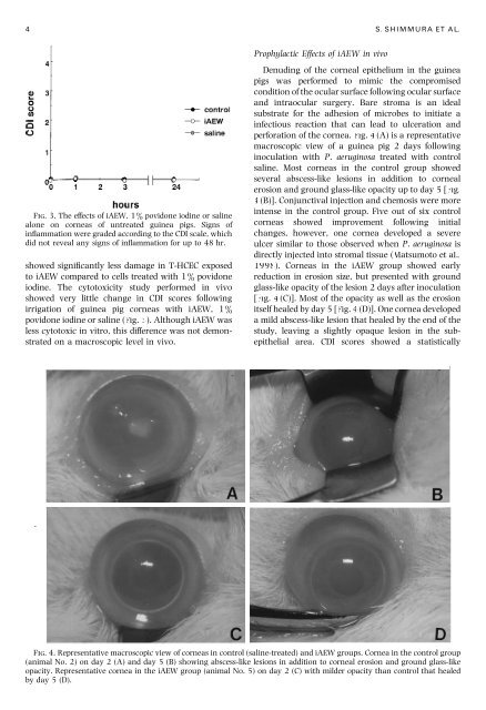
FIG. 4. Representative macroscopic view <strong>of</strong> corneas <strong>in</strong> control (sal<strong>in</strong>e-treated) and iAEW groups. Cornea <strong>in</strong> <strong>the</strong> control group<br />
(animal No. 2) on day 2 (A) and day 5 (B) show<strong>in</strong>g abscess-like lesions <strong>in</strong> addition to corneal erosion and ground glass-like<br />
opacity. Representative cornea <strong>in</strong> <strong>the</strong> iAEW group (animal No. 5) on day 2 (C) with milder opacity than control that healed<br />
by day 5 (D).


