Trochanteric flip osteotomy - Punjab Orthopaedic Association
Trochanteric flip osteotomy - Punjab Orthopaedic Association
Trochanteric flip osteotomy - Punjab Orthopaedic Association

You also want an ePaper? Increase the reach of your titles
YUMPU automatically turns print PDFs into web optimized ePapers that Google loves.
Review Article<br />
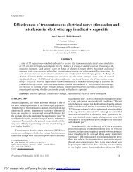
<strong>Trochanteric</strong> <strong>flip</strong> <strong>osteotomy</strong> : Revisited<br />
Vijay Sharma*, Davinder Pal Singh Makkar**, Kamran Farooque***, Amit Kapoor**<br />
* Assistant Professor; ** Senior Resident, ***Associate Professor<br />
Department Of <strong>Orthopaedic</strong>s, AIIMS, Ansari Nagar, New Delhi<br />
ABSTRACT<br />
Visualising high posterior wall fractures and removing any intra-articular fragments has always been a<br />
challenge to the surgeon because the vascularity of femoral head is at great risk due to close relation of<br />
medial circumflex femoral artery( MCFA ), the main vessel supplying the femoral head, with the external<br />
rotators of the hip. Posterior approach with Kocher-Langenbeck incision and surgical dislocation of the hip<br />
after trochanteric <strong>flip</strong> <strong>osteotomy</strong> and anterior capsulotomy preserves the vascularity as external rotators are<br />
not divided, which in turn protect the MCFA. We are reviewing the technique of digastric trochanteric <strong>flip</strong><br />
ostetomy. We had tried to clear its known indications, mentioned in the literature along with the complications<br />
of this procedure.<br />
Keywords: <strong>flip</strong> <strong>osteotomy</strong>, acetabular fracture, femoro-acetabular impingement<br />
INTRODUCTION<br />
There had been always the problem of exposure of high-T and<br />
high posterior wall fractures of the acetabulum.To allow<br />
complete visualization of the femur head and acetabulum, Ganz<br />
et al. described a safe method for surgical dislocation of the<br />
femoral head by means of a trochanteric <strong>flip</strong> ( digastric )<br />
<strong>osteotomy</strong> and anterior capsulotomy, thereby preserving the<br />
posteriorly based femoral head blood supply 1-3 . The technique<br />
is based on the detailed anatomical studies on the blood supply<br />
of the femoral head which is predominantly supplied by medial<br />
circumflex femoral artery( MCFA ) 4,5 . This technique of surgical<br />
dislocation of hip has now been used in various other hip<br />
preserving surgeries.<br />
Indications<br />
The use of surgical dislocation of hip with trochanteric <strong>flip</strong><br />
<strong>osteotomy</strong> has now been extended to various procedures<br />
involving the acetabulum and the head of femur. It has been<br />
used as an addition in standard Kocher-Langenbeck approach<br />
to have a better and direct view of the femur head and the<br />
acetabulum. The following are the main indications of digastric<br />
<strong>flip</strong> <strong>osteotomy</strong> :<br />
Corresponding Author :<br />
Dr Davinder Pal Singh Makkar* , M.B.B.S., M.S.( Ortho )<br />
Senior Resident, Department Of <strong>Orthopaedic</strong>s, AIIMS, Ansari Nagar,<br />
New Delhi-110029, India Fax : 011-26106826<br />
Email: dpsmakkar@yahoo.com, dpsingh.asr@gmail.com<br />
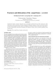
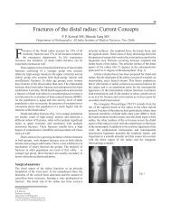
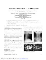
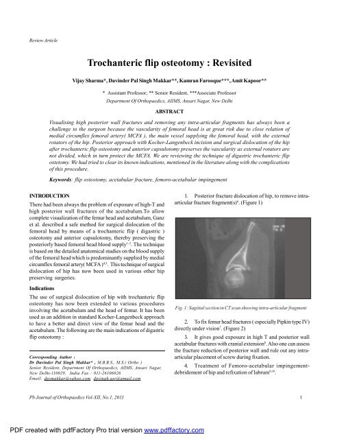
1. Posterior fracture dislocation of hip, to remove intraarticular<br />
fracture fragment(s) 6 . (Figure 1)<br />
Fig. 1: Sagittal section in CT scan showing intra-articular fragment<br />
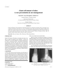
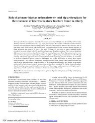
2. To fix femur head fractures ( especially Pipkin type IV)<br />
directly under vision 7 . (Figure 2)<br />
3. It gives good exposure in high T and posterior wall<br />
acetabular fractures with cranial extension 8 . Also one can assess<br />
the fracture reduction of posterior wall and rule out any intraarticular<br />
placement of screw during fixation.<br />
4. Treatment of Femoro-acetabular impingementdebridement<br />
of hip and refixation of labrum 9,10 .<br />
Pb Journal of <strong>Orthopaedic</strong>s Vol-XII, No.1, 2011<br />
1<br />
PDF created with pdfFactory Pro trial version www.pdffactory.com
Sharma et al<br />
Fig. 2: Fracture of head of the femur being reduced under direct<br />
vision after doing the trochanteric <strong>flip</strong> <strong>osteotomy</strong>.<br />
5. This approach can be used in resurfacing hip<br />
arthroplasties as it preserves vascularity and hence<br />
oxygenation to femoral head. Reduced oxygenation is one of<br />
the cause proposed for fracture neck femur after surface<br />
replacements 11 .<br />
Technique :<br />
Patient is placed in lateral decubitus position. One posterior<br />
support is applied over the sacrum while the anterior support<br />
should be over the pubic symphysis. Kocher - Langenbech<br />
incision is given and fascia is incised in the distal part of the<br />
incision while gluteus maximus is split in the proximal part. In<br />
obese patients, Gibson approach 12 is preferred to avoid the<br />
saddle back deformity with the curved incision. The trochanteric<br />
bursa is incised and reflected to expose the surface of the<br />
greater trochanter. The trochanteric branch of deep MCFA is<br />
seen at the upper border of the quadratus femoris and may be<br />
cauterized. The leg is internally rotated( 20 o to 30 o ) to view the<br />
posterior border of gluteus medius and posterosuperior edge<br />
of the greater trochanter. At this point, there is no need to<br />
visualize the piriformis tendon.<br />
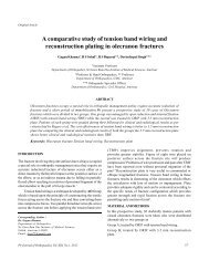
The <strong>osteotomy</strong> site is marked with the cautery, starting<br />
proximally from the posterosuperior edge of the greater<br />
trochanter to the posterior border of the vastus lateralis ridge<br />
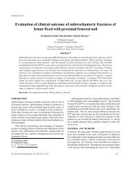
distally. Then, with a thin oscillating saw, <strong>osteotomy</strong> is done,<br />
with its plane in line with the direction of external rotators<br />
( Figure 3). The most important point to keep in mind that the<br />
proximal part of <strong>osteotomy</strong> should start just anterior ( ~ 5mm )<br />
to the most posterior portion of the gluteus medius muscle.<br />
This helps to preserve the external rotators and thus prevent<br />
the injury to the deep part of MCFA. The saw should stop at<br />
the anterior cortex and the osteotome is then used to complete<br />
the <strong>osteotomy</strong>. With this, the anterior cortex is fractured, which<br />
helps in reduction while refixing the trochanter. This <strong>osteotomy</strong><br />
should produce the trochanteric fragment of about 15mm in<br />
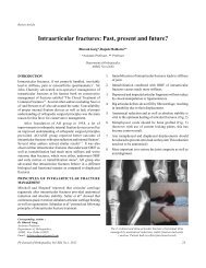
thickness. After releasing the remaining fibres of gluteus medius<br />
from the stable trochanter proximally and vastus lateralis fibres<br />
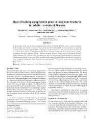
from the femur distally, the fragment is then <strong>flip</strong>ped anteriorly<br />
with the retractor ( Figure 4). This fragment has gluteus medius<br />
attached proximally and vastus lateralis distally.<br />
After retraction, the piriformis tendon and gluteus minimus<br />
muscle is exposed, and the interval within these two is used to<br />
approach the capsule of the hip. Care is required as the sciatic<br />
nerve is beneath the piriformis. The gluteus minimus is sharply<br />
dissected proximally from the underlying capsule, exposing<br />
the anterior and posterosuperior part of the capsule. The leg is<br />
now flexed and externally rotated. A Z-shaped capsulotomy is<br />
then done. The capsule is first incised along the axis of the<br />
femoral neck anterolaterally. Then, anteroinferior capsular<br />
Fig.3 : Osteotomy being done with thin oscillating saw. Note the<br />
direction of the <strong>osteotomy</strong> which is in line of external rotators.<br />
Fig.4 : The trochanteric fragment has been <strong>flip</strong>ped anteriorly along<br />
with vastus lateralis and gluteus medius.<br />
Pb Journal of <strong>Orthopaedic</strong>s Vol-XII, No.1, 2011<br />
2<br />
PDF created with pdfFactory Pro trial version www.pdffactory.com
<strong>Trochanteric</strong> <strong>flip</strong> <strong>osteotomy</strong> : Revisited<br />
incision is made. This incision should always remain anterior<br />
to the lesser trochanter because the MCFA lies posterior and<br />
superior to it. This may injure some branches of lateral circumflex<br />
femoral artery, but there is no risk of vascular compromise as it<br />
does not contribute to the vascularity of the femur head. The<br />
proximal part of the first incision is extended by turning it<br />
posterior and perpendicular along the acetabular rim, thus<br />
completing its Z shape. This part of the incision is limited by<br />
the piriformis tendon. Care is taken to avoid damaging the<br />
labrum. The hip is now dislocated anteriorly by flexing and<br />
externally rotating the leg.<br />
Thus, the vascularity of head is preserved as the external<br />
rotators are not divided which in turn cushions the deep MCFA,<br />
the main supplier to femoral head. A 2.0 mm drill hole made in<br />
the dislocated femoral head can document the preservation of<br />
its blood supply 13 .Bleeding from the drill hole supports that<br />
the vascularity has been preserved. Most of the times, the<br />
ligamentum teres needs to be incised to achieve full dislocation.<br />
This can be done with no harm, as it has insignificant<br />
contribution to femoral head blood supply in an adult.<br />
After dislocation of femoral head, we can have 360 0 view<br />
of acetabulum. Femoral head is directly visualised and so femur<br />
head fractures can be easily fixed. Furthermore, movements of<br />
head within acetabulum can be analysed for any<br />
femoroacetabular impingement.<br />
For closure of the capsule, a running suture can be used<br />
and any tension in it is avoided because this can stretch the<br />
retinaculum and can compromised the perfusion of the femoral<br />
head 2 . The <strong>flip</strong>ped part of trochanter is put back to its original<br />
position and refixed with two or three 4.5mm cortical or 6.5 mm<br />
cancellous screws with washer. Screws should be aimed towards<br />
the lesser trochanter( Fig.5, 6).<br />
Fig.5 : The trochanteric fragment has been put back and fixed with<br />
two 6.5mm cancellous screws used along with washers<br />
Fig.6 : Note that the direction of screws are towards the lesser<br />
trochanter.<br />
Post operatively, active abduction of hip is not allowed till 6-8<br />
weeks which is the required time for union of trochanteric<br />
fragment. Also, flexion beyond 70 0 is not permitted till 8 weeks.<br />
Passive range of motion at hip can be started a week after<br />
surgery.<br />
Complications<br />
If <strong>osteotomy</strong> is done taking care of landmarks, the complications<br />
should not happen. Sciatic nerve is in close proximity to external<br />
rotators and is beneath piriformis, and hence, injury can occur,<br />
if not taken care. Another issue is of trochanteric non union<br />
which is also very rare as the trochanteric fragment is stabilised<br />
by the digastrics muscle. Ganz et al 1 reported only 3 cases of<br />
failure of trochanteric fixation out of 213 procedures.<br />
Heterotopic ossification is reported but the incidence is<br />
less than when only Kocher-Langenbeck approach is done.<br />
This is because gluteus minimus is sharply dissected under<br />
vision and also overzealous retraction of gluteus medius is not<br />
required during visualising acetabular fractures.<br />
CONCLUSION<br />
We conclude that the trochanteric <strong>flip</strong> <strong>osteotomy</strong> is very useful<br />
procedure to assess and fix certain acetabular fractures and<br />
femoral head fractures more accurately. Furthermore, one can<br />
see the labral and cartilage pathologies directly, which are<br />
usually not seen in imaging techniques. As the vascularity of<br />
the head of femur is preserved, hip preserving surgeries can<br />
easily be accomplished.<br />
REFERENCES<br />
1. Ganz R, Gill TJ, Gautier E, Ganz K, Krugel N, Berlemann U. Surgical<br />
dislocation of the adult hip. A technique with full access to the<br />
Pb Journal of <strong>Orthopaedic</strong>s Vol-XII, No.1, 2011<br />
3<br />
PDF created with pdfFactory Pro trial version www.pdffactory.com
Sharma et al<br />
femoral head and acetabulum without the risk of avascular necrosis.<br />
J Bone Joint Surg Br. 2001;83:1119-24.<br />
2. Notzli HP, Siebenrock KA, Hempfing A, Ramseier LE, Ganz R.<br />
Perfusion of the femoral head during surgical dislocation of the hip.<br />
Monitoring by laser Doppler flowmetry. J Bone Joint Surg Br.<br />
2002;84:300-4.<br />
3. Siebenrock KA, Gautier E, Woo AK, Ganz R. Surgical dislocation of<br />
the femoral head for joint debridement and accurate reduction of<br />
fractures of the acetabulum. J Orthop Trauma. 2002;16:543-52.<br />
4. Gautier E, Ganz K, Krugel N, Gill T, Ganz R. Anatomy of the medial<br />
femoral circumûex artery and its surgical implications. J Bone Joint<br />
Surg Br. 2000;82-B:679-83<br />
5. Crock HV. Anatomy of the medial femoral circumXex artery and<br />
its surgical implications. J Bone Joint Surg Br. 2001; 83:149–150<br />
6. Naranje S, Shamshery P , Yadav CS, Gupta V, Nag HL. Digastric<br />
trochanteric <strong>flip</strong> <strong>osteotomy</strong> and surgical dislocation of hip in the<br />
management of acetabular fractures. Arch Orthop Trauma Surg.<br />
2010;130:93–101<br />
7. Droll KP, Broekhuyse H, O’Brien P. Fracture of the Femoral Head.<br />
J Am Acad Orthop Surg 2007;15:716-727<br />
8. Siebenrock KA, Gautier E , Ziran BH, Ganz R. <strong>Trochanteric</strong><br />
Flip Osteotomy for Cranial Extension and Muscle Protection in<br />
Acetabular Fracture Fixation Using a Kocher-Langenbeck<br />
Approach. J Orthop Trauma. 2006; 20(1): S52-S56<br />
9. Espinosa N, Beck M, Rothenfluh DA, Ganz R, Leunig M. Treatment<br />
of Femoro-Acetabular Impingement: Preliminary Results of Labral<br />
Refixation. J Bone Joint Surg Am. 2007;89:36-53.<br />
10. Christopher L. Peters and Jill A. Erickson. Treatment of<br />
Femoro-Acetabular Impingement with Surgical Dislocation and<br />
Débridement in Young Adults. J Bone Joint Surg Am. 2006;88:<br />
1735-1741.<br />
11. Steffen RT, Fern D, Norton M, Murray DW, Gill HS. Femoral<br />
Oxygenation During Hip Resurfacing Through the <strong>Trochanteric</strong><br />
Flip Approach. Clin Orthop Relat Res.2009;467:934–939.<br />
12. Gibson A. Posterior exposure of the hip joint. J Bone Joint Surg Br.<br />
1950;32-B:183-6.<br />
13. Gill TJ, Sledge JB, Ekkernkamp A, Ganz R. Intraoperative assessment<br />
of femoral head vascularity after femoral neck fracture. J Orthop<br />
Trauma 1998;12:474-8<br />
Pb Journal of <strong>Orthopaedic</strong>s Vol-XII, No.1, 2011<br />
4<br />
PDF created with pdfFactory Pro trial version www.pdffactory.com