What is synchrotron radiation?
What is synchrotron radiation?
What is synchrotron radiation?

Create successful ePaper yourself
Turn your PDF publications into a flip-book with our unique Google optimized e-Paper software.
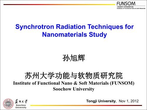
Synchrotron Radiation Techniques for<br />
Nanomaterials Study<br />
孙 旭 辉<br />
苏 州 大 学 功 能 与 软 物 质 研 究 院<br />
Institute of Functional Nano & Soft Materials (FUNSOM)<br />
Soochow University<br />
Tongji University, Nov 1, 2012
• SR techniques and its advantages in nano studies<br />
• Nanomaterials study by SR techniques<br />
o Electronic structure of nanomaterials (XAS, XES, STXM)<br />
o Optical property of nanomaterials (XEOL)<br />
o Crystal structure of nanomaterials (XRD)<br />
o Development of in-situ XAS techniques<br />
• Summary
<strong>What</strong> <strong>is</strong> <strong>synchrotron</strong> <strong>radiation</strong><br />
When an electron traveling at a speed close to the speed of light<br />
in an orbit, it emits a continuum of electromagnetic <strong>radiation</strong><br />
tangential to the orbit, which <strong>is</strong> Synchrotron light.<br />
3
Then they pass into the<br />
booster ring where they are<br />
accelerated to 99.9999% of<br />
the speed of light<br />
Electrons are<br />
generated here<br />
Creating the light<br />
And are finally transferred into the<br />
storage ring<br />
And initially<br />
accelerated in<br />
the LINAC<br />
Scheme of <strong>synchrotron</strong> <strong>radiation</strong> facility
NSRFII<br />
Diamond Light Source<br />
Shanghai Synchrotron Radiation Facility
Interior of the Canadian Light Source
Synchrotron Radiation Properties<br />
• Tunability (IR to hard x-rays, element specific)<br />
• Brightness (highly collimated, micro/nano beam)<br />
• Polarization (linear, circular, tunable, dichro<strong>is</strong>m)<br />
• Time structure (short pulse, dynamics)
Techniques of SR<br />
The fundamental parameters necessary for perception of physical world:<br />
• Energy (spectroscopy, state of matter)<br />
• Momentum (scattering)<br />
• Position (imaging, spatial d<strong>is</strong>tribution)<br />
• Time (dynamics)<br />
numerous techniques of SR<br />
Spectroscopy Scattering Imaging<br />
01 Low-Energy Spectroscopy 05 Hard X-Ray Diffraction 09 Hard X-Ray Imaging<br />
02 Soft X-Ray Spectroscopy 06 Macromolecular<br />
10 Soft X-Ray Imaging<br />
Crystallography<br />
03 Hard X-Ray Spectroscopy 07 Hard X-Ray Scattering 11 Infrared Imaging<br />
04 Optics/Calibration/Metrology 08 Soft X-Ray Scattering 12 Lithography
A Single Storage Ring Serves Many Scientific User Groups
Synchrotron Radiation Techniques at SSRF<br />
BL08U-A – Soft X-ray Spectromicroscopy (STXM)<br />
BL08U-B – X-ray Interference Lithography (XIL)<br />
BL14W – X-ray Absorption Fine Structure (XAFS)<br />
BL14B – X-ray Diffraction (XRD)<br />
BL15U – Hard X-ray Micro-focus<br />
BL16B – Small Angle X-ray Scattering (SAXS)<br />
BL10W – X-ray Imaging and Biomedical Application<br />
BL17U – Macromolecular Crystallography (MC)
<strong>What</strong> <strong>is</strong> X-ray absorption fine structures (XAFS) <br />
• As core electron <strong>is</strong> excited with hv the threshold<br />
(E o ), it <strong>is</strong> excited to a final state defined by the<br />
chemical environment, which modulates the<br />
absorption coefficient relative to that of a free<br />
atom. Th<strong>is</strong> modulation <strong>is</strong> known the XAFS,<br />
• XAFS contains all the information about the local<br />
structure and bonding of the absorbing atom<br />
• XAFS study requires a tunable X-ray source –<br />
<strong>synchrotron</strong> <strong>radiation</strong><br />
11
t<br />
<strong>What</strong> does XAFS look like<br />
Near edge<br />
XANES<br />
(NEXAFS)<br />
1s to LUMO<br />
(t3)<br />
EXAFS<br />
Si(CH3)4<br />
C<br />
Si<br />
Si K-edge<br />
12
Synchrotron Probe<br />
Spectroscopy<br />
Structure<br />
Valence<br />
Electrons<br />
Core Electrons<br />
Photon<br />
Energy<br />
Wavelength<br />
10eV 100eV 1keV 10keV<br />
100nm 10nm<br />
1nm 1Å<br />
Lithography<br />
Nanostructures<br />
Proteomics<br />
Protein<br />
Crystallography
Advantage of SR Techniques in Nano Study<br />
Comprehensive techniques (electron, ion,<br />
photon), various information<br />
New techniques: XAFS,<br />
XEOL, XES, RIXS,<br />
STXM, etc<br />
Individual nanomaterial<br />
characterization<br />
SR<br />
Techniques<br />
High Signal/No<strong>is</strong>e<br />
Ambient Conditions<br />
In-situ, dynamic<br />
Depth profile with SR X-ray<br />
(Surface & Interface)<br />
Less sample damage,<br />
reliable data
Absorption Techniques for Nano Studies<br />
• XAFS (X-ray Absorption Fine Structures)<br />
Measurement: Absorption coefficient above an edge<br />
Information: Conduction band, Local structure and<br />
bonding<br />
• XES (X-ray Em<strong>is</strong>sion Spectroscopy)<br />
Measurement: X-rays in, x-rays out<br />
Information: Valence band electron d<strong>is</strong>tribution<br />
• XEOL (X-ray Excited Optical Luminescence)<br />
Measurement: X-rays in, optical photons out<br />
Information: origin of luminescence, time dependent<br />
events (dynamics)
XANES<br />
XAFS, XEOL and XES<br />
E<br />
(III)<br />
PLY<br />
XEOL<br />
hv ex<br />
(I)<br />
Abs.<br />
CB<br />
VB<br />
(II)<br />
E F<br />
core<br />
Mono<br />
hv op<br />
hv f<br />
FLY<br />
Mono<br />
Channel<br />
Plate<br />
200 850<br />
Wavelength (nm)<br />
VB<br />
XES<br />
Energy (eV) E F
XANES and XES of Zn nanostructures<br />
500 nm
XEOL - Conversion of X-ray energy into optical em<strong>is</strong>sion<br />
X-ray phosphor<br />
thermalization<br />
hv >E o<br />
Recombination via<br />
exciton<br />
UV<br />
v<strong>is</strong>.<br />
Elliot & Gibson<br />
Solid State<br />
Physics Haper &<br />
Row, 1974<br />
o<br />
o<br />
hv ~ E o<br />
Core level<br />
Recombination via bound exciton,<br />
impurity and defect states
XEOL Techniques<br />
Energy Domain<br />
XEOL: Luminescence induced by selected excitation<br />
photon energy (usually across an absorption edge)<br />
Optical XAFS: Absorption across an edge monitored with<br />
the optical signal (photoluminescence yield )<br />
Time Domain<br />
Lifetime: Synchrotron pulse as the start/stop<br />
Time-gated XEOL: Luminescence within a selected time<br />
window between pulses<br />
Time-gated Optical XAFS: Photoluminescence yield<br />
with a time window
TRXEOL<br />
Photon Energy (eV)<br />
CLS-July 16, 2012<br />
Optical XAFS<br />
(time & wavelength selected)<br />
imaging
Normalized Intensity<br />
CdSe -Si Heteronanostructure<br />
Si<br />
CdSe<br />
CdSe-Si 1100 eV<br />
0.007<br />
0.006<br />
total<br />
Si CdSe<br />
0.005<br />
0.004<br />
0.003<br />
0.002<br />
slow<br />
(20-150 ns)<br />
fast<br />
(0-20 ns)<br />
0.001<br />
0.000<br />
300 400 500 600 700 800 900<br />
Wavelength (eV)<br />
hv = 1100 eV<br />
J. Phys. Chem. C, 111, 8475 (2007)
CdSe-Si Heterostructure: Se L 3,2 - edge<br />
Appl. Phys. Lett., 89, 243102 (2006)
• Advantages of SR techniques in nano studies<br />
• Nanomaterials study by SR techniques<br />
o Electronic structure of nanomaterials (XAS, XES, STXM)<br />
o Optical property of nanomaterials (XEOL)<br />
o Crystal structure of nanomaterials (XRD)<br />
o Development of in-situ XAS techniques<br />
• Summary
Electronic structure of nanomaterials<br />
by XAS, XES, and STXM
Probing solid state N-doping in graphene by XAS<br />
C K-edge<br />
GO: Graphene oxide<br />
GRA-x: Graphene-urea annealed<br />
at x degree<br />
A: C-C π* excitation<br />
B1: Amino C-N<br />
B2: C-O or C=O<br />
B3: Urea C=O π* excitation<br />
C: C-C σ* excitation<br />
Carbon 2012, 50, 321–341.
N K-edge<br />
GRA-x: Graphene-urea annealed<br />
at x degree<br />
N1: Pyridinic C-N<br />
N2: Amino C-N<br />
N3: Graphitic C-N<br />
N4: Urea C-N<br />
N5: C-N σ* excitation
Intensity(a.u.)<br />
Unpubl<strong>is</strong>hed<br />
Carbon nanocages (CNCs) studied by XAS and XES<br />
P-CNC<br />
N-CNC<br />
Pure CNCs<br />
282<br />
284<br />
286<br />
Photon Energy(eV)<br />
288<br />
290
Electronic Structure of Graphdiyne Probed by XAS and STXM<br />
JPCC, submitted
STXM and XANES study of N-doped CNTs sealed with N 2 gas<br />
J. Appl. Phys. 111, 124318 (2012)
Optical property of nanomaterials by XEOL
XEOL Study of PL Mechan<strong>is</strong>m of Nanostructures<br />
PL of Single Crystalline GeO2 Nanowires<br />
Ge:O≈1:1.8<br />
J. Phys. Chem. C, 115, 11420 (2011)
XAFS and XEOL of GeO2 Nanowires
Crystal structure of nanomaterials by XRD
SR-XRD study of In 2 Se 3 nanowires<br />
In:Se≈2:3<br />
TEM image and corresponding EDS spectra of an individual In 2 Se 3<br />
nanowire. Scare bar <strong>is</strong> 100 nm<br />
Appl. Phys. Lett., 89 (2006) 233121
SR-XRD of In 2 Se 3 nanowires<br />
JCPDS PDF 341279<br />
J. Mater. Chem., 21 (2011) 6944
In2Se3 Nanowires: HR-TEM Image<br />
(a)<br />
(b)<br />
α-phase<br />
κ-phase
In-situ phase transition of In 2 Se 3 nanowires
Electrical Res<strong>is</strong>tances of In 2 Se 3 Nanowires<br />
I-V character<strong>is</strong>tics of two devices fabricated with<br />
nanowires of as- synthesized and after annealing
Summary<br />
Synchrotron <strong>radiation</strong> techniques including absorption<br />
spectroscopy such as XAFS, (XANES and EXFAS), deexcitation<br />
spectroscopy such as XEOL, TRXEOL, and XES<br />
(RIXS), XRD and spectro-microscoy (STXM) have<br />
provided unprecedented research opportunities in probing<br />
the structure and electronic properties of nanomaterials.
Acknowledgements:<br />
Soochow-Western Joint Center for Synchrotron Radiation Research<br />
SR Team @ FUNSOM, Soochow University<br />
Prof. Suidong Wang<br />
Prof. Youyong Li<br />
Dr. Jun Zhong<br />
Dr. Xiujuan Zhang<br />
Prof. Steffen Duhm<br />
Dr. Keith Gilmore<br />
University of Western Ontario & Canadian Light Source<br />
Profs. T. K. Sham, X. L. Sun, Y. Song<br />
Dr. Yongfeng Hu, Tom Reger<br />
Shanghai Synchrotron Radiation Facility<br />
Dr. Xingtai Zhou<br />
Dr. Renzhong Tai<br />
Dr. Zheng Jiang<br />
Dr. Wen Wen<br />
Advanced Light Source, Berkeley Lawrence<br />
National Lab<br />
Dr. Jinghua Guo, Dr. Zhi Liu, Dr. Xiaosong Liu<br />
National Synchrotron Radiation Lab (Hefei)<br />
Prof. Ziyu Wu,Dr. Zhiyun Pan
Thank you for your attention!
Acknowledgements: