Saprolegnia - The iLumina Digital Library
Saprolegnia - The iLumina Digital Library
Saprolegnia - The iLumina Digital Library

Create successful ePaper yourself
Turn your PDF publications into a flip-book with our unique Google optimized e-Paper software.
internally; 40-220 x 18-28 µm. Spores dimorphic; discharge and behavior saprolegnoid,<br />
rarely aplanoid; primary spore cysts 10-14 µm in diameter. Gemmae very sparse;<br />
clavate or obpyriform; terminal, single. Oogonia spherical, obpyriform, napiform, or<br />
subspherical, rarely dolioform; lateral, occasionally terminal, rarely intercalary; (32-) 46-<br />
60 (-107) µm in diameter. Oogonial wall pitted or unpitted; smooth. Oogonial stalks<br />
( 1 / 4 -) 1-2 (-3 1 / 2 ) times the diameter of the oogonium, in length; stout; straight, curved,<br />
or bent; unbranched or with one or more short, lateral branches. Oospores centric;<br />
spherical, often nearly filling the oogonium; (1-) 6-16 (-28) per oogonium; (18-) 23-26 (-<br />
30) µm in diameter; at germination producing a germ hypha which may or may not<br />
bear a terminal sporangium. Antheridial branches androgynous or monoclinous, rarely<br />
diclinous; slender, usually contorted, twisted, or irregular and sparingly branched;<br />
persisting. Antheridial cells simple, generally tubular, occasionally clavate or slightly<br />
irregular; persisting; laterally appressed, very rarely attached apically; fertilization<br />
tubes present, not persisting.<br />
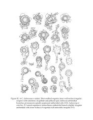
<strong>Saprolegnia</strong> glomerata is readily recognizable by the short, branched or<br />
unbranched, contorted, lateral (and often clustered) hyphal extensions (Fig. 96 G, I).<br />
Secondary characters of recognition are the contorted, branched antheridial filaments<br />
(Fig. 96 G, H, J) and the short, lateral evaginations on many of the oogonial stalks (Fig.<br />
96 H). In S. litoralis, the hypha immediately below a terminal oogonium, may bear<br />
short, lateral branches (Fig. 96 B) as does S. glomerata, but in the former, the hypha is<br />
usually swollen at its juncture with the oogonial septum (Fig. 96 A, B). In any case, the<br />
oospores in S. litoralis are occasionally subcentric, a condition not known to occur in S.<br />
glomerata.<br />
Although A. Lund recognized as early as 1934 that Tiesenhausen’s (loc. cit.)<br />
variety glomerata deserved specific rank, not all investigators followed (or were aware<br />
of) this disposition. For example, T. Ito (1944) retained the variety of glomerata and<br />
reduced <strong>Saprolegnia</strong> furcata and S. glomerata (Tiesenhausen) Lund to synonymy with it.<br />
Cejp (1959a) listed both Lund’s species and S. monoica as synonyms of Tiesenhausen’s<br />
variety. In addition, S. glomerata has been misidentified by some mycologists. Apinis<br />
(1929a, b) believed that Maurizio’s (1899) S. furcata was the same as Tiesenhausen’s<br />
variety glomerata. <strong>The</strong> specimens recorded by T. W. Johnson (1950b:399) as the variety<br />
glomerata were misidentified; he had collected forms of S. ferax. <strong>The</strong> S. monoica reported<br />
by Humphrey (1893) was not, as Coker (1923) maintained, identifiable as S. monoica var.<br />
glomerata; Humphrey’s material was very likely representative of S. ferax.<br />
CONFIRMED RECORDS: -- BRITISH ISLES: R.A. Couch (1951:161, p1s. 41-45);<br />
Forbes (1935a:226). CZECHOSLOVAKIA: Cejp (1959a:231, fig. 85 a-f). DENMARK:<br />
Lund (loc. cit.). GERMANY: Richter (1937:237). ICELAND: Howard et al. (1970: figs. 27,<br />
28). INDIA: Thakur Ji (1970:182, figs. 15-18). JAPAN: T. Ito (1942:124, fig. l e-g; 1944:52).<br />
LATVIA: Apinis (1929a: 213, text fig. 1). NETHERLANDS: Beverwijk (1948:232, fig. 3).<br />
POLAND: Szwanke (1938:8, pl. 2, figs. 10, 11; pl. 3, figs. 1-5). SWITZERLAND:<br />
Tiesenhausen (loc. cit.). UNITED STATES: Beneke (1948b:36); T. W. Johnson (1956a:186);<br />
618