Saprolegnia - The iLumina Digital Library
Saprolegnia - The iLumina Digital Library
Saprolegnia - The iLumina Digital Library

Create successful ePaper yourself
Turn your PDF publications into a flip-book with our unique Google optimized e-Paper software.
CHAPTER 38<br />
SAPROLEGNIA Nees von Esenbeck<br />
Nova Acta Phys.-Med. Acad. Caes. Leop.-Carol. Nat. Cur. 11:513. 1823<br />
Diplanes Leitgeb, Jahrb. Wiss. Bot. 7:385. 1869-70.<br />
Isoachlya Kauffman, Amer. J. Bot. 8:231. 1921.<br />
Cladolegnia Johannes, Feddes Repert. Spec. Nov. Regni. Veg. p. 211. 1955.<br />
Scoliolegnia Dick, J. Linn. Soc. Bot. 62:255. 1969.<br />
Monoecious. Sporangia fusiform, clavate, cylindrical, or irregular; renewed by<br />
internal proliferation, and also occasionally sympodially, cymosely, or in a basipetalous<br />
fashion. Spores dimorphic; sometimes polyplanetic; primary spores swimming away<br />
immediately upon release; in older cultures, and regularly in some species, discharge<br />
may be aplanoid or dictyuchoid. Gemmae present or absent. Oogonia lateral, terminal,<br />
intercalary, or sessile; variously shaped, but predominantly spherical or obpyriform.<br />
Oogonial wall pitted or unpitted; smooth or ornamented. Oogonial stalks of various<br />
lengths; usually simple. Oospores centric, subcentric, subeccentric, or eccentric; one to<br />
many; germinating to form a new mycelium, or to produce a germ hypha bearing a<br />
terminal sporangium. Antheridial branches, when present, androgynous, monoclinous,<br />
diclinous, or hypogynous. Antheridial cells predominantly tubular or clavate; attached<br />
laterally or apically or in a digitate manner to the oogonial wall.<br />
Type species: <strong>Saprolegnia</strong> ferax (Gruith.) Thuret, Ann. Sci. Nat. Bot. (3 e sér.) 14:229<br />
et sqq., pl. 22. 1850.<br />
Seymour (1970), reduced Isoachlya and Cladolegnia to synonymy with<br />
<strong>Saprolegnia</strong>, and the justification for that nomenclatural change need not be repeated<br />
here. Dick (1969b) seems to have retained Isoachlya Kauffman as a valid, genus, but (if<br />
we interpret his account correctly) did not accept Cladolegnia. Although the reference to<br />
Leitgeb’s Diplanes is usually cited as his 1868 publication, he did not formally describe a<br />
species to accompany the generic name. <strong>The</strong>refore, his paper of 1869-70 is taken as the<br />
proper original record of this genus.<br />
In 1969(a), Dick redefined <strong>Saprolegnia</strong> asterophora, a, species which he had<br />
commented upon earlier (1960c), and described (but did not name) two additional<br />
forms of the species with ornamented oogonia. <strong>The</strong>se three taxonomic entities he<br />
regarded (Dick, 1969a:255) as “...segregate species within an aggregate...”, raised the<br />
two forms to specific rank, and placed them in the new genus Scoliolegnia. In 1979,<br />
Mil’ko published a synopsis of the systematics of the genus <strong>Saprolegnia</strong>, and reduced<br />
Scoliolegnia to synonymy with the earlier taxon. As we agree fully with Mil’ko’s<br />
decision and basic premise, but recognize that his paper is not in translation, we are<br />
including our own analysis of the status of Dick’s genus. <strong>The</strong> priority for the<br />
nomenclatural changes in the species rests with Mil’ko.<br />
599
According to Dick (1969a), the delicate, flexuous mycelium and the “gritty”<br />
granular aspect of the hyphae were characteristic of the three species assigned to<br />
Scoliolegnia. He remarked that this mycelial texture contrasted with the stout, straight<br />
hyphae prominent in species of <strong>Saprolegnia</strong> and Isoachlya. <strong>The</strong>se characters alone,<br />
however, did not form the sole basis on which Scoliolegnia was established.<br />
Dick (1969a) concluded that the genera of <strong>Saprolegnia</strong>ceae were defined on too<br />
few characters. Accordingly, he compared and contrasted the species of Scoliolegnia on<br />
twenty structural criteria. <strong>The</strong> descriptive matter provided by Dick (particularly the<br />
characterization in Table 1 of his 1969a account), and the accompanying illustrations<br />
permit but one conclusion: there are essentially only two features that could possibly be<br />
used to distinguish Scoliolegnia species from members of <strong>Saprolegnia</strong>. <strong>The</strong>se are, for<br />
Scoliolegnia, the flexuous hyphae and the flared aspect of the rather prominent exit<br />
papilla after it has opened terminally at spore emergence. <strong>The</strong> first characteristic<br />
(mycelial texture) is, in fact, one by which de Bary’s <strong>Saprolegnia</strong> asterophora (one of the<br />
species assigned to Scoliolegnia) usually can be recognized in water culture; Dick’s two<br />
new species of Scoliolegnia also at times may be made out in gross culture on this<br />
feature. <strong>The</strong> prominently flared, opened exit papilla is not a consistent characteristic of<br />
<strong>Saprolegnia</strong> asterophora, and other species of <strong>Saprolegnia</strong> sometimes produce similar ones.<br />
Other structural features that Dick used to circumscribe his species, such as mean<br />
diameters of the exit papillae and of the oogonial stalks, are in our judgment too<br />
variable to be of value at the species level, to say nothing of the generic level. In any<br />
event, Dick did not mention the conspicuous nature of the exit papilla remnants in the<br />
formal description of Scoliolegnia.<br />
We find commendable Dick’s (1969a) argument that genera should be defined on<br />
the basis of several characters, but his contention falls short with respect to Scoliolegnia,<br />
since that genus rests in our view on only two criteria. In what we regard as a<br />
fundamental morphological characteristic essential for generic delimitation -- spore<br />
discharge pattern and sporangial renewal -- Scoliolegnia species cannot be distinguished<br />
from those of <strong>Saprolegnia</strong>. We interpret Dick’s argument favoring the recognition of<br />
Scoliolegnia basically to be a proposition that generic determinations in the<br />
<strong>Saprolegnia</strong>ceae may be made independent of reliance on sporangial renewal method.<br />
Admitting to <strong>Saprolegnia</strong> species with flexuous hyphae does not at all distort that<br />
genus into an unrecognizable complex. <strong>The</strong> same may be said for including species<br />
with subeccentric oospores. Indeed the concept of <strong>Saprolegnia</strong> is more firmly and<br />
competently established by the admission of Dick’s Scoliolegnia species.<br />
As with Achlya, <strong>Saprolegnia</strong> has been variously divided into infrageneric units.<br />
Schröter (1893) accepted three subgenera: Eusaprolegnia (the S. ferax group), Desmolegnia<br />
(the S. monoica complex), and Astrolegnia. Coker (1923) recognized Eusaprolegnia with<br />
four “Groups” -- Diclina, Ferax, Hypogyna, and Torulosa -- and Pseudosaprolegnia, with the<br />
single species, S. asterophora. In 1959(a), Cejp raised Coker’s Hypogyna and Diclina<br />
groups to section status (in the subgenus Eusaprolegnia, and did likewise with the Ferax<br />
group, which he attributed to de Bary. In addition, Cejp (1959a:251) proposed a fourth<br />
taxon in Eusaprolegnia, the Section Terrestres. Three subgenera of <strong>Saprolegnia</strong> --<br />
600
Asterophorae (ornamented oogonia), Leiothecae (smooth oogonia), and Agamae (lacking<br />
oogonia) -- were established by Naumov, in 1954. <strong>The</strong> subgenus Leiothecae was further<br />
divided into five sections, Hypogynae, Diclinae, Monoicae, Ferax, and Moniliferae. We see<br />
no reason (other than convenience) for retaining any subgeneric distinction, and are<br />
following the precedent set by Seymour (1970).<br />
<strong>The</strong> taxonomic monograph by Seymour (1970) provided a useful new account of<br />
<strong>Saprolegnia</strong> in that it solved some of the most troublesome problems in the genus. His<br />
unprecedented treatment of the S. ferax complex has been held to be too broadly<br />
encompassing (Dick, 1973); a more restrictive concept of <strong>Saprolegnia</strong> is apparent in<br />
Dick’s (1973) comments on the genus, and also is reflected in his treatment of<br />
Scoliolegnia. Neish and Green (1976) saw in Dick’s circumscription the recognition of<br />
twelve “morphospecies” [attributed to Scudder (1974), but first defined by Cain<br />
(1953:82) as a group of specimens sufficiently different morphologically from the most<br />
closely related forms known as to be given a specific name]. By experimental analyses,<br />
Neish and Green found that the species of <strong>Saprolegnia</strong> assayed for DNA base<br />
composition data could not be separated on the basis of this parameter. <strong>The</strong>y then<br />
suggested that the genus <strong>Saprolegnia</strong> appeared to be a homogeneous taxon, and any<br />
isolate whose DNA base composition fell outside the range of 55.5-60.5% GC should be<br />
excluded from this taxon. Miller and Ristanović (1975) commented upon the<br />
inadequacy and unreliability of the taxonomic markers customarily used in identifying<br />
species of <strong>Saprolegnia</strong>. Brief historical accounts of the genus <strong>Saprolegnia</strong> have been<br />
published by Dittrich (1956), and Seymour (1970).<br />
Key to the Species of <strong>Saprolegnia</strong><br />
1. Oogonial wall consistently ornamented . . . . . . . . . . . . . . . . . . . . . . . . . . . . . . . . . . . . . . . 2<br />
1. Oogonial wall predominantly smooth on outer<br />
surface, but if provided with projections these<br />
occur on few oogonia and are often inconspicuous . . . . . . . . . . . . . . . . . . . . . . . . . . . . . 5<br />
2. Ornamentations predominantly truncate . . . . . . . . . . . . . . . . . S. truncata (p. 604)<br />
2. Ornamentations predominantly narrowly<br />
or broadly papillate, sometimes cylindrical,<br />
and straight or curved, or bifurcate; some<br />
oogonia provided with an undulant or wavy wall . . . . . . . . . . . . . . . . . . . . . . . . 3<br />
3. Oospores subcentric . . . . . . . . . . . . . . . . . . . . . . . . . . . . . . . . . . . . . . .S. asterophora (p. 605)<br />
3. Oospores subeccentric . . . . . . . . . . . . . . . . . . . . . . . . . . . . . . . . . . . . . . . . . . . . . . . . . . . . . . 4<br />
4. Antheridial apparatus present; wall<br />
ornamentations dense and prominent . . . . . . . . . . . . . . . . S. subeccentrica (p. 607)<br />
4. Antheridial apparatus lacking; wall<br />
ornamentations sometimes sparse . . . . . . . . . . . . . . . . . . . . S. blelhamensis (p. 608)<br />
5. Oospores centric, subcentric, or frequently aborting<br />
or not maturing . . . . . . . . . . . . . . . . . . . . . . . . . . . . . . . . . . . . . . . . . . . . . . . . . . . . . . . . . . . . 6<br />
5. Oospores eccentric . . . . . . . . . . . . . . . . . . . . . . . . . . . . . . . . . . . . . . . . . . . . . . . . . . . . . . . . 21<br />
601
6. Oospores centric or subcentric, only rarely,<br />
if ever, aborting or failing to mature . . . . . . . . . . . . . . . . . . . . . . . . . . . . . . . . . . . . 7<br />
6. Oospores subcentric, but frequently<br />
aborting or not maturing . . . . . . . . . . . . . . . . . . . . . . . . . . . . . . S. australis (p. 609)<br />
7. Oospores predominantly fewer than three per oogonium . . . . . . . . . . . . . . . . . . . . . . 19<br />
7. Oospores predominantly more than three per oogonium . . . . . . . . . . . . . . . . . . . . . . . . 8<br />
8. Antheridial apparatus hypogynous . . . . . . . . . . . . . . . . . . . . . . . . . . . . . . . . . . . . . 9<br />
8. Antheridial apparatus androgynous,<br />
monoclinous, or diclinous, or absent . . . . . . . . . . . . . . . . . . . . . . . . . . . . . . . . . . . 10<br />
9. Oogonia predominantly obpyriform, spherical,<br />
or napiform . . . . . . . . . . . . . . . . . . . . . . . . . . . . . . . . . . . . . . . . . . . . . . . . S. hypogyna (p.610)<br />
9. Oogonia cylindrical, lobed, branched, irregular,<br />
or subspherical . . . . . . . . . . . . . . . . . . . . . . . . . . . . . . . . . . . . . . . . . . . S. irregularis (p. 612)<br />
10. Oogonial stalks short, their length usually<br />
less than the oogonial diameter . . . . . . . . . . . . . . . . . . . . . . . . . . . . . . . . . . . . . . .11<br />
10. Oogonial stalks predominantly as long<br />
as the oogonial diameter, in length, or longer . . . . . . . . . . . . . . . . . . . . . . . . . . 12<br />
11. Antheridial branches predominantly androgynous<br />
and originating near the oogonial septum;<br />
antheridial cells very long, curved, and tubular . . . . . . . . . . . . . . . . . . S. turfosa (p. 613)<br />
11. Antheridial branches predominantly monoclinous;<br />
antheridial cells short, clavate, or cylindrical . . . . . . . . . . . . . . . . . . S. uliginosa (p. 615)<br />
12. Oogonial stalks generally 1-7 times<br />
coiled, sometimes strongly recurved and<br />
oogonium pendulous . . . . . . . . . . . . . . . . . . . . . . . . . . . . . . . . . . S. furcata (p. 616)<br />
12. Oogonial stalks not coiled . . . . . . . . . . . . . . . . . . . . . . . . . . . . . . . . . . . . . . . . . . . .13<br />
13. Oogonial stalks and antheridial branches<br />
irregular, contorted and usually provided<br />
with short, lateral, cylindrical branches;<br />
hyphae developing clusters of short, twig-like<br />
branches . . . . . . . . . . . . . . . . . . . . . . . . . . . . . . . . . . . . . . . . . . . . . . . . . S. glomerata (p. 617)<br />
13. Oogonial stalks and antheridial branches seldom<br />
irregular or contorted and rarely provided with<br />
short, lateral, cylindrical branches; hyphae<br />
lacking clusters of twig-like branches . . . . . . . . . . . . . . . . . . . . . . . . . . . . . . . . . . . . . . . 14<br />
14. Antheridial apparatus absent or very<br />
infrequently produced . . . . . . . . . . . . . . . . . . . . . . . . . . . . . . . . . . . S. ferax (p. 619)<br />
14. Antheridial apparatus present on all<br />
or nearly all mature oogonia . . . . . . . . . . . . . . . . . . . . . . . . . . . . . . . . . . . . . . . . . 15<br />
15. Antheridial branches predominantly diclinous,<br />
rarely monoclinous or androgynous . . . . . . . . . . . . . . . . . . . . . . . . . . . . . . . . . . . . . . . . 16<br />
15. Antheridial branches predominantly<br />
monoclinous or androgynous, rarely or<br />
602
very infrequently diclinous . . . . . . . . . . . . . . . . . . . . . . . . . . . . . . . . . . . . . . . . . . . . . . . 17<br />
16. Oogonia frequently in toruloid chains<br />
which may or may not disarticulate with<br />
aging; antheridial branches not persisting,<br />
and not densely nesting around the oogonia;<br />
oospores centric . . . . . . . . . . . . . . . . . . . . . . . . . . . . . . . . . . . . . . S. torulosa (p. 625)<br />
16. Oogonia not in toruloid chains; antheridial<br />
branches persisting, sometimes nesting densely<br />
around the oogonium; oospores centric or<br />
subcentric . . . . . . . . . . . . . . . . . . . . . . . . . . . . . . . . . . . . . . . . . . . . S. diclina (p. 627)<br />
17. Oospores centric; oogonia predominantly terminal,<br />
and hypha or stalk often expanded at its juncture<br />
with the oogonium . . . . . . . . . . . . . . . . . . . . . . . . . . . . . . . . . . . . . . . . . . S. litoralis (p. 633)<br />
17. Oospores subcentric, or subcentric and centric;<br />
oogonia predominantly lateral . . . . . . . . . . . . . . . . . . . . . . . . . . . . . . . . . . . . . . . . . . . . . 18<br />
18. Antheridial branches associated with all<br />
or nearly all oogonia; oospores subcentric,<br />
rarely centric; antheridial branches<br />
predominantly androgynous . . . . . . . . . . . . . . . . . . . . . . . . . . S. terrestris (p. 635)<br />
18. Antheridial branches variable in abundance,<br />
but often sparse; oospores centric and subcentric;<br />
antheridial branches predominantly monoclinous . . . . . . . . . . S. ferax (p. 619)<br />
19. Antheridial branches absent . . . . . . . . . . . . . . . . . . . . . . . . . . . . . . . . . S. unispora (p. 637)<br />
19. Antheridial branches present . . . . . . . . . . . . . . . . . . . . . . . . . . . . . . . . . . . . . . . . . . . . . . 20<br />
20. Antheridial branches predominantly<br />
monoclinous or diclinous; oogonial<br />
stalks usually short; glomeruli lacking;<br />
oospores subcentric, rarely centric . . . . . . . . . . . . . . . . . . . S. megasperma (p. 638)<br />
20. Antheridial branches predominantly<br />
androgynous; oogonial stalks often<br />
branched and oogonia glomerulate;<br />
oospores centric and subcentric . . . . . . . . . . . . . . . . . . . . . . . . . . S. itoana (p. 640)<br />
21. Spores usually of three size ranges (classes)<br />
among the sporangia, but rarely within the<br />
same sporangium; generally more than<br />
three oospores per oogonium . . . . . . . . . . . . . . . . . . . . . . . . . . . . . . S. anisospora (p. 642)<br />
21. Spores of one size class only; generally 1-3<br />
oospores per oogonium . . . . . . . . . . . . . . . . . . . . . . . . . . . . . . . . . . . . . . . . . . . . . . . . . . . 22<br />
22. Antheridial branches present . . . . . . . . . . . . . . . . . . . . . . . . . . . . . . . . . . . . . . . . 23<br />
22. Antheridial branches absent . . . . . . . . . . . . . . . . . . . . . . . . . . S. eccentrica (p. 644)<br />
23. Predominantly a single oospore per oogonium;<br />
oospores generally 35-38 µm in diameter;<br />
oogonial wall unpitted . . . . . . . . . . . . . . . . . . . . . . . . . . . . . . . . . . . . . . . S. richteri (p. 645)<br />
603
23. Predominantly 2-3 oospores per oogonium;<br />
oospores generally 16-22 µm in diameter;<br />
pitted, at least at the region of attachment<br />
of antheridial cells . . . . . . . . . . . . . . . . . . . . . . . . . . . . . . . . . . . . . . . . . S. luxurians (p. 646)<br />
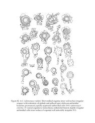
<strong>Saprolegnia</strong> truncata Seymour<br />
Mycotaxon 92:1-10, figs. 41-51. 2005<br />
(Figure 91 C-M)<br />
Monoecious. Mycelium limited, dense; hyphae moderately stout (16-) 24–26<br />
(-35) µm in diameter, sparingly branched. Sporangia rare; naviculate or cylindrical,<br />
appearing very early or proliferating from immature oogonia after prolonged culture,<br />
then subspherical with a long, cylindrical exit tube; straight or slightly curved; renewal<br />
not observed; not persistent; averaging 120 x 28 µm; discharge papilla with thickened,<br />
flaring wall; basal portion often thick-walled. Spores dimorphic; discharge and<br />
behavior saprolegnoid; cysts 9-13 µm in diameter. Gemmae, when present, naviculate<br />
or spherical; terminal or intercalary; single; smooth. Oogonia abundant; terminal or<br />
lateral, rarely intercalary; single; spherical, occasionally ovoid; (18-) 34-38 (-45) µm in<br />
diameter, exclusive of papillae. Oogonial wall unpitted; ornamented with prominent,<br />
truncate projections: (9-) 14-16 (-29) µm long, occasionally intermixed with papillae.<br />
Oogonial stalks up to 8 times the diameter of oogonium, in length; straight, slender,<br />
unbranched. Oospores subcentric; spherical or ovoid, occasionally ellipsoidal; 1, rarely<br />
2 per oogonium, and plerotic or aplerotic; (36-) 29-33 (-42) µm in diameter; germination<br />
unknown. Antheridial branches rare; androgynous, usually arising immediately below<br />
the oogonium to which attached, single; not persistent. Antheridial cells simple;<br />
laterally appressed; persisting; fertilization tubes unknown.<br />
Holotype: Fig. 91 (C-M); Accession Nr. MS 242, Randall <strong>Library</strong> Special<br />
Collection, Univ. of North Carolina at Wilmington (USA), isolated from roadside forest<br />
soil, highway BR 174 to Boa Vista, 60 km from Manaus, Brazil 5 March 1978.<br />
Among the species of <strong>Saprolegnia</strong> with ornamented oogonia, S. truncata is<br />
distinctive by reason of its stout, prominent wall protrusions. <strong>The</strong>se generally are<br />
truncate (Fig. 91 G, H, L) but on some oogonia (Fig. 91 J) a few rounded papillae also<br />
may be produced.<br />
604
Essentially two types of sporangia are formed by <strong>Saprolegnia</strong> truncata. <strong>The</strong><br />
terminal, thin-walled sporangia (Fig. 91E) usually associated with species in the genus<br />
are rare, and we have not seen how they are renewed. <strong>The</strong> second type of sporangium<br />
-- more commonly developed than the “typical” ones in our specimens -- is in essence a<br />
reversal of an oogonial initial to a function as an asexual cell (Fig. 91 C, D). <strong>The</strong>se<br />
sporangia therefore are thick-walled and subspherical at the base, but thin-walled<br />
distally (except in the vicinity of the exit orifice). Release of spores from these sporangia<br />
is characteristically saprolegnoid. So far as we are aware, the sporangia developed from<br />
converted oogonium initials are not found in any other species in the genus. <strong>The</strong><br />
reversion of antheridial branches into vegetative hyphae or oogonia has been<br />
documented (Moreau and Moreau, 1935c; T. W. Johnson, 1973a).<br />
Developmentally, <strong>Saprolegnia</strong> truncata is unique among members of the<br />
<strong>Saprolegnia</strong>ceae. In most species propagated on hempseed in water culture, immature<br />
oogonia appear on colonies after 36 hours, but in S. truncata, the oogonia generally are<br />
formed and become mature within 8-12 hours following infestation of fresh hempseed.<br />
This extremely rapid appearance of oogonia may in some way be associated with<br />
suppression of the sporangial phase even during the early stages of mycelial<br />
development. This phenomenon commands further study from both a biochemical<br />
(regulatory) and developmental standpoint.<br />
<strong>The</strong> discovery of <strong>Saprolegnia</strong> truncata is the first instance in which we have<br />
collected a member of this genus in low-elevation, humid tropical areas. Some<br />
representatives of <strong>Saprolegnia</strong> have been recovered from tropical countries in soils at<br />
high elevations, but Fajola and his associates (1978) found S. ferax and S. litoralis in river<br />
soils in Nigeria.<br />
SPECIMEN EXAMINED: -- SOUTH AMERICA (1), RLS.<br />
<strong>Saprolegnia</strong> asterophora de Bary<br />
Jahrb. Wiss. Bot. 2:189, pl. 20, figs. 25-27. 1860<br />
(Figure 92 A-F)<br />
Cladolegnia asterophora (de Bary) Johannes, Feddes Repert. Spec. Nov. Regni Veg., p. 215.<br />
1955.<br />
Scoliolegnia asterophora (de Bary) Dick, J. Linn. Soc. Bot. 62:257, pl. 1, figs. G, J, M; fig. 1.<br />
1969.<br />
Monoecious. Mycelium delicate, hyphae often flaccid and flexuous. Sporangia<br />
rare or abundant; clavate or fusiform, usually short, renewed internally; 120-821 x 12-43<br />
µm. Spores dimorphic; discharge and behavior saprolegnoid; primary spore cysts 12-14<br />
µm in diameter. Gemmae sparse; clavate or irregular, terminal or intercalary, Oogonia<br />
lateral, occasionally terminal, rarely intercalary; spherical, subspherical or oval,<br />
infrequently napiform or obpyriform; (25-) 42-56 (-78) µm in diameter, inclusive of wall<br />
ornamentations. Oogonial wall unpitted; densely papillate or provided with papillae<br />
605
and tuberculate projections. Oogonial stalks ( 1 / 2 -) 1-1 1 / 2 (-3) times the diameter of the<br />
oogonium in length; straight, branched, unbranched, or irregular. Oospores subcentric;<br />
spherical or subspherical; 1-2 (-6) per oogonium and usually not filling it; (17-) 26-33<br />
(-44) µm in diameter; germination not observed. Antheridial branches predominantly<br />
androgynous, rarely monoclinous, very rarely diclinous; sometimes arising in a<br />
clustered fashion below the oogonium; unbranched or branched, usually irregular in<br />
general configuration; persisting. Antheridial cells simple; short-clavate or tuberous;<br />
apically or laterally appressed; persisting; fertilization tubes present or absent, not<br />
persisting.<br />
<strong>The</strong> densely papillate oogonia that generally contain only one or two subcentric<br />
oospores (Fig. 92 B, C, E) make this species easily identifiable. In gross culture,<br />
specimens sometimes can be recognized by reason of the very diffuse, flaccid nature of<br />
the mycelium.<br />
Dick (1969a) placed this species in Scoliolegnia, a genus that we do not recognize.<br />
An unusual “form” of <strong>Saprolegnia</strong> asterophora first described by Dick in 1960(c) later was<br />
placed by him (1969a) in Scoliolegnia and renamed Scoliolegnia subeccentrica. We<br />
recognize Dick’s species as a valid taxon of <strong>Saprolegnia</strong>.<br />
Of the three taxa that Dick (1969a) considered to be an aggregate species --<br />
Scoliolegnia (=<strong>Saprolegnia</strong>) asterophora, S. subeccentrica, and S. blelhamensis -- only S.<br />
asterophora has subcentric oospores. With respect to the nature and density of its<br />
oogonium wall ornamentations, S. asterophora (Fig. 92 B) very closely approaches S.<br />
subeccentrica (Fig. 92 N, O).<br />
CONFIRMED RECORDS: -- BRITISH ISLES: Dick (loc. cit.); Forbes<br />
(1935b:7, fig. 2a-f). CANADA: Maestres (1977:150, figs. 46, 47). CZECHOSLOVAKIA:<br />
Cejp (1959a:253,1 fig. 96) DENMARK: Petersen (1909a: fig. 3h; 1910: fig. 3h). FINLAND:<br />
Häyrén (1927:50). GERMANY: de Bary (loc. cit.; 1881:269 et sqq., pl. 6, figs. 18-29;<br />
1888:614); A. Fischer (1892: 343); Minden (1912:529, fig. 1h); Zopf (1890:297). ICELAND:<br />
Howard et al. (1970: fig. 26). JAPAN: Kobayasi and Konno (1969:732, fig. 4 I-M);<br />
Kobayashi and Ôkubo (1954:567, fig. 9); Shibuya (1959:148, fig. 3). LATVIA: Apinis<br />
(1929a:218). MIDDLE EUROPE: Migula (1903:66). NETHERLANDS: Beverwijk<br />
(1948:233, fig. 4). POLAND: Staniak (1971:366). REPUBLIC OF CHINA: Chiou and<br />
Chang (1976:44, pl. 3, figs. 1-9). SWITZERLAND: Maurizio (1895d:13). UNITED<br />
STATES: Beneke (1948b:46); R. L. Butler (1975: figs. 9-12); Coker (1923:64, pl. 19);<br />
Humphrey (1893:110, pl. 17, figs. 54, 55); Seymour (1970:23, figs. 12-23). USSR:<br />
Morochkovs’kiĭ et al. (1967:126, fig. 110); Naumov (1954:66). [Istvánffi (1895: pl. 35, figs.<br />
19-21; pl. 36, fig. 22) studied this species cytologically, but gave no record of its source.]<br />
RECORDED COLLECTIONS: -- BRITISH ISLES: Cook and Morgan (1934); Dick<br />
(1962, 1963, 1964, 1966); Dick and Newby (1961); Perrott (1960); R. E. Roberts (1963);<br />
Sparrow (1936). CANADA: Dick (1971c); Maestres and Nolan (1978). DENMARK: A.<br />
Lund (1934, 1978). FINLAND: Häyrén (1956). GERMANY: Höhnk (1935a). JAPAN:<br />
Shirai et al. (1927); Suzuki (1961f). UNITED STATES: Coker (1927); Klich (1980);<br />
606
Kauffman (1915); V. D. Matthews (1927); C. E. Miller (1965); Monsma (1936). USSR:<br />
Érgashev and Kirgizhaeva (1978); Osipyan et al. (1974).<br />
SPECIMENS EXAMINED: -- ICELAND (3), NORWAY (1), SWEDEN (9), TWJ.<br />
UNITED STATES (3), RLS. Centraalbureau (1); MWD (1).<br />
<strong>Saprolegnia</strong> subeccentrica (Dick) Mil’ko<br />
Mikol. i Fitopatol. 13:290. 1979<br />
(Figures 91 A, B; 92 N, O)<br />
Scoliolegnia subeccentrica Dick, J. Linn. Soc. Bot. 62:257, pl. 1, figs. B, D, F, H, K, N; fig. 2.<br />
1969.<br />
Monoecious. Mycelium delicate; hyphae flaccid and flexuous. Sporangia<br />
abundant in young and old cultures; cylindrical, clavate, or long-fusiform, often curved<br />
or slightly irregular; proliferating internally; or renewed sympodially or in a<br />
basipetalous fashion; 116-883 x 14-33 µm. Spores dimorphic; discharge and behavior<br />
saprolegnoid; primary spore cysts 10-13 µm in diameter. Gemmae absent. Oogonia<br />
terminal, occasionally lateral, rarely intercalary; spherical, subspherical or ellipsoidal,<br />
dolioform when intercalary; (28-) 39-57 (-76) µm in diameter, inclusive of wall<br />
ornamentations. Oogonial wall unpitted, thin; densely provided with papillae, or with<br />
cylindrical, curved, irregular, or bifurcate ornamentations each rounded at the apex.<br />
Oogonial stalks variable in length; slender, irregular, unbranched. Oospores<br />
subeccentric; spherical or subspherical; 1-2 (-4) per oogonium, and nearly filling it; (16-)<br />
26-36 (-52) µm in diameter; germination not observed. Antheridial branches generally<br />
sparse; when present, androgynous or monoclinous; unbranched or very sparingly<br />
branched; persisting. Antheridial cells simple; clavate or tubular; persisting; attached<br />
apically or laterally; fertilization tubes present but not persisting.<br />
With respect to the abundance of oogonium wall ornamentations <strong>Saprolegnia</strong><br />
subeccentrica most nearly resembles S. asterophora. <strong>The</strong> wall projections are generally far<br />
more numerous in S. subeccentrica (Fig. 91 A) than in S. blelhamensis (Fig. 92 K). <strong>The</strong><br />
oospore type in S. subeccentrica separates this species from S. asterophora, but is identical<br />
to that in S. blelhamensis.<br />
<strong>Saprolegnia</strong> subeccentrica has antheridial branches, but S. blelhamensis lacks such<br />
structures. Because the degree of branch development in the former is related to culture<br />
conditions, it is possible that S. subeccentrica is more closely akin to S. blelhamensis than<br />
the gross morphology of the oogonial wall ornamentations would suggest. We were<br />
unable to isolate our specimens of S. subeccentrica, hence could not study variability in<br />
this species.<br />
Dick (loc. cit.) emphasized in the description of Scoliolegnia subeccentrica the<br />
narrow, tubular or bulbous (flaring) aspect to the papilla involved in sporangium<br />
dehiscence. Such exit papillae occur in our specimens of the species, but similar ones<br />
also are found in S. asterophora and S. blelhamensis. Dick (loc. cit.) maintained that the<br />
607
shape of the sporangial papillae in S. subeccentrica and S. blelhamensis was not the same,<br />
but we have not found this to be the case.<br />
<strong>The</strong> specimens of <strong>Saprolegnia</strong> asterophora described and illustrated by Dick in 1960<br />
were later (Dick, loc. cit.) renamed Scoliolegnia (=<strong>Saprolegnia</strong>) subeccentrica. Specimens of<br />
S. subeccentrica obtained from Dick’s collections have failed to develop any sexual<br />
apparatus in culture.<br />
cit.)<br />
CONFIRMED RECORDS: -- BRITISH ISLES: Dick (1960:597, figs. 1, 2, 3 M; loc.<br />
SPECIMENS EXAMINED: -- NORWAY (2), TWJ. MWD (2).<br />
<strong>Saprolegnia</strong> blelhamensis (Dick) Mil’ko<br />
Mikol. i Fitopatol. 13:290. 1979<br />
(Figure 92 G-M)<br />
Scoliolegnia blelhamensis Dick, J. Linn, Soc. Bot. 62:259, pl. 1, figs. A, C, E, I, L, O; fig. 3.<br />
1969.<br />
Monoecious. Mycelium delicate; hyphae flaccid and flexuous. Sporangia formed<br />
only in young cultures; clavate or cylindrical; renewed internally; 50-584 x 15-29 µm.<br />
Spores dimorphic; discharge and behavior saprolegnoid; primary spore cysts 10-16 µm<br />
in diameter. Gemmae absent. Oogonia terminal or lateral, frequently or only<br />
occasionally intercalary; spherical, subspherical, or oval, but dolioform when<br />
intercalary; (23-) 43-66 (-97) µm in diameter, inclusive of wall ornamentations. Oogonial<br />
wall unpitted; thin; densely or sparsely papillate, and occasionally provided with<br />
cylindrical or elongate-papillate projections as well, or nearly smooth. Oogonial stalks<br />
1 / 2 -2 times the oogonial diameter in length; straight, bent, or crooked, often irregular;<br />
unbranched or once-branched. Oospores subeccentric; spherical, subspherical, or oval;<br />
(1-) 1-3 (-12) per oogonium and nearly filling it; (16-) 26-32 (-41) µm in diameter,<br />
germination not observed. Antheridial apparatus absent.<br />
Dick (loc. cit.) recorded in detail the differences among this species, S. asterophora,<br />
and S. subeccentrica. Chiefly, S. blelhamensis differs from S. asterophora in oospore<br />
structure (Fig. 92B-E, G-J), and, of course, in the absence of an antheridial apparatus (in<br />
the former). While the wall ornamentations of some oogonia in S. blelhamensis (Fig. 92<br />
G, H) approach the configuration of those produced by S. asterophora (Fig. 92 C, D),<br />
there generally are fewer projections in the former species. A few oogonia of S.<br />
blelhamensis are intact, devoid of all but one or two papillations (Fig. 92 L). Contrary to<br />
the condition in S. asterophora, the wall of each of the oogonia in S. blelhamensis is very<br />
thin (Fig. 92 E, H).<br />
<strong>The</strong> species name is misspelled (blelhamenis) in the original description (Dick, loc.<br />
cit.), but is used correctly in the accompanying text.<br />
608
CONFIRMED RECORD: -- BRITISH ISLES: Dick (loc. cit.).<br />
RECORDED COLLECTION: -- BRITISH ISLES: Dick (1966, pro parte).<br />
SPECIMENS EXAMINED: -- NORWAY (1), TWJ. MWD (3).<br />
<strong>Saprolegnia</strong> australis Elliott<br />
New Zealand J. Bot. 6:103, figs. 2, 4 c-f. 1968<br />
(Figure 93 A-E)<br />
Monoecious. Mycelium dense, diffuse; hyphae slender or stout. Sporangia<br />
cylindrical or clavate; renewed internally or cymosely; primary ones 20-460 x 15-33 µm;<br />
secondary ones usually shorter, but up to 600 µm long. Spores dimorphic; discharge<br />
and behavior generally saprolegnoid, occasionally dictyucoid; primary spore cysts 10.5-<br />
11.7 µm in diameter. Gemmae sparse or abundant; cylindrical, clavate, spherical, or<br />
irregular; terminal or intercalary, usually single. Oogonia generally terminal,<br />
occasionally lateral or intercalary, sometimes sessile; obovate, obpyriform, spherical, or<br />
napiform, sometimes slightly irregular, fusiform or dolioform when intercalary;<br />
(35-) 59-80 (-121) µm in diameter. Oogonial wall pitted, smooth. Oogonial stalks<br />
generally ( 1 / 3 -) 1-3 (-4) times the diameter of the oogonium, in length; straight, curved,<br />
twisted, or irregular; unbranched. Oospores may or may not mature, or may abort;<br />
when mature, subcentric; spherical to subspherical; (1-) 4-12 (-32) per oogonium, but<br />
usually not filling it; (10-) 22-27 (-36) µm in diameter; germination not observed.<br />
Antheridial branches, when present, predominantly diclinous, occasionally or rarely,<br />
monoclinous or androgynous; persisting or deliquescing; sparingly to moderately<br />
branched; usually slightly irregular, sometimes conspicuously contorted. Antheridial<br />
cells simple or branched, persisting; tubular or attached in a digitate fashion;<br />
fertilization tubes present or absent, not persisting.<br />
Until the report by Padgett (1976) of the recovery of specimens from eastern<br />
North Carolina, <strong>Saprolegnia</strong> australis was known only from the type locality. <strong>Saprolegnia</strong><br />
australis appears to be most variable with respect to sporangium size and the<br />
predominating oospore number and diameter (Padgett, 1976: Table 1; Nolan and<br />
Maestres, 1978: Table 1).<br />
<strong>The</strong> oospores of <strong>Saprolegnia</strong> australis are subcentric with the refractive droplets<br />
surrounding the ooplasm (Fig. 93 B) or only partially so (Fig. 93 E); such oospores are<br />
referred to by T. W. Johnson (1956b) and Seymour (1970) as types I and III, and both<br />
types can occur in the same oogonium. <strong>The</strong> oospheres of S. australis may or may not<br />
mature, or the oospores may develop and then abort (Fig. 93 A, C, D). In any event,<br />
nearly all oogonia contain at least a few disintegrated cells or imperfectly formed<br />
oospheres. Budding of oospheres -- resulting in small, spherical cells -- has been<br />
reported (Padgett, 1976).<br />
<strong>The</strong> preponderance of diclinous antheridial branches in <strong>Saprolegnia</strong> australis<br />
recalls a like condition in S. diclina. <strong>The</strong> oospheres of the latter generally mature,<br />
however, yet it must be recognized that some of the “parasitica-like” forms of S. diclina<br />
609
(see this species) also produce oogonia with imperfectly developed oospores<br />
(Willoughby, 1971a). While the oospores of S. australis are subcentric, those of S. diclina<br />
are predominantly centric. Both species have pitted, generally obpyriform oogonia that,<br />
in S. australis are primarily terminal, but are predominantly lateral in S. diclina. <strong>The</strong>se<br />
two species are thus separable on few characters, and those that may be used show<br />
degrees of variation that encompass features of both. Further study of additional<br />
isolates may show the two to be variants of one species.<br />
Padgett (1978b) demonstrated that <strong>Saprolegnia</strong> australis is capable of actively<br />
invading mesohaline estuarine waters (see Chapter 3). <strong>The</strong> fungus also has been found<br />
associated with fish (Pickering and Willoughby, 1977, on Perca fluviatilis; Hatai, Egusa,<br />
and Nomura, 1977, on fingerlings of rainbow trout); such a habitat further relates<br />
Elliott’s species to S. diclina (which includes S. parasitica sensu Kanouse).<br />
<strong>The</strong> chief experimental work with <strong>Saprolegnia</strong> australis has been that by R. F.<br />
Elliott (1968). She propagated the species under a variety of culture conditions noting<br />
that oospore number and colony and oogonium sizes could be modified. On the other<br />
hand, oospore structure and the origin and degree of branching of the antheridial<br />
filaments were not changed by manipulating the environmental parameters during<br />
incubation. At 25 o C colonies of S. australis produced larger oogonia containing more<br />
oospores than did isolates grown at 20 o C. Larger oogonia, and ones with fewer<br />
oospores, developed on mycelium propagated in larger culture vessels rather than in<br />
smaller ones.<br />
CONFIRMED RECORDS: -- CANADA: Maestres (1977:144, 145, figs. 21-23);<br />
Nolan and Maestres (1978:892, figs. 1, 2). JAPAN: Hatai, Egusa, and Nomura (1977:204,<br />
figs. 6-9). NEW ZEALAND: R. F. Elliott (loc. cit.). UNITED STATES: Padgett<br />
(1976:1260, figs. 1, 2); Shipman (1977: fig. 10).<br />
RECORDED COLLECTIONS: -- BRITISH ISLES: Pickering et al. (1979); Pickering<br />
and Willoughby (1977). CANADA: Maestres and Nolan (1978). UNITED STATES:<br />
Klich (1980); Padgett (1978a); Shipman (1979).<br />
SPECIMENS EXAMINED: -- CANADA (1), NEW ZEALAND (1), R. F. Elliott,<br />
preserved specimens. NORWAY (1), TWJ. UNITED STATES (1), DEP.<br />
<strong>Saprolegnia</strong> hypogyna (Pringsheim) de Bary<br />
Bot. Zeitung (Berlin) 41:56. 1883<br />
(Figure 94 A-D)<br />
<strong>Saprolegnia</strong> ferax var. hypogyna Pringsheim, Jahrb. Wiss. Bot. 9:196, pl. 18, figs. 9, 10.<br />
1873-74.<br />
<strong>Saprolegnia</strong> hypogyna var. I Maurizio, Flora 79:126, pl. 4, figs. 5-12. 1894.<br />
<strong>Saprolegnia</strong> hypogyna var. II. Maurizio, ibid., p. 128, pl. 4, figs. 13-16. 1894.<br />
<strong>Saprolegnia</strong> hypogyna var. III. Maurizio, ibid., p. 129, pl. 4, figs. 17-20a. 1894.<br />
<strong>Saprolegnia</strong> hypogyna var. IV. Maurizio, ibid., p. 131, pl. 4, figs. 21-23. 1894.<br />
<strong>Saprolegnia</strong> hypogyna var. V. Maurizio, ibid., p. 132, pl. 4, figs. 24-27. 1894.<br />
610
<strong>Saprolegnia</strong> intermedia Maurizio, Jahrb. Wiss. Bot. 29:97, pl. 2, figs. 37-51a. 1896.<br />
<strong>Saprolegnia</strong> hypogyna var. coregoni Maurizio, Z. Fischerei und deren Hilfswiss., Mitt.<br />
Deutsch. Fischerei-Vereins 7:55. 1899.<br />
Monoecious. Mycelium diffuse; hyphae slender, sparingly branched. Sporangia<br />
cylindrical, clavate, or fusiform; generally straight or curved, occasionally irregular;<br />
renewed internally, rarely by sympodial branching; 76-480 x 13-38 µm. Spores<br />
dimorphic; discharge and behavior saprolegnoid; primary spore cysts 10-13 µm in<br />
diameter. Gemmae sparse or abundant, obpyriform or clavate, infrequently spherical;<br />
terminal or intercalary. Oogonia terminal or lateral, infrequently intercalary;<br />
obpyriform or spherical, occasionally napiform, rarely dolioform; (28-) 68-75 (-101) µm<br />
in diameter. Oogonial wall pitted; smooth. Oogonial stalks variable in length; stout or<br />
slender, straight or slightly irregular; unbranched. Oospores centric; spherical; (1-) 8-12<br />
(-24) per oogonium; filling the oogonium or clustering in the upper, expanded portion;<br />
(18-) 21-26 (-44) µm in diameter; at germination producing a slender germ hypha.<br />
Antheridial branches absent. Antheridial cells hypogynous; fertilization tubes arising<br />
apically from the hypogynous cells, branched or unbranched, persisting.<br />
This is one of two species of <strong>Saprolegnia</strong> producing hypogynous antheridial cells.<br />
<strong>The</strong> smooth, generally spherical or obpyriform oogonia (Fig. 94 A, B) of S. hypogyna<br />
distinguish it immediately from S. irregularis (Fig. 94 G-N).<br />
Whether or not <strong>Saprolegnia</strong> hypogyna has an androgynous antheridial apparatus<br />
in addition to the hypogynous ones is an open question. Maurizio (1894: fig. 17)<br />
illustrated a branched hypogynous cell for his variety III of S. hypogyna. He commented<br />
on the resemblance of this lateral protrusion to an androgynous antheridium.<br />
Hypogynous cells branched at the side were induced in specimens of S. hypogyna grown<br />
in some of the nutrient solutions used by Kauffman (1908). As these lateral protrusions<br />
evidently were not cut off from the hypogynous cell, such an antheridial apparatus now<br />
would be termed hemihypogynous.<br />
As to the function of the hypogynous cell in <strong>Saprolegnia</strong> hypogyna, Maurizio<br />
(1894:149) contended that the projections from the basal cell into the oogonial cavity (see<br />
Fig. 94 A, D) were not fertilization tubes. He regarded these filamentous protrusions as<br />
expressions of a tendency toward secondary growth, paralleling, as it were, internal<br />
proliferation in sporangia. Kauffman (1908) was of a like mind regarding the<br />
fertilization tubes in S. hypogyna. He also thought that the hypogynous cells were not<br />
true antheridia, but were aborted or latent cells that had lost (or never had attained) a<br />
sexual function. Should functional androgynous antheridial branches (or ones of other<br />
origins as well) be found in S. hypogyna, the species then would approach S. ferax.<br />
<strong>The</strong> relatively extensive synonymy of <strong>Saprolegnia</strong> hypogyna is documented<br />
adequately by Seymour (1970).<br />
CONFIRMED RECORDS: -- BRITISH ISLES: R. A. Couch (1951:156, pls. 39, 40)<br />
[his illustrations are very doubtfully of this species]. CZECHOSLOVAKIA: Cejp<br />
611
(1959a:214, fig. 77). DENMARK: A. Lund (1934:17); Petersen (1909a:381; 1910:521).<br />
GERMANY: de Bary (loc. cit., 1888:615); A. Fischer (1892:334); Minden (1912:526, 528,<br />
529; fig. lf); Pringsheim (loc. cit.). ICELAND: Seymour (1970:24, figs. 24-31). LAPLAND:<br />
Gäumann (1918:158). LATVIA: Apinis (1929a:210). MIDDLE EUROPE: Migula (1903:64,<br />
67). RUMANIA: Moruzi and Toma (1968: pl. 4, figs. 31, 32); Toma (1971:9, pl. 1).<br />
SWITZERLAND: Maurizio (locc. citt.; 1899:55); Tiesenhausen (1912:268 et sqq., figs. 2, 3).<br />
UNITED STATES: R. L. Butler (1975: figs. 21-24.); Kauffman (1908: pl. 23); Shipman<br />
(1977: fig. 14), USSR: Dudka and Logvinenko (1968:271, fig. 2.3); Naumov (1954:66).<br />
RECORDED COLLECTIONS: -- BRITISH ISLES: O’Sullivan (1965); Perrott<br />
(1960); Pickering et al. (1979); Willoughby (1974, 1977). DENMARK: Obel (1910a, b).<br />
GERMANY: Höhnk (1956a). ICELAND: Howard et al. (1970); T. W. Johnson (1968).<br />
INDIA: Khulbe (1977, 1980a); Khulbe and Bhargava (1977). NETHERLANDS: Bootsma<br />
(1973). ROMANIA: Toma (1969). UNITED STATES: Clausz (1970, 1974); Kauffman<br />
(1906); Klich (1980); Monsma (1936); Rooney and McKnight (1972); Shipman (1979).<br />
USSR: Érgashev and Kirgizhaeva (1978); Logvinenko (1971); Logvinenko and<br />
Meshcheryakova (1971); Osipyan et al. (1974). YUGOSLAVIA: Ristanović (1970a, 1973).<br />
SPECIMENS EXAMINED: -- BRITISH ISLES (1), L. G. Willoughby. ICELAND<br />
(4), RLS. NORWAY (1), TWJ.<br />
<strong>Saprolegnia</strong> irregularis Johnson and Seymour<br />
Svensk Bot. Tidskr. 69:90, figs. 1, 2. 1975<br />
(Figure 94 E-N)<br />
Monoecious. Mycelium dense, compact; hyphae sparingly branched. Sporangia<br />
clavate or cylindrical, irregular and secondary ones constricted in passage through exit<br />
pore of emptied sporangium, occasionally spherical or subspherical; renewed internally<br />
or infrequently in a cymose or sympodial manner; primary ones 60-381 x 17-33 µm.<br />
Spores dimorphic; discharge and behavior saprolegnoid; primary or secondary spore<br />
cysts 8-12 µm in diameter. Gemmae abundant; cylindrical, lobed, irregular, sparingly<br />
papillate, simple or branched; terminal or intercalary, single or catenulate; sometimes<br />
converting directly into oogonia. Oogonia terminal or intercalary; subspherical,<br />
cylindrical, branched, lobed, or irregular; (51-) 90-157 (-168) x (21-) 66-140 (-231) µm,<br />
including lobes or branches. Oogonial wall conspicuously pitted; smooth, except where<br />
wall substance protrudes through pits in a truncate fashion. Oospores centric or<br />
subcentric, very infrequently subeccentric; occasionally aborting; spherical,<br />
subspherical, or ellipsoidal; 1-40 per oogonium, and occasionally filling it; (l8-) 23-27 (-<br />
33) µm in diameter; germination not observed. Antheridial branches absent. Antheridia,<br />
when present, hypogynous, and often producing conspicuous unbranched or branched<br />
fertilization tubes; evidently proliferating internally, but successive ones may not have a<br />
poroid apex and are nonfunctional; fertilization tubes persisting.<br />
612
<strong>Saprolegnia</strong> irregularis is recognized by its prominently irregular oogonia and<br />
often profusely developed fertilization tubes (Fig. 94 H, K, M). No other known species<br />
in the genus has this combination of characters.<br />
<strong>The</strong> hypogynous antheridial cells (not all oogonia are attended by these) of<br />
<strong>Saprolegnia</strong> irregularis are of two types. Generally, the apex of the cell is prolonged into a<br />
fertilization tube (Fig. 94 M), but occasionally the apex is open (into the oogonial<br />
cavity), and secondary antheridial cells proliferate internally (Fig. 94 J, L, N). Precisely<br />
how the antheridial apparatus functions in this species is not known, and it is<br />
conceivable that the distally poroid cells (Fig. 94 H) simply may represent ones from<br />
which the fertilization tube has deliquesced. In any event, the internally proliferated<br />
cells recall Maurizio’s (1894) belief that the hypogynous cells and fertilization tubes in S.<br />
hypogyna were merely expressions of renewed growth.<br />
CONFIRMED RECORD: -- ICELAND: Johnson and Seymour (loc. cit.). INDIA: J.<br />
N. Rai and Misra (1977b:76, fig. 3b).<br />
SPECIMENS EXAMINED: - ICELAND (7), TWJ.<br />
<strong>Saprolegnia</strong> turfosa (Minden) Gäumann<br />
Bot. Not. 1918:154. 1918<br />
(Figure 95 A-C)<br />
<strong>Saprolegnia</strong> spec. (2) Reinsch, Jahrb. Wiss. Bot. 11:295, pl. 14, figs. 7-13. 1878.<br />
<strong>Saprolegnia</strong> paradoxa Petersen Bot. Tiddskr. 29:379, fig. l d, e. 1909 (also in Ann. Mycol.<br />
8:520, fig. l d, e. 1910); non S. paradoxa Maurizio, Z. Fischerei und deren<br />
Hilfswiss., Mitt. Deutsch. Fischerei-Vereins 7:46, figs. 10-12. 1899.<br />
<strong>Saprolegnia</strong> monoica var. turfosa Minden, Kryptogamenfl. Mark Brandenburg 5:516. 1912.<br />
Aplanes turfosus (Minden) Coker, J. Elisha Mitchell Sci. Soc. 42:216. 1927.<br />
Aplanes ozeensis Kobayasi, in Kobayasi and Ookubo, J. Jap. Bot. 27:181, figs. 9, 10. 1952.<br />
Monoecious. Mycelium stout; hyphae sparingly branched. Sporangia fusiform,<br />
clavate, or cylindrical; straight, curved, or moderately irregular; renewed internally;<br />
102-627 x 18-66 µm. Spores dimorphic; discharge and behavior saprolegnoid,<br />
infrequently aplanoid; primary spore cysts 9-11 µm in diameter. Gemmae sparse or<br />
absent; when present, cylindrical, obpyriform, irregular, or branched; terminal, rarely<br />
intercalary; predominantly single. Oogonia lateral, rarely terminal, intercalary, or<br />
sessile; spherical, rarely ovoid or subspherical; (18-) 60-85 (-134) µm in diameter,<br />
including wall ornamentations. Oogonial wall conspicuously pitted; smooth or rarely<br />
with a few short, broad papillae. Oogonial stalks very short, predominantly ( 1 / 8 -) 1 / 4 -<br />
1 / 2 (-2) times the diameter of the oogonium, in length; stout, straight or curved;<br />
unbranched. Oospores centric; spherical or ellipsoidal; (1-) 6-16 (-42) per oogonium, and<br />
usually filling it; (16-) 26-31 (-37) µm in diameter; germination not observed.<br />
Antheridial branches androgynous, infrequently monoclinous and arising near the<br />
oogonial stalk; short, curved or bent, unbranched or branched; persisting. Antheridial<br />
613
cells simple; long, cylindrical or tubular, rarely long-clavate; branched or unbranched;<br />
laterally appressed, rarely attached in a digitate fashion; persisting; fertilization tubes<br />
not observed.<br />
Aside from the members of <strong>Saprolegnia</strong> with ornamented oogonia, S. turfosa is<br />
one of the species of the genus that is most easy to recognize. <strong>The</strong> large, very shortstalked<br />
oogonia attended by androgynous antheridial branches each terminated by a<br />
long, tubular antheridial cell (Fig. 95 A) mark this species. <strong>Saprolegnia</strong> uliginosa also has<br />
short-stalked oogonia (Fig. 95 F) but its antheridial branches are frequently<br />
monoclinous (these are infrequent in S. turfosa).<br />
In view of the ease with which <strong>Saprolegnia</strong> turfosa can be recognized, its<br />
taxonomic history is surprisingly complicated. Seymour (1970:28, 29) reviewed the<br />
systematics of this species particularly thoroughly, and only a few additional comments<br />
are necessary. Reinsch (loc. cit.) was apparently the first to see S. turfosa, but chose not<br />
to name it. He (loc. cit., p. 311) referred to figure 15 on plate 17 as an illustration of<br />
<strong>Saprolegnia</strong> sp. 2, but this was an error; that figure depicts sporangia of a Gonapodya.<br />
Coker (1923) apparently had difficulty in recognizing S. turfosa, a situation which he<br />
corrected in his 1927 paper. It appears that A. Lund (1934) misidentified S. turfosa,<br />
equating it with Aplanes treleaseanus (see Achlya androgyna). He listed both S. treleaseana<br />
of Humphrey (1893) and Achlya treleaseana (Humphrey) Kauffman as synonyms of what<br />
is here recognized as S. turfosa. In addition, A. Lund (1934:21) also considered that<br />
Petersen’s S. paradoxa was synonymous with Aplanes treleaseanus; Petersen’s species, on<br />
the contrary, is conspecific with S. turfosa. It is very likely that the Aplanes turfosus<br />
reported from the Soviet Union by Morochkovs’kiĭ et al. (1967) was Achlya androgyna. In<br />
their account of the Russian material, papillate oogonia are described for the specimen<br />
they encountered.<br />
<strong>Saprolegnia</strong> turfosa is evidently not common (although it is widely distributed). In<br />
our experience, individuals appear most frequently in water or debris from Sphagnum<br />
spp. bogs. We have recovered S. turfosa repeatedly from acid waters (not bogs) in<br />
Norway and Sweden. A specimen collected on hempseed baited in a bog water sample<br />
from Norway was infected with Olpidiopsis saprolegniae var. saprolegniae.<br />
CONFIRMED RECORDS: -- CZECHOSLOVAKIA: Cejp (1959a:271, figs. 104,<br />
105). DENMARK: A. Lund (1934:20, fig. 7); Petersen (loc. cit.). GERMANY: Minden (loc.<br />
cit.); Reinsch (loc. cit.); Richter (1937:259, fig. 17). ICELAND: Howard et al. (1970: fig. 36).<br />
JAPAN: Kobayasi and Ookubo (loc. cit.). LAPLAND: Gäumann (loc. cit.). LATVIA:<br />
Apinis (1929a:227). UNITED STATES: Beneke (1948b:38, pl. 3); R. L. Butler (1975: figs.<br />
29-32); Coker (1923:79, pl. 20; loc. cit.); Kobayasi et al. (1967:10, pl. 14, fig. M; text fig. 3);<br />
Milanez (1966:110, pl. 12, fig. d; pl. 13); Milanez and Beneke (1968:19, pl. 2, fig. 10);<br />
Overman (1970:36); Seymour (1970:27, figs. 40-48); A. W. Ziegler (1948b:16, pl. 1, figs.1-<br />
9; 1952:13, pl. 5, fig. 4). USSR: Logvinenko and Meshcheryakova (1971: fig. 7).<br />
RECORDED COLLECTIONS: -- BRITISH ISLES: Dick (1962, 1963, 1966); Dick<br />
and Newby (1961). CANADA: Dick (1971c). CZECHOSLOVAKIA: Cejp (1931).<br />
614
DENMARK: A. Lund (1978). ICELAND: T. W. Johnson (1968). JAPAN: Kobayashi and<br />
Ôkubo (1954); Ookubo (1954); Suzuki (1961f). UNITED STATES: Coker (1927); Klich<br />
(1980); V. D. Matthews (1927).<br />
SPECIMENS EXAMINED: -- BRITISH ISLES (1), W. R. I. Cook (Centraalbureau,<br />
Baarn). ICELAND (12), NORWAY (6), SWEDEN (11), TWJ. UNITED STATES (22), RLS.<br />
<strong>Saprolegnia</strong> uliginosa Johannes<br />
Archiv Mikrobiol. 14:595, fig. 1. 1950<br />
(Figure 95 D-F)<br />
Monoecious. Mycelium moderately dense, extensive; hyphae slender, flaccid.<br />
Sporangia clavate, cylindrical, or fusiform, sometimes irregular, renewed internally,<br />
rarely in basipetalous succession; 108-266 x 12-42 µm. Spores dimorphic; discharge and<br />
behavior saprolegnoid; primary and secondary spore cysts 9-12 µm in diameter.<br />
Gemmae sparse; spherical, obpyriform, cylindrical, fusiform, or irregular; terminal or<br />
intercalary, single or catenulate. Oogonia sparse or abundant; lateral, rarely terminal,<br />
occasionally intercalary; spherical, infrequently napiform or obpyriform; (32-) 60-68 (-<br />
91) µm in diameter. Oogonial wall pitted under the region of attachment of antheridial<br />
cells; smooth. Oogonial stalks ( 1 / 12 -) 1 / 8 –1 (-2) times the diameter of the oogonium, in<br />
length; stout, straight, curved or recurved, infrequently bent; unbranched. Oospores<br />
centric, rarely subcentric; spherical; (2-) 5-7 (-25) per oogonium, and nearly filling it;<br />
(21-) 25-33(-36) µm in diameter; germination not observed. Antheridial branches<br />
predominantly monoclinous, and arising very near the oogonial stalk; infrequently<br />
androgynous; rarely diclinous; slender, slightly irregular, occasionally producing one or<br />
two short, lateral branches; persisting. Antheridial cells simple; generally tubular,<br />
straight or curved, occasionally faintly cylindro-clavate; laterally appressed, persisting;<br />
fertilization tubes present, not persisting.<br />
Like <strong>Saprolegnia</strong> turfosa, S. uliginosa usually has spherical oogonia borne laterally<br />
on short stalks (Fig. 95 E, F) and containing predominantly centric oospores. <strong>The</strong><br />
similarity between these two species does not go beyond these features since the<br />
antheridial branches of S. uliginosa generally are monoclinous (Fig. 95 A) whereas<br />
androgynous ones clearly predominate in S. turfosa.<br />
<strong>Saprolegnia</strong> uliginosa seems allied to S. glomerata -- with respect to antheridial<br />
branch origin, for example -- but can be distinguished from that species because it lacks<br />
the short, lateral twig-like evaginations (Fig. 96 I) arising from the hyphae and<br />
antheridial filaments. Moreover, in S. glomerata the oogonia are most often obpyriform;<br />
spherical ones are usually encountered in S. uliginosa. While the oogonia of S. uliginosa<br />
are pitted under the region of antheridial cell contact, those of S. glomerata usually are<br />
pitted elsewhere as well. In view of the characteristics of the Iceland specimens<br />
reported by Howard et al. (1970), S. uliginosa and S. glomerata cannot be separated with<br />
confidence on predominating oospore diameter.<br />
615
It would appear on the surface that <strong>Saprolegnia</strong> uliginosa is a relatively rare<br />
species. One of the Norwegian isolates in our collections is latent in the development of<br />
its sporangia and the sexual apparatus, and grows very slowly in water culture. This<br />
suggests that the species may be overgrown quickly by more rapidly developing<br />
species, and thus not appear at all in gross cultures.<br />
<strong>The</strong> description and illustrations of a <strong>Saprolegnia</strong> sp. recovered by Shibuya (1959)<br />
suggest that he may have collected Johannes’ species, but we cannot be certain; we<br />
include the Japanese record as an unconfirmed one.<br />
CONFIRMED RECORDS: -- GERMANY: Johannes (loc. cit.). ICELAND: Howard<br />
et al. (1970:77, fig.37). INDIA: J. N. Rai and Misra (1977b:76, fig. 3c). UNITED STATES:<br />
Seymour (1970:47, figs. 78-84).<br />
RECORDED COLLECTION: -- JAPAN: Shibuya (1959:149, fig. 4)().<br />
SPECIMENS EXAMINED: -- ICELAND (1), NORWAY (2), TWJ. UNITED<br />
STATES (5), RLS.<br />
<strong>Saprolegnia</strong> furcata Maurizio<br />
Z. Fischerei und deren Hilfswiss. Mitt. Deutsch. Fischerei-Vereins 7:48 et seq., figs. 13-15.<br />
1899<br />
(Figure 97 E-I)<br />
<strong>Saprolegnia</strong> retorta Horn, Ann. Mycol. 2:233, fig. 21. 1904.<br />
Monoecious. Mycelium moderately delicate, sparingly branched. Sporangia<br />
clavate, cylindrical, or fusiform, straight, curved, or irregular; renewed internally or by<br />
cymose branching; 60-420 x 16-40 µm. Spores dimorphic; discharge and behavior<br />
saprolegnoid; primary spore cysts 10-13 µm in diameter. Gemmae sparse; clavate,<br />
spherical, subspherical, pyriform, or irregular; terminal, single. Oogonia lateral,<br />
occasionally terminal, very rarely intercalary; obpyriform or napiform, infrequently<br />
spherical, very rarely dolioform; (33-) 43-48 (-81) µm in diameter. Oogonial wall<br />
unpitted or sparsely pitted; smooth. Oogonial stalks ( 1 / 2 -) 1-6 (-8) times the diameter of<br />
the oogonium, in length; slender, branched or unbranched; usually coiled, strongly<br />
bent, or irregular and twisted. Oospores centric, rarely subcentric; spherical; (1-) 4-10<br />
(-28) per oogonium, and usually nearly filling it; (16-) 20-24 (-45) µm in diameter;<br />
germination not observed. Antheridial branches predominantly androgynous, rarely<br />
monoclinous or diclinous; slender, occasionally branched, usually twisted or irregular;<br />
infrequently bearing oogonia; persisting. Antheridial cells simple; tubular, clavate, or<br />
irregular, rarely once-branched; persisting; laterally or apically appressed; fertilization<br />
tubes present, not persisting.<br />
<strong>The</strong> strongly bent (Fig. 97 F) or conspicuously coiled (Fig. 97 E), relatively long<br />
oogonial stalks constitute the chief feature by which this species can be recognized.<br />
Maurizio (loc. cit.) emphasized that the antheridial branches in <strong>Saprolegnia</strong> furcata could<br />
616
evert to oogonium production. However, as Seymour (1970) pointed out, this feature<br />
is too variable among isolates to be of much taxonomic value.<br />
<strong>Saprolegnia</strong> furcata has been reported under several names. Cornu (1872) possibly<br />
described this species as S. spiralis, but the fungus as he reported it was infected, and<br />
the taxon is not valid. Horn’s (loc. cit.) <strong>Saprolegnia</strong> retorta appears to have been identical<br />
to Cornu’s S. spiralis, yet apparently was not infected. Quite likely S. monoica var.<br />
tortipes Shkorbatov (1923) was S. furcata as well, but as there are no illustrations of the<br />
variety, this supposition cannot be confirmed. Apinis (1929a) gives as a synonym of S.<br />
furcata Tiesenhausen’s S. monoica var. glomerata. That variety is synonymous with S.<br />
glomerata, however, as A. Lund (1934) pointed out. We are including Cejp’s (1959a)<br />
account of S. spiralis in S. furcata, but only with reservation. <strong>The</strong> illustrations he<br />
provided (Cejp, 1959a; fig. 95) do not show a fungus infected by Rozella (as Cornu had<br />
illustrated S. spiralis), hence it is difficult to see how Cejp could have identified his<br />
Czechoslovakian specimens with Cornu’s species. <strong>The</strong> one good illustration by A.<br />
Braun (1856: pl. 5, fig. 22) of an uninfected filament of S. ferax shows oogonia on<br />
strongly recurved stalks; he may well have observed what was later to be described by<br />
Maurizio as S. furcata.<br />
<strong>Saprolegnia</strong> furcata has been used extensively in research on ultrastructure. <strong>The</strong><br />
accounts by Beakes and Gay (1977: 1978a, b), Heath and Greenwood (1968), Heath et al.<br />
(1971), and Howard and Moore (1970) are notable in this respect (see Chapters 13-15).<br />
CONFIRMED RECORDS: -- CZECHOSLOVAKIA: Cejp (1959a:236, 251, fig.<br />
95)(). GERMANY: A. Braun (1856: pl. 5, fig. 22); Horn (loc. cit.); Minden (1912:517);<br />
Richter (1937:238, fig. 2). ICELAND: T.W. Johnson (1974b: figs. 160-162). LATVIA:<br />
Apinis (1929a:213, fig. 1). MIDDLE EUROPE: Migula (1903:67). SWITZERLAND:<br />
Maurizio (loc. cit.).<br />
RECORDED COLLECTIONS: -- BRITISH ISLES: Dick and Newby (1961),<br />
O’Sullivan (1965). GERMANY: Höhnk (1935a). USSR: Logvinenko and Meshcheryakova<br />
(1971).<br />
SPECIMENS EXAMINED: -- BRITISH ISLES (2), A. E. Apinis, M. W. Dick.<br />
ICELAND (2), UNITED STATES (13), RLS.<br />
<strong>Saprolegnia</strong> glomerata (Tiesenhausen) Lund<br />
Kongel. Danske Vidensk. Selsk. Naturvidensk. Math. Afh. 9 6:14 fig. 4. 1934<br />
(Figure 96 G-J)<br />
<strong>Saprolegnia</strong> monoica var. glomerata Tiesenhausen, Arch. Hydrobiol. Planktonk. 7:277, figs.<br />
6-8. 1912.<br />
Monoecious. Mycelium delicate; some principal hyphae stout and provided<br />
with numerous short, scattered or clustered lateral, often twig-like branches. Sporangia<br />
abundant or sparse; cylindrical, fusiform, clavate, or irregular; rarely pyriform; straight,<br />
curved, or bent, occasionally twisted, and sometimes enlarged at base; renewed<br />
617
internally; 40-220 x 18-28 µm. Spores dimorphic; discharge and behavior saprolegnoid,<br />
rarely aplanoid; primary spore cysts 10-14 µm in diameter. Gemmae very sparse;<br />
clavate or obpyriform; terminal, single. Oogonia spherical, obpyriform, napiform, or<br />
subspherical, rarely dolioform; lateral, occasionally terminal, rarely intercalary; (32-) 46-<br />
60 (-107) µm in diameter. Oogonial wall pitted or unpitted; smooth. Oogonial stalks<br />
( 1 / 4 -) 1-2 (-3 1 / 2 ) times the diameter of the oogonium, in length; stout; straight, curved,<br />
or bent; unbranched or with one or more short, lateral branches. Oospores centric;<br />
spherical, often nearly filling the oogonium; (1-) 6-16 (-28) per oogonium; (18-) 23-26 (-<br />
30) µm in diameter; at germination producing a germ hypha which may or may not<br />
bear a terminal sporangium. Antheridial branches androgynous or monoclinous, rarely<br />
diclinous; slender, usually contorted, twisted, or irregular and sparingly branched;<br />
persisting. Antheridial cells simple, generally tubular, occasionally clavate or slightly<br />
irregular; persisting; laterally appressed, very rarely attached apically; fertilization<br />
tubes present, not persisting.<br />
<strong>Saprolegnia</strong> glomerata is readily recognizable by the short, branched or<br />
unbranched, contorted, lateral (and often clustered) hyphal extensions (Fig. 96 G, I).<br />
Secondary characters of recognition are the contorted, branched antheridial filaments<br />
(Fig. 96 G, H, J) and the short, lateral evaginations on many of the oogonial stalks (Fig.<br />
96 H). In S. litoralis, the hypha immediately below a terminal oogonium, may bear<br />
short, lateral branches (Fig. 96 B) as does S. glomerata, but in the former, the hypha is<br />
usually swollen at its juncture with the oogonial septum (Fig. 96 A, B). In any case, the<br />
oospores in S. litoralis are occasionally subcentric, a condition not known to occur in S.<br />
glomerata.<br />
Although A. Lund recognized as early as 1934 that Tiesenhausen’s (loc. cit.)<br />
variety glomerata deserved specific rank, not all investigators followed (or were aware<br />
of) this disposition. For example, T. Ito (1944) retained the variety of glomerata and<br />
reduced <strong>Saprolegnia</strong> furcata and S. glomerata (Tiesenhausen) Lund to synonymy with it.<br />
Cejp (1959a) listed both Lund’s species and S. monoica as synonyms of Tiesenhausen’s<br />
variety. In addition, S. glomerata has been misidentified by some mycologists. Apinis<br />
(1929a, b) believed that Maurizio’s (1899) S. furcata was the same as Tiesenhausen’s<br />
variety glomerata. <strong>The</strong> specimens recorded by T. W. Johnson (1950b:399) as the variety<br />
glomerata were misidentified; he had collected forms of S. ferax. <strong>The</strong> S. monoica reported<br />
by Humphrey (1893) was not, as Coker (1923) maintained, identifiable as S. monoica var.<br />
glomerata; Humphrey’s material was very likely representative of S. ferax.<br />
CONFIRMED RECORDS: -- BRITISH ISLES: R.A. Couch (1951:161, p1s. 41-45);<br />
Forbes (1935a:226). CZECHOSLOVAKIA: Cejp (1959a:231, fig. 85 a-f). DENMARK:<br />
Lund (loc. cit.). GERMANY: Richter (1937:237). ICELAND: Howard et al. (1970: figs. 27,<br />
28). INDIA: Thakur Ji (1970:182, figs. 15-18). JAPAN: T. Ito (1942:124, fig. l e-g; 1944:52).<br />
LATVIA: Apinis (1929a: 213, text fig. 1). NETHERLANDS: Beverwijk (1948:232, fig. 3).<br />
POLAND: Szwanke (1938:8, pl. 2, figs. 10, 11; pl. 3, figs. 1-5). SWITZERLAND:<br />
Tiesenhausen (loc. cit.). UNITED STATES: Beneke (1948b:36); T. W. Johnson (1956a:186);<br />
618
Rose (1932:25, pl. 1, figs. 6-9); Sorenson (1962: pl. 7, figs. A, B); A. W. Ziegler (1952:7, pl.<br />
2, figs. 7,8).<br />
RECORDED COLLECTIONS: -- BRITISH ISLES: Apinis (1960, 1964); Cook and<br />
Forbes (1933); Cook and Morgan (1934). GERMANY: Höhnk (1935a). JAPAN: Suzuki<br />
(1961f). POLAND: Zaborowska (1965). RUMANIA: Toma (1969). UNITED STATES:<br />
Cotner (1930b); G. C. Hughes (1959, 1962); Poitras (1955); Sparrow (1952b); A. W.<br />
Ziegler (1958b). YUGOSLAVIA: Ristanović (1973).<br />
SPECIMENS EXAMINED: -- ICELAND (66), NORWAY (4), TWJ. UNITED<br />
STATES (26), RLS.<br />
<strong>Saprolegnia</strong> ferax (Gruith.) Thuret<br />
Ann. Sci. Nat. Bot. (3 e sér.) 14:229 et sqq., pl. 22. 1850<br />
(Figure 98)<br />
Conferva ferax Gruithuisen, Nova Acta Phys.-Med. Acad. Caes. Leop-Carol. Nat. Cur.<br />
10:445, pl. 38, figs. 14-16. 1821.<br />
<strong>Saprolegnia</strong> molluscorum Nees von Esenbeck, Nova Acta Phys.-Med. Acad. Caes. Leop.-<br />
Carol. Nat. Cur. 11:514. 1823.<br />
Leptomitus ferax Agardh, Systema Algarum, p. 49. 1824.<br />
Achlya prolifera Pringsheim, Nova Acta Phys.-Med. Acad. Caes. Leop-Carol. Nat. Cur.<br />
23:399 et sqq. pls. 46-50. 1851.<br />
<strong>Saprolegnia</strong> monoica Pringsheim, Jahrb. Wiss. Bot. 1:292, pls. 19, 20. 1858.<br />
Achlya intermedia Bail, Amtl. Ber. Versamml. Deutsch. Naturf. Aerzte, Konigsberg<br />
35:257. 1860.<br />
Diplanes saprolegnioides Leitgeb, Jahrb. Wiss, Bot. 7:385, pl. 24. 1869-70.<br />
Achlya ferax (Kützing) Duncan, Proc. Roy. Soc. London 25:253, pl. 7, figs. 36-38, 40-42.<br />
1876.<br />
<strong>Saprolegnia</strong> thureti de Bary, Abh. Senckenberg Naturf. Ges. 12:326, pl. 5, figs. 1-10.<br />
1881. (Also in de Bary and Woronin, Beitr. Morphol. Physiol. Pilze, 4: 102, pl. 5,<br />
figs. 1-10. 1881.)<br />
<strong>Saprolegnia</strong> mixta de Bary, Bot. Zeitung (Berlin) 41:56. 1883.<br />
<strong>Saprolegnia</strong> monoica var. montana de Bary, ibid., 46:617. 1888.<br />
<strong>Saprolegnia</strong> esocina Maurizio, Jahrb. Wiss. Bot. 29:82, pl. 1, figs. 4-17. 1896.<br />
<strong>Saprolegnia</strong> heterandra Maurizio, ibid., p. 87, pl. 1, figs. 18-27. 1896.<br />
<strong>Saprolegnia</strong> bodanica Maurizio, ibid., p. 107, pl. 2, figs. 52-59a. 1896.<br />
<strong>Saprolegnia</strong> paradoxa Maurizio, Z. Fischerei und deren Hilfswiss. Deutsch. Fischerei-<br />
Vereins 7:46 et seq., figs. 10-12. 1899; non S. paradoxa Petersen, Bot. Tidsskr.<br />
29:379. 1909. (Also in Petersen, Ann. Mycol. 8:520. 1910.)<br />
<strong>Saprolegnia</strong> floccosa Maurizio, ibid., p. 50 et seq., figs. 16, 17. 1899.<br />
<strong>Saprolegnia</strong> semidioica Petersen, Bot. Tidsskr. 29:378, fig. 1f. 1909. (Also in Petersen, Ann.<br />
Mycol. 8:519, fig. lf. 1910.)<br />
<strong>Saprolegnia</strong> monoica var. vexans Pieters, Bot. Gaz. (Crawfordsville) 60:489. 1915.<br />
<strong>Saprolegnia</strong> mixta var. asplundii Gäumann, Bot. Not. 1918:155. 1918.<br />
619
<strong>Saprolegnia</strong> lapponica Gäumann, ibid., p. 156. 1918.<br />
<strong>Saprolegnia</strong> tokugawana Emoto, Bot. Mag. (Tokyo) 37:15, pl. 1. 1923.<br />
<strong>Saprolegnia</strong> monoica var. floccosa (Maurizio) Cejp, Oomycetes I, Flora ČSR, Ser. B, Part 2,<br />
p. 234, figs. 85 g-j, 86. 1959.<br />
<strong>Saprolegnia</strong> ferax var. lapponica (Gäumann) Cejp, ibid., p. 245. 1959.<br />
<strong>Saprolegnia</strong> ferax var. esocina (Maurizio) Cejp, ibid., p. 246. 1959.<br />
<strong>Saprolegnia</strong> monoica var. acidamica Suzuki, J. Jap. Bot., 36:234. 1961.<br />
Monoecious. Mycelium stout, hyphae moderately to sparingly branched.<br />
Sporangia clavate, fusiform, cylindrical, or slightly irregular, sometimes nearly<br />
spherical; straight or bent; renewed internally with secondary ones nesting inside<br />
discharged primary ones, or partially emerged through orifices of empty sporangia and<br />
forming bead-like chains or cylindrical segments, or emerging fully through orifices of<br />
previously emptied sporangia; rarely renewed in a basipetalous or cymose manner; 31-<br />
624 x 18-67 µm. Spores dimorphic; discharge and behavior saprolegnoid, rarely<br />
aplanoid or dictyucoid, very rarely achlyoid or leptolegnoid; primary spore cysts 9-12<br />
µm in diameter. Gemmae, when present, variable in shape and position. Oogonia<br />
lateral, terminal, or intercalary and then single or catenulate, infrequently occurring in<br />
emptied sporangia or sessile; spherical, obpyriform, napiform, sometimes obovate or<br />
clavate, cylindrical when in discharged sporangia, often dolioform when intercalary;<br />
(28-) 60-80 (-194) µm in diameter. Oogonial wall generally conspicuously and<br />
abundantly pitted, rarely unpitted; smooth or rarely with one or two short, papilliform<br />
evaginations, or apiculate. Oogonial stalks ( 1 / 3 -) 1-2 (3 1 / 2 ) times the diameter of the<br />
oogonium, in length; stout, straight, bent, or curved; very rarely once-branched.<br />
Oospores centric or subcentric, spherical or ellipsoidal; (1-) 10-18 (-54) per oogonium<br />
and nearly filling it, (12-) 22-28 (-44) µm in diameter; germinating by a slender germ<br />
hypha that may or may not bear a small, apical, clavate sporangium. Antheridial<br />
branches variable in abundance and density, and sometimes absent; when present,<br />
monoclinous or androgynous, rarely diclinous; slender, slightly to prominently<br />
irregular, unbranched or very sparingly branched; persisting. Antheridial cells simple;<br />
generally tubular or clavate, occasionally irregular, infrequently once-branched; usually<br />
persisting; laterally appressed, very rarely attached apically; fertilization tubes present,<br />
not persisting.<br />
Based on a survey of existing literature and the characterization of a generous<br />
collection of living material, Seymour (1970) demonstrated that <strong>Saprolegnia</strong> ferax was a<br />
complex of morphological variants. Many of the various forms that had from time to<br />
time been described, he concluded, were recognizable as individuals, but at the same<br />
time occupied some point in a long series of morphological gradients. Accordingly, he<br />
proposed a very extensive synonymy (Seymour, 1970:29, 30) for S. ferax; this is adopted<br />
here with minor corrections. Discussion of the synonymous species beyond that<br />
provided by Seymour is unnecessary. Florinskaya (1968) has also studied extensively<br />
the degree of morphological variability in S. ferax, particularly that to be found in<br />
620
oogonial shape and position. She did not propose any taxonomic changes, however, as<br />
a result of the analyses she performed.<br />
<strong>Saprolegnia</strong> ferax is a ubiquitous and very common species. Generally it is most<br />
easily recognized by reliance on a combination of predominating characters: large,<br />
conspicuously or sparsely pitted oogonia, centric and subcentric oospores (sometimes<br />
in the same oogonium), occasional development of oogonia in discharged sporangia,<br />
and a preponderance of androgynous or monoclinous antheridial branches (when these<br />
filaments are present at all). Identification of the species often is complicated by the fact<br />
that the abundance of antheridial branches is especially variable among cultures.<br />
Antheridial filaments are usually sparse, but specimens nearly devoid of an antheridal<br />
apparatus, or ones generally provided with such branches are common as well. In any<br />
event, even when a large percentage of the oogonia in any colony are attended by<br />
antheridial branches the filaments are seldom branched, and are usually not long and<br />
extensively developed. In the general configuration of its oogonia, S. diclina approaches<br />
S. ferax. Cultures of S. diclina consistently have a generous production of predominantly<br />
diclinous antheridia (such a branch origin is rare in S. ferax).<br />
<strong>The</strong> discovery of oogonia in <strong>Saprolegnia</strong> ferax is attributable to Thuret (1850:229 et<br />
sqq., pl. 22) although he believed these cells were only lateral sporangia. <strong>The</strong> status of<br />
the species was early called into question, and several names appeared in the literature.<br />
Kützing (1843, 1849), for example, who thought the fungus was an alga, listed eleven<br />
synonyms for S. ferax. <strong>The</strong> species was put into Leptomitus by Agardh (1824), and into<br />
Achlya by Duncan in 1876(b). Without a doubt some of the earliest reports of the<br />
collection of S. ferax were based on misidentifications, and curious conclusions about<br />
the species were fashioned. In some remarks on this species published in 1871, M. C.<br />
Cooke concluded that Sporendonema muscae Fries or Empusa musca Cohn were<br />
imperfectly formed “terrestrial conditions” of S. ferax. De Bary (1881:254 et sqq.)<br />
understood S. ferax to be a collective name only.<br />
<strong>Saprolegnia</strong> ferax has been implicated as an active agent in saprolegniosis (R. C.<br />
Srivastava, 1976; Nolard-Tintigner, 1970, among others). Some investigators, however,<br />
deduced that representatives of the species simply acted as secondary invaders of<br />
already diseased or injured fish (Drew, 1909). <strong>Saprolegnia</strong> ferax also is associated with<br />
ulcerative dermal necrosis (Chapter 29).<br />
Because of its ready availability and rapid growth in culture, <strong>Saprolegnia</strong> ferax has<br />
proven to be a useful experimental subject in a variety of studies, examples of which<br />
follow. Nolan’s (1976) physiological study of S. ferax demonstrated for the first time the<br />
use and release by a fungus of cadaverine (Chapter 22). <strong>Saprolegnia</strong> ferax also has been<br />
employed extensively in ultrastructural work (Chapters 13-15), notably by such<br />
investigators as Manton et al. (1951, 1952), Heath (1975a, b), Heath and Greenwood<br />
(1968; 1970a, c), and Holloway and Heath (1977b). Under the binomial S. monoica, S.<br />
ferax was made use of in an investigation of hyphal elongation and branching by<br />
Larpent (l963a-c, 1964), and in experimental analyses by Borkowski (1967; 1968a, b;<br />
1969; 1970) of the axial ordination of primary spores during release.<br />
621
Using <strong>Saprolegnia</strong> ferax among other fungi, Ho (1978) performed a quantitative<br />
analysis of hyphal branching at the periphery of colonies grown on an agar medium.<br />
He selected ten parameters for the analysis: apical segment ratio, diameter ratio, and<br />
hyphal density, among others. <strong>The</strong> mycelium of S. ferax typically produced very long<br />
apical segments (distance from apex of hypha to first “internode” or branch hypha), and<br />
a very low total branch ratio (total number of branches on a primary hypha in relation<br />
to the total number of primary or “leading” hyphae in the mycelium). <strong>The</strong> ratio<br />
between the sum of the length of all filaments and the total number of hyphal apices<br />
indicated that this species displayed a very large hyphal growth unit (one of the ten<br />
parameters measured). Ho suggested that the nature of the hyphal branching system<br />
might be characteristic enough of some species to have taxonomic value.<br />
Among the numerous records of the collection of <strong>Saprolegnia</strong> ferax (in its broadest<br />
sense) there are four that must remain suspect, even though the specimens so identified<br />
were described or illustrated, or both. <strong>The</strong> following reports are in this very doubtful<br />
category: Almeida et al. (1943: pl. 21 fig. 6), Richter (1937:241, fig. 3; see S. latvica),<br />
Stirling (1878: 1 unnumbered fig.), and Walz (1870a:556, pl. 9, figs. 20, 21; also in Val’ts,<br />
1870: figs. 20, 21; conidia are described and illustrated).<br />
CONFIRMED RECORDS: -- ASIA: Sorokine (1889-90:142). AUSTRALIA:<br />
Crooks (1937:211, fig. 1A-G). AUSTRIA: Keissler (1917: fig. 2). BELGIUM: Nolard-<br />
Tintigner (1970:765, fig. 1; 1973:25, figs. 9, 10). BRITISH ISLES: E. M. Brown (1938:163,<br />
166, figs. 1, 2.4-2.6); M. C. Cooke (1871: figs. 308, 309); R. A. Couch (1951:139, 144, 172,<br />
175, pls. 32-38, 46-48); Duncan (loc. cit.); Forbes (1935a:223-225; 1935b: 10 figs. la, b);<br />
Massee (1891:132, 137, figs. 38; pl. 5, figs. 91-93); Manton et al. (1951: pls. 1-8; 1952; pls.<br />
1-3); Newby (1948b:266, figs. 1-6); O’Sullivan (1965: fig. 1-1- 3); W. G. Smith (1878a: figs.<br />
104-106; 1878b: 3 unnumbered figs.); Walpole and Huxley (1882: figs. 1-5). CANADA:<br />
Maestres (1977:146, 147, fig. 45); C. L. Moore (1908-09:222, figs. 1, 2).<br />
CZECHOSLOVAKIA: Cejp (1934:187, pl. 1, figs. 5, 6; loc. cit.; 1957: fig. 118.3 and 119.2;<br />
1959a:229, 231, 235, 236, 241, 245, 247, figs. 76, 84, 85a-f, 88-94). DENMARK: A. Lund<br />
(1934:12); Petersen (loc. cit.; 1909a:380, figs. 1a, b; 1910:519, 521, figs. 1a, b). EAST<br />
GERMANY: Borkowski (1967: figs. 1-3; 1968a: figs. 1, 5, 9; 1968b: figs. 1-3; 1969: figs. 1, 2;<br />
1970: figs. 1-4). FRANCE: Lechmere (1910:308, pls. 1, 2; 1911a: 178-188, 201, figs. 1-14 (p.<br />
180), figs. 1-15 (p. 184), figs. 1-17 (p. 186); 1911b:376, figs. 2-43; Moreau and Moreau<br />
(1938:227, 228, pl. 1, figs. 1-28; pl. 6, figs. 1-5; pl. 20; 1948:225, 226); Thuret (loc. cit.).<br />
GERMANY: Bail (loc. cit.); de Bary (loc. cit.; 1852: pl. 7, figs. 24, 25; 1881:254 et sqq., pl. 5,<br />
figs. 11-19; pl. 6, figs. 1, 2; 1883:56; 1888:615, 616, 617); Bock (1956:32); Claussen (1908:<br />
pls. 6, 7); A. Fischer (1892:337, 339, figs. 51a, b, e, d; 52); Gotze (1918: figs. 7-9); Hagedorn<br />
and Weinert (1971: figs. 1-36); Höhnk (1935b: figs. 1-7; 1953a: 56, pl. 1; 1969:136); Klebs<br />
(1899: fig. 1); Kützing (1843:157, pl. 1); Leitgeb (loc. cit.); Mäckel (1928: figs. 1, 2, 3c-i, 4-<br />
21, 25, 26); Maurizio (loc. cit.); Minden (1912:514, 515, 519, 521, figs. 1b-d, i); Pieters<br />
(1915b:307 et sqq., pl. 170); Pringsheim (loc. cit.); Reinke (1869: pl. 12); Schröter<br />
(1889:252); Wemmer (1954: fig. 1); Zopf (1890:297, fig. 68). INDIA: Chaudhuri et al.<br />
622
(1947:27, 29, fig. 8); Dayal and Thakur Ji (1968c:34, figs. 14-20; Khulbe (1977:22, 23, pl. 2,<br />
figs. 1-6; pl. 7, figs. 7-11; pl. 10, fig. 4; 1980c:79, 80, figs. 7, 8); S. B. Saksena and<br />
Rajagopalan (1958:11, figs. 8-12); Shah et al. (1977:143); G. C. Srivastava (1967a:283, pl. 1,<br />
figs. 4-8); R. C. Srivastava (1976: pl. 5, fig. A); Sydow et al. (1907:511); Talukdar and<br />
Baruah (1952:125); Thakur Ji (1970:182, figs. 10-14). IRAQ: Ismail et al. (1975:85, figs. 1-3);<br />
Muhsin [1977:30, figs. A-C (on two plates)]. JAPAN: Emoto (loc. cit.); S. Ito (1936:77, 78,<br />
79); T. Ito (1942:121, 1221 fig. 1a-d; 1944:52); Kobayasi and Konno (1969:732, fig. 5);<br />
Kobayasi and Ookubo (1952b:184, fig. 12); Nagai (1931: 6, pl. 2, figs. 2-15); Okane<br />
(1976:2, fig. 4; 1977: figs. U, V); Suzuki (loc. cit.). LAPLAND: Gäumann (loc. cit.;<br />
1918:152). LATVIA: Apinis (1929a:213, 215). MIDDLE EUROPE: Migula (1903:65, 66,<br />
67); Reinke (1869: pl. 12). NEPAL: Bhatt and Manandhar (1971:56, figs. 4-7); Joshi<br />
(1967:45). NETHERLANDS: Boedjin (1921:118, pl. 3, figs. 1-6; 1923:85). PEOPLE’S<br />
REPUBLIC OF CHINA: Er (1973:37); Ou (1940:434); Shen and Siang (1948:191, 192).<br />
POLAND: Stpiczyńska (1962:101, 102, 104, figs. 4, 6); Stpiczyńska-Tober (1965:69, fig. 4);<br />
Zaborowska (1965:41, fig. 4). REPUBLIC OF CHINA: Chiou et al. (1975:167, pl. 2, figs.<br />
20, 21); Chung (1973: figs. 5, 6). RUMANIA: Moruzi and Toma (1968:172, pl. 1, figs. 5-10;<br />
pl. 4., figs. 28, 29); Toma (1968:113, pl. 3., figs. 24-27; pl. 7, figs. 56-61; 1969:215). SOUTH<br />
AMERICA: Beneke and Rogers (1970:58); Milanez (1968:103, fig. 6); A. L. Rogers et al.<br />
(1970: 103). SWITZERLAND: Maurizio (loc. cit.; 1895d:11, figs. 1-3); Tiesenhausen<br />
(1912:277, 280, 283, fig. 9). UNITED STATES: Beers and Sherwood (1966: figs. 8-14);<br />
Beneke (1948b:27, 32 34, 35, pl. 2); Bretsnyder (1943:10, 11); R. L. Butler (1975: figs. 33-<br />
36); Coker [1923:40, 46, 51, pls. 4 (upper fig.), 11-13]; Fox and Wolf (1977a:102, fig. 5);<br />
Graff (1928:171); J. V. Harvey (1942:20, 21, pl. 1, figs. 1-5); Holland (1958:21, 22);<br />
Humphrey (1893:104, 105, 106, pl. 16, figs. 37-45); Hutchison (1940:8, 9, figs. 5, 8); T. W.<br />
Johnson (1950b:397, 399; 1956a:186); Kauffman (1908:368, 369); Milanez (1966:103);<br />
Neish (1975a:1424, figs. 1-16); O’Bier (1960:23, 28, pl. 1, figs. a-d; pl. 3, figs. a-m);<br />
Overman (1970:35); Pieters (loc. cit.; 1915b:307 et sqq., pl. 170); Rajagopalan (1963:117, fig.<br />
2A-H); Rose (1932:24, 26, 28, 29, pl. 1, fig. 5; pl. 2, figs. 10-14, 17-19); Shipman (1977: fig.<br />
13); Sorenson (1962: pl. 5, figs. D, G-I; pl. 6, figs. C, D; pl. 7, fig. C; pl. 8, figs. B, C); W. N.<br />
Tiffney (1936:19); Wolf (1937a: figs, 1-6; 1944:21, 23, pl. 1, figs. 3, 4). USSR: Domashova<br />
(1971:190, figs. 3, 4); Dudka and Florinskaya (1971:432, figs. 1, 2); Dudka and<br />
Logvinenko (1968: fig. 1.1); Florinskaya (1968:151 et sqq., figs. 1-6; 1969:108-111, figs. 3-6;<br />
1971: figs. 2,4); Milovtsova (1935a:32, 34, pl. 2-a, figs. F l , F 2 , G l , G 2 ); Morochkovs’kiĭ et al.<br />
(1967:127, 128, figs. 111-113); Naumov (1954:67, fig. 34); Naumova (1955:136, fig. 5);<br />
Shkorbatov (1927:74, 75, 77).<br />
RECORDED COLLECTIONS: -- AFRICA: Fajola et al. (1978). AUSTRALIA:<br />
Johnston (1917). BELGIUM: Bommer and Rousseau (1884); Nolard-Tintigner (1971).<br />
BRITISH ISLES: Apinis (1960, 1964); Berkeley (1944); Dick (1962, 1963, 1964, 1966);<br />
Beakes (1980a); M. J. Berkeley (1864); Cook and Forbes (1933); Cook and Morgan (1934);<br />
Dick and Newby (1961); Drew (1909); Greenhalgh (1974); Hallett and Dick (1976);<br />
Hartog (1895); Hunter (1975); Massee and Crossland (1905); Murray (1885); J. H.<br />
Patterson (1903); Perrott (1960); Pickering et al. (1979); R. E. Roberts (1963); Rushton<br />
(1926); W. G. Smith (1878b); Stirling (1880a,b); Stuart and Fuller (1968a); Swan (1889);<br />
623
Trow (1895); Willoughby (1965, 1971a, 1974, 1977); Willoughby and Collins (1966);<br />
Willoughby and Pickering (1977). BULGARIA: Valkanov (1931a). CANADA: Dick<br />
(1971c); Maestres and Nolan (1978); Nolan (1976). CZECHOSLOVAKIA: Cejp (1931;<br />
1959b). DENMARK: A. Lund (1934, 1978); Obel (1910a, b). FAEROES: Höhnk (1960).<br />
FINLAND: Häyrén (1902-03; 1943-44, 1956). FRANCE: Dop (1905a); Radais (1898);<br />
Schnetzler (1887; 1888a, b). GERMANY: Bock (1956); Dubitzky (1934); Höhnk (1935a;<br />
1952c, d; 1956a, b; 1957a; 1958); Krause (1960); Lindstedt (1872); F. Müller (1911); Pieters<br />
(1915a); Remy (1950); Schröter (1893); Stoll (1936); Walentowicz (1885). GREENLAND:<br />
Höhnk (1956b). HUNGARY: Moesz (1937-38). ICELAND: Larsen (1932); Howard et al.<br />
(1970). INDIA: Bhargava et al. (1971); Bhattacharya and Baruah (1953); E. J. Butler and<br />
Bisby (1931); Dayal and Tandon (1962, 1963); Dayal and Thakur Ji (1965, 1966); Khulbe<br />
(1980a); Khulbe and Bhargava (1977); Mekrani (1980); Mer et al. (1980); G. C. Srivastava<br />
(1967b); G. C. Srivastava and R. C. Srivastava (1976b; 1977a, e); R. C. Srivastava and G.<br />
C. Srivastava (1978a); Thakur Ji (1967). IRAQ: Al-Saadi et al. (1979). JAPAN: Hoshina et<br />
al. (1958, 1960); S. Ito and Nagai (1931); Kobayashi and Okubo (1954); Okane (1978);<br />
Ookubo (1954); Ookubo and Kobayasi (1955); Shirai et al. (1927); Suzuki (1960a-d, f-h;<br />
1961b, d, e, g-j; 1962 a-c, e; 1964); Suzuki and Hatakeyama (1960); Suzuki and Nimura<br />
(1960; 1961 b, d; 1962); Suzuki and Nishita (1963); Suzuki and Tatsuno (1964; 1965a, b).<br />
NETHERLANDS: Beverwijk (1948). NEW ZEALAND: Karling (1966f). OCEANIA:<br />
Karling (1968b). POLAND: Dominik (1936); Walentowicz (1885); Żebrowska (1976a).<br />
RUMANIA: Toma (1971). SOUTH AMERICA: Beneke and Rogers (1962); Herter<br />
(1930:33); Karling (1946); Milanez (1970); Upadhyay (1967). SWITZERLAND: Blanc<br />
(1887). TASMANIA: Rodway (1897). UNITED STATES: Agersborg (1933); Atkinson<br />
(1897); Beneke and Schmitt (1961); Clausz (1970, 1974); Coker (1927); Coker and Braxton<br />
(1926); W. B. Cooke and Bartsch (1959, 1960); W. B. Cooke and Matsuura (1969); Crane<br />
and Vermillion (1966); J. J. Davis (1914); Farr and Paterson (1974); Galloway (1891);<br />
Gaertner and Sparrow (1966); Gerard (1879); Harkness and Moore (1980); J. V. Harvey<br />
(1925a, b; 1927b, c; 1930; 1952); Höhnk (1935a); G. C. Hughes (1959; 1962); T. W. Johnson<br />
(1950b); Kauffman (1906; 1915); Klich (1980); Kuehn (1960); Lounsbury (1927); R. Martin<br />
(1968a); V. D. Matthews (1927); Monsma (1936); C. E. Miller (1965); Pieters (1915a);<br />
Poitras (1955); Ricker (1902); Ristanović et al. (1973); Rooney and McKnight (1972);<br />
Schmitt and Beneke (1962); Scott (1960b); Scott and O’Bier (1962); Scott et al. (1963);<br />
Shipman (1979); Slifkin (1964); Sparrow (1952b); W. N. Tiffney (1939); Trelease (1882);<br />
Webber (1889); Whiffen (1945); Wolf (1937b); Wolf and Wolf (1941); Ziegler (1948b,<br />
1958b). USSR: Domashova (1974b); Dudka (1965, 1966); Érgashev and Kirgizbaeva<br />
(1978); Kirgizbaeva (1976); Kirgizbaeva et al. (1975); Logvinenko (1970, 1971);<br />
Logvinenko and Meshcheryakova (1971); Meshcheryakova 1970, 1974, 1977); Mikheeva<br />
(1969); Mil’ko and Belyakova (1968); Mil’ko and Dudka (1968); Mil’ko and Zakharova<br />
(1976); V. Miller (1906); Osipyan et al. (1974); Shcherbak’ (1910); Walz (1870b);<br />
Yachevskim’ (1895). WEST INDIES: Sparrow (1952a). YUGOSLAVIA: Ristanović (1970a,<br />
b; 1973).<br />
624
SEPCIMENS EXAMINED: -- ENGLAND (1), CMI (A. Apinis). ICELAND (37),<br />
NORWAY (61), SWEDEN (57), TWJ. UNITED STATES (154), TWJ, RLS. Centraalbureau<br />
(1).<br />
<strong>Saprolegnia</strong> torulosa de Bary<br />
Abh. Senckenberg Naturf. Ges. 12:326, pl. 6, figs., 3-17. 1881<br />
(Figure 97A-D)<br />
<strong>Saprolegnia</strong> sp. Lindstedt, Synopsis der <strong>Saprolegnia</strong>ceen . . ., p. 48, pl. 4. 1872.<br />
<strong>Saprolegnia</strong> monilifera de Bary, Bot. Zeitung (Berlin) 41:56. 1883.<br />
<strong>Saprolegnia</strong> rhaetica Maurizio, Flora 79:112, pl. 3, figs. 1-16; pl. 4, figs. 1-4. 1894.<br />
<strong>Saprolegnia</strong> variabilis Minden, Kryptogamenfl. Mark Brandenburg 5:524. 1912.<br />
Isoachlya toruloides Kauffman and Coker, in Kauffman, Amer. J. Bot. 8:231, pls. 13, 14.<br />
1921.<br />
Isoachlya monilifera (deBary) Kauffman, ibid., p. 231. 1921.<br />
Isoachlya torulosa var. paucispora Moreau and Moreau, Bull. Soc. Mycol. France 64:228,<br />
fig. 2. 1948.<br />
Isoachlya rhaetica (Maurizio) Cejp, Oomycetes I, Flora ČSR, Ser. B, Part 2, p. 201, fig. 71.<br />
1959.<br />
Isoachlya torulosa (de Bary) Cejp, ibid., p. 205. 1959.<br />
Monoecious. Mycelium dense or sparse; hyphae usually sparingly to moderately<br />
branched. Sporangia sparse or abundant, cylindrical, clavate, fusiform, or irregular;<br />
straight, curved, or bent; terminal or intercalary and moniliform; renewed internally, or<br />
in a basipetalous or cymose succession; 80-457 x 44-39 µm. Spores from primary<br />
sporangia dimorphic, but monomorphic or dimorphic from secondary ones; discharge<br />
and behavior saprolegnoid, occasionally achlyoid; primary spore cysts 10-12 µm in<br />
diameter. Gemmae, when present, sparse or abundant; spherical, pyriform, obpyriform,<br />
cylindrical, clavate, or irregular; terminal or intercalary, single or catenulate. Oogonia<br />
lateral, terminal or intercalary and single or catenulate; sometimes found in empty<br />
sporangia; catenulate ones variable in frequency and degree of disarticulation;<br />
spherical, subspherical, ovoid, obpyriform, napiform, or cylindrical, rarely irregular;<br />
(40-) 50-65 (-108) µm in diameter. Oogonial wall unpitted, or with a few inconspicuous<br />
pits; smooth, but wall substance sometimes protruding through the pits giving a<br />
pseudopapillate aspect. Oogonial stalks ( 1 / 2 -) 1-2 (-3 1 / 2 ) times the diameter of the<br />
oogonium, in length; stout, straight, curved, or irregular; unbranched. Oospores centric;<br />
spherical or nearly ellipsoidal; (1-) 2-8 (-14) per oogonium and usually nearly filling it;<br />
(12-) 23-28 (-35) µm in diameter; at germination forming a branched or unbranched<br />
hyphal system, some branches bearing small, terminal sporangia. Antheridial branches<br />
diclinous, infrequently androgynous; slender, sparingly branched or unbranched;<br />
persistent or evanescent. Antheridial cells simple; clavate or cylindrical, occasionally<br />
slightly irregular, rarely once-branched; persisting; laterally appressed; fertilization<br />
tubes uncommon, not persisting.<br />
625
<strong>Saprolegnia</strong> torulosa is an exceptionally variable species, but all the isolates we<br />
have examined express two characteristics, namely catenulate oogonia (which may or<br />
may not disarticulate; Fig. 97 A), and predominantly diclinous antheridial branches<br />
(Fig. 97 A, B) of varying densities. In a few instances watermolds identified as S. torulosa<br />
have been described as having oogonia positioned in terminal, discharged, primary<br />
sporangia (Fig. 97 D). This feature, however, is also quite characteristics of S. ferax. In<br />
his description of S. monilifera (=torulosa) from Japan, Nagai (1931:8) wrote that an<br />
antheridium-like “body” formed from the basal septum of the oogonium but was not<br />
involved in fertilization. Prominent protrusions or extensions of the septum into the<br />
oogonial cavity are commonly encountered in species of <strong>Saprolegnia</strong>, but are of no<br />
consequence taxonomically.<br />
<strong>The</strong> lengthy synonymy suggests that <strong>Saprolegnia</strong> torulosa has had a less than<br />
tranquil taxonomic history. For the most part, contradictory reports on the morphology<br />
of the species seem to have centered on the abundance of antheridial hyphae in isolates,<br />
and whether or not the catenulate oogonia disarticulated. Seymour (1970) first directed<br />
attention to the uncertain status principally of three species -- S. torulosa, Isoachlya<br />
(=<strong>Saprolegnia</strong>) toruloides, and S. monilifera de Bary. He was unable to resolve the<br />
taxonomy of these species, and considered them imperfectly known. From a<br />
comparative study of a number of specimens from Iceland, Howard and his<br />
collaborators (1970) succeeded in defining S. torulosa in a broad sense, and justifying a<br />
lengthy synonymy. Reed (1973:43) did not follow the nomenclature proposed by<br />
Howard and his associates. In a thesis manuscript Reed made a new combination (I.<br />
toruloides was the basionym): <strong>Saprolegnia</strong> toruloides (Kauffman and Coker). As this name<br />
has not been published it is excluded.<br />
Howard et al. (1970) established experimentally that antheridial branch<br />
abundance and the tendency of oogonia to disarticulate could be modified in<br />
<strong>Saprolegnia</strong> torulosa. It is reasonable to suppose that the reported absence of antheridial<br />
branches in some specimens (earlier identified as I. toruloides, for example) was<br />
attributable to the evanescent nature of these filaments. <strong>The</strong> analysis of <strong>Saprolegnia</strong><br />
torulosa by Howard et al. (1970) leaves no doubt that there are no sharp distinctions<br />
among the elements of the torulosa complex. Seymour’s (1970:64) views support the<br />
disposition of taxa proposed by Howard and his associates.<br />
In connection with a watermold isolated from Madeira Island Höhnk (1962) used<br />
the name Cladolegnia toruloides (Kauffman et Coker) Johannes. This is an error since<br />
Johannes (1955a:212) included Isoachlya toruloides with <strong>Saprolegnia</strong> torulosa, and not as a<br />
new combination in his genus Cladolegnia.<br />
CONFIRMED RECORDS: -- BRITISH ISLES: E. M. Brown (1938:166); R. A. Couch<br />
(1951:115, pls. 22-24); O’Sullivan (1965:323, 343, figs. II-50- 60, II-62-74). CANADA:<br />
Maestres (1977:147, figs. 14-20). CZECHOSLOVAKIA: Cejp (1934:88; 1957: fig. 119, 1;<br />
1959a:203, 207, figs. 72-75). DENMARK: A. Lund (1934:17, 18, figs. 6a-e). FRANCE:<br />
Lechmere (1911a:169-174, 175, figs. 1-19 (p. 170), 1-16 (p. 172); 1911b: fig. 1); Moreau and<br />
626
Moreau (loc. cit.). GERMANY: de Bary (loc. cit.; 1883:56; 1888:618, 629, pl. 9, fig. 6); Bock<br />
(1956:33); A. Fischer (1892:340, 342); Götze (1918: fig. 10); Richter (1937:241). ICELAND:<br />
Howard et al. (1970:73, figs. 29-33). INDIA: Chaudhuri and Banerjee (1942:224, pl. 3);<br />
Chaudhuri and Kochhar (1935:140, pl. 6, figs. 1-9); Chaudhuri et al. (1947:27, 29, figs. 8,<br />
9); Dayal (1958:50, pl. 3); Khulbe (1977:18, 19, pl. 6, figs. 8-11); 1980c:78, 79). JAPAN:<br />
Kobayashi and Ôkubo (1954:567, fig. 8); Nagai (1931:8, pl. 2, figs. 16-26). LATVIA:<br />
Apinis (1929a:223). MIDDLE EUROPE: Migula (1903:66). PEOPLE’S REPUBLIC OF<br />
CHINA: Shen and Siang (1948:194). POLAND: Staniak (1971:363); Stpiczyńska<br />
(1962:112, fig. 12). RUMANIA: Toma (1971:11, pls, 2, 9). SWITZERLAND: Maurizio (loc.<br />
cit.); Tiesenhausen (1912:283). UNITED STATES: Beneke (1948b:51); R. L. Butler (1975:<br />
figs. 29-32); Coker (1923:82, 88, pls. 21, 25; pl. 50, figs. 6-12); Graff (1928:172); Humphrey<br />
(1893:107, pl. 16, figs. 46-49); T. W. Johnson (1956a:189); Kauffman and Coker (in<br />
Kauffman, loc. cit); Milanez (1966:109, pl. 12, figs. a-c); Milanez and Beneke (1968:19, pl.<br />
2, figs. 8, 9); Reed (1973:43: fig. 17); Rose (1932:31, pl. 2, figs. 15, 16); Wolf (1944:23, 25, pl.<br />
1, fig. 8). USSR: Domashova (1971:192) (); Milovtsova (1935a:32, pl. 2, fig. E 1 );<br />
Morochkovs’kiĭ et al. (1967:133, figs. 118, 119); Naumov (1954:68); Shkorbatov (1927:75,<br />
76).<br />
RECORDED COLLECTIONS: -- BRITISH ISLES: Cook and Morgan (1934); Dick<br />
(1962, 1963, 1966); Dick and Newby (1961); Hartog (1895); Hodkinson and Dalton<br />
(1973); Hunter (1975); Perrott (1960); R. E. Roberts (1963). CANADA: Dick (1971c);<br />
Maestres and Nolan (1978). CZECHOSLOVAKIA: Cejp (1931). DENMARK: Obel<br />
(1910a, b). FINLAND: Häyrén (1902-03, 1956). FRANCE: Volkonsky (1934). GERMANY:<br />
Höhnk (1933, 1935a, 1956a); Stoll (1936). INDIA: Dayal (1959); Dayal and Tandon (1962,<br />
1963); Dayal and Thakur Ji (1965, 1966); Khulbe (1980a); Khulbe and Bhargava (1977);<br />
Thakur Ji (1967). JAPAN: S. Ito and Nagai (1931); Suzuki (1961f). LAPLAND: Gäumann<br />
(1918). MADEIRA: Höhnk (1962). NETHERLANDS: Beverwijk (1948). POLAND:<br />
Dominik (1936); Stpiczyńska-Tober (1965); Żebrowska (1976a). SOUTH AMERICA:<br />
Upadhyay (1967). UNITED STATES: Coker and Braxton (1926); V. D. Matthews (1927);<br />
K. B. Raper (1928); Scott (1960b); Slifkin (1964); Sparrow (1952b); A. W. Ziegler (1958b).<br />
USSR: Kirgizbaeva et al. (1975); Logvinenko (1970, 1971); Logvinenko and<br />
Meshcheryakova (1971); Mil’ko and Belyakova (1968).<br />
SPECIMENS EXAMINED: -- ICELAND (16), TWJ, RLS.<br />
<strong>Saprolegnia</strong> diclina Humphrey<br />
Trans. Amer. Phil. Soc. (N.S.) 17:109, pl. 17, figs. 50-53. 1893<br />
(Figure 99)<br />
<strong>Saprolegnia</strong> dioica de Bary, Bot. Zeitung (Berlin) 41:56. 1883.<br />
<strong>Saprolegnia</strong> crustosa var. I. Maurizio, Z. Fischerei und deren Hilfswiss., Mitt. Deutsch.<br />
Fischerei-Vereins 7:52.1899.<br />
<strong>Saprolegnia</strong> crustosa var. II. Maurizio ibid., p. 53. 1899.<br />
<strong>Saprolegnia</strong> crustosa var. III Maurizio, ibid., p. 54. 1899.<br />
<strong>Saprolegnia</strong> stagnalis Tiesenhausen, Arch. Hydrobiol. Planktonk. 7:276. 1912.<br />
627
<strong>Saprolegnia</strong> delica Coker, <strong>Saprolegnia</strong>ceae, p. 30, p1s. 4 (lower photo), 5, 6, 14 (lower<br />
photo). 1923.<br />
<strong>Saprolegnia</strong> parasitica sensu Coker, ibid., p. 57, pl. 18. 1923.<br />
Isoachlya parasitica (Coker) Nagai, J. Fac. Agric. Hokkaido Imp. Univ. 32:12, pl. 2, figs.<br />
27-34. 1931.<br />
<strong>Saprolegnia</strong> parasitica Coker emend. Kanouse, Mycologia 24:447, p1s. 12, 13. 1932.<br />
<strong>Saprolegnia</strong> pseudocrustosa Lund, Kongel. Danske Vidensk. Selsk. Naturvidensk. Math.<br />
Afh. 9 6:9, fig. 2. 1934.<br />
Isoachlya anisospora var. indica Saksena and Bhargava, Curr. Sci. 13:79, figs. 1, 2. 1944.<br />
Cladolegnia stagnalis (Tiesenhausen) Johannes, Feddes Repert. Spec. Nov. Regni Veg., p.<br />
212. 1955.<br />
<strong>Saprolegnia</strong> diclina var. numerosa Cejp, Oomycetes I, Flora ČSR, Ser. B, Part 2, p. 219.<br />
1959.<br />
<strong>Saprolegnia</strong> diclina var. minima Cejp, ibid., p. 219, fig. 78h. 1959.<br />
<strong>Saprolegnia</strong> crustosa var. similis Cejp, ibid., p. 224. 1959.<br />
<strong>Saprolegnia</strong> crustosa var. punctulata Cejp, ibid., p. 225. 1959.<br />
<strong>Saprolegnia</strong> shikotsuensis Hatai, Egusa, and Awakura, Fish Pathol. 12:105, figs. 1-9. 1977.<br />
Monoecious. Mycelium sparingly to moderately branched. Sporangia cylindrical,<br />
clavate, fusiform, or irregular; straight or slightly curved, sometimes faintly sigmoid;<br />
renewed internally, or in cymose or basipetalous succession; 75-1050 x 20-80 µm. Spores<br />
dimorphic; discharge and behavior saprolegnoid, or very rarely aplanoid; encysted<br />
spores 9-12 µm in diameter. Gemmae, when present, pyriform, obpyriform, cylindrical,<br />
clavate, or irregular; terminal or intercalary, single or catenulate. Oogonia sparse to<br />
moderately abundant, and often appearing in culture only after prolonged incubation;<br />
terminal, lateral, or intercalary, single or catenulate; spherical, subspherical,<br />
obpyriform, napiform, or slightly irregular, dolioform or subspherical when intercalary;<br />
spherical ones (30-) 50-70 (-130) µm in diameter, subspherical or obpyriform ones 54-146<br />
x 18-72 µm. Oogonial wall unpitted, pitted, or with pits only under the region of<br />
attachment of antheridial cells; pits sometimes inconspicuous; smooth. Oogonial stalks<br />
( 1 / 3 -) 1-1 1 / 2 (-3) times the diameter of the oogonium, in length; stout, straight or curved;<br />
unbranched or once-branched. Oospores centric or subcentric, both types occurring in<br />
some oogonia; spherical, (1-) 8-16 (>100) per oogonium, and may or may not fill it; (12-)<br />
18-26 (-44) µm in diameter; at germination forming a slender, irregular germ tube<br />
terminating in a small, cylindrical or clavate sporangium. Antheridial branches<br />
diclinous, rarely monoclinous or androgynous; slender, irregular, sparingly branched or<br />
unbranched; frequently, occasionally, or infrequently wrapping about or closely<br />
applied to the oogonium; persisting or deliquescing. Antheridial cells simple, very<br />
rarely compound; tubular or clavate, often irregular, sometimes branched or lobed;<br />
persisting or deliquescing; laterally appressed, very rarely apically attached to the<br />
oogonial wall; fertilization tubes, when present, persisting or deliquescing.<br />
628
<strong>The</strong> predominance of diclinous antheridial branches (Fig. 99 A-C) -- these are<br />
often very abundant and may indeed enwrap oogonia partially or fully (Fig. 99 G) --<br />
sets <strong>Saprolegnia</strong> diclina apart readily from all species in the genus except S. australis (see<br />
discussion, this species). In the general configuration of its oogonia, S. diclina certainly<br />
resembles S. ferax, but in the latter, the antheridial branches (when present) are<br />
predominantly monoclinous or androgynous, and are not as prominently displayed.<br />
Like S. ferax, S. diclina produces both centric and subcentric oospores, and the two types<br />
may occur in the same oogonia. Milanez (1968:100) observed subcentric oospores in<br />
some oogonia of his specimens of Humphrey’s species.<br />
A characteristic of <strong>Saprolegnia</strong> diclina not amply emphasized in previous<br />
treatments of the species is its proclivity toward latent development of the oogonia.<br />
Gross cultures hastily examined -- and as hastily discarded as nonsexual specimens --<br />
may indeed account for the apparent absence of this very common and widely<br />
distributed species in accounts of surveys and floristic studies. Nonsexual cultures of<br />
<strong>Saprolegnia</strong> should be kept for at least 4-5 weeks, and preferably incubated at 10-18 o C<br />
(Coker, 1923; Kanouse, 1932; O’Bier, 1960; Willoughby, 1971a).<br />
It was Humphrey (1893) who first recognized that de Bary’s <strong>Saprolegnia</strong> dioica<br />
was a distinct species but one with a preempted name. Pringsheim (1858) had used the<br />
epithet dioica for a watermold parasitized by a chytridiaceous fungus. <strong>The</strong> name dioica<br />
also appears as a new name in a publication by Schröter and Schneider (1870) but<br />
without description or illustrations (nomen nudum). Humphrey renamed de Bary’s<br />
species diclina in reference to the preponderance of diclinous antheridial branches.<br />
A thorough analysis and circumscription of <strong>Saprolegnia</strong> diclina appears in<br />
Seymour’s (1970:45-46) monograph and the justification for the taxonomic changes he<br />
proposed is adequate and need not be repeated here. Additionally, Seymour (1970:47)<br />
commented on the similarities between S. diclina and S. parasitica (as Kanouse had<br />
defined this taxon). He emphasized that these two species shared four rather<br />
prominent characteristics, and suggested that the relationship between S. diclina and S.<br />
parasitica was not fully understood. Evidence accumulated since Seymour’s monograph<br />
-- notably in accounts by Neish (1976, 1977) and Willoughby (1971a, 1978) and our own<br />
study of specimens -- convinces us that S. parasitica (sensu Kanouse, not Coker) no<br />
longer can be retained as a species separate from S. diclina.<br />
Neish (1976, 1977) divided 20 isolates of <strong>Saprolegnia</strong> (from fish) into four<br />
categories based on sporangium and oogonium production at 10 o , 14 o , and 21 o C.<br />
Isolates in one category produced oogonia consistently, and were assignable, he<br />
concluded, to the S. diclina-S. parasitica complex. Included in a second group (his<br />
category D) were those isolates that were sporadic in their production of the sexual<br />
apparatus, but oogonia could be induced by incubating the cultures at 10 o C for four<br />
months. Neish emphasized that these isolates in category D closely resembled<br />
saprolegnians recovered from English salmonids by Willoughby (1968b, 1969, 1971a).<br />
Further, Neish commented that the majority of his isolates could not be identified<br />
precisely. He found that values for the DNA base composition (buoyant density<br />
629
determinations) of isolates was not useful in separating the species that had been<br />
recognized classically on morphological grounds.<br />
In evaluating his observations and experimental data, Neish (1976) defined the<br />
<strong>Saprolegnia</strong> diclina-S. parasitica complex as one consisting of specimens with<br />
predominantly diclinous antheridial branches, and thin-walled, unpitted or<br />
inconspicuously pitted oogonia. He proposed (Neish, 1976:118) that the name parasitica<br />
be discarded as a nomen ambiguum, and a much-broadened interpretation of S. diclina be<br />
adopted.<br />
Detailed morphological analyses by Willoughby (1968b, 1969 1970, 1971a),<br />
Willoughby and Pickering (1977), and Pickering and Willoughby (1977) of watermolds<br />
associated with fish led to a decision that isolates could be grouped into types.<br />
Willoughby (1978) recognized three types of <strong>Saprolegnia</strong> diclina (parasitic forms from<br />
salmonids and perch, and strictly saprophytic ones) based upon the ratio of oogonium<br />
length to diameter. He concluded that it was impossible to maintain a two-species<br />
concept consisting of S. diclina and S. parasitica, and conserved the former name. We<br />
agree with his decision and are herewith formally reducing S. parasitica to synonymy.<br />
In his study on salmonid fish saprolegnians, Willoughby (1978) called attention<br />
to the fact that some isolates of the diclina-parasitica complex produced very extensive<br />
antheridial branches that closely invested the oogonia to which they were attached.<br />
Much earlier, Tiesenhausen (1912) had referred to profuse antheridial branch<br />
development, and illustrated clearly what Willoughby (1978: fig. 2e-h) was to refer to as<br />
a “bird nest” condition. We find the heavily enwrapped oogonia to be produced (Fig.<br />
99 F, G) in some specimens of S. diclina recovered from water and mud samples.<br />
Willoughby also discovered that the oospheres did not mature consistently. This<br />
condition recalls immediately the infrequency of mature oospores in S. australis, and<br />
strengthens the view that R. F. Elliott’s (1968) species might prove to be only a variant<br />
of S. diclina.<br />
In sum, it may be stated that a broadened, flexible interpretation of <strong>Saprolegnia</strong><br />
diclina emerges from an analysis of all the data at hand. Specimens isolated from fish<br />
may show profuse antheridial development, but sparse development is characteristic of<br />
other specimens. <strong>The</strong> same may be said for “saprophytic” (saprotrophic) forms of the<br />
species, as is shown by Willoughby’s (1978: fig. 4) S. diclina Type 3, and our own isolates<br />
from water (Fig. 99 G). With respect to oospore size -- Willoughby (1978) analyzed this<br />
character thoroughly in his specimens -- there is noticeable overlap and intergrading<br />
among isolates. It may be recalled, however, that the oospores of S. parasitica sensu<br />
Kanouse (1932) were smaller than those of S. diclina (sensu Seymour, 1970). This<br />
distinction fades when additional isolates are examined (see Willoughby, 1978, fig. 5).<br />
<strong>The</strong>re remains the troublesome matter of a name to be applied to the nonsexual<br />
specimens of <strong>Saprolegnia</strong> associated with fish. To these, of course, Coker (1923) had<br />
applied the name parasitica, and this subsequently was widely adopted even after<br />
Kanouse (loc. cit.) had discovered the sexual stage of individuals she chose to identify<br />
with Coker’s species. <strong>The</strong>re is no way to be certain that the watermolds studied by<br />
Kanouse were indeed identical to Coker’s specimens. In the final analysis, it appears to<br />
630
us best to identify any nonoogonial isolates of <strong>Saprolegnia</strong> from fish simply as<br />
<strong>Saprolegnia</strong> sp., and all accounts of nonsexual S. parasitica referred to in the literature<br />
subsequent to Kanouse’s redefinition of the taxon should be so treated.<br />
<strong>The</strong> affinities of Pieters’ (1915c) ill-defined <strong>Saprolegnia</strong> kauffmaniana to other taxa<br />
in the genus were analyzed amply by Seymour (1970); he retained Pieters’ species in the<br />
category of imperfectly known species. We now see no justification for continuing to<br />
recognize S. kauffmaniana since its critical characters still remain unknown. Although<br />
Seymour (1970:39) included S. parasitica var. kochhari Chaudhuri (Chaudhuri and<br />
Kochhar, 1935:139, pl. 5, figs. 1-6) as a synonym of S. parasitica, the variety has no status<br />
(since it is known from the asexual stage only), and the name may be excluded from the<br />
genus. <strong>The</strong> material described by Chaudhuri properly could only be identified as<br />
<strong>Saprolegnia</strong> sp.<br />
<strong>Saprolegnia</strong> shikotsuensis (Hatai, Egusa, and Awakura, loc. cit.), the most recent<br />
addition to the genus, is but one of many forms of S. diclina. With respect to the usual<br />
characteristics that are taxonomically critical (oogonial wall pitting, antheridial branch<br />
origin, oospore structure, and sizes of the oogonia and oospores), S. shikotsuensis does<br />
not differ from either S. diclina or S. australis. Like S. diclina (and the “S. parasitica<br />
forms” of this species), the Japanese species exhibits latent production of oogonia.<br />
Hatai and his associates report their species to have predominantly 1-8 oospores per<br />
oogonium - in S. diclina the usual number is larger - but if the illustrations<br />
accompanying the description of S. shikotusuensis are of the “typical” morphology, the<br />
oogonia are in fact generally provided with more than this modest number of oospores.<br />
Some of the photographs of the sexual apparatus of S. shikotsuensis are indistinguishable<br />
from illustrations of Willoughby’s (1978) S. diclina type 3. A subculture of the type<br />
(deposited at Sankyo, Ltd., Tokyo) has failed to produce oogonia and antheridia even<br />
with prolonged incubation. With the merging of S. shikotsuensis into S. diclina, the host<br />
range of the latter may be extended to include kokanee salmon, Oncorhynchus nerka var.<br />
adonis (see Chapter 29).<br />
CONFIRMED RECORDS: -- BELGIUM: Nolard-Tintigner (1973:25, 29, 30, figs.<br />
11-13; 1974:767, fig. 2). BRITISH ISLES: E. M. Brown (1938:164, 166, figs. 2.1-2.3); R. A.<br />
Couch (1951:123, pls. 25-31); Forbes (1935a:226); J. L. Harrison and Jones (1971: fig. 5); F.<br />
E. V. Smith (1923:64); Willoughby (1968b:872, figs. 1-5; 1977: 133 et sqq., figs. 1, 2; 1978:<br />
figs, 1-4). CANADA: Chong (1973: 19); Lester (1974:196); McKay (1967: pls. 2, 3);<br />
Maestres (1977:145, 146, 148, figs. 27, 58); C. L. Moore (1908-09:222, fig. 3); Neish (1976:<br />
pls. 3, 5, 7; pl. 8, figs. 4-6; 1977: pl. 2) CZECHOSLOVAKIA: Cejp (1931:97; 1934:186, pl. 1,<br />
figs. 1-4; loc. cit.; 1957: fig. 118, 4; 1959a:217, 219, 220, 223-225, figs. 78 a-g, 79-83).<br />
DENMARK: A. Lund (loc. cit.; 1934:10, 11, 21, figs. 3a, 3b, 8); Petersen (1909a: fig. 1c;<br />
1910: fig. 1c). FINLAND: Häyrén (l942: figs. 1, 2; 1954: figs. 1-27). FRANCE: Aleem et al.<br />
(1953:45-47, figs. 1-16); Moreau and Moreau (1938:231, pl. 2, pl. 6, figs. 8, 9; 1948:227).<br />
GERMANY: de Bary (loc. cit.; 1888:619, pl. 10, figs. 12, 13); Bock (1956:31); A. Fischer<br />
(1892:335); Minden (1912:512, fig. 1e); Richter (1937:235, 236, 239); Zopf (1890:298).<br />
ICELAND: Howard et al. (1970: fig. 35). INDIA: Chaudhuri and Kochhar (1935:139);<br />
631
Chaudhuri et al. (1947:25, 26, 30, figs. 6, 7); Dayal (1958:49, pl. 1; 1959); Khulbe (1977:17,<br />
19-21, 24, pl. 1, figs. 3-7; pl. 6, figs. 4-7; pl. 7, figs. 1-6, 12-15; pl. 9, figs. 4, 5; pl. 10, figs. 1-<br />
3; 1980c: 78, 79, 80, figs. 3, 4); Prabhuji (1979:76, pl. 8, figs. 14-18); Saksena and Bhargava<br />
(loc. cit.); Shah et al. (1977:143); G. C. Srivastava (1967a:282, pl. 1, figs. 1-3); R. C.<br />
Srivastava (1976: pl. 2, fig. A; pl. 5, fig. A; pl. 6, fig. A; pl. 7, fig. B); Thakur Ji (1970:180,<br />
figs. 1-9). IRAQ: Ismail et al. (1979:87, 90, figs. 8, 9, 12-15); Muhsin (1977:35, 40, figs. A-C,<br />
A-D). JAPAN: Hatai and Egusa (1977:188, figs. 1-4); Hatai, Egusa, and Awagura (loc.<br />
cit.); Hoshina et al. (1960:61, 70, figs. 2, 6); S. Ito (1936:74, 75, figs. 32.2, 32.3b, 32.4, 32.7-<br />
32.11); Kobayasi and Ôkubo (1954:569, fig. 10); Nagai (loc. cit.; 1931:4, pl. 1, figs. 12-21);<br />
Nagai and Takahashi (1962: figs. 1-19); Okane (1977:5, figs. R-T); Ookubo (1954:53, 54,<br />
figs. 42, 43); Shibuya (1959:146, fig. 1). LATVIA: Apinis (1929a:210, 221). MIDDLE<br />
EUROPE: Migula (1903:65). NEPAL: S. C. Singh (1968a:13). PEOPLE’S REPUBLIC OF<br />
CHINA: Er (1973:37); Ou (1940: 434, fig. 6); Shen and Siang (1948:192). POLAND:<br />
Stpiczyńska (1962:99, 100, 101, figs. 2, 3); Stpiczyńska-Tober (1965:68); Szwanke (1938:4,<br />
5, 9, pl. 1, figs. 1-6; pl. 3, figs, 6, 7); Zaborowska (1965:39, fig. 1). PORTUGAL:<br />
Vasconcellos and d’Oliveira (1947: 2 unnumbered figs.). REPUBLIC OF CHINA: Chiou<br />
and Chang (1976:49, pl. 6, figs. 1-5); Chiou et al. (1975:168, pl. 2, fig. 23). RUMANIA:<br />
Moruzi and Toma (1968: pl. 1, figs. 1-4; pl. 4, fig. 30); Toma (1969:215, pl. 1). SOUTH<br />
AMERICA: Beneke and Rogers (1970:59); Milanez (1968:102, figs. 7, 8; 1970: 32); A. L.<br />
Rogers et al. (1970:103). SWITZERLAND: Maurizio (locc. citt.); Tiesenhausen (loc. cit.;<br />
1912:273 et sqq., figs. 4, 5). UNITED STATES Beneke (1948b:25, 26, 41); Bretsnyder<br />
(1943:9, 10); Coker (loc. cit.; 1923:26, 57, pls. 3, 4, 14, 18); Graff (1928:170); J. V. Harvey<br />
(1942:19); Hubschman and Schmitt (1969: fig. 1); Humphrey (loc. cit.); Hutchison (1940:6,<br />
fig. 6); T. W. Johnson (1950b:398, 399; 1956a: 185, 186); Kanouse (loc. cit.); Milanez<br />
(1966:100, 107); Milanez and Beneke (1966:175, p1s. 1, 2); O’Bier (1960:21, 26, pl. 1, figs. f-<br />
i; pl. 2; pl. 4, figs a-g); Rose (1932:21, 22, pl. 1, figs. 1-4); Rossy-Valderrama (1955:39, pl. 7,<br />
figs. 1-7); Rucker (1944: figs. 1-5, 7, 8, 10-13, 15, 17-19, 21); Seymour (1970:43, figs. 71-77);<br />
Shipman (1977: fig. 12); Slifkin (1967a: figs. 1-26, 28, 29, 31-70); Sorenson (1962: pl. 6,<br />
figs. A, B; pl. 8, figs, A, D); Szaniszlo (1965: fig. 1); W. N. Tiffney (1936:19); Wolf<br />
(1944:25, pl. 1, figs. 5, 6); A. W. Ziegler (1948b: pl. 2, figs. 4-6; 1952:7, pl. 2, figs. 1,2).<br />
USSR: Domashova (1971:191, 192, fig. 5); Dudka and Florinskaya (1971:433, fig. 3);<br />
Dudka and Logvinenko (1968: figs. 1.1, 1.2); Florinskaya (1969:107, 112, 113, figs. 1, 2, 7,<br />
8; 1971: fig. 3); Milovtsova (1935:33, pl. 2-a, fig. M); Morochkovs’kiĭ et al. (1967:129-131,<br />
figs. 114-116); Shkorbatov (1927:75). WEST INDIES: Volz and Beneke (1972:2).<br />
RECORDED COLLECTIONS (this section contains records of S. parasitica sensu<br />
Coker and are therefore unconfirmed and questionable): -- AFRICA: El-Hissy (1974).<br />
BRITISH ISLES: M. E. Brown (1968); M. E. Brown and Collins (1966); Cook and Forbes<br />
(1933); Cook and Morgan (1934); Dick (1963, 1964, 1966); Dick and Newby (1961);<br />
Forbes (1935b); B. R. Green and Dick (1972); Hallett and Dick (1976); Hunter (1975);<br />
H. Meier and Webster (1954); Newton (1971); O’Sullivan (1965); Perrott (1960);<br />
Pickering et al. (1979); Pickering and Willoughby (1977); R. H. Richards and Pickering<br />
(1978); R. E. Roberts (1963); Stuart and Fuller (1968a); Trow (1895); Willoughby (1969,<br />
1970, 1971a, 1972); Willoughby and Collins (1966); Willoughby and Pickering (1977).<br />
632
CANADA: Chong (1973); Dick (1970, 1971c); Maestres and Nolan (1978); Neish and<br />
Green (1976). CZECHOSLOVAKIA: Cejp (1931). DENMARK: A. Lund. (1978); Obel<br />
(1910a, b). FINLAND: Häyrén (1902-03, 1956) FRANCE: Aleem et al. (1953); Rioux and<br />
Achard (1956); Volkonsky (1934.). GERMANY: Höhnk (1935a, 1953a, 1956a); Schlösser<br />
(1929); Stoll (1936). ICELAND: T. W. Johnson (1974b). INDIA: Bhargava et al. (1971);<br />
Chaudhuri et al. (1947); Dayal (1959); Dayal and Thakur Ji (1965, 1966); Gopalakrishnan<br />
(1964, 1965); Jhingran (1966); Khulbe (1980a); Khulbe and Bhargava (1977); Mer et al.<br />
(1980); Mekrani (1980); Prabhuji and Srivastava (1977); G. C. Srivastava (1967b); G. C.<br />
Srivastava and Srivastava (1976b; 1977e, f); R. C. Srivastava and G. C. Srivastava<br />
(1978a); Thakur Ji (1967). IRAQ: Al-Saadi et al. (1979). JAPAN: Arasaki et al. (1958a);<br />
Egusa (1963, 1965); Egusa and Nishikawa (1965); Hoshina and Ookubo (1956); S. Ito and<br />
Nagai (1931); Miyazaki et al. (1977); Nagai and Takahashi (1962); Okane (1967, 1978);<br />
Suzuki (1960c, d, h, g; 1961a, b, d, f, h-j; 1962a, b, d); Suzuki and Hatakeyama (1960,<br />
1961); Suzuki and Nimura (1960, 1961a, 1962); Suzuki and Tatsuno (1964; 1965,a, b).<br />
LAPLAND: Gäumann (1918). NETHERLANDS: Boedijn (1923). PHILIPPINES: Dogma<br />
(1975). POLAND: Staniak (1971). PORTUGAL: Camara et al. (1936). RUMANIA: Toma<br />
(1971). SOUTH AMERICA: Beneke and Rogers (1962). UNITED STATES: Allison (1950);<br />
Atkinson (1897); Bangham (1933); Bangham and Hunter (1939); Beneke and Schmitt<br />
(1961); Clausz (1970, 1974); W. B. Cooke and Bartsch (1959, 1960); Coker and Braxton<br />
(1926); Crane and Vermillion (1966); Fox and Wolf (1977a); Graff (1928); J. V. Harvey<br />
(1952); Hoffman (1949); G. C. Hughes (1959, 1962); Kauffman (1915); Klich (1980);<br />
Kuehn (1960); Lennon (1955); McLain (1951); Martin (1968a); V. D. Matthews (1935);<br />
Mohr (1901); Monsma (1936); Paterson (1967); Pieters (1915b); Poitras (1955); Powell et<br />
al.(1972); Ristanović et al. (1973); Rooney and McKnight (1972); Rossy-Valderrama<br />
(1956); Salvin (1941); Schmitt and Beneke (1962); Scott (1960b); Scott and O’Bier (1962);<br />
Scott et al. (1963); Shipman (1979); Slifkin (1964, 1967a); Sorenson (1964); Sparrow<br />
(1952b); S. G. Taylor and Bailey (1979); TeStrake (1958, 1959); W. N. Tiffney 1939); W. N.<br />
Tiffney and Wolf (1937); Vishniac and Nigrelli (1957); M. W. Ward (1939); Wolf and<br />
Wolf (1941); Wu (1979); A. W. Ziegler (1948b, 1958b). USSR: Domashova (1974b);<br />
Dudka (1964a, b; 1965; 1966); Érgashev and Kirgizbaeva (1978); Florinskaya (1971);<br />
Kirgizbaeva (1976); Kirgizbaeva et al. (1975); Logvinenko (1970, 1971); Logvinenko and<br />
Meshcheryakova (1971); Mikheeva (1969); Mil’ko (1965); Mil’ko and Belyakova (1968);<br />
Mil’ko and Dudka (1968); Mil’ko and Zakharova (1976); Naumova (1955: 136, fig. 4)();<br />
Osipyan et al. (1974); Suchevyanu (1966). YUGOSLAVIA: Ristanović (1970b, 1973).<br />
SPECIMENS EXAMINED: -- ICELAND (11), NORWAY (7), SWEDEN (38), TWJ.<br />
UNITED STATES (231), TWJ, RLS. Centraalbureau (3).<br />
<strong>Saprolegnia</strong> litoralis Coker<br />
<strong>Saprolegnia</strong>ceae, p. 54, pls. 15, 16. 1923<br />
(Figure 95 G, H; 96 A, B)<br />
Monoecious. Hyphae stout, sparingly branched; in some specimens, hypha<br />
subtending terminal oogonia may be conspicuously irregular and provided with short,<br />
633
cylindrical or papilliform lateral evaginations. Sporangia sparse; cylindrical, fusiform,<br />
or irregular, straight, bent or curved; sometimes branched or lobed, rarely subspherical<br />
or hemispherical; renewed internally; cylindrical or fusiform ones 80-270 x 16-35 µm.<br />
Spores dimorphic; discharge and behavior saprolegnoid; primary spore cysts 10-12 µm<br />
in diameter. Gemmae abundant; spherical, obpyriform, or cylindrical; terminal or<br />
intercalary; single or catenulate. Oogonia terminal, occasionally lateral, rarely<br />
intercalary; spherical, obpyriform, or napiform, dolioform when intercalary, terminal<br />
ones sometimes apiculate; (42-) 51-57 (-80) µm in diameter. Oogonial wall sparsely but<br />
conspicuously pitted; smooth. Oogonial stalks ( 1 / 4 -) 1-1 1 / 2 (-3) times the diameter of the<br />
oogonium, in length; stout; straight, curved, or irregular; unbranched or sparingly<br />
branched; occasionally with 4 to a few cylindrical or papilliform lateral protrusions.<br />
Oospores centric; spherical or ellipsoidal; (1-) 2-6 (-24) per oogonium, and usually filling<br />
it, or nearly so; (18-) 30-36 (-58) µm in diameter; at germination producing a slender<br />
hypha which may or may not bear a terminal sporangium. Antheridial branches<br />
androgynous or monoclinous, very rarely diclinous; stout, slightly irregular,<br />
unbranched or sparingly branched; some specimens producing gnarled, twisted<br />
branches provided with one or more short, papilliform lateral protrusions; persisting.<br />
Antheridial cells simple, tubular or tubular and slightly irregular, unbranched;<br />
persisting; laterally appressed; fertilization tubes rarely observed, and evidently not<br />
persisting.<br />
<strong>The</strong> predominance of androgynous antheridial branches arising from just below<br />
the oogonium (Fig. 95 G, H) and the occasional flaring of the hypha subtending<br />
terminal oogonia (Fig. 96 A, B) constitute the chief features by which <strong>Saprolegnia</strong> litoralis<br />
is recognized. Some specimens have a branched cluster of antheridial filaments below<br />
the oogonium (Coker, ibid., pl. 16, figs. 3, 7); others form conspicuously gnarled and<br />
irregular hyphae immediately below the oogonium (Fig.96 B).<br />
Neither Coker (loc. cit.) nor Seymour (1970) gave a size range for the sporangia of<br />
S. litoralis. <strong>The</strong> measurements in the foregoing description are taken from a single<br />
isolate from Norwegian bog soil. Coker (loc. cit.) described some of the terminal oogonia<br />
as apiculate cells; we have not seen such structures in any of our collections of the<br />
species.<br />
<strong>The</strong> lateral, short-stalked oogonia of <strong>Saprolegnia</strong> litoralis resemble in their general<br />
aspect those of S. uliginosa (compare Fig. 95 F, G). In the latter species, however, the<br />
antheridial branches are predominantly monoclinous (Fig. 95 D, F) and the oogonial<br />
wall is pitted only under the region of attachment of antheridial cells. Although some<br />
oogonia of S. ferax are equipped with androgynous antheridial branches these are not as<br />
abundant as in S. litoralis, and are seldom as extensively branched. Other characteristics<br />
of S. ferax also distinguish it readily from Coker’s species.<br />
Cejp (1959a:238) considered (through apparently without firm assurance) that<br />
<strong>Saprolegnia</strong> spiralis Cornu sensu Minden, and S. spiralis var. charkoviensis Shkorbatov<br />
were synonymous with S. litoralis. <strong>Saprolegnia</strong> spiralis is properly excluded from the<br />
valid taxa of the genus.<br />
634
CONFIRMED RECORDS: -- AFRICA: El-Hissy (1974: pl. 2, figs. b, c). BRITISH<br />
ISLES: Dick (1969b: pl. 1, fig. 1: pl. 2, fig. 2) (collection site not recorded).<br />
CZECHOSLOVAKIA: Cejp (1959a:238, fig. 87). DENMARK: A. Lund (1934:13, fig. 3c-e).<br />
FRANCE: Moreau and Moreau (1938:229, pl. 1, figs. 29-34; pl. 6, figs. 6, 7). GERMANY:<br />
Richter (1937:237, fig. 1). GREENLAND: Kobayasi et al. (1971:22, fig. 11). ICELAND:<br />
Howard et al. (1970: fig. 38). INDIA: Dayal and Thakur Ji (1965:317, pls. 1, 2). JAPAN:<br />
Hoshina et al. (1958:36, figs. 1, 2). LATVIA: Apinis (1929a:216, text fig. 2). POLAND:<br />
Stpiczyńska (1962:103, fig. 5); Zaborowska (1965: fig. 3). UNITED STATES: Beneke<br />
(1948b:44); R. L. Butler (1975: figs. 45-48); Coker (loc. cit.); Hutchison (1940:7, fig. 7); A.<br />
W. Ziegler (1948b: pl. 2, fig. 3; 1952: 7, pl. 2, figs. 5, 6). USSR: Dudka and Logvinenko<br />
(1968: fig. 2.2).<br />
RECORDED COLLECTIONS: -- AFRICA: Fajola et al. (1978). BRITISH ISLES:<br />
Dick (1962, 1963, 1966); Dick and Newby (1961); Perrott (1960); R. E. Roberts (1963).<br />
CANADA: Dick (1971c). HUNGARY: Bérczi (1940a: 87, fig. 45; 1940b)(). INDIA: Dayal<br />
and Thakur Ji (1966); Thakur Ji (1967). JAPAN: Suzuki and Hatakeyama (1961). NEW<br />
ZEALAND: Karling (1966f). POLAND: Stpiczyńska-Tober (1965). SOUTH AMERICA:<br />
Upadhyay (1967). UNITED STATES: Coker (1927); Coker and Braxton (1926); Fox and<br />
Wolf (1977a); G. C. Hughes (1959, 1962); T. W. Johnson (1956a); V. D. Matthews (1927,<br />
1935); C. E. Miller (1965); Poitras (1955); Sparrow (1952b); M. W. Ward (1939); A. W.<br />
Ziegler (1958b). USSR: Logvinenko and Meshcheryakova (1971); Mil’ko and Zakharova<br />
(1976).<br />
SPECIMENS EXAMINED: -- ICELAND (1), NORWAY (1), SWEDEN (2), TWJ.<br />
UNITED STATES (19), TWJ, RLS. Centraalbureau (1), CMI (1).<br />
<strong>Saprolegnia</strong> terrestris Cookson ex Seymour<br />
Nova Hedwigia (Beiheft) 19:37, figs. 56-64. 1970<br />
(Figure 96 C-F)<br />
<strong>Saprolegnia</strong> terrestris Cookson, Proc. Royal Soc. Victoria (N.S.) 49:235, pl. 11; figs. 1,2.<br />
1937.<br />
Monoecious. Hyphae slender, sparingly branched. Sporangia fusiform, pyriform,<br />
obpyriform, or clavate, frequently spherical or nearly so, often irregular and contorted;<br />
renewed internally, by cymose branching, or in basipetalous succession; 60-400 x 16-48<br />
µm. Spores dimorphic; discharge and behavior saprolegnoid; primary spore cysts 6-11<br />
µm in diameter. Gemmae, when present, fusiform or obpyriform, infrequently<br />
conspicuously irregular or branched; terminal or intercalary. Oogonia lateral or<br />
terminal, infrequently intercalary; spherical or obpyriform, sometimes oval or apiculate,<br />
infrequently dolioform; (35-) 60-65 (9l) µm in diameter, inclusive of papillae, if any.<br />
Oogonial wall pitted or unpitted; smooth or occasionally very sparsely papillate.<br />
Oogonial stalks ( 1 / 2 -) 1-2 (-11) times the diameter of the oogonium in length; stout,<br />
straight or curved, and slightly irregular; unbranched. Oospores subcentric, rarely<br />
635
centric; spherical or ellipsoidal; (1-) 2-11(-18) per oogonium, and nearly filling it; (20-)<br />
24-32 (-41) µm in diameter; germination not observed. Antheridial branches<br />
androgynous and sometimes arising close to the oogonial septum, or infrequently<br />
monoclinous; stout or delicate, irregular, infrequently branched; persisting. Antheridial<br />
cells simple; generally clavate; not persisting; apically or laterally appressed;<br />
fertilization tubes sometimes present, not persisting.<br />
Like <strong>Saprolegnia</strong> litoralis, S. terrestris commonly has androgynous antheridial<br />
branches (compare Fig. 95 H, 96 E), and the general configuration of the laterally<br />
produced sexual apparatus in both is very similar. <strong>The</strong> oospores of S. litoralis are<br />
generally slightly larger than those of S. terrestris, but the latter usually has subcentric<br />
ones while those of the former are consistently centric. In Cookson’s species (loc. cit.)<br />
lateral oogonia prevail, but terminal ones are most frequently encountered in Coker’s S.<br />
litoralis.<br />
That <strong>Saprolegnia</strong> terrestris may be somewhat variable is borne out by the<br />
specimens recovered from Iceland soil by Howard et al. (1970). <strong>The</strong> oogonia in those<br />
isolates commonly were attended by three or more androgynous antheridial branches,<br />
recalling to a degree the condition in S. glomerata. Furthermore, the oospores were more<br />
numerous in the Iceland material (6-11 predominantly; 2-6, Cookson, loc. cit.), and their<br />
average diameter smaller (24 µm as opposed to 29 µm) than in Cookson’s specimens.<br />
In a paper published in 1968 Elliott reported that the culture conditions in which<br />
<strong>Saprolegnia</strong> terrestris was grown influenced the expression of colony and oogonium size,<br />
and number of oospores. Colony age also was a determining factor in the<br />
morphological expression of Cookson’s species. For example, the oogonia of S. terrestris<br />
were larger and had more oospores if the mycelium was grown at 25 o C than at 20 o C.<br />
When a large culture vessel was used colonies produced large oogonia with few<br />
oospores. Oospore structure, antheridial branch origin, and degree of branching by<br />
these filaments were not modified by culture condition manipulation (Elliott, 1968).<br />
<strong>The</strong> concentration of calcium chloride in the growth medium has an effect on certain<br />
aspects of the sexual apparatus in S. terrestris, according to J. Fletcher (1979c); see<br />
Chapter 20.<br />
Bryant and Howard [1969: figs. 2-24, 26 (diagrammatic)] using the Iceland<br />
specimens recovered by Howard et al. (1970) concluded that meiosis in <strong>Saprolegnia</strong><br />
terrestris took place in the formation of gametes and not in germination of the zygote.<br />
<strong>The</strong> same specimens also were employed by Howard and Moore [1970: figs. 1-57; 58, 59,<br />
(diagrammatic)] in an analysis of the subcellular structure of oospores. In their 1974<br />
paper Holloway and Heath described asynchronous retraction of flagella in the<br />
primary planonts of S. terrestris. However, in a later paper (1977b) Holloway and Heath<br />
concluded that their specimens were probably representative of S. ferax rather than S.<br />
terrestris.<br />
In her treatment of <strong>Saprolegnia</strong> terrestris, Cookson (1937:236, 237) made special<br />
mention of the presence of oospores in which a lunate series of refractive droplets was<br />
present. She concluded that these oospores were quite different from subcentric ones,<br />
636
and proposed that they be called eccentric. <strong>The</strong> latter term, however, traditionally had<br />
been applied to a very different oospore type, but Cookson made no mention of this<br />
fact. <strong>The</strong> eccentric oospore described for S. terrestris by its author was merely one of the<br />
two possible types of subcentric refractive droplet disposition.<br />
Although the reports of <strong>Saprolegnia</strong> terrestris by Bhatt and Manandhar (1971) and<br />
Shipman (1977) are accompanied by illustrations, the figures are not entirely convincing<br />
evidence that they had in fact collected Cookson’s species. Thus we cannot confirm the<br />
identity of their specimens.<br />
CONFIRMED RECORDS: -- AUSTRALIA: Cookson [loc. cit.; also in Seymour (loc.<br />
cit.)]. BRITISH ISLES: Willoughby (1974: fig. 13d). CANADA: Maestres (1977:148, 149).<br />
ICELAND: Howard et al. (1970:71, fig. 34). IRAQ: Ismail et al. (1979:87, figs. 10, 11);<br />
Muhsin (1977:39, figs. A-C). NEW ZEALAND: R. F. Elliott (1968:102, figs. 1, 4 A, B).<br />
REPUBLIC OF CHINA: Chiou et al. (1975:167, pl. 2, fig. 22).<br />
RECORDED COLLECTIONS: -- BRITISH ISLES: Dick (1964, 1966). CANADA:<br />
Dick (1971c); Maestres and Nolan (1978). JAPAN: Suzuki (1961 f, h; 1962a). NEPAL:<br />
Bhatt and Manandhar (1971: 56, figs. 1-3)(). UNITED STATES: Klich (1980); Shipman<br />
(1977: fig. 15; 1979)(). YUGOSLAVIA: Ristanović (1973).<br />
SPECIMENS EXAMINED: -- CANADA (1), RLS. ICELAND (3), NORWAY (1),<br />
TWJ. Centraalbureau (1); subculture of type.<br />
<strong>Saprolegnia</strong> unispora (Coker and Couch) Seymour<br />
Nova Hedwigia (Beiheft) 19:57, figs. 130-136. 1970<br />
(Figure 100 I-K)<br />
Isoachlya unispora Coker and Couch, in Coker, <strong>Saprolegnia</strong>ceae, p. 85, pls. 22, 23. 1923.<br />
Cladolegnia unispora (Coker and Couch) Johannes, Feddes Repert. Spec. Nov. Regni Veg.,<br />
p. 217. 1955.<br />
Monoecious. Mycelium extensive; hyphae stout, sparingly branched. Sporangia<br />
clavate, fusiform, pyriform, cylindrical, or subspherical basally and attenuated apically;<br />
often slightly irregular or curved; renewed internally or in a cymose fashion, rarely<br />
sympodially; 88-317 x 21-127 µm. Spores dimorphic, or rarely monomorphic; discharge<br />
and behavior saprolegnoid, rarely dictyucoid; primary spore cysts 10-12 µm in<br />
diameter. Gemmae abundant, fusiform, pyriform, obpyriform, cylindrical, spherical,<br />
subspherical, or irregular; terminal or intercalary; single or catenulate. Oogonia lateral,<br />
occasionally terminal or sessile, rarely intercalary; frequently clustered on the hyphae or<br />
arranged in a sympodially branched glomerulus; spherical or obpyriform, occasionally<br />
subspherical, oval, or broadly clavate, very rarely angular, sometimes cylindrical (in old<br />
sporangia); (18-) 45-55 (-91) µm in diameter, inclusive of wall ornamentations. Oogonial<br />
wall pitted or unpitted; smooth or very rarely with one to a few short, inconspicuous<br />
broad papillae, or a single apiculus. Oogonial stalks ( 1 / 8 -) ½ -1 1 / 4 (-3) times the<br />
diameter of the oogonium, in length; stout, straight or curved, sometimes slightly<br />
637
irregular; unbranched, branched, or forming a glomerulus. Oospores centric or<br />
subcentric; spherical or compressed at one side, or ellipsoidal; 1-2 (-4) per oogonium,<br />
and generally not filling it; (l6-) 32-38 (-43) µm in diameter; germination not observed.<br />
Antheridial apparatus absent.<br />
<strong>Saprolegnia</strong> unispora is recognized chiefly by its short-stalked, large, generally<br />
spherical oogonia (often in glomeruli or on once-branched stalks) usually containing a<br />
single, large, centric or subcentric oospore, but having no attendant antheridial<br />
filaments. Thus, in all essential features but one -- the lack of an antheridial apparatus --<br />
S. unispora is hardly distinguishable from S. megasperma (compare Figs. 93 H, 100 K).<br />
Nolan (1975a), in fact, theorized that S. unispora might represent part of an evolutionary<br />
trend from S. megasperma through a form of the latter possessing what were alleged to<br />
be parasitic antheridial branches.<br />
Although <strong>Saprolegnia</strong> unispora shares with S. itoana the characteristic of a single<br />
oospore (predominantly) in each oogonium (Fig. 100 C-H), these species are unlike in<br />
two other features of taxonomic value. <strong>The</strong> oogonial stalks of S. itoana (formerly<br />
recognized as S. subterranea by Seymour, 1970) are consistently longer than those of S.<br />
unispora. Moreover, S. itoana usually forms antheridia whereas S. unispora is not known<br />
to do so.<br />
Coker and Couch (Coker, loc. cit., pl. 23, fig. 12) describe the oospores of Isoachlya<br />
(=<strong>Saprolegnia</strong>) unispora as centric, but the illustration of the “...ripe...” eggs depicts them<br />
unmistakably as subcentric. This error is repeated in Coker and Matthews (1937). In all<br />
probability the S. unisperma referred to by Dick (1973:138) was S. unispora.<br />
CONFIRMED RECORDS: -- AUSTRALIA: Cookson (1937:241). BRITISH ISLES:<br />
R. A. Couch (1951:107, p1s. 19-21). CANADA: Maestres (1977:149, fig. 64).<br />
CZECHOSLOVAKIA: Cejp (1959a:199, fig. 70g-l). INDIA: Dayal (1958:50, pl. 2). JAPAN:<br />
Okane (1977:6, figs. W, X). REPUBLIC OF CHINA: Volz et al. (1974:231, fig. 8). UNITED<br />
STATES: R. L. Butler (1975: figs. 49-52); Coker and Couch (loc. cit.); Seymour (loc. cit.); A.<br />
W. Ziegler (1948b:19, pl. 2, figs. 7-11; pl. 3, fig. 1; 1952: 8, pl. 2, fig. 10). USSR:<br />
Morochkovs’kii et al. (1967: 134, fig. 120).<br />
RECORDED COLLECTIONS: -- BRITISH ISLES: Apinis (1964); Cook and<br />
Morgan (1934). CANADA: Dick (1971c); Maestres and Nolan (1978). GERMANY:<br />
Höhnk (1935a; 1956a). INDIA: Dayal (1959). JAPAN: Okane (1978); Suzuki (1961f, i).<br />
NEW ZEALAND: Karling (1966f). REPUBLIC OF CHINA: Liu and Volz (1977). SOUTH<br />
AMERICA: Upadhyay (1967). UNITED STATES: G. C. Hughes (1959, 1962); T. W.<br />
Johnson (1956a); T. W. Johnson and Surratt (1955); V. D. Matthews (1935); Scott (1960b);<br />
Slifkin (1964); A. W. Ziegler (1958b). USSR: Dudka (1965, 1966); Logvinenko and<br />
Meshcheryakova (1971).<br />
SPECIMENS EXAMINED: -- UNITED STATES (11), TWJ, RLS. Centraalbureau<br />
(2).<br />
<strong>Saprolegnia</strong> megasperma Coker<br />
638
<strong>Saprolegnia</strong>ceae, p. 56, pl. 17. 1923<br />
(Figure 93 F-H)<br />
Monoecious. Hyphae sparingly branched. Sporangia filiform, cylindrical,<br />
pyriform, or irregularly clavate; straight, curved, or irregular; renewed internally, or by<br />
cymose or sympodial branching, or in basipetalous succession; 40-432 x 10-63 µm.<br />
Spores dimorphic, or rarely monomorphic; discharge and behavior saprolegnoid, rarely<br />
dictyucoid or aplanoid; primary spore cysts 10-13 µm in diameter. Gemmae abundant;<br />
spherical, oval, or irregular; terminal, rarely intercalary; catenulate or single. Oogonia<br />
lateral, occasionally terminal, very rarely intercalary; occasionally glomerulate;<br />
spherical, napiform, or obpyriform, rarely () apiculate; (36-) 42-56 (-106) µm in<br />
diameter. Oogonial wall pitted or unpitted; smooth. Oogonial stalks stout; ( 1 / 4 -) 1 (-4)<br />
times the diameter of the oogonium, in length; straight, curved, or irregular;<br />
unbranched or branched. Oospores subcentric, rarely centric; 1-2 (-12) per oogonium,<br />
and usually not filling it; (14-) 32-38 (-52) µm in diameter; germination not observed.<br />
Antheridial branches, when present, monoclinous or diclinous, occasionally<br />
androgynous; slender, curved or slightly irregular, unbranched or branched; persisting.<br />
Antheridial cells simple; persisting; apically or laterally appressed; fertilization tubes<br />
usually present, not persisting.<br />
Generally, each oogonium of <strong>Saprolegnia</strong> megasperma has only one large and<br />
prominent oospore. As Coker (loc. cit.) mentioned, the oogonia of this species<br />
occasionally are arranged racemosely in clusters near the ends of hyphae. Furthermore,<br />
Seymour (1970) found the oogonia to be glomerulate at times. Both of these authors<br />
reported the monoclinous antheridial filaments to be unbranched, but in the specimen<br />
we have from Sweden, they are occasionally once-branched.<br />
Insofar as we can determine, the only difference between <strong>Saprolegnia</strong> megasperma<br />
and S. unispora is that the former has antheridia but the latter does not. <strong>The</strong><br />
arrangement of oogonia in S. unispora in a scorpioid cyme (Coker, 1923: pl. 23, figs. 2, 3)<br />
is a characteristic found also occasionally in S. megasperma.<br />
An isolate identified as S. megasperma deposited at the Centraalbureau, Baarn,<br />
lacks antheridial filaments. That specimen must therefore be identified as S. unispora.<br />
CONFIRMED RECORDS: --AUSTRALIA: Cookson (1937:241). CANADA: Nolan<br />
(1975a: fig. 1; 1975b: figs. 5-7). JAPAN: Shibuya (1959:147, fig. 2). LATVIA: Apinis<br />
(1929a:218). SOUTH AMERICA: Beneke and Rogers (1970:59). UNITED STATES:<br />
Beneke (1948b:45); R. L. Butler (1975: figs. 53-56); Coker (loc. cit.); A. W. Ziegler<br />
(1948b:18, pl. 2, figs. 1, 2; 1952: pl. 2, fig. 3). USSR: Logvinenko and Meshcheryakova<br />
(1971: fig. 4).<br />
RECORDED COLLECTIONS: -- BRITISH ISLES: Dick (1962, 1963, 1966); Dick<br />
and Newby (1961); Willoughby and Collins (1966). JAPAN: Suzuki (1961f); Suzuki and<br />
Hatakeyama (1960). SOUTH AMERICA: Upadhyay (1967). UNITED STATES: Farr and<br />
Paterson (1974); G. C. Hughes (1959, 1962); Poitras (1955); A. W. Ziegler (1958b).<br />
639
SPECIMENS EXAMINED: -- SWEDEN (1), TWJ. UNITED STATES (12), RLS.<br />
Centraalbureau (1).<br />
<strong>Saprolegnia</strong> itoana (Nagai) Seymour<br />
Mycotaxon 92:11-32, figs. 57-64. 2005, and present fig. 100 A-H<br />
Isoachlya subterranea Dissmann, Beih. Bot. Centralbl. 48:110, figs. 1-7. 1931.<br />
Isoachlya itoana Nagai, J. Fac. Agric. Hokkaido Imp. Univ. 32:11, pl. 2, figs. 35-37; pl. 3,<br />
figs. 1-8. 1931. Basionym<br />
Isoachlya glomerata Richter, Flora 131:241, fig. 4. 1937.<br />
Cladolegnia itoana (Nagai) Johannes, Feddes Repert. Spec. Nov. Regni Veg., p. 213. 1955.<br />
Cladolegnia subterranea (Dissmann) Johannes, ibid., p. 213. 1955.<br />
Cladolegnia glomerata (Richter) Johannes, ibid., p. 214. 1955.<br />
<strong>Saprolegnia</strong> subterranea (Dissmann) Seymour, Nova Hedwigia (Beiheft) 19:59, figs. 146-<br />
153. 1970.<br />
Monoecious. Mycelium dense, extensive; hyphae stout, moderately branched.<br />
Sporangia fusiform or cylindrical, frequently curved, bent, or irregular, infrequently<br />
branched; sometimes having short, lateral protrusions basally; apex usually narrow and<br />
tapering; renewed internally or by sympodial branching, internally renewed ones<br />
usually formed outside the discharged sporangium; 161-560 x 21-52 µm. Spores<br />
dimorphic, or rarely monomorphic; discharge and behavior saprolegnoid rarely<br />
dictyucoid. Gemmae variable in abundance; fusiform, spherical, obpyriform, or<br />
irregular; terminal or intercalary, single or catenulate. Oogonia lateral, occasionally<br />
terminal, rarely intercalary and single; frequently in clusters on the hyphae; spherical or<br />
obpyriform, rarely cylindrical; (19-) 40-50 (-60) µm in diameter, including any wall<br />
projections. Oogonial wall pitted, unpitted, or pitted only under the region of<br />
attachment of antheridial cells; smooth; occasionally with one to a few papillate<br />
ornamentations. Oogonial stalks ( 1 / 4 -) 1-3 (-5) times the diameter of the oogonium, in<br />
length; straight, curved, bent, or irregular; unbranched or branched and bearing<br />
oogonia in a glomerulate fashion. Oospores centric or subcentric; spherical, but<br />
ellipsoidal or flattened at one side when crowded in the oogonium; 1 (-2-4) per<br />
oogonium and usually nearly filling it; (18-) 30-40 (-46) µm in diameter; germinating to<br />
form a long, slender, unbranched germ hypha bearing a small, terminal sporangium.<br />
Antheridial branches, when present, predominantly androgynous, occasionally<br />
monoclinous, infrequently or rarely diclinous; when androgynous, arising from any<br />
point on the oogonial stalk, and occasionally very short and attached near the oogonial<br />
septum; slender, curved or slightly irregular, unbranched or sparingly branched;<br />
persisting. Antheridial cells simple; tubular or slightly clavate, sometimes irregular;<br />
persisting; laterally appressed, very rarely attached apically or in a digitate fashion;<br />
fertilization tubes present in some oogonia, and persisting.<br />
Basionym: Isoachlya itoana Nagai.<br />
640
<strong>The</strong> choice of Nagai’s (loc. cit.) Isoachlya itoana as the basionym rather than<br />
Dissmann’s I. subterranea is arbitrary. <strong>The</strong>re evidently is no record of when, in 1931, the<br />
individual numbers of volume 48 (Abteilung 2) of the Beihefte zum Botanischen<br />
Centralblatt were issued, hence the publication date of Dissmann’s paper is unknown.<br />
Circumstantial evidence (Merxmüller, communication) suggests that Dissmann’s<br />
account appeared in the latter part of 1931, but perhaps as early as July 31 of that year.<br />
Part 1 of volume 32 of the Journal of the Faculty of Agriculture, Hokkaido Imperial<br />
University, carries the issue date August 1931, but Nagai’s article is dated July 1931.<br />
Since there is at least a date on Nagai’s paper but not Dissmann’s, we have chosen the<br />
former as having priority.<br />
A combination of two characteristics sets <strong>Saprolegnia</strong> itoana apart from other taxa<br />
in the genus, namely, the production of only one oospore in the majority of oogonia<br />
(Fig. 100 C, D), and the predominance of androgynous antheridial branches (Fig. 100 D-<br />
G). <strong>The</strong> species most nearly resembles S. unispora (Fig.100 K) in general configuration of<br />
the oogonia (including the formation of glomeruli), and the presence of centric and<br />
subcentric oospores. <strong>Saprolegnia</strong> itoana however, has an antheridial apparatus which S.<br />
unispora does not. Other species -- as, for example, S. eccentrica -- may have a small<br />
number of oospores per oogonium, but these are eccentric.<br />
<strong>The</strong> close proximity of some androgynous antheridial branches in <strong>Saprolegnia</strong><br />
itoana to the oogonium to which they are attached (Fig. 100 F) is a feature recalling<br />
some species of Pythiopsis (Fig. 85 J, K, P). <strong>The</strong> same may be said for the strongly<br />
tapering configuration of the sporangia (Fig. 100 A, B). <strong>The</strong> spores of Pythiopsis species,<br />
however, are monomorphic (monoplanetic according to Coker, 1923, and others) as<br />
opposed to the dimorphic condition in S. itoana. Both Nagai (loc. cit.) in describing<br />
Isoachlya itoana, and Dissmann (loc. cit.) in his account of I. subterranea referred to the<br />
diplanetic condition or emergence of two spore types. Accordingly, their species are not<br />
assignable to Pythiopsis in spite of some obvious similarities with members of that<br />
genus.<br />
Although Kelman (1947), who isolated Isoachlya (=<strong>Saprolegnia</strong>) itoana from North<br />
Carolina soils, recognized the similarities between it and Dissmann’s (loc. cit.) I.<br />
subterranea, he did not choose to modify the taxonomic status of the two. Somewhat<br />
later, T. W. Johnson and Surratt (1955) redefined certain species of Isoachlya, and<br />
reduced Nagai’s species to a synonym of Dissmann’s taxon. <strong>The</strong>re is no doubt that<br />
some specimens identified as Nagai’s or Dissman’s species were misidentified, and in<br />
reality were representative of Pythiopsis humphreyana. This was true, for example, of<br />
isolate 569 illustrated by T. W. Johnson and Surratt (1955) as figures 14, 16, and 24.<br />
Pythiopsis intermedia Coker and Harvey (J. V. Harvey, 1925b) was believed by<br />
Seymour (1970:59) to be synonymous with Dissmann’s Isoachlya subterranea, a<br />
conclusion certainly supported by Coker and Matthews (1937) who placed P. intermedia<br />
into Isoachlya. We have analyzed the characteristics of a large number of isolates of<br />
Pythiopsis in comparison with existing descriptive and illustrative matter. We are<br />
satisfied that Isoachlya intermedia and Pythiopsis intermedia were merely new names<br />
applied to Pythiopsis humphreyana, a valid and recognizable species as Coker (1914)<br />
641
originally thought it to be. Accordingly, the names Pythiopsis intermedia and Isoachlya<br />
intermedia are not considered to be synonymous (Seymour, loc. cit.) with <strong>Saprolegnia</strong><br />
itoana.<br />
<strong>The</strong> <strong>Saprolegnia</strong> sp. No. 28 described by Kobayasi and Konno (1971a) appears to<br />
have some affinities with S. itoana in general configuration of the oogonia. As their<br />
species had catenulate oogonia and seldom formed antheridial branches, it cannot be<br />
equated confidently with S. itoana. Kobayasi and Konno, moreover, did not describe<br />
the oospore type of their isolate. We are listing their fungus among the unidentified<br />
members of the genus.<br />
In their study of DNA base compositions, B. R. Green and Dick (1973) stated that<br />
the GC values obtained by them supported Seymour’s (loc. cit.) removal of Isoachlya<br />
subterranea to <strong>Saprolegnia</strong>.<br />
CONFIRMED RECORDS: -- BRITISH ISLES: O’Sullivan (1965:233, 272, figs. I-4-6,<br />
I-13, I-14, II-1-19, II-21-34). GERMANY: Dissmann (loc. cit.); Richter (loc. cit.). JAPAN: S.<br />
Ito (1936:73, fig. 31); Nagai (loc. cit.). REPUBLIC OF CHINA: Chiou et al. (1975:165, pl. 2,<br />
fig. 19). UNITED STATES: R. L. Butler (1975: figs. 13-16); Fox and Wolf (1977:102, fig. 6);<br />
T.W. Johnson and Surratt (1955:124, figs. 1-13, 15, 17-21, 23, 25-34); Kelman (1947:210, pl.<br />
27); Seymour (loc. cit.); A. W. Ziegler (1948b:20).<br />
RECORDED COLLECTIONS: -- BRITISH ISLES: Dick (1964, 1966); Pickering and<br />
Willoughby (1977). JAPAN: S. Ito and Nagai (1931); Suzuki (1961f). UNITED STATES:<br />
T. W. Johnson (1956a).<br />
SPECIMENS EXAMINED: -- UNITED STATES (14) RLS. Centraalbureau (1);<br />
MWD (2).<br />
<strong>Saprolegnia</strong> anisospora deBary<br />
Bot. Zeitung (Berlin) 46:619, pl. 9, fig. 4. 1888<br />
(Figure 101 D-G)<br />
Isoachlya anisospora (de Bary) Coker and Matthews, North Amer. Fl. 2(1):26. 1937.<br />
Monoecious. Sporangia cylindrical, clavate, or fusiform, but usually curved or<br />
irregular in general configuration; renewed internally or by cymose branching or<br />
basipetalous succession; 60-227 x 16-37 µm. Spores dimorphic or rarely monomorphic;<br />
discharge and behavior saprolegnoid, rarely dictyucoid; primary spores commonly of<br />
three size groups: 25-33 x 10-12 µm, 20-24 x 10-13 µm, or 11-14 x 8-11 µm, such groups<br />
are usually in separate sporangia rather than being intermixed in the sporangium.<br />
Oogonia lateral, terminal, or intercalary; spherical, obpyriform, or dolioform,<br />
occasionally napiform; (36-) 40-60 (-93) µm in diameter. Oogonial wall pitted under<br />
region of attachment of antheridial cells, very infrequently elsewhere; smooth. Oogonial<br />
stalks ( 1 / 4 -) 1 / 2 (-3) times the diameter of the oogonium, in length; stout, straight or<br />
curved, unbranched or once-branched. Oospores eccentric, but sometimes aborting;<br />
spherical, usually nearly filling the oogonium; wall thin or thickened; (1-) 4-6 (-l8) per<br />
642
oogonium, and filling it or not; (14-) l8-26 (-42) µm in diameter; germination not<br />
observed. Antheridial branches predominantly diclinous, rarely or infrequently<br />
monoclinous or androgynous; sparingly branched or unbranched; generally somewhat<br />
irregular in contour; persisting. Antheridial cells simple; irregularly tubular or clavate;<br />
occasionally once-branched; persisting; laterally appressed; fertilization tubes usually<br />
present, but not persisting.<br />
This is one of four species of <strong>Saprolegnia</strong> which produce eccentric oospores (see S.<br />
richteri, S. eccentrica, S. luxurians); all were, until the study by Seymour (1970), placed in<br />
Isoachlya or Cladolegnia, genera now not recognized as valid ones. <strong>Saprolegnia</strong> anisospora<br />
is distinguishable from these species (as well as all others in the genus) by reason of the<br />
three sizes of primary planonts produced.<br />
Although the oospores of <strong>Saprolegnia</strong> anisospora are generally eccentric, both<br />
Newby (1948a) and Seymour (1970) called attention to a variant type. In some isolates<br />
(Fig. 101 D) some oospores had two or more refractive droplets arranged in an eccentric<br />
fashion in the ooplasm. Newby remarked that such oospores were likely to be<br />
immature, and Seymour agreed. Coker (1923: pl. 9, fig. 4) illustrated an oogonium of<br />
S. anisospora with one oospore of a type that has since been termed subeccentric. Two<br />
other oospores which Coker figured in the same oogonium are shown as centric.<br />
Indeed, Coker (1923:34, 35) thought de Bary (loc. cit.) had erred in describing and<br />
figuring the oospores of S. anisospora as eccentric.<br />
Planont size is taxonomically important in <strong>Saprolegnia</strong> anisospora. Coker (1923)<br />
recognized two size “classes” and subdivided one of these (the small-size group) into<br />
two “subclasses”. Newby (1948a) concluded that although there were three sizes of<br />
motile primary spores, only two sizes were recognizable in the encysted primary<br />
planonts. <strong>The</strong> most extensive and critical study of spore dimensions in S. anispora was<br />
that by Hallett and Dick (1976). <strong>The</strong>y determined that spores of all isolates at hand of<br />
this species had either a bimodal or trimodal distribution as to size. Moreover, the<br />
relative frequencies of the three size classes responded to the level of incubation<br />
temperature (Chapter 8). Hallett and Dick calculated that the overall ratio among the<br />
three classes (mean spore diameters of 12.9, 16.6, and 21.1 µm, respectively, and mean<br />
spore volumes of 1120, 2400, and 4920 µm 3 ) was 1:2:4. In his specimens of S. anisospora<br />
Newby (1948a) found some “double” planonts with four flagella.<br />
Isoachlya anisospora var. indica (R. K. Saksena and Bhargava, 1944) is not a variety<br />
of Isoachlya (=<strong>Saprolegnia</strong>) anisospora, but is a variant of S. diclina (Seymour, 1970). A<br />
watermold identified by Goldsmith (1948:137, pl. 15, figs. 1-3) as S. delica differed from<br />
S. anisospora only by reason of the absence of various sized spores. Seymour (1970)<br />
attributed this discrepancy to a strain difference (Monsma, 1936), and considered<br />
Goldsmith’s fungus to be a representative of S. anisospora. Although no new evidence<br />
has come to light to support this view, we are following Seymour’s decision.<br />
<strong>Saprolegnia</strong> anisospora has been isolated from fish eggs (Chapter 29) and dead fish<br />
(Monsma, 1936; Newby, 1961), and has been reported (S. Ito and Nagai, 1931) as one of<br />
several saprotrophic fungi that can infect rice seeds and seedlings (Oryza sativa L.).<br />
643
CONFIRMED RECORDS: --AFRICA: Goldsmith (1948:137, pl. 15, figs. 1-3).<br />
AUSTRALIA: Cookson (1937:241). BRITISH ISLES: Hallett and Dick (1976:179 et sqq.);<br />
Newby (1948a:264, figs. 1-23). CANADA: Maestres (1977:144). CZECHOSLOVAKIA:<br />
Cejp (1934:188, pl. 1, fig. 7; 1959a:193, figs. 67, 68). GERMANY: de Bary (loc. cit.); A.<br />
Fischer (1892:337); Richter (1937:235). INDIA: G. C. Srivastava (1967a:283, pl. 2, figs. 1-<br />
4). IRAQ: Ismail et al. (1979:87, figs. 4-7); Muhsin 1977:34, figs. A-D). JAPAN: S. Ito<br />
(1936:76, fig. 32.1-32.3a, 32.5, 32.6); Nagai (1931:5, pl. 1, figs. 22-30; pl. 2, fig. 1). LATVIA:<br />
Apinis (1929a:221, text fig. 3). MIDDLE EUROPE: Migula (1903:65). NETHERLANDS:<br />
Beverwijk (1948: 235). RUMANIA: Toma (1969:217, pl. 2). UNITED STATES: Coker<br />
(1923:33, pls. 7-10); Coker and Matthews (1937:26); Fox and Wolf (1977:102); Overman<br />
(1970:34); Setzer (1941: pls. 1, 2); Seymour (1970:51, figs. 102-110). USSR: Naumov<br />
(1954:67).<br />
RECORDED COLLECTIONS: -- BRITISH ISLES: Apinis (1964); Dick (1963, 1966);<br />
Dick and Newby (1961); R. E. Roberts (1963). CANADA: Maestres and Nolan (1978).<br />
CZECHOSLOVAKIA: Cejp (1931). DENMARK: Obel (1910a, b). GERMANY: Höhnk<br />
(1935a). INDIA: G. C. Srivastava (1967b). IRAQ: Al-Saadi et al. (1979). JAPAN: S. Ito and<br />
Nagai (1931); Suzuki (1961f). REPUBLIC OF CHINA: Liu and Volz (1977). RUMANIA:<br />
Toma (1971). UNITED STATES: R. L. Butler (1975)(); Monsma (1936); Slifkin (1964).<br />
USSR: Logvinenko (1971); Logvinenko and Meshcheryakova (1971).<br />
SPECIMENS EXAMINED: -- NORWAY (1), TWJ. UNITED STATES (6), W. W.<br />
Scott, RLS. Centraalbureau (1).<br />
<strong>Saprolegnia</strong> eccentrica (Coker) Seymour<br />
Nova Hedwigia (Beiheft) 19:53, figs. 111-121. 1970<br />
(Figure 101 A-C)<br />
Isoachlya eccentrica Coker, <strong>Saprolegnia</strong>ceae, p. 87, pl. 24. 1923.<br />
Cladolegnia eccentrica (Coker) Johannes, Feddes Repert. Spec. Nov. Regni Veg., p. 217.<br />
1955.<br />
Monoecious. Mycelium delicate; hyphae sparingly branched. Sporangia<br />
cylindrical or clavate, infrequently obpyriform, pyriform, or fusiform; renewed<br />
sympodially, occasionally in basipetalous succession, rarely internally; 130-380 x 25-36<br />
µm. Spores dimorphic or rarely monomorphic; discharge and behavior saprolegnoid,<br />
rarely dictyucoid; primary spore cysts 10-12 µm in diameter. Gemmae abundant;<br />
clavate, pyriform, or obpyriform, frequently irregular; terminal or intercalary, single or<br />
catenulate. Oogonia lateral, infrequently terminal; spherical, obpyriform, or napiform,<br />
infrequently irregular; (15-) 30-36 (-47) µm in diameter. Oogonial wall pitted or<br />
unpitted; smooth. Oogonial stalks ( 1 / 2 -) 1-2 (-3) times the diameter of the oogonium, in<br />
length; stout, straight or curved, infrequently irregular, rarely once-branched. Oospores<br />
eccentric; spherical or ellipsoidal; 1 (-4) per oogonium; usually not filling it; (12-) 24-30<br />
(-32) µm in diameter; germination not observed. Antheridial apparatus lacking.<br />
644
Among the species of <strong>Saprolegnia</strong> with eccentric oospores, S. eccentrica is easily<br />
recognized because it lacks antheridial branches (Figs. 101 A-C). Its chief characteristics<br />
are shared with S. richteri, save for the presence of antheridia in the latter. We have not<br />
seen specimens of S. richteri, but should the presence or absence of antheridial branches<br />
prove to be variable, S. richteri properly would be merged with S. eccentrica. Like S.<br />
eccentrica, S. luxurians has predominantly one oospore in the oogonium; the latter has<br />
larger oospores (and oogonia) than the former, and possesses a well-developed<br />
antheridial apparatus.<br />
Dick (1960c: fig. 3 H-K) figures subeccentric oospores for Isoachlya (=<strong>Saprolegnia</strong>)<br />
eccentrica. None of the oospores in the specimens of S. eccentrica that we have examined<br />
possessed such a refractive body disposition pattern, and certainly Coker (loc. cit.) had<br />
described and illustrated the oospores of I. eccentrica as eccentric. It is possible that Dick<br />
had at hand another species. Although V. D. Matthews (1935) reported seeing<br />
internally proliferated sporangia (saprolegnoid renewal), in specimens of I. eccentrica,<br />
Coker (loc. cit.) had earlier stated that his species never displayed such a pattern.<br />
CONFIRMED RECORDS: -- BRITISH ISLES: O’Sullivan (1965:301, fig. II-36-48).<br />
CANADA: Maestres (1977:146). CZECHOSLOVAKIA: Cejp (1959a:198, fig. 70).<br />
POLAND: Staniak (1971:362). UNITED STATES: Coker (loc. cit.); V. D. Matthews<br />
(1935:307); Seymour (loc. cit.). USSR: Morochkovs’kiĭ et al. (1967:132, fig. 117).<br />
RECORDED COLLECTIONS: -- BRITISH ISLES: Cook and Morgan (1934); Dick<br />
(1962, 1963); Dick and Newby (1961); Hunter (l975). CANADA: Maestres and Nolan<br />
(1978). GERMANY: Höhnk (1935a). INDIA: J. N. Rai and Misra (1977b:76, fig. 3a).<br />
JAPAN: Suzuki (1960c). UNITED STATES: R. L. Butler (1975: figs. 17-20); Coker (1927);<br />
J. V. Harvey (1925a, b; 1927b, c; 1930); G. C. Hughes (1959, 1962); Klich (1980); V. D.<br />
Matthews (1927); A. W. Ziegler (1958b).<br />
SPECIMENS EXAMINED: -- SWEDEN (1), TWJ. UNITED STATES (14), RLS.<br />
Centraalbureau (1).<br />
<strong>Saprolegnia</strong> richteri Richter ex Seymour<br />
Nova Hedwigia (Beiheft) 19:53. 1970<br />
Isoachlya terrestris Richter, Flora 131:242, figs. 5, 6. 1937.<br />
Cladolegnia terrestris (Richter) Johannes, Feddes Repert. Spec. Nov. Regni Veg., p. 216.<br />
1955.<br />
Monoecious. Hyphae delicate, sparingly branched. Sporangia cylindrical or<br />
slightly irregular, or fusiform and tapering toward the apex; producing lateral,<br />
cylindrical or fusiform branches; renewed internally or by sympodial branching; 293-<br />
543 x 25-44 µm. Spores apparently dimorphic; discharge and behavior saprolegnoid;<br />
primary() spore cysts 8-10 µm in diameter. Gemmae unknown. Oogonia lateral;<br />
globose, seldom somewhat irregular; 32-44 µm in diameter. Oogonial wall unpitted;<br />
645
smooth or with a few irregular evaginations 10-12 µm long. Oogonial stalks 1 / 2 - 3 times<br />
the diameter of the oogonium, in length; straight, seldom curved; unbranched or<br />
branched and bearing oogonia in a glomerulate fashion. Oospores eccentric; globose or<br />
ovoid; (1-) 2-3 (-5) per oogonium and filling it; 16-27 µm in diameter. Antheridial<br />
branches present on about half of the oogonia; monoclinous or diclinous; slender,<br />
irregular, branched or unbranched; often arising at a distance from the oogonium to<br />
which attached. Antheridial cells laterally appressed; fertilization tubes not observed.<br />
(Adapted from Richter, loc. cit.)<br />
<strong>Saprolegnia</strong> richteri resembles S. eccentrica, but may be separated from that species<br />
by two features primarily. Antheridial branches are produced by S. richteri (although<br />
not all oogonia are so attended), but none is formed in S. eccentrica. <strong>Saprolegnia</strong> richteri<br />
generally has 2-3 oospores per oogonium, while in Coker’s species, a single oospore<br />
predominates. <strong>The</strong>re also are noticeable similarities between S. richteri and S. luxurians;<br />
see discussion of the latter species.<br />
<strong>The</strong> species was not validly published (Richter, loc. cit.) when placed in Isoachlya.<br />
Johannes (1955a) transferred Richter’s species to Cladolegnia, but he also failed to<br />
validate the species with a Latin diagnosis. Seymour (loc. cit.) chose to accept Richter’s<br />
species, but not recognizing either Isoachlya or Cladolegnia, assigned it to <strong>Saprolegnia</strong>. A<br />
new name was chosen (Seymour, 1970:54) since the epithet terrestris had been used by<br />
Cookson (1937) for another <strong>Saprolegnia</strong>.<br />
Richter (loc. cit.) described his species as having pitted sporangia. <strong>The</strong>se were not<br />
pits in the sense of the thin areas common in the oogonial walls of watermolds, but<br />
were orifices through which spores escaped during discharge.<br />
<strong>Saprolegnia</strong> richteri is known from only two collections, that by Richter, and a<br />
later one by Willoughby (1971a). <strong>The</strong> latter investigator did not provide a description<br />
or any illustrations of his specimens, but such would have been helpful in assessing the<br />
status of the species.<br />
Hoshina and his associates (1960:71, fig. 8) reported the isolation of an Isoachlya<br />
sp. from eel culture ponds in Japan. <strong>The</strong>y thought their watermold was similar to I.<br />
terrestris (=richteri) but differed from Richter’s species in having much smaller oospores<br />
(10-15 µm in diameter) that lacked an oil reserve globule. <strong>The</strong>re is no way to be certain<br />
of the identification of Isoachlya sp., but from what is provided in their report it<br />
assuredly was not Richter’s species.<br />
CONFIRMED RECORD: -- GERMANY: Richter (loc. cit.).<br />
RECORDED COLLECTION: -- BRITISH ISLES: Willoughby (1971a).<br />
<strong>Saprolegnia</strong> luxurians (Bhargava and Srivastava) Seymour<br />
Nova Hedwigia (Beiheft) 19:55: figs. 122-129. 1970<br />
(Figure 102 A-C)<br />
646
Isoachlya luxurians Bhargava and Srivastava. Trans. Brit. Mycol. Soc. 49:269, pl. 8; text<br />
fig. 1. 1966.<br />
Monoecious. Mycelium diffuse, luxuriant; hyphae slender, delicate, sparingly<br />
branched. Sporangia fusiform, cylindrical, or clavate; straight, curved, or irregular;<br />
renewed internally, by cymose branching or occasionally in basipetalous succession,<br />
rarely by sympodial branching; 110-926 x 14-60 µm. Spores dimorphic; discharge and<br />
behavior saprolegnoid; primary spore cysts 10-14 µm in diameter. Gemmae rare; when<br />
present, generally very irregular, lobed, or branched; terminal or lateral. Oogonia<br />
lateral, rarely terminal or intercalary; spherical, subspherical, or obpyriform, rarely<br />
cylindrical or dolioform; (16-) 45-58 (-74) µm in diameter. Oogonial wall moderately and<br />
inconspicuously pitted, or pitted only under the region of attachment of the antheridial<br />
cells, rarely unpitted; smooth on outer surface, rarely very sparingly papillate;<br />
infrequently irregular on inner surface. Oogonial stalks ( 1 / 4 -) 1-3 (-4 1 / 2 ) times the<br />
diameter of the oogonium, in length; stout; straight or curved; unbranched or very<br />
sparingly branched. Oospores eccentric; spherical or rarely ellipsoidal; 1 (-6) per<br />
oogonium, and sometimes filling it; (14-) 35-38 (-62) µm in diameter; germination not<br />
observed. Antheridial branches monoclinous, androgynous, or diclinous; slender,<br />
curved or slightly irregular; unbranched, infrequently very sparingly branched;<br />
persisting. Antheridial cells simple or compound; tubular, clavate, or irregular;<br />
persisting; laterally appressed, sometimes apically attached; fertilization tubes<br />
observed, not persisting.<br />
One of four species of <strong>Saprolegnia</strong> with eccentric oospores, S. luxurians is by<br />
reason of its antheridial filaments separable from S. eccentrica. <strong>Saprolegnia</strong> luxurians can<br />
be distinguished from S. anisospora (also having antheridial branches) on the basis of the<br />
predominance of a single oospore in the oogonia of the former. Additionally, S.<br />
anisospora produces three sizes of planonts; S. luxurians does not. We have not seen<br />
specimens of S. richteri (Seymour, 1970:54), but it would appear to differ from S.<br />
luxurians chiefly in oogonium and oospore size: S. luxurians has proportionately larger<br />
cells than S. richteri. It should be emphasized that one of Richter’s (1937: fig. 6a)<br />
illustrations of his Isoachlya terrestris (=S. richteri; Seymour, 1970) shows a luxuriantly<br />
branched portion of the mycelium. If S. richteri should prove to have such a growth<br />
pattern consistently, and to be somewhat variable in antheridial branch origin, it might<br />
properly be merged with S. luxurians.<br />
Seymour (1970:56) examined a subculture of type material of Isoachlya luxurians.<br />
He found that some characteristics of the isolate were more variable than Bhargava and<br />
Srivastava (loc. cit.) had observed. This variation was particularly noticeable in sizes of<br />
the oogonia, and diameter and number of oospores.<br />
CONFIRMED RECORD: -- INDIA: Bhargava and Srivastava (loc. cit.).<br />
RECORDED COLLECTIONS: -- INDIA: Prabhuji (1979); G. C. Srivastava (1967b).<br />
647
SPECIMENS EXAMINED: -- INDIA (1), K. S. Bhargava (subculture of type).<br />
UNITED STATES (2), RLS.<br />
<strong>Saprolegnia</strong> sp. 12241<br />
Mycotaxon 2:190, figs. 1-15. 1975<br />
Monoecious. Sporangia oval, ovoid, pyriform, or broadly fusiform, occasionally<br />
subspherical; apex a broad, nearly truncate papilla; renewed internally and confined<br />
within an old sporangium or forming outside the orifice, or infrequently by sympodial<br />
branching; 44-81 x 20-51 µm. Spores dimorphic; escaping slowly through a broad, apical<br />
orifice as laterally biflagellate cells; swimming sluggishly and by slight amoeboid<br />
motion becoming subapically (anteriorly) biflagellate; encysting before germinating to<br />
produce a new hyphal system. Gemmae very sparse; pyriform; confined to discharged<br />
sporangia. Oogonia sparse, terminal on hyphae proliferating through discharged<br />
sporangia; spherical or obpyriform; (38-) 41-77 (-121) µm in diameter, including wall<br />
ornamentations. Oogonial wall pitted, at first smooth, then becoming papillate or<br />
undulate as maturation proceeds; papillae usually formed by the protrusion of wall<br />
substance through the pits. Oospores centric; spherical; (3-) 12-24 (-64) per oogonium<br />
and generally filling it; (14-) 16 (-19) µm in diameter; germination not observed.<br />
Antheridial branches diclinous; slender, unbranched or sparingly branched; originating<br />
as hyphae proliferating through discharged sporangia or as sympodial branches;<br />
persisting. Antheridial cells simple; clavate or tubular; persisting; apically or laterally<br />
appressed; fertilization tubes not observed.<br />
This unusual watermold, tentatively assigned to <strong>Saprolegnia</strong>, is known only from<br />
four sites (in Norway). <strong>The</strong> fungus was collected on chironomid exuviae, and on bits of<br />
snakeskin baited in samples of water and sediment. One group of specimens (TWJ<br />
12255) was parasitized by a species of Phlyctochytrium, but the chytrid disappeared as<br />
the watermold was subcultured (in unifungal culture).<br />
As the description shows, the planont behavior pattern in this as yet unnamed<br />
watermold is unlike that of any other member of the family. <strong>The</strong> planonts are laterally<br />
biflagellate on emergence, and only become anteriorly biflagellate as they move about<br />
with amoeboid (and weak flagellar) motion upon release from the sporangium. In all<br />
other members of the family where the primary spores are not suppressed, these cells<br />
emerge as apically biflagellate swarmers.<br />
A further unusual feature of the Norwegian fungus is that of the configuration of<br />
the sexual apparatus. <strong>The</strong> oogonia are formed (always) terminally on hyphal branches<br />
proliferating through old sporangia, and the diclinous antheridial filaments may have a<br />
like origin. <strong>The</strong> majority of the oogonia in all collections were papillate. However, the<br />
papillate condition was in most instances the result of wall substance() protruding<br />
partially from pits. Such a condition has been detected in other species of <strong>Saprolegnia</strong>,<br />
but it is by no means of common occurrence.<br />
648
<strong>The</strong> sporangium discharge mechanism (not planont behavior), renewal by<br />
internal proliferation, and oogonia with multiple centric oospores are characteristics<br />
that ally this unidentified fungus to <strong>Saprolegnia</strong>, and exclude it from Pythiopsis (its<br />
sporangia resemble those of members of this latter genus) and Protoachlya, two other<br />
genera whose representatives have motile spores at discharge.<br />
Until the fungus is propagated in axenic culture and its growth pattern on other<br />
substrates determined, it must remain unidentified. However, we may speculate that<br />
the affinities of sp. 12241 would seem to be with <strong>Saprolegnia</strong> ferax or S. diclina. <strong>The</strong>se two<br />
species may be found (although infrequently) on snakeskin bait in gross cultures, and<br />
while they have a much-reduced mycelium (as does this <strong>Saprolegnia</strong> from Norway) on<br />
this substratum, the behavior of their sporangia and spores is not modified. It will be<br />
necessary to confirm the spore behavior pattern in the unidentified <strong>Saprolegnia</strong> before it<br />
can be assigned to any genus with finality. <strong>The</strong> fungus should be sought in Sphagnum<br />
bogs.<br />
CONFIRMED RECORD: -- NORWAY: T. W. Johnson (loc. cit.).<br />
SPECIMENS EXAMINED: -- NORWAY (4), TWJ.<br />
<strong>Saprolegnia</strong> sp.<br />
<strong>The</strong> citations to follow record with or without descriptions or illustrations<br />
unidentified (in some instances unidentifiable) specimens of <strong>Saprolegnia</strong>. Most such<br />
records are simply of asexually reproducing individuals, a number of which were<br />
found on living fish, in fish eggs, or associated with other aquatic animals. <strong>The</strong>se latter<br />
records are designated by an asterisk. (*); reference to the specific publication will<br />
provide the name of the invaded animal. <strong>The</strong> citations also include the unidentified<br />
specimens assigned to Isoachlya.<br />
AFRICA: Eicker (1976); El-Hissy (1974); Karling (1976); Nolard-Tintigner (1974:<br />
figs. 8, 9*). AUSTRALIA: M. I. Collins (1920: figs. 1-11); Hardy (1910*); Johnston (1917*);<br />
Johnston and Bancroft (1921*); M c Kenzie and Hall (1976*; reported merely as<br />
“<strong>Saprolegnia</strong>ceae”). BELGIUM: Nolard-Tintigner (1971*; 1973: figs. 14 -16*). BRITISH<br />
ISLES: Ajmal and Hobbs (1967*); Apinis (1964); C. J. A. Berkeley (1944: fig. 1); J. C.<br />
Collins and Willoughby (1962); Dick (1963, 1966); Dick and Newby (1961); Edington<br />
(1889: 372 et seq.*); H. J. Fletcher (1979); Hodkinson and Dalton (1973); Hodkinson and<br />
Hunter (1970a*, 1970b*); Lechmere (1910: pls. 1, 2; in 1911a paper reported as S. thureti,<br />
but as he saw no oogonia, this identification is doubtful); Newton (1971); O’Sullivan<br />
(1965); Park (1972a); Pickering et al. (1979); R. H. Richards and Pickering (1978*); R. J.<br />
Roberts et al. (1969*, 1973*); R. J. Roberts, Leckie, and Slack (1970*); R. J. Roberts and<br />
Shepherd (1974); Rushton (1933*, 1935*, 1936*); Stenton (1953); Willoughby (1962,<br />
1971a*); Willoughby and Collins (1966). CANADA: Chong (1973*); Dick (1970, 1971c);<br />
Duff (1929*); Golini and Sherry (1979); Graham (1956*); F. C. Harrison (1918*);<br />
Huntsman (1918*); Lester (1974*); D. L. McKay (1967*); Maestres (1977: figs. 59-63, in<br />
649
part); N. V. Martin (1956*); Neish (1977*); Neish and Green (1976*); Pippy and Hare<br />
(1969*). CZECHOSLOVAKIA: Cejp (1959a: 33, 191). DENMARK: A. Lund (1978);<br />
Petersen (1903, 1909a*, 1910*). “EAST” GERMANY: Johannes (1939: fig. 18). FAEROES:<br />
Höhnk (1960). FINLAND: Häyrén (1930; 1955*). FRANCE: Chaze (1925*); Chiappelli<br />
(1933*; as “saprolegnacea”); P.-A. Dangeard (1916); Griffon and Maublanc (1911*);<br />
Guilliermond [1923; reported from decaying fungus (agaric) among other unusual<br />
substrates]. GERMANY: Höhnk (1935a; 1939: 351, fig. 7; 19-56b; 1958); Lenz (1924);<br />
Lindstedt (1872); Maurizio (1896b:80, 81, pl. 1, figs. 1-3); Minden (1916); Reinsch (1878);<br />
Rothert (1890: pl. 10*); Schlösser (1929:538); Unger (1843: pl. 4*; 1844: pl. 1*; as Achlya<br />
prolifera); Vlk (1939: fig. la); Wunder (1938). GREENLAND: Höhnk (1960); Kobayasi et<br />
al. (1971: 25, fig. 12). HONG KONG: Yung and Stenton (1964); INDIA: Bhargava (1943;<br />
possibly same as Isoachlya anisospora var. indica of R. K. Saksena and Bhargava, 1944);<br />
Chaudhuri and Kochhar (1935:140, pl. 5, figs. 20-29; reported erroneously as Aplanes<br />
sp.); Chaudhuri et al. (1947:19, fig. 3; reported erroneously as Aplanes sp.);<br />
Chidambaram (1942*); Khulbe (1977, 1980a); Khulbe and Bhargava (1977); Mer et al.<br />
(1980); A. K. Srivastava (1978b*); G. C. Srivastava (1976*); R. C. Srivastava and G. C.<br />
Srivastava (1978a*); Thakur Ji and Dayal (1966). IRAQ: Ismail et al. (1979); Muhsin<br />
[1977:48, figs. A-D (two plates)]. JAPAN: Hoshina et al. (1960:71, fig. 8*; possibly S.<br />
ferax); Kobayasi and Konno (1971a:13, 14, fig. 5A-G); Kobayasi and Watari (1934:722,<br />
figs. 67, 68); Nakamura (1962*); Okane (1978); Shibuya (1959:149, 151, fig. 4; possibly S.<br />
ferax); Suzuki (1960a, b, d, f; 196lb, d, h, i, k, 1; 1962c, e); Suzuki and Hatakeyama (1960,<br />
1961); Suzuki and Nimura (1960; 1961a, d, e; 1962); Suzuki and Nishita (1963); Suzuki<br />
and Tatsuno (1964, 1965a); Suzuki et al. (1960, 1961); Tanaka (1935*). MADEIRA: Höhnk<br />
(1962). NETHERLANDS: Beverwijk (1948); Bootsma (1973: figs. 1-6*). NEW ZEALAND:<br />
Jolly (1967*); NORWAY: Kobayasi et al. (1968:36, fig. 2). PAKISTAN: Ali (1968*).<br />
PEOPLE’S REPUBLIC OF CHINA: Jettmar (1947*). POLAND: Sakowicz and Gottwald<br />
(1958*). REPUBLIC OF CHINA: Chung (1973*); Sawada (1912:72, pl. 10, figs. 1-10;<br />
1919:44, 66; 1961). SOUTH AMERICA: Conroy and Vasquez (1976); Milanez (1968:103,<br />
figs. 1-5); Möller (1901); Viegas and Teixeira (1943). SWEDEN: Areschoug (1844*).<br />
SWITZERLAND: Maurizio (1895a: fig. 3*; 1896b: 111, pl. 2, figs. 60-65; 1897b*).<br />
TASMANIA: Johnston (1917*). UNITED STATES: Armstrong (1955, 1956); G. C. Atkins<br />
(1897: pl. 18, lower fig.*); Bandoni et al. 1975); G. C. Becker and Shaw (1955); Beneke and<br />
Schmitt (1961); Benoit and Matlin (1966*); E. H. Brown (1960*, as “fungus”; 1968*); M. C.<br />
Carlson (1920: pls. 1-3); Clausz (1970, 1974); Clinton (1893*); Coker (1927); Collins and<br />
Dechtiar (1974*); W. B. Cooke (1976a); W. B. Cooke and Bartsch (1959, 1960); W. B.<br />
Cooke and Matsuura (1969); Cummins (1954*); Farr and Paterson (1974); Fox and Wolf<br />
(1977); Fuller and Poyton (1964); Gaertner and Sparrow (1966); Haenseler et al. (1923);<br />
J.V. Harvey (1927b; 1942:24, 25, pl. 1, figs. 6-9; pl. 2; 1952); Hine (1878: pl. 4*; pl. 5 also<br />
alleged to be of a <strong>Saprolegnia</strong> sp., but the figures leave no doubt that a mixture of<br />
representatives of other genera was illustrated); Ho (1975a); Humphrey (1893: pl. 14,<br />
figs. 1, 3, 8, 14, 15); T. W. Johnson (1950b); Kanouse (1925); Karling (1977); Kauffman<br />
(1908); Klich (1980); D. L. Koch (1973*; 1976*); Kuehn (1960); S. Lockwood (1890*; mixed<br />
culture specimens); Lounsbury (1927:545, pls. 1-3); Marnell and Hunsaker (1970*);<br />
650
Milanez (1966:112); C. E. Miller (1965); O’Bier (1960*); Oseid (1977*); Overman (1970;<br />
37); Padgett (1978a); Paterson (1967); Povah (1935); Raney (1942*); Salvin (1941); Schmitt<br />
and Beneke (1962); Scott (1960b; Scott and O’Bier (1962); Scott and Warren (1964: fig. 1*);<br />
Shipman (1979); Slifkin (1964); Sorenson (1962: pl. 5, figs. E, F); Sparrow (1965); Stanley<br />
and Colby (1971*); Stevens and Keil (1931*); TeStrake (1958); D. A. Webster (1945*, as<br />
“fungus”); White (1975); Wolf (1944); A. W. Ziegler (1952). USSR: Astakhova and<br />
Martino (1968*); De-La-Rue (1873: figs. 1-9); Florinskaya (1969); Ivankov (1971*);<br />
Mikheeva (1969: fig. 1); Mil’ko and Belyakova (1968); Mil’ko and Zakharova (1976);<br />
Shereshevskaya (1932: figs. 1-5*). YUGOSLAVIA: Chorine and Baranoff (1929*);<br />
Ristanović (1970a); Wunder (1954*).<br />
IMPERFECTLY KNOWN SPECIES OF SAPROLEGNIA<br />
<strong>Saprolegnia</strong> curvata Minden<br />
Kryptogamenfl. Mark Brandenburg 5:609. 1915<br />
Isoachlya curvata (Minden) Cejp, Oomycetes I, Flora ČSR, Ser. B, Part 2, p. 197, figs. 69c,<br />
d. 1959.<br />
Monoecious. Hyphae 40-60 µm in diameter, 1 -1.5 mm long. Sporangia<br />
cylindrical, seldom weakly fusiform, straight, clavate, or [some] other shape;<br />
predominantly curved, hook-like, bent, or spiral; renewed internally; 200-400 x 40-60<br />
µm. Spores dimorphic; discharge and behavior saprolegnoid; encysted spores 25 µm in<br />
diameter. Gemmae not observed. Oogonia intercalary, lateral, terminal or sessile; single<br />
or catenulate; irregular, asymmetrical, elongate, seldom globose. Oogonial wall<br />
unpitted; smooth but having irregular contour, or some papilla-like processes. Oogonial<br />
stalks short. Oospores spherical, seldom more or less elongate or ellipsoidal; (1-) 3-5<br />
(-10) per oogonium; germination not observed. Antheridial branches abundant, long,<br />
sinuous, wrapping about the oogonium. Antheridial cells irregularly clavate or tubular;<br />
apically or laterally appressed. (Adapted from Minden loc. cit.; 1916.)<br />
This species, known only from the original collection by Minden (loc. cit.; see also<br />
1916:218, figs. 23a, b; pl. 5, figs. 38-44), has two distinctive features. <strong>The</strong> sporangia are<br />
bent or curved processes (“hirtenstabartig”), a characteristic shared with no other<br />
species in the genus, and the oogonia are conspicuously irregular and asymmetrical.<br />
Some characteristics necessary for a critical evaluation of Minden’s species are not<br />
known; chiefly these are the type of oospore structure and the origin of the antheridial<br />
branches. One oospore depicted by Minden (1916: pl. 5, fig. 41) might be interpreted as<br />
a centric one, but not with any degree of certainty. <strong>The</strong> species must again be recovered<br />
(originally collected on the eggs of Bunynia sp.) and examined at least in unifungal<br />
culture before it can be identified properly.<br />
Cejp’s (loc. cit.) transfer of <strong>Saprolegnia</strong> curvata to Isoachlya was done without<br />
benefit of a study of specimens.<br />
651
CONFIRMED RECORD: -- GERMANY: Minden (loc. cit.).<br />
EXCLUDED TAXA<br />
<strong>Saprolegnia</strong> androgyna Archer<br />
Quart. J. Microscop. Sci. (N. S.) 7:123, pl. 6, fig. 1. 1867<br />
Humphrey (1893) assigned this species to Aplanes, but it is doubtless an Achlya,<br />
and is so treated here.<br />
<strong>Saprolegnia</strong> bernardensis Harvey<br />
J. Elisha Mitchell Sci. Soc. 58:22, pl. 3. 1942<br />
This is an illegitimate name, having been published without an accompanying<br />
Latin diagnosis. <strong>The</strong> specimens as described by Harvey were probably only forms of<br />
<strong>Saprolegnia</strong> ferax. Upadhay (1967) reported finding this species in Brazil.<br />
<strong>Saprolegnia</strong> candida Kützing<br />
Species Algarum, p. 159. 1849<br />
<strong>The</strong> filaments (hyphae) were described as having obtuse apices. Kützing (loc.<br />
cit.) placed Agardh’s (1824) Leptomitus candidus into synonymy with <strong>Saprolegnia</strong> candida,<br />
but from Agardh’s description, it would appear likely that he was indeed describing a<br />
Leptomitus. This being the case, and assuming that Kützing was correct in his decision,<br />
S. candida was probably a Leptomitus.<br />
<strong>Saprolegnia</strong> capitulifera Braun<br />
Betrachtungen über die Erscheinung der Verjüngung in der Natur..., p. 20l. 1851<br />
<strong>The</strong> fungus described by A. Braun (loc. cit.; 1853) was doubtless an Achlya, but in<br />
writing of the “fruit clubs” (sporangia), he stated that they were imperfect because new<br />
ones did not grow through the emptied ones as in <strong>Saprolegnia</strong> ferax. Clearly, Braun saw<br />
spore formation and discharge, and oogonium and oospore development, but was<br />
unaware of the significance of the latter. <strong>The</strong>re is insufficient information in his account<br />
to identify this Achlya with certainty. Schröter’s (1889) contention that Braun’s species<br />
was Achlya prolifera is not supported by direct evidence.<br />
<strong>Saprolegnia</strong> corcagiensis Hartog<br />
Quart. J. Microscop. Sci. (N. S.) 27:429 f. 1886-87<br />
It is likely that Hartog was dealing with a mixed culture, since the sporangia of<br />
this species as he described them were of a leptomitaceous fungus, and the oogonia of<br />
652
some unidentifiable saprolegnian. Ramsbottom (1915b) believed Hartog’s species to be<br />
Apodya lactea (=Leptomitus lacteus). Coker and Matthews (1937:61) list S. corcagiensis as a<br />
synonymy of L. lacteus (Roth) Agardh.<br />
<strong>Saprolegnia</strong> debari Walz<br />
Bot. Zeitung (Berlin) 28:545, pl. 9, figs. 1-12. 1870<br />
Reported to occur in Spirogyra densa Kützing, Walz’s species is a Pythium. It is<br />
described also in the Russian edition of this paper: Valt’s, 1870:27, figs. 1-12.<br />
<strong>Saprolegnia</strong> dichotoma Suhr<br />
In, Brentel, Flora Germanicae Exsiccati No. 206<br />
A specimen of Leptomitus lacteus (Lechmere, 1911b; Cejp, 1959a) described as a<br />
species of <strong>Saprolegnia</strong>.<br />
<strong>Saprolegnia</strong> dioica Pringsheim<br />
Jahrb. Wiss. Bot. 2:206 et sqq., pl. 22, figs. 1-6. 1860<br />
<strong>The</strong> <strong>Saprolegnia</strong> dioica described by Pringsheim (loc. cit.) is not the species of the<br />
same name circumscribed by Schröter (Schröter and Schneider, 1870) or de Bary (1883).<br />
Pringsheim’s S. dioica was based on parasitized specimens and therefore has no validity.<br />
In reference to a fungus he identified as <strong>Saprolegnia</strong> dioica Pringsheim, M. J.<br />
Berkeley (1864:150) followed Pringsheim’s conclusion and conjectured that the minute,<br />
uniflagellate bodies “produced” by the organism passed through the “... apertures of<br />
the Oögonia, and thus vivify the resting spores.” Pringsheim contended that<br />
fertilization was accomplished by motile male gametes. In reality, of course, the<br />
“sperms” were the planonts of a parasitic chytrid.<br />
Walz (1870a:556, pl. 9, figs. 20, 21) collected in Russia a watermold which he<br />
referred to Pringsheim’s species (also in the Russian edition of this paper, as Valt’s,<br />
1870, figs. 20, 21). It is quite unlikely that Walz even saw specimens similar to the<br />
parasitized “<strong>Saprolegnia</strong> dioica”; one illustration (fig. 21) is suspiciously like a<br />
representation of the columella of a mucoraceous fungus.<br />
<strong>Saprolegnia</strong> dioica Schröter<br />
In, Schröter and Schneider Jahresber. Schles. Ges. Vaterl. Cult. 47:143. 1810<br />
As there are no illustrations or description accompanying this name, the species<br />
is a nomen nudum. Accordingly, it cannot be disposed of as a synonym of a known (and<br />
valid) species, although Seymour (1970:30) so treats it under <strong>Saprolegnia</strong> ferax. His<br />
listing of S. dioica Schröter among the synonyms of S. diclina also is corrected by<br />
excluding this species from the genus. Cejp (1959a:248) lists S. dioica Schröter with S.<br />
mixta (= ferax), although he also clearly held this synonymy in question.<br />
653
<strong>Saprolegnia</strong> dioica form pseudodioica Schlösser<br />
Planta 8:537. 1929<br />
<strong>The</strong>re is no formal description of this form, and the name appears merely to<br />
represent Schlösser’s attempt to single out a collection that could not with certainty be<br />
identified with <strong>Saprolegnia</strong> dioica de Bary (itself synonymous with S. diclina).<br />
<strong>Saprolegnia</strong> dioica-racemosa De-La-Rue<br />
Bjull. Moskovsk. Obshch. Isp. Prir., Otd. Biol. 42:469. 1869<br />
This name -- it was not written as the variety racemosa of <strong>Saprolegnia</strong> dioica as has<br />
been thought (Seymour, 1970:30) -- was applied to a specimen described only as being<br />
much branched. <strong>The</strong>re are no illustrations, and the description is far too brief to allow<br />
any judgment as to the correct identification. Furthermore, it cannot be listed as a<br />
synonymous species (A. Fischer, 1892; Seymour, 1970) for the same reason.<br />
<strong>Saprolegnia</strong> divisa Pringsheim<br />
Phycomyceteae, Berlese and De Toni, in P. A. Saccardo, Sylloge<br />
fungorum onnium hucusque cognitorum. Vol. 7, p. 269. 1888<br />
This name is an orthographic error committed by Berlese and De Toni (loc. cit.),<br />
presumably in transcribing <strong>Saprolegnia</strong> dioica Pringsheim.<br />
<strong>Saprolegnia</strong> elongata Massee<br />
British Fungi, p. 271. 1891<br />
As Coker (1923:76) pointed out, Massee’s species was based in part on a<br />
Pythium and in part on a <strong>Saprolegnia</strong>. Goldie-Smith (1952:287n) thought the Pythium<br />
component might have been P. undulatum Petersen. <strong>The</strong> other component is not<br />
identifiable.<br />
<strong>Saprolegnia</strong> exigua Murgoci<br />
Bull. Acad. Roumâna, Bucharest Seçt. Stünifica 30:158 et sqq., figs. 5-11. 1947<br />
Without a doubt, Murgoci observed a fungus in the notocord and cartilaginous<br />
portions of the vertebra (and in adjacent musculature and connective tissue) of sterlet<br />
from Rumania, but its identity is obscure. She reported that secondary sporangia were<br />
formed in empty ones, and illustrated the spores as biflagellate cells. However, she<br />
gave the dimensions of the oospores as 3.5-4.8 µm in diameter. <strong>The</strong> description is<br />
inadequate, and, moreover, unaccompanied by a Latin diagnosis. <strong>The</strong>se are sufficient<br />
grounds on which to exclude the species.<br />
654
<strong>Saprolegnia</strong> invaderis Davis and Lazar<br />
Trans. Amer. Fish. Soc. 70:264 et sqq., figs. 11-6. 1940<br />
<strong>The</strong>re is no precise, formal description of this species in the paper by H. S. Davis<br />
and Lazar, and nowhere in the account does a Latin diagnosis appear. <strong>The</strong> morphology<br />
of the disease condition caused by this species (Davis and Lazar, loc. cit.; Chapter 29)<br />
identifies the fungus as a form of <strong>Saprolegnia</strong> ferax.<br />
<strong>Saprolegnia</strong> Kauffmaniana Pieters<br />
Bot. Gaz. (Crawfordsville) 60:488, pl. 21, figs. 5-7. 1915<br />
As the critical features required for identification of Pieters’ species are absent<br />
from the description, it is excluded. Seymour (1970) retained the taxon, but in the<br />
category of doubtful ones. See discussion of <strong>Saprolegnia</strong> diclina.<br />
A fungus identified as <strong>Saprolegnia</strong> kauffmaniana was collected by T. W. Johnson<br />
(1956a), but he thought it might be only a strain of S. ferax. Fungi under the name of<br />
Pieters’ species have also been reported (but without description or illustrations) by<br />
Slifkin (1961) and Ristanović et al. (1973). <strong>The</strong>se records cannot now be confirmed.<br />
Khulbe’s (1980c:79, figs. 5, 6) report of this species probably is incorrect; S. diclina seems<br />
the correct identification.<br />
<strong>Saprolegnia</strong> lactea (Roth) Braun<br />
Betrachtungen über die Erscheinung der Verjüngung in der Natur..., p. 287. 1851<br />
Humphrey (1893), Coker and Matthews (1937), Cejp (1959a), and Sparrow (1960)<br />
list this species as a synonym of Leptomitus lacteus.<br />
<strong>Saprolegnia</strong> lactea Pringsheim<br />
Jahrb. Wiss. Bot. 2:228, pl. 23, figs. 6-10; pl. 25, figs. 1-6. 1860<br />
Although Pringsheim preserved the name lactea within <strong>Saprolegnia</strong>, his account<br />
leaves the impression that he equated S. lactea with Agardh’s Leptomitus lacteus.<br />
<strong>Saprolegnia</strong> latvica Apinis<br />
Acta Horti Bot. Univ. Latv. 4:211, pl. 1. 1929<br />
As is evident from Apinis’ later (1933a, b) account of Archilegnia, a new genus<br />
erected to accommodate this species, the watermold was infected, and therefore must<br />
be excluded as a species. See remarks, Archilegnia. Aage Lund (1978) has reported<br />
isolating this species from Danish soils.<br />
<strong>Saprolegnia</strong> libertiae Kützing<br />
Species Algarum, p. 160. 1849<br />
655
Kutzing described <strong>Saprolegnia</strong> libertia as an organism consisting of tubular,<br />
nonseptate, simple, hyaline filaments aggregated in tufts, and evidently took the<br />
characterization directly from Agardh (1824:49). <strong>The</strong> latter described Leptomitus<br />
libertiae, a species which has since been reduced to synonymy with L. lacteus (Sparrow,<br />
1960). <strong>Saprolegnia</strong> libertiae cannot be placed in either the genus <strong>Saprolegnia</strong> or the family<br />
because it is too sketchily circumscribed.<br />
<strong>Saprolegnia</strong> minor Kützing<br />
Phycologia generalis, p. 157. 1843<br />
Perhaps <strong>Saprolegnia</strong> minor is, as Seymour (1970) thought, an Entomphthora. In<br />
their account of E. culicis (Braun) Fresenius, D. M. MacLeod et al. (1976) remarked that<br />
there was little evidence to suggest that S. minor was identical to this<br />
entomophthoraceous fungus. <strong>The</strong> later descriptions of S. minor provided by Kützing in<br />
1845 and 1849 give no clue to its actual identity. This species also was reported by<br />
Robin in 1853.<br />
<strong>Saprolegnia</strong> mixta forma agama Lotsy<br />
Botanische Stammsgeschichte, Vol. 1, p. 168, fig. 98. 1907<br />
This is not a formally described taxon, and the name is merely Lotsy’s<br />
designation of the apogamous form of <strong>Saprolegnia</strong> mixta used by B. M. Davis (1903a, b)<br />
in his study of sporogenesis and oogenesis.<br />
<strong>Saprolegnia</strong> monoica Pringsheim var. ocellata Shkorbatov<br />
Bot. Mater. Inst. Sporov. Rast. Glavn. Bot. Sada RSFSR 2:34. 1923<br />
It is impossible, from the brief descriptive matter provided by Shkorbatov, to<br />
determine the affinities of this variety, just as Seymour (1970) remarked. A very brief<br />
account of the taxon appears in a later paper by Shkorbatov (1927, 82). <strong>The</strong> description<br />
of the oogonial and antheridial apparatus suggests that the variety may have been near<br />
<strong>Saprolegnia</strong> turfosa or S. uliginosa.<br />
<strong>Saprolegnia</strong> monoica Pringsheim var. tortipes Shkorbatov<br />
Bot. Mater. Inst. Sporov. Rast. Glavn. Bot. Sada RSFSR 2:35. 1923<br />
<strong>The</strong> variety tortipes is so poorly described that it cannot be evaluated. Seymour<br />
(1970) thought it approached <strong>Saprolegnia</strong> furcata; Shkorbatov’s later description (1927:82)<br />
of the variety strengthens this view.<br />
<strong>Saprolegnia</strong> mucophaga Smith<br />
Gard. Chron. (N.S.) 22:245, figs. 49, 50. 1884<br />
656
Although both Humphrey (1893) and Coker (1923) thought that <strong>Saprolegnia</strong><br />
mucophaga was a Pythium, this seems very unlikely. <strong>The</strong> hyphae are illustrated by W. G.<br />
Smith (loc. cit.) as septate filaments, and two of the figures of reproductive structures<br />
alleged to be of this species are strongly suggestive of drawings of conidia. Possibly<br />
Smith’s species was one of the Fungi Imperfecti; it is not a <strong>Saprolegnia</strong>. Ramsbottom<br />
(1915b) simply noted that the description and figures provided by Smith were<br />
unsatisfactory.<br />
<strong>Saprolegnia</strong> papillosa (Humphrey) Apinis<br />
Acta Horti Bot. Univ. Latv. 4:218, pl. 2. 1929<br />
In his treatment of Achlya papillosa, T. W. Johnson (1956b) was in doubt about the<br />
status of <strong>Saprolegnia</strong> papillosa, but Seymour (1970) rejected the latter species. <strong>The</strong> fungus<br />
which Apinis saw was assuredly not the same as Humphrey’s Achlya papillosa. <strong>The</strong><br />
oogonia of Humphrey’s and Apinis’ species are very different. In fact, were it not for<br />
oospore number in S. papillosa, a position for it near S. blelhamensis might be<br />
appropriate.<br />
Apinis (1929a: pl. 2, figs. 1-4) illustrated saprolegnoid renewal and pyriform<br />
primary spores for <strong>Saprolegnia</strong> papillosa. It is not certain that Apinis (1929a, b) had in<br />
fact isolated the fungus on which the transfer to <strong>Saprolegnia</strong> was made.<br />
<strong>Saprolegnia</strong> parasitica var. kochhari Chaudhuri<br />
In, Chaudhuri and Kochhar, Proc. Indian Acad. Sci. (Sect. B)<br />
2:139, pl. 5, figs. 1-6. 1935<br />
This variety is known only from the original collection and a later report by<br />
Chaudhuri et al. in 1947 (p. 26, fig. 7). As the variety was described only from the<br />
asexual phase, its relationship to S. parasitica sensu Kanouse is purely conjectural.<br />
Indeed, the Indian specimens fall into precisely the same category as most specimens<br />
reported as S. parasitica Coker, that is, identifiable only as <strong>Saprolegnia</strong> sp. <strong>The</strong> name has<br />
no validity.<br />
<strong>Saprolegnia</strong> philomukes Smith<br />
Gard. Chron. (N.S.) 22:245. 1884<br />
<strong>The</strong> events surrounding the publication of this name are curious enough to<br />
warrant reporting. In his 1884 paper, W. G. Smith first published this name to attach to<br />
a fungus which he stated he had described and illustrated in 1882. <strong>The</strong>re is no such<br />
description or figure applicable to this fungus in the 1882 paper. Also in the 1884 (p.<br />
781) account, W. G. Smith stated that the December 22, 1883, issue of “Gardner’s<br />
Chronicle” contained an illustration of a fungus (found on the side of a silo). This<br />
fungus, he said, he had named elsewhere as <strong>Saprolegnia</strong> philomukes. <strong>The</strong> 1883 account is<br />
657
y Pim, not Smith, and what Pim describes (and Smith later is alleged to have named) is<br />
not saprolegniaceous, nor is it a Pythium as Coker (1923) concluded. At best guess, S.<br />
philomukes is a name applied to a Zygorhynchus or some allied form. According to Pim<br />
(1883), the organism he found on ensilage conjugated as in the Spirogyras, but was<br />
allied to Pythium.<br />
<strong>Saprolegnia</strong> prolifera Braun<br />
Betrachtuiagen über die Erscheinung der Verjüngung in der Natur..., p. 268. 1851<br />
This is Achlya prolifera Nees von Esenbeck, and is so treated.<br />
<strong>Saprolegnia</strong> quisquiliarum Roumequère<br />
Fungi selecti exsiccati, cent. LX. 1891<br />
We have not seen the specimens in this exsiccat. According to Humphrey (1893)<br />
there was nothing saprolegniaceous about them.<br />
<strong>Saprolegnia</strong> saccata Kützing<br />
Species algarum, p. 159. 1849<br />
Kützing described this species as having filaments each with a saccate apex and a<br />
general mucronate aspect. <strong>The</strong>re is nothing to identify the specimens (found in<br />
stagnant water, in slime) with the <strong>Saprolegnia</strong>ceae.<br />
Saprolenia schachtii Frank<br />
In, Schenk, Handbuch der Botanik, Vol. 1, p. 477. 1879<br />
Other than a brief description derived by Berlese and deToni (1888) apparently<br />
from Schacht’s (1856: pl. 3, fig. 8) single illustration, there is no formal, published<br />
account of this species; Frank (1897) simply applied the name but saw no specimens so<br />
far as we can determine. Both Minden (1912) and Coker (1923) considered Frank’s<br />
species to be a Pythium (A. Fischer, 1892, believed it was synonymous with P.<br />
debaryanum), but curiously neither cited Schacht’s illustration to support that view.<br />
<strong>The</strong> fungus which Schacht (1856) depicted cannot be identified; the one terminal<br />
swelling (Berlese and deToni called it an oogonium) figured is pythiaceous, but not far<br />
removed from the configuration of a small, immature sporangium of a Pythiopsis.<br />
<strong>Saprolegnia</strong> siliquaeformis Reinsch<br />
Jahrb. Wiss. Bot. 11:293, pl. 15, figs. 12, 13. 1878<br />
Although Cornu (1877b) considered <strong>Saprolegnia</strong> siliquaeformis to be a<br />
Monoblepharis and named it M. prolifera, A. Fischer (1892) created the genus Gonopodya<br />
658
to accommodate Cornu’s species. Thaxter (1895) put Reinsch’s species in Gonapodya --<br />
as G. siliquaeformis -- but this name also is synonymous with G. prolifera (Sparrow, 1960).<br />
<strong>Saprolegnia</strong> spiralis Cornu<br />
Ann. Sci. Nat. Bot. (5 e sér.) 15:10, pl. 6, figs. 10-12, 15-17; pl. 7, figs. 1-4, 10, 1872<br />
Simultaneous with the report and description of the new taxa Rozella septigena<br />
and Woronina polycistis, Cornu (loc. cit.) described <strong>Saprolegnia</strong> spiralis as one of the host<br />
fungi. Since the species was based on obviously infected material, it is excluded.<br />
Curiously, Cornu’s <strong>Saprolegnia</strong> spiralis has been reported three times (since the<br />
original account) and evidently as a valid species. Höhnk (1935a), recorded S. spiralis in<br />
some collections from Germany. As S. spiralis Cornu sensu Minden, the species was<br />
reported from Czechoslovakia by Cejp (1959a:251, fig. 95). He thought Horn’s S. retorta<br />
was possibly synonymous with Cornu’s species. Neither the description nor<br />
illustration of S. spiralis Cornu sensu Minden published by Logvinenko and<br />
Meshcheryakova (1971:87, fig. 2) provide a clue as to what watermold species (from<br />
Russia) was actually at hand.<br />
Johannes (1955a) transferred Cornu’s <strong>Saprolegnia</strong> spiralis to Cladolegnia as a new<br />
combination, but evidently did not do so on the basis of a study and analysis of<br />
specimens. Cejp (1959a) cited C. spiralis (Cornu) Johannes as a synonym of Cornu’s<br />
species.<br />
<strong>Saprolegnia</strong> spiralis Cornu var. Charkoviensis Shkorbatov<br />
This epithet appears in Cejp’s compilation of the Oomycetes of Czechoslovakia,<br />
published in 1959(a). It is evidently a name resulting from a misinterpretation or an<br />
error in transcription. <strong>The</strong> variety described by Shkorbatov (1923, 1927) was applied to<br />
Minden’s <strong>Saprolegnia</strong> variabilis.<br />
<strong>Saprolegnia</strong> tenuis Kützing<br />
Species Algarum, p. 159. 1849<br />
<strong>The</strong> characteristics said to define this species -- branched, attenuate, flaccid,<br />
nonseptate filaments -- are too scanty to be useful in identifying its affinities. Possibly<br />
the organism which Kützing saw was not even saprolegniaceous.<br />
<strong>Saprolegnia</strong> toruloides (Kauffman and Coker) Reed<br />
In, thesis, An Illustrated Guide..., p. 43, fig. 17. 1973<br />
Reed (loc. cit.) assigned Isoachlya toruloides (Kauffman, 1921) to <strong>Saprolegnia</strong>. <strong>The</strong><br />
species had earlier been relegated to synonymy with S. torulosa (Howard et al., 1970).<br />
<strong>Saprolegnia</strong> variabilis Minden var. Charkoviensis Shkorbatov<br />
659
Bot. Mater. Inst. Sporov. Rast. Glavn. Bot. Sada RSFSR 2:35. 1923<br />
<strong>The</strong> description has nothing in the way of critical details to permit a decision on<br />
the taxonomic status of the variety. From Shkorbatov’s later (1927:82) account, this<br />
variety is probably nothing more than a representative of <strong>Saprolegnia</strong> ferax.<br />
<strong>Saprolegnia</strong> xylophila Kützing<br />
Phycologia Generalis, p. 157, pl. 2. 1843<br />
It is very likely, if one may judge from illustrations, that Kützing had indeed<br />
observed a watermold (perhaps a mixture) to which he applied this name. <strong>The</strong> apices of<br />
some filaments, for example, are illustrated as containing spore-like bodies. <strong>The</strong><br />
description does not convey the impression of a watermold, and nothing in later<br />
accounts (1845, 1849) by Kützing adds any information on which to base a decision on<br />
the affinities of the organism. <strong>The</strong> fasciculate nature of the filaments of S. xylophila<br />
recall some of the Fungi Imperfecti, and this is strengthened by taking into account the<br />
substratum on which the fungus was found: decaying wood (in stagnant water). Too<br />
ill-defined to be identified.<br />
Scoliolegnia depauperata Dick<br />
J. Gen. Microbiol. 65:326. 1971<br />
<strong>The</strong>re are no illustrations or description of this species. <strong>The</strong> name first appears in<br />
Table 1 of Dick’s 1971(c) publication, and in 1973 (p. 141), he refers to it as a nomen<br />
nudum.<br />
660