Biomimetics in endodontics - Health Sciences
Biomimetics in endodontics - Health Sciences
Biomimetics in endodontics - Health Sciences

You also want an ePaper? Increase the reach of your titles
YUMPU automatically turns print PDFs into web optimized ePapers that Google loves.
Jojo Kottoor<br />
Technology - Biomimetic <strong>endodontics</strong>: barriers and strategies<br />
TECHNOLOGY<br />
BIOMIMETIC ENDODONTICS : BARRIERS<br />
AND STRATEGIES<br />
JOJO KOTTOOR<br />
Department of Conservative Dentistry and Endodontics,<br />
Mar Baselios Dental College, Kothamangalam, Kerala, India<br />
Correspondence to: drkottoor@gmail.com<br />
Abstract<br />
Biomaterials used <strong>in</strong> the medical field lack the ability to <strong>in</strong>tegrate with biological<br />
systems through a cellular pathway. Conversely, biomimetic materials transcend<br />
the regular biomaterial <strong>in</strong> utility and will suitably perform the functions of the<br />
biological molecule that needs to be replaced. However, certa<strong>in</strong> practical obstacles<br />
are yet to be overcome before biomimetic approaches can be applied as evidencebased<br />
approach <strong>in</strong> cl<strong>in</strong>ics. The article highlights on the past achievements, current<br />
developments and future prospects of tissue eng<strong>in</strong>eer<strong>in</strong>g and regenerative therapy<br />
<strong>in</strong> the field of <strong>endodontics</strong> and bioeng<strong>in</strong>eered teeth.<br />
Introduction<br />
<strong>Biomimetics</strong> is def<strong>in</strong>ed as the study of the<br />
formation, structure, or function of<br />
biologically produced substances and<br />
materials and biological mechanisms and<br />
processes especially for the purpose of<br />
synthesiz<strong>in</strong>g similar products by artificial<br />
mechanisms which mimic natural ones. A<br />
material fabricated by biomimetic technique<br />
based on natural process found <strong>in</strong> biological<br />
systems is called a biomimetic material. 1,2<br />
Biomimicry or biomimetics (from bios,<br />
mean<strong>in</strong>g life, and mimesis, mean<strong>in</strong>g to<br />
imitate) <strong>in</strong>volves study<strong>in</strong>g nature’s most<br />
successful developments and then imitat<strong>in</strong>g<br />
these designs to create new materials. The<br />
ma<strong>in</strong> disadvantage with traditional<br />
biomaterials used <strong>in</strong> the medical field is that<br />
they lack the ability to <strong>in</strong>tegrate with<br />
<strong>Health</strong> <strong>Sciences</strong> 2013;2(1):JS007 1 An Open Access Peer Reviewed E-Journal
Jojo Kottoor<br />
Technology - Biomimetic <strong>endodontics</strong>: barriers and strategies<br />
biological systems through a cellular<br />
pathway which can lead to failure of the<br />
material. However, biomimetic materials<br />
transcend the regular biomaterial <strong>in</strong> utility<br />
and will suitably perform the functions of<br />
the biological molecule that needs to be<br />
replaced. 3 A biomimetic approach to restore<br />
tooth structure is based on regenerative<br />
endodontic procedures by application of<br />
tissue eng<strong>in</strong>eer<strong>in</strong>g which opens up a whole<br />
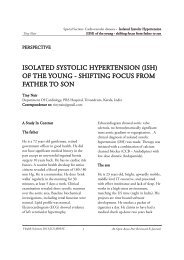
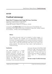
new arena for the practioner. The key<br />
elements of tissue eng<strong>in</strong>eer<strong>in</strong>g are stem cells,<br />
morphogen, and a scaffold of extracellular<br />
matrix (Figure 1). 4<br />
Figure 1. Tissue eng<strong>in</strong>eer<strong>in</strong>g triad<br />
Stem cells are def<strong>in</strong>ed as cells that have the<br />
ability to cont<strong>in</strong>uously divide and produce<br />
progeny cells that develop <strong>in</strong>to various other<br />
cells or tissues. 5 There are two major types of<br />
stem cells, Embryonic and Adult stem cells<br />
their most important properties be<strong>in</strong>g their<br />
ability of self renewal and their ability to<br />
grow <strong>in</strong>-vitro. Compar<strong>in</strong>g the different stem<br />
cell types, adult stem cells, which have the<br />
least amount of ethical concerns, are<br />
presently be<strong>in</strong>g used <strong>in</strong> medical therapies<br />
and are readily accessible. 6 Postnatal stem<br />
cells have been found <strong>in</strong> almost all body<br />
tissues, <strong>in</strong>clud<strong>in</strong>g dental tissues. To date,<br />
eight types of human dental stem cells have<br />
been isolated and characterized: i) Dental<br />
pulp stem cells (DPSCs), ii) Stem cells from<br />
human exfoliated deciduous teeth (SHED),<br />
iii) Stem cells from apical papillae (SCAP),<br />
iv) Periodontal ligament stem cells<br />
(PDLSCs), v) Epithelium-orig<strong>in</strong>ated dental<br />
stem cells (EpSC), vi) Mesenchymal stem<br />
cells (BMSC), vii) Stem cells from the<br />
dental follicle (DFSC), and viii) Endothelial<br />
progenitor cells (EPCs).<br />
Signal<strong>in</strong>g molecules or morphogens are<br />
extracellular secreted signall<strong>in</strong>g molecules<br />
that play a key role <strong>in</strong> signal<strong>in</strong>g many of the<br />
events of repair and regeneration <strong>in</strong>clud<strong>in</strong>g<br />
tertiary dent<strong>in</strong>ogenesis, a response of pulpdent<strong>in</strong><br />
repair. These signall<strong>in</strong>g networks can<br />
<strong>Health</strong> <strong>Sciences</strong> 2013;2(1):JS007 2 An Open Access Peer Reviewed E-Journal
Jojo Kottoor<br />
Technology - Biomimetic <strong>endodontics</strong>: barriers and strategies<br />
be generally classified <strong>in</strong>to growth factors<br />
and <strong>in</strong>flammatory cytok<strong>in</strong>es. 7 Growth<br />
factors are soluble prote<strong>in</strong>s that act as<br />
signal<strong>in</strong>g agents for cells, and <strong>in</strong>fluence<br />
critical functions, such as cell division,<br />
matrix synthesis and tissue differentiation.<br />
Primarily, five em<strong>in</strong>ent families of growth<br />
factors appear to regualate the process of<br />
odontogenesis: Fibroblast growth factor,<br />
Bone morphogenic prote<strong>in</strong> (BMP),<br />
Hedgehog, W<strong>in</strong>gless (WNT) and<br />
Transform<strong>in</strong>g growth factor. Inflammatory<br />
cytok<strong>in</strong>es (Interleuk<strong>in</strong> and Tumor necrosis<br />
factor) are molecules that regulate cellular<br />
behaviour of bone under <strong>in</strong>flammation,<br />
<strong>in</strong>fection and wound heal<strong>in</strong>g. 8<br />
The scaffold or the extracellular matrix is a<br />
mixture of prote<strong>in</strong>s <strong>in</strong>clud<strong>in</strong>g collagen,<br />
fibronect<strong>in</strong>, polysaccharide hyaluronic acid,<br />
proteoglycans and lam<strong>in</strong><strong>in</strong>s form<strong>in</strong>g an<br />
elastic network surround<strong>in</strong>g most cells and<br />
tissue structures. 9 Current scaffolds used <strong>in</strong><br />
tissue eng<strong>in</strong>eer<strong>in</strong>g can be grouped <strong>in</strong>to three<br />
ma<strong>in</strong> categories. Natural scaffolds like<br />
Collagen, lyophilized bone and coral are the<br />
most commonly used natural scaffold. The<br />
ma<strong>in</strong> disadvantage of natural scaffolds is that<br />
they often lack the desired structural<br />
<strong>in</strong>tegrity for its <strong>in</strong>dependent use <strong>in</strong> load<br />
bear<strong>in</strong>g areas. M<strong>in</strong>eral based scaffolds<br />
usually are made of calcium phosphates <strong>in</strong><br />
the form of hydroxyapatite or beta<br />
Tricalcium phosphate and by vary<strong>in</strong>g the<br />
content of calcium the rate of degradation of<br />
these scaffolds can be controlled. They lack<br />
the strength of natural scaffolds and are<br />
brittle mak<strong>in</strong>g it susceptible to fracture 10 and<br />
hence were <strong>in</strong>troduced the synthetic<br />
scaffolds. These <strong>in</strong>clude the porous<br />
ceramics, spongiosus collagen, fibrous<br />
titanium mesh, poly lactic acid (PLA), poly<br />
glycolic acid (PGA), and their copolymers,<br />
poly lactic-co-glycolic acid (PLGA) which<br />
are all polyester material that degrade with<strong>in</strong><br />
the human body. They have the advantage<br />
of be<strong>in</strong>g able to function <strong>in</strong> load bear<strong>in</strong>g;<br />
but have the disadvantage of lack<strong>in</strong>g<br />
osteo<strong>in</strong>ductiveness and an <strong>in</strong>herent difficulty<br />
<strong>in</strong> obta<strong>in</strong><strong>in</strong>g high porosity and regular pore<br />
size. 11 This has led researchers to concentrate<br />
efforts to eng<strong>in</strong>eer scaffolds at the<br />
nanostructural level to modify cellular<br />
<strong>in</strong>teractions with the scaffold. 12<br />
Biomimetic approaches for<br />
regeneration<br />
The creation and delivery of new tissues to<br />
replace diseased, miss<strong>in</strong>g, or traumatized<br />
pulp is referred to as regenerative<br />
<strong>endodontics</strong>. Although current root canal<br />
treatment modalities offer high levels of<br />
success for many conditions, an ideal form<br />
of therapy might consist of regenerative<br />
approaches <strong>in</strong> which diseased or necrotic<br />
pulp tissues are removed and replaced with<br />
healthy pulp tissues to revitalize the teeth.<br />
However, the challenge lies <strong>in</strong> design<strong>in</strong>g and<br />
fabricat<strong>in</strong>g biomimetic materials like<br />
enamel, dent<strong>in</strong>, cementum, pulp, bone and<br />
periodontal ligament and focus should be<br />
toward regenerat<strong>in</strong>g the diseased and<br />
necrotic tissues rather than replac<strong>in</strong>g them<br />
<strong>Health</strong> <strong>Sciences</strong> 2013;2(1):JS007 3 An Open Access Peer Reviewed E-Journal
Jojo Kottoor<br />
Technology - Biomimetic <strong>endodontics</strong>: barriers and strategies<br />
with some conventional replacement<br />
materials. This article reviews current<br />
biomimetic approaches for regeneration<br />
tooth and its associated structures.<br />
a. Root canal revascularization<br />
Treatment of the young permanent tooth<br />
with a necrotic root canal system and an<br />
<strong>in</strong>completely developed root is fraught with<br />
difficulty. Not only is the root canal system<br />
often difficult to fully debride, but the th<strong>in</strong><br />
dent<strong>in</strong>al walls <strong>in</strong>crease the risk of a<br />
subsequent fracture. Other than the<br />
procedure like maturogenesis or<br />
apexogenisis, root canal revascularization is a<br />
procedure to establish the vitality <strong>in</strong> a<br />
nonvital tooth to allow repair and<br />
regeneration of tissues. The typical<br />
revascularization protocol advocates that the<br />
immature tooth, diagnosed with apical<br />
periodontitis, should be accessed and<br />
irrigated with either 5% NaOCl _ 3%<br />
H2O2 or 5.25% NaOCl and Peridex TM<br />
(Procter & Gamble, C<strong>in</strong>c<strong>in</strong>nati, OH). An<br />
antimicrobial agent (either an antibiotic<br />
such as metronidazole, ciprofloxac<strong>in</strong> or<br />
ciprofloxac<strong>in</strong>, metronidazole, m<strong>in</strong>ocycl<strong>in</strong>e<br />
or Ca (OH)2 should be then applied <strong>in</strong>to<br />
the root canal system, and the access cavity<br />
is sealed. After an average of 3 weeks, <strong>in</strong> the<br />
absence of symptoms, the tooth is reentered,<br />
the tissue is irritated until bleed<strong>in</strong>g<br />
is started and a blood clot produced, and<br />
then MTA is placed over the blood clot, and<br />
the access is sealed. With<strong>in</strong> the next 2 years<br />
a gradual <strong>in</strong>crease <strong>in</strong> root development can<br />
be observed. 13 However, revascularization<br />
procedures lack standardization of treatment<br />
protocols with a myriad of reported<br />
techniques, <strong>in</strong>tracanal medicaments and<br />
irrigants.<br />
b. Stem cell therapy<br />
The simplest method to adm<strong>in</strong>ister cells of<br />
appropriate regenerative potential is to <strong>in</strong>ject<br />
the postnatal stem cells <strong>in</strong>to the dis<strong>in</strong>fected<br />
root canal system. Autologous dental stem<br />
cells are the most accessible stem cells for<br />
this therapy. Among the eight different post<br />
natal dental stem cells Dental pulp stem cells<br />
(DPSCs), Stem cells from human exfoliated<br />
deciduous teeth (SHED), and Stem cells<br />
from the apical papilla (SCAP) were more<br />
commonly used <strong>in</strong> the field of regenerative<br />
<strong>endodontics</strong>. 14<br />
DPSCs are the stem cells isolated from<br />
human dental pulp. The most strik<strong>in</strong>g<br />
feature of DPSCs is their ability to<br />
regenerate a dent<strong>in</strong>-pulp-like complex that is<br />
composed of m<strong>in</strong>eralized matrix with<br />
tubules l<strong>in</strong>ed with odontoblasts, and fibrous<br />
tissue conta<strong>in</strong><strong>in</strong>g blood vessels <strong>in</strong> an<br />
arrangement similar to the dent<strong>in</strong>-pulp<br />
complex found <strong>in</strong> normal human teeth. 15<br />
Stem cells from human exfoliated deciduous<br />
teeth (SHED) have become an attractive<br />
alternative for dental tissue eng<strong>in</strong>eer<strong>in</strong>g. The<br />
use of SHED might br<strong>in</strong>g advantages for<br />
tissue eng<strong>in</strong>eer<strong>in</strong>g over the use of stem cells<br />
from adult human teeth as follows: (a)<br />
<strong>Health</strong> <strong>Sciences</strong> 2013;2(1):JS007 4 An Open Access Peer Reviewed E-Journal
Jojo Kottoor<br />
Technology - Biomimetic <strong>endodontics</strong>: barriers and strategies<br />
SHED were reported to have higher<br />
proliferation rate compared with stem cells<br />
from permanent teeth, which might<br />
facilitate the expansion of these cells <strong>in</strong> vitro<br />
before replantation. (b) SHED cells are<br />
retrieved from a tissue that is "disposable"<br />
and readily accessible <strong>in</strong> young patients. ie,<br />
exfoliated deciduous teeth. It also has an<br />
added advantage of abundant cell supply,<br />
and pa<strong>in</strong>less stem cell collection with<br />
m<strong>in</strong>imal <strong>in</strong>vasion. 16<br />
A recent f<strong>in</strong>d<strong>in</strong>g is the presence of a new<br />
population of a mesenchymal stem cells<br />
resid<strong>in</strong>g <strong>in</strong> the apical papilla of <strong>in</strong>completely<br />
developed teeth. They are termed stem cells<br />
from the apical papilla (SCAP). It is<br />
hypothesised that SCAP appear to be the<br />
source of primary odontoblast that are<br />
responsible for the formation of root<br />
dent<strong>in</strong>e, whereas DPSCs are likely the<br />
source of replacement odontoblast. 17 S<strong>in</strong>ce<br />
these stem cells are <strong>in</strong> the apical papilla, they<br />
are benefited by its collateral circulation,<br />
which enables it to survive dur<strong>in</strong>g the<br />
process of pulp necrosis.<br />
There are several advantages to an approach<br />
us<strong>in</strong>g postnatal stem cells. First, autogenous<br />
stem cells are relatively easy to harvest and<br />
to deliver by syr<strong>in</strong>ge, and the cells have the<br />
potential to <strong>in</strong>duce new pulp regeneration.<br />
Second, this approach is already used <strong>in</strong><br />
regenerative medical applications, <strong>in</strong>clud<strong>in</strong>g<br />
bone marrow replacement, and a recent<br />
review has described several potential<br />
endodontic applications. 18 However, there<br />
are several disadvantages to a delivery<br />
method of <strong>in</strong>ject<strong>in</strong>g cells. First, the cells may<br />
have low survival rates. Second, the cells<br />
might migrate to different locations with<strong>in</strong><br />
the body, possibly lead<strong>in</strong>g to aberrant<br />
patterns of m<strong>in</strong>eralization.<br />
c. Pulp implantation<br />
Dental pulp tissue is vulnerable to <strong>in</strong>fection.<br />
Currently, entire pulp amputation followed<br />
by pulp-space dis<strong>in</strong>fection and fill<strong>in</strong>g with<br />
an artificial rubber-like material is employed<br />
to treat the <strong>in</strong>fection - commonly known as<br />
root-canal therapy. In pulp implantation,<br />
replacement pulp tissue is produced by<br />
tissue eng<strong>in</strong>eer<strong>in</strong>g triad and is transplanted<br />
<strong>in</strong>to cleaned and shaped root canal systems.<br />
Rebecca et al had generated Dental pulp like<br />
tissue by us<strong>in</strong>g the tissue eng<strong>in</strong>eer<strong>in</strong>g triad,<br />
the Dental Pulp Stem Cells (DPSCs), a<br />
Collagen Scaffold, and Dent<strong>in</strong> Matrix<br />
prote<strong>in</strong> 1 after subcutaneous transplantation<br />
<strong>in</strong> mice. Collagen served as the scaffold, and<br />
dent<strong>in</strong> matrix prote<strong>in</strong> 1 (DMP1) was the<br />
growth factor. The result concluded that the<br />
triad of DPSCs, a collagen scaffold, and<br />
DMP1 can <strong>in</strong>duce an organized matrix<br />
formation similar to that of pulpal tissue,<br />
which might lead to hard tissue formation. 19<br />
Similarly pulp tissue has also been<br />
successfully regenerated <strong>in</strong>-vitro us<strong>in</strong>g the<br />
tissue eng<strong>in</strong>eer<strong>in</strong>g triad, but by us<strong>in</strong>g a<br />
different stem cell, scaffold and<br />
morphogens. 20<br />
<strong>Health</strong> <strong>Sciences</strong> 2013;2(1):JS007 5 An Open Access Peer Reviewed E-Journal
Jojo Kottoor<br />
Technology - Biomimetic <strong>endodontics</strong>: barriers and strategies<br />
One of the potential problems associated<br />
with the implantation of cultured pulp<br />
tissue is that specialized procedures may be<br />
required to ensure that the cells properly<br />
adhere to root canal walls. When implant<strong>in</strong>g<br />
pulp <strong>in</strong>to the root canals that have blood<br />
supply only from the apical end, enhanced<br />
vascularization is needed <strong>in</strong> order to support<br />
its vitality. As a result, microscale<br />
technologies that provide open channels or<br />
the ability to guide vascular <strong>in</strong>gress from the<br />
apex through the pulp may be of particular<br />
benefit. 21 Recent efforts <strong>in</strong> develop<strong>in</strong>g<br />
scaffold systems for tissue eng<strong>in</strong>eer<strong>in</strong>g have<br />
been focus<strong>in</strong>g on creat<strong>in</strong>g a system that<br />
promotes angiogenesis for the formation of a<br />
vascular network. 22 These scaffolds are<br />
impregnated with growth factors such as<br />
VEGF (vascular endothelial growth factor)<br />
and/or platelet derived growth factor or<br />
further, with the addition of endothelial<br />
cells.<br />
d. Injectable scaffold delivery<br />
Rigid tissue eng<strong>in</strong>eered scaffold structures<br />
provide excellent support for cells used <strong>in</strong><br />
bone and other body areas where the<br />
eng<strong>in</strong>eered tissue is required to provide<br />
physical support. However, <strong>in</strong> root canal<br />
systems a tissue eng<strong>in</strong>eered pulp is not<br />
required to provide structural support of the<br />
tooth. 23 This will allow tissue eng<strong>in</strong>eered<br />
pulp tissue to be adm<strong>in</strong>istered <strong>in</strong> a soft<br />
three-dimensional scaffold matrix. Among<br />
the <strong>in</strong>jectable biomaterials <strong>in</strong>vestigated so<br />
far, hydrogels are more and more attractive<br />
<strong>in</strong> the field of tissue eng<strong>in</strong>eer<strong>in</strong>g. Hydrogels<br />
are <strong>in</strong>jectable scaffolds that can be delivered<br />
by syr<strong>in</strong>ge and have the potential to be non<strong>in</strong>vasive<br />
and easy to deliver <strong>in</strong>to root canal<br />
systems. In theory, the hydrogel may<br />
promote pulp regeneration by provid<strong>in</strong>g a<br />
substrate for cell proliferation and<br />
differentiation <strong>in</strong>to an organized tissue<br />
structure. Past problems with hydrogels<br />
<strong>in</strong>cluded limited control over tissue<br />
formation and development, but advances <strong>in</strong><br />
formulation have dramatically improved<br />
their ability to support cell survival. 24<br />
Despite these advances, hydrogels at are at<br />
an early stage of research, and this type of<br />
delivery system, although promis<strong>in</strong>g, has yet<br />
to be proven to be functional <strong>in</strong> vivo. 52 To<br />
make hydrogels more practical, research is<br />
focus<strong>in</strong>g on mak<strong>in</strong>g them<br />
photopolymerizable to form rigid structures<br />
once they are implanted <strong>in</strong>to the tissue site. 25<br />
e. Three-dimensional cell pr<strong>in</strong>t<strong>in</strong>g<br />
One of the most promis<strong>in</strong>g approaches <strong>in</strong><br />
tissue eng<strong>in</strong>eer<strong>in</strong>g is the application of 3D<br />
scaffolds, which provide cell support and<br />
guidance <strong>in</strong> the <strong>in</strong>itial tissue formation<br />
stage. The porosity of the scaffold and<br />
<strong>in</strong>ternal pore organization <strong>in</strong>fluence cell<br />
migration and play a major role <strong>in</strong> its<br />
biodegradation dynamics, nutrient diffusion<br />
and mechanical stability. In order to control<br />
cell migration and cellular <strong>in</strong>teractions<br />
with<strong>in</strong> the scaffold, novel technologies<br />
capable of produc<strong>in</strong>g 3D structures <strong>in</strong><br />
accordance with predef<strong>in</strong>ed design are<br />
<strong>Health</strong> <strong>Sciences</strong> 2013;2(1):JS007 6 An Open Access Peer Reviewed E-Journal
Jojo Kottoor<br />
Technology - Biomimetic <strong>endodontics</strong>: barriers and strategies<br />
required. In theory, an <strong>in</strong>k-jet-like device is<br />
used to dispense layers of cells suspended <strong>in</strong><br />
a hydrogel to recreate the structure of the<br />
tooth pulp tissue. 26,27 The three-dimensional<br />
cell pr<strong>in</strong>t<strong>in</strong>g technique can be used to<br />
precisely position cells, and this method has<br />
the potential to create tissue constructs that<br />
mimic the natural tooth pulp tissue<br />
structure. This may <strong>in</strong>volve position<strong>in</strong>g of<br />
odontoblasts around the periphery, with<br />
fibroblasts <strong>in</strong> the core. The major challenge<br />
<strong>in</strong>volved is the precise orientation of cellular<br />
suspensions accord<strong>in</strong>g to the apical and<br />
coronal asymmetry of pulp. 28 Theoretically,<br />
the disadvantage of us<strong>in</strong>g the threedimensional<br />
cell pr<strong>in</strong>t<strong>in</strong>g technique is that<br />
careful orientation of the pulp tissue<br />
construct accord<strong>in</strong>g to its apical and coronal<br />
asymmetry would be required dur<strong>in</strong>g<br />
placement <strong>in</strong>to cleaned and shaped root<br />
canal systems.<br />
f. Gene Therapy<br />
Gene therapy is a method of deliver<strong>in</strong>g<br />
genes with the help of viral or non-viral<br />
vectors. The gene delivery <strong>in</strong> <strong>endodontics</strong><br />
would be to deliver m<strong>in</strong>eraliz<strong>in</strong>g genes <strong>in</strong>to<br />
pulp tissue to promote tissue m<strong>in</strong>eralization.<br />
Viral vectors are genetically altered to<br />
elim<strong>in</strong>ate ability of caus<strong>in</strong>g disease, without<br />
los<strong>in</strong>g <strong>in</strong>fectious capacity to the cell. At<br />
present adenoviral, retroviral,<br />
adenoassociated virus, herpes simplex virus,<br />
lentivirus are be<strong>in</strong>g developed. Nonviral<br />
delivery systems uses plasmids, peptides,<br />
cationic liposomes, DNA-ligand complex,<br />
gene guns, electroporation, and<br />
sonoporation to address safety concerns such<br />
as immunogenicity and mutagenesis. 29<br />
Most of the risks of gene therapy may arise<br />
from the vector system rather than the gene<br />
expressed. Widespread cl<strong>in</strong>ical application<br />
still awaits the development of vectors that<br />
are safe, affordable, efficient, simple for<br />
application, and that have ability to express<br />
the required level of transgene for the<br />
sufficient long term. 30<br />
Both <strong>in</strong> vivo and ex vivo approaches can be<br />
used for gene therapy. In the <strong>in</strong> vivo<br />
approach, the gene is delivered systemically<br />
<strong>in</strong>to the blood stream or locally to target<br />
tissues by <strong>in</strong>jection or <strong>in</strong>halation. In the field<br />
of conservative dentistry, BMPs or BMP<br />
genes are directly applied to the exposed<br />
pulp. The ex vivo approach <strong>in</strong>volves genetic<br />
manipulation of cells <strong>in</strong> vitro, which are<br />
subsequently transplanted to the<br />
regeneration site. In conservative dentistry,<br />
DPSCs were isolated, differentiated <strong>in</strong>to<br />
odontoblasts with recomb<strong>in</strong>ant BMPs or<br />
BMP genes, and f<strong>in</strong>ally autogenously<br />
transplanted to regenerate dent<strong>in</strong>. 32<br />
The ma<strong>in</strong> challenges for gene therapy <strong>in</strong> the<br />
next decade will be the requirements to<br />
demonstrate that gene therapy can provide<br />
cost-effective and safe long-term treatments<br />
for conditions that would otherwise lead to<br />
significant pulp necrosis. This <strong>in</strong>dicates the<br />
potential of add<strong>in</strong>g growth factors before<br />
pulp capp<strong>in</strong>g, or <strong>in</strong>corporat<strong>in</strong>g them <strong>in</strong>to<br />
<strong>Health</strong> <strong>Sciences</strong> 2013;2(1):JS007 7 An Open Access Peer Reviewed E-Journal
Jojo Kottoor<br />
Technology - Biomimetic <strong>endodontics</strong>: barriers and strategies<br />
restorative and endodontic materials to<br />
stimulate dent<strong>in</strong> and pulp regeneration.<br />
g. Bioeng<strong>in</strong>eered tooth<br />
The ultimate goal of regenerative therapy is<br />
to develop fully function<strong>in</strong>g bioeng<strong>in</strong>eered<br />
organs that can replace lost or damaged<br />
organs follow<strong>in</strong>g disease, <strong>in</strong>jury, or ag<strong>in</strong>g.<br />
Research on whole tooth regeneration is also<br />
advanc<strong>in</strong>g us<strong>in</strong>g a strategy of transplant<strong>in</strong>g<br />
artificial tooth germ and allow<strong>in</strong>g it to<br />
develop <strong>in</strong> the adult aral environment.<br />
Tissue eng<strong>in</strong>eer<strong>in</strong>g builds a tissue such as<br />
sk<strong>in</strong>, bone and cartilage, by seed<strong>in</strong>g cells<br />
<strong>in</strong>to a scaffold. 33 Research on the fabrication<br />
of teeth from dissociated cells was first<br />
performed us<strong>in</strong>g tooth germ cells. 34 When<br />
explanted, seeded with porc<strong>in</strong>e third<br />
impacted tooth bud cells, were implanted<br />
for 20-30 weeks <strong>in</strong>to omentum,<br />
bioeng<strong>in</strong>eered teeth were visible with<strong>in</strong> the<br />
explants. 35 However, the regenerated teeth<br />
were not identical to their naturally formed<br />
counterparts.<br />
Ikeda et al reported a fully function<strong>in</strong>g tooth<br />
replacement achieved by transplantation of a<br />
bioeng<strong>in</strong>eered tooth germ <strong>in</strong>to the alveolar<br />
bone of a lost tooth region <strong>in</strong> an adult<br />
mouse. 36 Bioeng<strong>in</strong>eered tooth, which was<br />
erupted and occluded, had the correct tooth<br />
structure, hardness of m<strong>in</strong>eralized tissues for<br />
mastication. However, the bioeng<strong>in</strong>eered<br />
tooth was smaller than the other normal<br />
teeth. In addition, the authors couldn’t<br />
regulate the crown width, cusp position, and<br />
tooth pattern<strong>in</strong>g <strong>in</strong>clud<strong>in</strong>g<br />
anterior/posterior and buccal/l<strong>in</strong>gual<br />
structures. However, <strong>in</strong> a more recent study<br />
Oshima et al showed that the crown widths<br />
and the cusp numbers of bioeng<strong>in</strong>eered<br />
molar could be regulated by cell<br />
manipulation method. 37 Tooth regeneration<br />
is an important stepp<strong>in</strong>g stone <strong>in</strong> the<br />
establishment of eng<strong>in</strong>eered organ<br />
transplantation, which is one of the ultimate<br />
goals of regenerative therapies.<br />
Conclusions<br />
The practice of <strong>endodontics</strong> has grown by<br />
leap and bounds <strong>in</strong> the past few decades.<br />
Replacement of diseased or lost tooth<br />
structure with biocompatible restorative<br />
materials is currently the order of today but<br />
each of these procedures does have their own<br />
limitations and drawbacks. Regeneration of<br />
the lost tooth structure rather than<br />
replacement dur<strong>in</strong>g treatment will ensure<br />
better prognosis and higher rate of success.<br />
Hence the future of <strong>endodontics</strong> would<br />
<strong>in</strong>volve the use of such biomimetic materials<br />
which could successfully replace lost enamel,<br />
dent<strong>in</strong>e, cementum and even the pulp<br />
tissue.<br />
References<br />
1. Benyus JM. Biomimicry: Innovation<br />
Inspired by Nature. New York:<br />
William Morro 1997.<br />
<strong>Health</strong> <strong>Sciences</strong> 2013;2(1):JS007 8 An Open Access Peer Reviewed E-Journal
Jojo Kottoor<br />
Technology - Biomimetic <strong>endodontics</strong>: barriers and strategies<br />
2. Dillow A, Lowman A. Biomimetic<br />
Materials And Design. USA: Taylor &<br />
Francis LLC 2002.<br />
3. Slavk<strong>in</strong> HC. <strong>Biomimetics</strong> :<br />
replac<strong>in</strong>g body parts is no longer<br />
science fiction. J Am Dent Assoc<br />
1996;127:1254-7.<br />
4. Craig RB. Tissue Eng<strong>in</strong>eer<strong>in</strong>g:<br />
Restorative Dental Materials, 12 th<br />
edition, 2007.<br />
5. Rao M S. Stem sense: a proposal for<br />
the classification of stem cells. Stem<br />
Cells Dev 2004; 13:452-5.<br />
6. Reznick JB. Stem cells: Emerg<strong>in</strong>g<br />
Medical and Dental Therapies for<br />
Dental Professionals. Dental Town<br />
Oct 2008; 44-52.<br />
7. Bartold PM, Shi S, Gronthos S.<br />
Stem cells and periodontal<br />
regeneration.<br />
Periodontol 2006;40:164-72.<br />
8. Nakashima M, Akam<strong>in</strong>e A. The<br />
application of tissue eng<strong>in</strong>eer<strong>in</strong>g to<br />
regeneration of pulp and dent<strong>in</strong> <strong>in</strong><br />
<strong>endodontics</strong>. J Endod 2005;31:711-8.<br />
9. Prescott RS, Alsanea R, Fayad MI,<br />
Johnson BR, Wenckus CS, Hao J, et<br />
al. In-Vivo generation of dental pulplike<br />
tissue by us<strong>in</strong>g dental pulp stem<br />
cells, a collagen scaffold, and dent<strong>in</strong>e<br />
matrix prote<strong>in</strong> 1 after subcutaneous<br />
transplantation <strong>in</strong> mice. J Endod 2008;<br />
34: 421-426.<br />
10. Sharma B, Elisseeff JH. Eng<strong>in</strong>eer<strong>in</strong>g<br />
structurally organized cartilage and<br />
bone tissues. Ann Biomed Eng<br />
2004;32:148-59.<br />
11. Van Amerongen MJ, Harmsen MC,<br />
Petersen AH, Kors G, van Luyn MJ.<br />
The enzymatic degradation of scaffolds<br />
and their replacement by vascularized<br />
extracellular matrix <strong>in</strong> the mur<strong>in</strong>e<br />
myocardium. Biomaterials 2006; 27:<br />
2247-57.<br />
12. Tuzlakoglu K, Bolgen N, Salgado<br />
AJ, Gomes ME, Pisk<strong>in</strong> E, Reis RL.<br />
Nano- and micro-fiber comb<strong>in</strong>ed<br />
scaffolds: a new architecture for bone<br />
tissue eng<strong>in</strong>eer<strong>in</strong>g. J Mater Sci Mater<br />
Med 2005;16:1099-104.<br />
13. Kottoor J, Velmurugan N.<br />
Revascularization for a necrotic<br />
immature permanent lateral <strong>in</strong>cisor: a<br />
case report and literature review. Int J<br />
Paediatr Dent 2013 (In Press) doi:<br />
10.1111/ipd.12000.<br />
14. Garcia-Godoy F, Murray PE. Status<br />
and potential commercial impact of<br />
stem cell-based treatments on dental<br />
and craniofacial regeneration. Stem<br />
Cells Dev 2006;15:881-7.<br />
<strong>Health</strong> <strong>Sciences</strong> 2013;2(1):JS007 9 An Open Access Peer Reviewed E-Journal
Jojo Kottoor<br />
Technology - Biomimetic <strong>endodontics</strong>: barriers and strategies<br />
15. Gronthos S, Brahim J, Li W, Fisher<br />
LW, Cherman N, Boyde A, et al. Stem<br />
cell properties of human dental pulp<br />
stem cells. J Dent Res 2002;81:531-5.<br />
16. Miura M, Gronthos S, Zhao M, Lu<br />
B, Fisher LW, Robey PG, et al. SHED:<br />
stem cells from human exfoliated<br />
deciduous teeth. Proc Natl Acad Sci U<br />
S A 2003;100:5807-12.<br />
17. Bakopoulou A, Leyhausen G, Volk<br />
J, Tsiftsoglou A, Garefis P, Koidis P, et<br />
al. Comparative analysis of <strong>in</strong> vitro<br />
osteo/odontogenic differentiation<br />
potential of human dental pulp stem<br />
cells (DPSCs) and stem cells from the<br />
apical papilla (SCAP). Arch Oral Biol<br />
2011;56:709-21.<br />
18. Bluteau G, Luder HU, De Bari C,<br />
Mitsiadis TA. Stem cells <strong>in</strong> tooth<br />
eng<strong>in</strong>eer<strong>in</strong>g. Eur Cell Mater 2008;16:1-<br />
9.<br />
19. Prescott RS, Alsanea R, Fayad MI,<br />
Johnson BR, Wenckus CS, Hao J, et<br />
al. In-Vivo generation of dental pulplike<br />
tissue by us<strong>in</strong>g dental pulp stem<br />
cells, a collagen scaffold, and dent<strong>in</strong>e<br />
matrix prote<strong>in</strong> 1 after subcutaneous<br />
transplantation <strong>in</strong> mice. J Endod<br />
2008;34:421-6.<br />
20. Cordeiro MM, Dong Z, Kaneko T,<br />
Zhang Z, Miyazawa M, Shi S, et al.<br />
Dental pulp tissue eng<strong>in</strong>eer<strong>in</strong>g with<br />
stem cells from exfoliated deciduous<br />
teeth. J Endod 2008;34:962-9.<br />
21. Gauv<strong>in</strong> R, Khademhosse<strong>in</strong>i A.<br />
Microscale technologies and modular<br />
approaches for tissue eng<strong>in</strong>eer<strong>in</strong>g:<br />
mov<strong>in</strong>g toward the fabrication of<br />
complex functional structures. ACS<br />
Nano 2011;5:4258-64.<br />
22. Sun Q, Chen RR, Shen Y, Mooney<br />
DJ, Rajagopalan S, Grossman PM.<br />
Susta<strong>in</strong>ed vascular endothelial growth<br />
factor delivery enhances angiogenesis<br />
and perfusion <strong>in</strong> ischemic h<strong>in</strong>d limb.<br />
Pharm Res 2005;22:1110-6.<br />
23. Tziafas D, Alvanou A,<br />
Papadimitriou S, Gasic J, Komnenou<br />
A. Effects of recomb<strong>in</strong>ant basic<br />
fibroblast growth factor, <strong>in</strong>sul<strong>in</strong>-like<br />
growth factor-II and transform<strong>in</strong>g<br />
growth factor-beta 1 on dog dental<br />
pulp cells <strong>in</strong> vivo. Arch Oral Biol<br />
1998;43:431– 44.<br />
24. Desgrandchamps F. Biomaterials <strong>in</strong><br />
functional reconstruction. Curr Op<strong>in</strong><br />
Urol 2000;10:201-6.<br />
25. Luo Y, Shoichet MS. Light-activated<br />
immobilization of biomolecules to<br />
agarose hydrogels for controlled<br />
cellular response. Biomacromolecules<br />
2004;5:2315-23.<br />
<strong>Health</strong> <strong>Sciences</strong> 2013;2(1):JS007 10 An Open Access Peer Reviewed E-Journal
Jojo Kottoor<br />
Technology - Biomimetic <strong>endodontics</strong>: barriers and strategies<br />
26. Dusseiller MR, Schlaepfer D, Koch<br />
M, Kroschewski R, Textor M. An<br />
<strong>in</strong>verted microcontact pr<strong>in</strong>t<strong>in</strong>g method<br />
on topographically structured<br />
polystyrene chips for arrayed micro-3-<br />
D cultur<strong>in</strong>g of s<strong>in</strong>gle cells. Biomaterials<br />
2005;26:5917–25.<br />
27. Sanjana NE, Fuller SB. A fast<br />
flexible <strong>in</strong>k-jet pr<strong>in</strong>t<strong>in</strong>g method for<br />
pattern<strong>in</strong>g dissociated neurons <strong>in</strong><br />
culture. J Neurosci Methods<br />
2004;136:151-63.<br />
28. Michna S, Wu W, Lewis JA.<br />
Concentrated hydroxyapatite <strong>in</strong>ks for<br />
direct-write assembly of 3-D periodic<br />
scaffolds. Biomaterials 2005;26:5632-9.<br />
29. Roemer K, Friedmann T. Concepts<br />
and strategies for human gene therapy.<br />
Eur J Biochem 1992;208:211-25.<br />
30. Baum B J, O'Connell B C. The<br />
impact of gene therapy on dentistry. J<br />
Am Dent Assoc 1995;126:179-89.<br />
31. Nakashima M. Induction of dent<strong>in</strong><br />
formation on can<strong>in</strong>e amputated pulp<br />
by recomb<strong>in</strong>ant human bone<br />
morphogenetic prote<strong>in</strong>s (BMP)-2 and<br />
-4. J Dent Res 1994;73:1515-22.<br />
32. Liu L, L<strong>in</strong>g J, Wei X, Wu L, Xiao Y.<br />
Stem cell regulatory gene expression <strong>in</strong><br />
human adult dental pulp and<br />
periodontal ligament cells undergo<strong>in</strong>g<br />
odontogenic/osteogenic differentiation.<br />
J Endod 2009;35:1368-76.<br />
33. Jernvall J, Jung HS. Genotype,<br />
phenotype, and developmental biology<br />
of molar tooth characters. Am J Phys<br />
Anthropol 2000;Suppl 31:171-90.<br />
34. Peters H, Ball<strong>in</strong>g R. Teeth - Where<br />
and how to make them. Trends Genet.<br />
1999;15:59-65.<br />
35. Duailibi MT, Duailibi SE, Young<br />
CS, Bartlett JD, Vacanti JP, Yelick PC.<br />
Bioeng<strong>in</strong>eered teeth from cultured rat<br />
tooth bud cells. J Dent Res<br />
2004;83:523–8.<br />
36. Ikeda E, Morita R, Nakao K, Ishida<br />
K, Nakamura T, Takano-Yamamoto<br />
T, Ogawa M, Mizuno M, Kasugai S,<br />
Tsuji T. Fully functional<br />
bioeng<strong>in</strong>eered tooth replacement as an<br />
organ replacement therapy. Proc Natl<br />
Acad Sci U S A 2009;106:13475-80.<br />
37. Oshima M, Mizuno M, Imamura A,<br />
Ogawa M, Yasukawa M, Yamazaki H,<br />
et al. Functional tooth regeneration<br />
us<strong>in</strong>g a bioeng<strong>in</strong>eered tooth unit as a<br />
mature organ replacement regenerative<br />
therapy. PLoS One 2011;6:e21531.<br />
<strong>Health</strong> <strong>Sciences</strong> 2013;2(1):JS007 11 An Open Access Peer Reviewed E-Journal