2002 - University of Washington Bone and Joint Sources
2002 - University of Washington Bone and Joint Sources
2002 - University of Washington Bone and Joint Sources

Create successful ePaper yourself
Turn your PDF publications into a flip-book with our unique Google optimized e-Paper software.
Evidence that FGF-18 is a Growth Factor for Mature Human<br />
Articular Chondrocytes<br />
RUSSELL J. FERNANDES, PH.D., JEFF L. ELLSWORTH, PH.D., EMMA E. MOORE, PH.D.,<br />
STEVEN D. HUGHES, PH.D., AND DAVID R. EYRE, PH.D.<br />
Articular cartilages are specialized<br />
connective tissues that dissipate<br />
loads as the bearing surfaces in<br />
joints. Cartilage cells, the chondrocytes,<br />
synthesize a complex extracellular<br />
matrix containing collagens,<br />
proteoglycans, <strong>and</strong> noncollagenous<br />
proteins. Type II collagen, the<br />
predominant collagen <strong>of</strong> cartilage is<br />
polymerized into a network <strong>and</strong> is<br />
responsible for the tensile strength <strong>of</strong><br />
cartilage.<br />
Trauma <strong>and</strong> disease <strong>of</strong> the synovial<br />
joint can cause structural damage to the<br />
articular cartilage. Changes include<br />
age-related fibrillation, cartilage<br />
degeneration due to osteoarthritis, <strong>and</strong><br />
focal chondral <strong>and</strong> osteochondral<br />
defects. In most other tissues, such<br />
lesions would be rapidly repaired.<br />
Adult articular cartilage, however, has<br />
only a very limited capacity to heal.<br />
Efforts to stimulate cartilage healing<br />
have focussed on two general areas:<br />
surgical procedures, designed to<br />
stimulate the endogenous repair<br />
response, <strong>and</strong> biological repair,<br />
designed to stimulate chondrocyte<br />
proliferation <strong>and</strong>/or matrix production<br />
by the application <strong>of</strong> peptide growth<br />
factors or by cell transplantation.<br />
The fibroblast growth factors<br />
(FGF’s) play roles in tissue repair,<br />
embryonic development, <strong>and</strong> growth in<br />
70<br />
60<br />
50<br />
40<br />
30<br />
20<br />
10<br />
0<br />
FGF18<br />
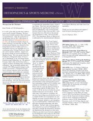
Figure 1: Effect <strong>of</strong> FGF-18 on chondrocyte proliferation.<br />
animals. FGF-18, a novel member <strong>of</strong><br />
the FGF family has been shown to<br />
stimulate hepatocytes <strong>and</strong> intestinal<br />
cells to proliferate. As part <strong>of</strong> a screen<br />
for factors that regulate angiogenesis,<br />
we expressed Fgf18 by adenovirusmediated<br />
gene transfer in the pinnae <strong>of</strong><br />
nude mice. Although an angiogenic<br />
response was not observed, a surprising<br />
phenotype developed: compared to<br />
pinnae that received null adenovirus,<br />
those that received adenovirusexpressing<br />
Fgf18 became visibly thicker.<br />
The increase in thickness was largely<br />
due to an Fgf18-mediated increase in<br />
chondrocyte proliferation, type-II<br />
collagen synthesis, <strong>and</strong> extracellular<br />
matrix production.<br />
These interesting observations<br />
prompted us to investigate if Fgf18 was<br />
expressed by articular chondrocytes<br />
<strong>and</strong> examine the effects <strong>of</strong> highly<br />
purified FGF-18 protein on articular<br />
chondrocytes in vivo <strong>and</strong> vitro. Our<br />
results suggest that Fgf18 may play a<br />
role in the biology <strong>of</strong> normal cartilage.<br />
METHODS<br />
Cell <strong>and</strong> tissue culture<br />
Full thickness adult human<br />
cartilage, <strong>and</strong> chondrocytes (high<br />
density monolayer or micromass)<br />
isolated from talus joints (Northwest<br />
Tissue Center, Seattle) were maintained<br />
+FGF18<br />
Control<br />
0 1 2 3 4 5 6<br />
Week in culture<br />
in serum free culture (DMEM) in the<br />
presence or absence <strong>of</strong> 100ng/ml<br />
recombinant FGF-18 protein. Cell<br />
numbers from monolayer cultures were<br />
determined weekly with the aid <strong>of</strong> a<br />
Neubauer haemocytometer. Cell layer<br />
collagen from monolayer or micromass<br />
cultures was solubilized by pepsin<br />
digestion <strong>and</strong> the a1(II)chains were<br />
identified by western blotting after<br />
SDS-PAGE.<br />
Histochemistry<br />
<strong>and</strong><br />
Immunohistochemistry<br />
Micromass cultures were fixed,<br />
embedded in paraffin, sectioned <strong>and</strong><br />
stained with saffranin O, H&E <strong>and</strong><br />
immunostained with type II collagen<br />
antibody <strong>and</strong> proliferating cell nuclear<br />
antigen (PCNA) antibody.<br />
RESULTS AND DISCUSSION<br />
In situ hybridizations revealed that<br />
Fgf18 mRNA <strong>and</strong> mRNA for two <strong>of</strong> its<br />
receptors, Fgfr3-(IIIc) <strong>and</strong> Fgfr2-(IIIc),<br />
were localized within chondrocytes <strong>of</strong><br />
human talus articular cartilage.<br />
Incubation <strong>of</strong> primary cultures <strong>of</strong><br />
adult human talus articular<br />
chondrocytes with FGF-18 protein<br />
increased the proliferation <strong>of</strong> these cells<br />
(Figure 1).<br />
Western blot analysis <strong>of</strong> collagen<br />
deposited in the extracellular matrix<br />
showed an increase in type II collagen<br />
accumulation within one week after<br />
culture in the presence <strong>of</strong> FGF18<br />
(Figure 2).<br />
In high-density micromass cultures,<br />
(Figure 3) the thickness <strong>of</strong> the cell layer,<br />
cell numbers, staining <strong>of</strong> chondrocyte<br />
nuclei with antibodies to proliferating<br />
cell nuclear antigen (PCNA) (Figure 3,<br />
panels B, D) <strong>and</strong> type II collagen<br />
accumulation in the extracellular<br />
matrix (Figure 3, panels A, C) were<br />
increased by incubation with media<br />
containing 100ng FGF-18/ml for 4<br />
weeks. Increased PCNA staining was<br />
also seen in the nuclei <strong>of</strong> chondrocytes<br />
from explant cultures treated with FGF-<br />
18 (data not shown).<br />
These data imply that FGF-18 can<br />
act as a trophic factor for adult human<br />
articular chondrocytes in primary cell<br />
40 <strong>2002</strong> ORTHOPAEDIC RESEARCH REPORT