2002 - University of Washington Bone and Joint Sources
2002 - University of Washington Bone and Joint Sources
2002 - University of Washington Bone and Joint Sources

You also want an ePaper? Increase the reach of your titles
YUMPU automatically turns print PDFs into web optimized ePapers that Google loves.
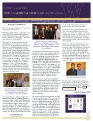
Figure 5: Deep cut in loaded ligament. 12X.<br />
resolution electron microscopy.<br />
This study also confirms that the<br />
patellar ligament is composed <strong>of</strong><br />
independent parallel units. When<br />
transected, the anterior third <strong>of</strong> the<br />
ligament remains fully crimped even<br />
when the intact ligament is under<br />
sufficient tension to completely ablate<br />
crimp. This condition would not occur<br />
in the face <strong>of</strong> any functional crossover<br />
between cut <strong>and</strong> uncut segments. It<br />
also strongly implies that mechanical<br />
coupling between collagen fibrils does<br />
exist within the anterior b<strong>and</strong>, since<br />
partial section <strong>of</strong> that segment does not<br />
cause full recoil <strong>of</strong> the transected part.<br />
Collagen fiber recruitment is thought<br />
to be important to how ligaments resist<br />
elongation under load. This evidence<br />
for lateral support between collagen<br />
fibers further advances our<br />
underst<strong>and</strong>ing <strong>of</strong> the functional ultrastructure<br />
<strong>of</strong> normal ligaments.<br />
RECOMMENDED READING<br />
Gathercole <strong>and</strong> Keller, Matrix, 1991<br />
Woo et al. J. Orthop Res., 1993<br />
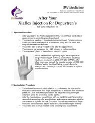
Figure 6: Superficial cut in loaded ligament. 60X.<br />
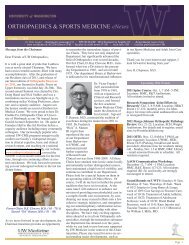
Figure 7: SEM superficial cut in loaded ligament.<br />
34 <strong>2002</strong> ORTHOPAEDIC RESEARCH REPORT