2002 - University of Washington Bone and Joint Sources
2002 - University of Washington Bone and Joint Sources
2002 - University of Washington Bone and Joint Sources

Create successful ePaper yourself
Turn your PDF publications into a flip-book with our unique Google optimized e-Paper software.
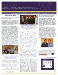
Figure 2: Loaded- 36 Newtons. Partial un-crimping- 12X.<br />
(Figure 2 <strong>and</strong> 3). The 67, 100, <strong>and</strong> 220<br />
N loaded specimens displayed no<br />
evidence <strong>of</strong> crimping in any region<br />
(Figure 4). The blinded observer<br />
correctly identified the loading history<br />
in 14/16 specimens. Two <strong>of</strong> the 9N<br />
loaded specimens were incorrectly<br />
identified as un-loaded specimens.<br />
Collagen Fiber Interaction<br />
Loading the uncut ligaments at<br />
150N resulted in complete un-crimping<br />
<strong>of</strong> the ligament. “Deep” section through<br />
the anterior 1/3 <strong>of</strong> the patellar ligament<br />
resulted in a crimping <strong>of</strong> the cut fibers<br />
which was visible to the full depth <strong>of</strong><br />
the incision, <strong>and</strong> which was visible from<br />
insertion to insertion (Figure 5). The<br />
uncut fibers immediately deep to the<br />
incision were loaded <strong>and</strong> completely<br />
un-crimped (Figure 5). The cut fibers<br />
at the deepest point <strong>of</strong> the incision were<br />
retracted to approximately the same<br />
distance as the superficial fibers.<br />
Superficial section, partially through<br />
the superficial b<strong>and</strong> <strong>of</strong> the patellar<br />
ligament, resulted in a localized<br />
retraction <strong>and</strong> crimping <strong>of</strong> fibers near<br />
the cut site (Figure 6). However, the cut<br />
fibers remained uncrimped along the<br />
majority <strong>of</strong> the length <strong>of</strong> the ligament.<br />
A triangular shaped lesion was visible<br />
on SEM, as the cut fibers adjacent to<br />
the loaded fibers were not as retracted,<br />
nor as crimped as the superficial cut<br />
fibers (Figure 7).<br />
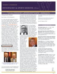
Figure 3: Loaded- 36 Newtons. Partial un-crimping- 60X.<br />
Figure 4: Loaded- 67 Newtons. No Crimp 12X.<br />
DISCUSSION<br />
This study shows that freezefixation<br />
can preserve ligament collagen<br />
in an un-crimped state, <strong>and</strong> thus loaded<br />
fibers can be distinguished from nonloaded<br />
fibers in which a crimp remains.<br />
Using this technique, the rabbit patellar<br />
ligament showed a consistent pattern<br />
<strong>of</strong> strain dispersion with increasing<br />
load. Under these loading conditions,<br />
the central 1/3 <strong>of</strong> the ligament uncrimped<br />
at lower applied loads than the<br />
deep <strong>and</strong> superficial regions. The crimp<br />
in the entire specimen was extinguished<br />
at approximately 67N which, based<br />
upon ligament cross-sectional<br />
estimates, correlates closely with the<br />
previously reported toe-region <strong>of</strong> the<br />
stress strain curve <strong>of</strong> the rabbit patellar<br />
ligament (approx 4.5 MPa) (Woo et al,<br />
1993). The strength <strong>of</strong> this technique<br />
is that it enables investigators to<br />
examine entire ligaments or tendons<br />
preserved in a functionally loaded state<br />
using various forms <strong>of</strong> microscopy,<br />
including the potential use <strong>of</strong> high<br />
<strong>2002</strong> ORTHOPAEDIC RESEARCH REPORT 33