2002 - University of Washington Bone and Joint Sources
2002 - University of Washington Bone and Joint Sources
2002 - University of Washington Bone and Joint Sources

Create successful ePaper yourself
Turn your PDF publications into a flip-book with our unique Google optimized e-Paper software.
Using a Freeze Fixation Technique <strong>and</strong> Histological Crimp<br />
Analysis for Mapping the Strain in Functionally Loaded<br />
Ligaments<br />
RICHARD BOORMAN, M.D., M.SC., TONY NORMAN, B.S.E., FREDERICK A. MATSEN III, M.D.,<br />
AND JOHN CLARK, M.D., PH.D.<br />
In a non-loaded state, type I collagen<br />
fibrils in tendons <strong>and</strong> ligaments<br />
assume a sinusoidal wave shape that<br />
disappears with modest tensile loads<br />
(Gathercole <strong>and</strong> Keller, 1991). This<br />
“crimp” is visible by polarized light<br />
microscopy because collagen fibrils are<br />
aligned in uniform register. A better<br />
underst<strong>and</strong>ing <strong>of</strong> how ligaments<br />
distribute load may yield critical<br />
information on how they fail with<br />
injury <strong>and</strong> disease. Yet the response <strong>of</strong><br />
crimp to load has, by necessity, been<br />
studied primarily in subunits <strong>of</strong><br />
tendons <strong>and</strong> ligaments small enough to<br />
transilluminate <strong>and</strong> not under<br />
functional loading conditions. Freezesubstitution<br />
fixation <strong>of</strong> tissues plungefrozen<br />
under load permits conventional<br />
polarized light <strong>and</strong> electron microscopy<br />
examination <strong>of</strong> collagen fibers<br />
preserved in a loaded state. This study<br />
examines whether the distribution <strong>of</strong><br />
strain can be mapped in loaded<br />
ligaments frozen in situ <strong>and</strong> fixed by<br />
freeze-substitution. In a second line <strong>of</strong><br />
investigation crimping behavior <strong>of</strong><br />
partially sectioned, loaded ligaments<br />
preserved by freeze-fixation was used<br />
to determine whether ligaments are<br />
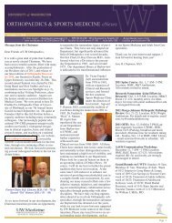
Figure 1: Unloaded ligament- Crimped - 12X.<br />
composed <strong>of</strong> independent bundles <strong>of</strong><br />
interconnected fibrils. We hypothesized<br />
that (A) the crimping pattern <strong>of</strong> patellar<br />
ligaments under polarized light would<br />
progressively extinguish with increasing<br />
loads, <strong>and</strong> (B) that fibrils within a<br />
loaded “functional b<strong>and</strong>” would not<br />
recoil into a uniformly un-crimped<br />
state unless the entire b<strong>and</strong> is<br />
transected.<br />
METHODS AND MATERIALS<br />
Hind-limbs <strong>of</strong> 11 mature New<br />
Zeal<strong>and</strong> white rabbits sacrificed<br />
according to the ethical guidelines <strong>of</strong><br />
the local animal care committee were<br />
harvested, <strong>and</strong> the s<strong>of</strong>t tissues dissected<br />
leaving the femur- intact stifle joint<strong>and</strong><br />
tibia <strong>and</strong> extensor mechanism<br />
completely intact. The joint was fixed<br />
on to a frame while flexed at 90 o , <strong>and</strong> a<br />
simulated isometric quadriceps pull<br />
was applied by means <strong>of</strong> a wire passed<br />
through the patella, thus functionally<br />
loading the patellar ligament.<br />
Specimens were loaded at 9N (n=3),<br />
18N (n=3), 36N (n=3), 67N (n=3),<br />
100N (n=1), <strong>and</strong> 220N (n=1). After 30<br />
seconds a loaded joint was immersed<br />
in isopentane cooled to –165 0 C with<br />
liquid nitrogen. These knees <strong>and</strong> 2 unloaded<br />
control specimens were then<br />
fixed by freeze-substitution in<br />
methanol/acetone solutions <strong>of</strong> acrolein<br />
(-80 o C for 4 days) <strong>and</strong> glutaraldehyde<br />
(-20 o C for a further 4 days). Once<br />
brought to room temperature, the<br />
patellar ligaments were prepared for<br />
histological examination by paraffin<br />
embedding, 10um sectioning in the<br />
mid-sagittal plane <strong>and</strong> staining with<br />
Masson’s trichrome. The specimens<br />
were analyzed under polarized light<br />
microscopy, <strong>and</strong> the crimp pattern was<br />
analyzed by an observer blinded to the<br />
loading history. Based upon the relative<br />
amount <strong>of</strong> un-crimped tissue a<br />
speculation was recorded by the<br />
blinded observer as to whether no load,<br />
minimal load (9N), moderate load (18<br />
<strong>and</strong> 36N), or high load (>67N) was<br />
applied.<br />
Collagen Fiber Interaction<br />
Three specimens had a “deep”<br />
transverse scalpel cut made through the<br />
anterior b<strong>and</strong> <strong>of</strong> the patellar ligament<br />
at the mid-point. Three specimens had<br />
a“superficial” cut partially through the<br />
anterior b<strong>and</strong>. All these specimens were<br />
loaded at 150N. Two uncut controls<br />
were similarly loaded at 150N.<br />
Specimens were prepared for polarized<br />
light microscopy <strong>and</strong> scanning electron<br />
microscopy.<br />
RESULTS<br />
Unloaded ligaments displayed a<br />
consistent, regular pattern <strong>of</strong> crimp<br />
along the entire length <strong>of</strong> the specimens<br />
(Figure 1). The specimens loaded at 9<br />
Newtons were virtually<br />
indistinguishable from the unloaded<br />
specimens, with the exception <strong>of</strong> one<br />
specimen which had some uncrimping<br />
near the insertions. The specimens<br />
loaded at 18N had a b<strong>and</strong> <strong>of</strong> uncrimping<br />
in the central 1/3 <strong>of</strong> the<br />
ligament.<br />
The specimens loaded at 36 N had<br />
an increased area <strong>of</strong> un-crimping but<br />
crimping was still evident in the deep<br />
<strong>and</strong> superficial fibers <strong>of</strong> the ligaments<br />
32 <strong>2002</strong> ORTHOPAEDIC RESEARCH REPORT