

Plasmodium ovale
Plasmodium ovale
Plasmodium ovale

Create successful ePaper yourself
Turn your PDF publications into a flip-book with our unique Google optimized e-Paper software.
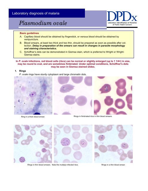
Laboratory diagnosis of malaria<br />
<strong>Plasmodium</strong> <strong>ovale</strong><br />
Basic guidelines<br />
A. Capillary blood should be obtained by fingerstick, or venous blood should be obtained by<br />
venipuncture.<br />
B. Blood smears, at least two thick and two thin, should be prepared as soon as possible after collection.<br />
Delay in preparation of the smears can result in changes in parasite morphology<br />
and staining characteristics.<br />
C. Schüffner’s dots can be demonstrated in Giemsa stain, which is preferred to Wright or Wright-<br />
Giemsa stains.<br />
In P. <strong>ovale</strong> infections, red blood cells (rbcs) can be normal or slightly enlarged (up to 1 1/4×) in size,<br />
may be round to oval, and are sometimes fimbriated. Under optimal conditions, Schüffner's dots<br />
may be seen in Giemsa stained slides.<br />
1. Rings<br />
P. <strong>ovale</strong> rings have sturdy cytoplasm and large chromatin dots.<br />
Ring in a thick blood smear.<br />
Rings in fimbriated rbcs in thin blood smears.<br />
Rings in thin blood smears. Note the multiply-infected rbcs. Rings in a thin blood smear.
Laboratory diagnosis of malaria<br />
<strong>Plasmodium</strong> <strong>ovale</strong><br />
2. Trophozoites<br />
P. <strong>ovale</strong> trophozoites have sturdy cytoplasm, large chromatin dots, and can be compact to slightly irregular.<br />
Trophozoite in a thick blood smear. Compact trophozoites in fimbriated rbcs in thin blood smears. Schüffner’s dots are<br />
also visible.<br />
Ring forms and developing trophozoites in thin blood smears.<br />
Compact trophozoites showing Schüffner’s dots. The image on the left also shows<br />
prominent fimbriation.<br />
Compact trophozoite in a fimbriated rbc<br />
in a thin blood smear.<br />
Schüffner’s dots are also visible.<br />
Ring forms, developing and compact<br />
trophozoites in a thin blood smear.
Laboratory diagnosis of malaria<br />
<strong>Plasmodium</strong> <strong>ovale</strong><br />
3. Gametocytes<br />
P. <strong>ovale</strong> gametocytes are round to oval and may almost fill the red blood cells. Pigment is brown and<br />
more coarse in comparison to P. vivax.<br />
Gametocyte in a thick blood smear. Gametocyte in a thin blood smear. Gametocyte in a thin blood smear. The<br />
infected rbc shows some fimbriation.<br />
Macrogametocytes in thin blood smears. Notice how they nearly fill the infected rbcs. Course pigment, a discrete red nucleus and<br />
Schüffner’s dots can be seen.<br />
Microgametocyte in thin blood smear.<br />
Note the diffuse pigment.<br />
Gametocytes in thin blood smears.
Laboratory diagnosis of malaria<br />
<strong>Plasmodium</strong> <strong>ovale</strong><br />
4. Schizonts<br />
P. <strong>ovale</strong> schizonts have 6 to 14 merozoites with large nuclei, clustered around a mass of dark-brown<br />
pigment.<br />
Schizonts in thick blood smears. Schizont in a thin blood smear.<br />
Schizonts in thin blood smears. Note the infected rbcs are oval.<br />
Schizont in a thin blood smear with a<br />
developing trophozoite.