

A Review of the Genus Eunice - Smithsonian Institution Libraries
A Review of the Genus Eunice - Smithsonian Institution Libraries
A Review of the Genus Eunice - Smithsonian Institution Libraries

You also want an ePaper? Increase the reach of your titles
YUMPU automatically turns print PDFs into web optimized ePapers that Google loves.
NUMBER 523 267<br />
These two species are very similar; branchiae have single<br />
filaments in <strong>the</strong> first 10 setigers in E. aciculata and only in two<br />
to three setigers in E. perrieri; in addition, <strong>the</strong> latter has twice<br />
as many (10) branchial filaments where <strong>the</strong> branchiae are best<br />
developed as <strong>the</strong> former.<br />
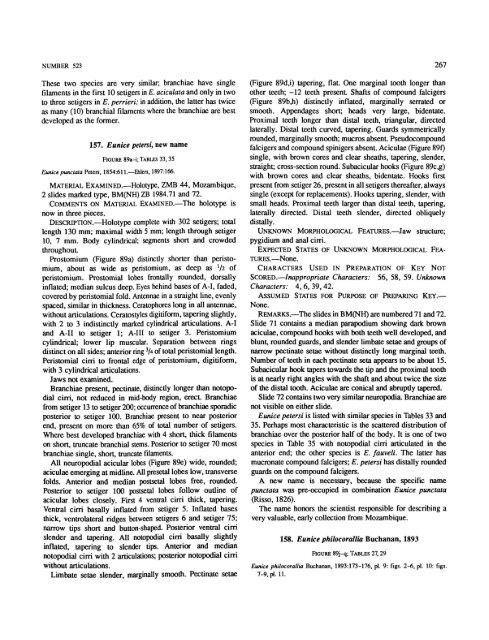
157. <strong>Eunice</strong> petersi, new name<br />
FIGURE 89a-i; TABLES 33,35<br />
<strong>Eunice</strong> punclala Peters. 1854:611.—Ehlers, 1897:166.<br />
MATERIAL EXAMINED.—Holotype, ZMB 44, Mozambique,<br />
2 slides marked type, BM(NH) ZB 1984.71 and 72.<br />
COMMENTS ON MATERIAL EXAMINED.—The holotype is<br />
now in three pieces.<br />
DESCRIPTION.—Holotype complete with 302 setigers; total<br />
length 130 mm; maximal width 5 mm; length through setiger<br />
10, 7 mm. Body cylindrical; segments short and crowded<br />
throughout.<br />
Prostomium (Figure 89a) distinctly shorter than peristomium,<br />
about as wide as pcristomium, as deep as l /i <strong>of</strong><br />
peristomium. Prostomial lobes frontally rounded, dorsally<br />
inflated; median sulcus deep. Eyes behind bases <strong>of</strong> A-I, faded,<br />
covered by pcristomial fold. Antennae in a straight line, evenly<br />
spaced, similar in thickness. Ceratophores long in all antennae,<br />
without articulations. Ceratostyles digitiform, tapering slightly,<br />
with 2 to 3 indistinctly marked cylindrical articulations. A-I<br />
and A-II to setiger 1; A-III to setiger 3. Peristomium<br />
cylindrical; lower lip muscular. Separation between rings<br />
distinct on all sides; anterior ring 3 A <strong>of</strong> total peristomial length.<br />
Peristomial cirri to frontal edge <strong>of</strong> peristomium, digitiform,<br />
with 3 cylindrical articulations.<br />
Jaws not examined.<br />
Branchiae present, pectinate, distinctly longer than notopodial<br />
cirri, not reduced in mid-body region, erect. Branchiae<br />
from setiger 13 to setiger 200; occurrence <strong>of</strong> branchiae sporadic<br />
posterior to setiger 100. Branchiae present to near posterior<br />
end, present on more than 65% <strong>of</strong> total number <strong>of</strong> setigers.<br />
Where best developed branchiae with 4 short, thick filaments<br />
on short, truncate branchial stems. Posterior to setiger 70 most<br />
branchiae single, short, truncate filaments.<br />
All neuropodial acicular lobes (Figure 89e) wide, rounded;<br />
aciculae emerging at midline. All presetal lobes low, transverse<br />
folds. Anterior and median postsetal lobes free, rounded.<br />
Posterior to setiger 100 postsetal lobes follow outline <strong>of</strong><br />
acicular lobes closely. First 4 ventral cirri thick, tapering.<br />
Ventral cirri basally inflated from setiger 5. Inflated bases<br />
thick, ventrolateral ridges between setigers 6 and setiger 75;<br />
narrow tips short and button-shaped. Posterior ventral cirri<br />
slender and tapering. All notopodial cirri basally slightly<br />
inflated, tapering to slender tips. Anterior and median<br />
notopodial cirri with 2 articulations; posterior notopodial cirri<br />
without articulations.<br />
Limbate setae slender, marginally smooth. Pectinate setae<br />
(Figure 89d,i) tapering, flat. One marginal tooth longer than<br />
o<strong>the</strong>r teeth; -12 teeth present Shafts <strong>of</strong> compound falcigers<br />
(Figure 89b,h) distinctly inflated, marginally serrated or<br />
smooth. Appendages short; heads very large, bidentate.<br />
Proximal teeth longer than distal teeth, triangular, directed<br />
laterally. Distal teeth curved, tapering. Guards symmetrically<br />
rounded, marginally smooth; mucros absent. Pseudocompound<br />
falcigers and compound spinigers absent. Aciculae (Figure 89f)<br />
single, with brown cores and clear sheaths, tapering, slender,<br />
straight; cross-section round. Subacicular hooks (Figure 89c,g)<br />
with brown cores and clear sheaths, bidentate. Hooks first<br />
present from setiger 26, present in all setigers <strong>the</strong>reafter, always<br />
single (except for replacements). Hooks tapering, slender, with<br />
small heads. Proximal teeth larger than distal teeth, tapering,<br />
laterally directed. Distal teeth slender, directed obliquely<br />
distally.<br />
UNKNOWN MORPHOLOGICAL FEATURES.—Jaw structure;<br />
pygidium and anal cirri.<br />
EXPECTED STATES OF UNKNOWN MORPHOLOGICAL FEA-<br />
TURES.—None.<br />
CHARACTERS USED IN PREPARATION OF KEY NOT<br />
SCORED.—Inappropriate Characters: 56, 58, 59. Unknown<br />
Characters: 4,6, 39,42.<br />
ASSUMED STATES FOR PURPOSE OF PREPARING KEY.—<br />
None.<br />
REMARKS.—The slides in BM(NH) are numbered 71 and 72.<br />
Slide 71 contains a median parapodium showing dark brown<br />
aciculae, compound hooks with both teeth well developed, and<br />
blunt, rounded guards, and slender limbate setae and groups <strong>of</strong><br />
narrow pectinate setae without distinctly long marginal teeth.<br />
Number <strong>of</strong> teeth in each pectinate seta appears to be about 15.<br />
Subacicular hook tapers towards <strong>the</strong> tip and <strong>the</strong> proximal tooth<br />
is at nearly right angles with <strong>the</strong> shaft and about twice <strong>the</strong> size<br />
<strong>of</strong> <strong>the</strong> distal tooth. Aciculae are conical and abruptly tapered.<br />
Slide 72 contains two very similar neuropodia. Branchiae are<br />
not visible on ei<strong>the</strong>r slide.<br />
<strong>Eunice</strong> petersi is listed with similar species in Tables 33 and<br />
35. Perhaps most characteristic is <strong>the</strong> scattered distribution <strong>of</strong><br />
branchiae over <strong>the</strong> posterior half <strong>of</strong> <strong>the</strong> body. It is one <strong>of</strong> two<br />
species in Table 35 with notopodial cirri articulated in <strong>the</strong><br />
anterior end; <strong>the</strong> o<strong>the</strong>r species is E. fauveli. The latter has<br />
mucronate compound falcigers; E. petersi has distally rounded<br />
guards on <strong>the</strong> compound falcigers.<br />
A new name is necessary, because <strong>the</strong> specific name<br />
punctata was pre-occupied in combination <strong>Eunice</strong> punctata<br />
(Risso, 1826).<br />
The name honors <strong>the</strong> scientist responsible for describing a<br />
very valuable, early collection from Mozambique.<br />
158. <strong>Eunice</strong> philocorallia Buchanan, 1893<br />
FIGURE 89j-q; TABLES 27,29<br />
<strong>Eunice</strong> philocorallia Buchanan, 1893:173-176, pi. 9: figs. 2-6, pi. 10: figs.<br />
7-9, pi. 11.


