ANA Screen ELISA ANA Screen ELISA - Montwell.com.tr
ANA Screen ELISA ANA Screen ELISA - Montwell.com.tr
ANA Screen ELISA ANA Screen ELISA - Montwell.com.tr

Create successful ePaper yourself
Turn your PDF publications into a flip-book with our unique Google optimized e-Paper software.
<s<strong>tr</strong>ong>ANA</s<strong>tr</strong>ong> <s<strong>tr</strong>ong>Screen</s<strong>tr</strong>ong> <s<strong>tr</strong>ong>ELISA</s<strong>tr</strong>ong><br />
25012<br />
Background<br />
Circulating antibodies to in<strong>tr</strong>a-cellular<br />
s<strong>tr</strong>uctures especially to nuclear antigens<br />
represent a characteristic feature of<br />
systemic autoimmune diseases. Among<br />
the most important ones are doubles<strong>tr</strong>anded<br />
DNA (dsDNA), Ro52, Ro60, La,<br />
cen<strong>tr</strong>omere proteins, Scl-70<br />
(topoisomerase I, topo I), RNP/Sm, Sm,<br />
Jo-1 and PM/Scl. Many of those antigens<br />
are considered as specific marker for a<br />
certain autoimmune disease whereas<br />
others show moderate specificity (see<br />
table 1). Since in the indirect<br />
immunofluorescence (IIF) test a few<br />
clinically relevant antibodies (e.g. Ro60<br />
(SS-A), Ro52, Jo-1) are hardly<br />
detectable, the <s<strong>tr</strong>ong>ANA</s<strong>tr</strong>ong> <s<strong>tr</strong>ong>Screen</s<strong>tr</strong>ong> <s<strong>tr</strong>ong>ELISA</s<strong>tr</strong>ong><br />
represents a useful alternative method to<br />
IIF.<br />
Table 1 Overview of the autoantigens<br />
Antigen<br />
dsDNA (r)<br />
Ro52 (r)<br />
Ro60 (r)<br />
La (r)<br />
RNP/Sm (n)<br />
Sm (n)<br />
Jo-1 (r)<br />
Scl-70 (r)<br />
CENP (r)<br />
PM1-Alpha (s)<br />
Disease association<br />
Systemic lupus erythematosus (SLE)<br />
SjS, PM, SLE, SSc<br />
Sjögren Syndrome (SjS)<br />
Sjögren Syndrome (SjS)<br />
Mixed connective tissue disease<br />
SLE<br />
Polymyositis (PM)<br />
Systemic sclerosis (SSc)<br />
Systemic sclerosis (SSc)<br />
PM/SSc overlap syndrome<br />
r = re<s<strong>tr</strong>ong>com</s<strong>tr</strong>ong>binant, n = native, s = synthetic<br />
Intended use<br />
The <s<strong>tr</strong>ong>ANA</s<strong>tr</strong>ong> <s<strong>tr</strong>ong>Screen</s<strong>tr</strong>ong> <s<strong>tr</strong>ong>ELISA</s<strong>tr</strong>ong> is intended for<br />
the semi-quantitative determination of<br />
antinuclear antibodies (<s<strong>tr</strong>ong>ANA</s<strong>tr</strong>ong>s). The<br />
results of the <s<strong>tr</strong>ong>ANA</s<strong>tr</strong>ong> <s<strong>tr</strong>ong>Screen</s<strong>tr</strong>ong> <s<strong>tr</strong>ong>ELISA</s<strong>tr</strong>ong> aid to<br />
the diagnosis of systemic autoimmune<br />
diseases. The test should be used as<br />
initial screening test.<br />
La (SS-B)<br />
SmD<br />
CENP<br />
Scl-70<br />
RNP<br />
TMB<br />
SmB<br />
Jo-1<br />
Measurement at<br />
450 nm<br />
Anti-IgG-HRP<br />
Conjugate<br />
PM1-Alpha<br />
dsDNA<br />
Ro52<br />
Ro60 (SS-A)<br />
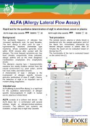
Figure 1 Test principle <s<strong>tr</strong>ong>ANA</s<strong>tr</strong>ong> <s<strong>tr</strong>ong>Screen</s<strong>tr</strong>ong> <s<strong>tr</strong>ong>ELISA</s<strong>tr</strong>ong><br />
The wells of the <s<strong>tr</strong>ong>ANA</s<strong>tr</strong>ong> <s<strong>tr</strong>ong>Screen</s<strong>tr</strong>ong> <s<strong>tr</strong>ong>ELISA</s<strong>tr</strong>ong> are coated with a<br />
mixture of native, re<s<strong>tr</strong>ong>com</s<strong>tr</strong>ong>binant and synthetic autoantigens<br />
(see Table 1). During the first step diluted patient sample is<br />
incubate in the wells. During the incubation time, the<br />
autoantibodies contained in the sample will bind to coated<br />
antigens. After washing steps and conjugate- and<br />
subs<strong>tr</strong>ate–incubation these <s<strong>tr</strong>ong>com</s<strong>tr</strong>ong>plexes can be quantified<br />
by photome<strong>tr</strong>ic analysis.
General features<br />
• CE marked<br />
• User-friendly<br />
• Colored reagents<br />
• Ready to use reagents (except washing<br />
buffer)<br />
• Breakapart mircotiter s<strong>tr</strong>ips<br />
Technical information<br />
• Assay time: < 1.5 h at RT<br />
(30 min /30 min /15 min)<br />
• 3 µL serum or plasma per test<br />
• Detection System: HRP/TMB<br />
(OD 450 nm / 620 nm )<br />
• Wide measuring range<br />
• Low detection limit<br />
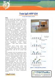
ID Target <s<strong>tr</strong>ong>ELISA</s<strong>tr</strong>ong> (RU) Interpretation<br />
CDC 1 DNA 3.8 positive<br />
CDC 2 SS-B/La 2.6 positive<br />
CDC 3<br />
RNP/Sm,<br />
SS-A/Ro, SS-B/La<br />
5.0 positive<br />
CDC 4 U-1 RNP 5.2 positive<br />
CDC 5 Sm 4.4 positive<br />
CDC 6 Fibrillarin 1.0 borderline<br />
CDC 7 SS-A/Ro 1.9 positive<br />
CDC 8 Cen<strong>tr</strong>omere 2.6 positive<br />
CDC 9 Scl-70 2.6 positive<br />
CDC 10 Jo-1 4.6 positive<br />
CDC 11 PM/Scl (PM 1) 1.3 borderline<br />
CDC 12 Rib-P 0.7 negative<br />
Figure 2<br />
Results of the CDC <s<strong>tr</strong>ong>ANA</s<strong>tr</strong>ong> reference sera. 12 reference serum<br />
samples, available from the “Center for Disease Con<strong>tr</strong>ol and<br />
Prevention (CDC)” were tested in the <s<strong>tr</strong>ong>ANA</s<strong>tr</strong>ong> <s<strong>tr</strong>ong>Screen</s<strong>tr</strong>ong> <s<strong>tr</strong>ong>ELISA</s<strong>tr</strong>ong><br />
(REF: 25012). All sera, except CDC 6, 11 and 12 have been<br />
tested positive. CDC 6 and 11 are borderline and CDC 12 is<br />
negative. The borderline result of CDC 6 and the negative<br />
result of CDC 12 can be explained by the absence of<br />
Fibrillarin and Rib-P in the <s<strong>tr</strong>ong>ANA</s<strong>tr</strong>ong> <s<strong>tr</strong>ong>Screen</s<strong>tr</strong>ong>.<br />
Assay performance<br />
• Good correlation to reference <s<strong>tr</strong>ong>ELISA</s<strong>tr</strong>ong><br />
systems<br />
• Excellent “lot to lot” correlation R 2 > 0.9<br />
• Low in<strong>tr</strong>a- and inter-assay variation<br />
• The clinical specificity against apparently<br />
healthy blood donors has been<br />
determined as 100% in a cohort of<br />
Caucasian SLE patients<br />
• The clinical sensitivity for SLE in a<br />
Caucasian SLE cohort has been found at<br />
85% (cut-off 1.0 RU)<br />
Table 2 Precision (in<strong>tr</strong>a-assay variation) of the <s<strong>tr</strong>ong>ANA</s<strong>tr</strong>ong><br />
<s<strong>tr</strong>ong>Screen</s<strong>tr</strong>ong> <s<strong>tr</strong>ong>ELISA</s<strong>tr</strong>ong><br />
Serum Mean RU CV %<br />
<s<strong>tr</strong>ong>ANA</s<strong>tr</strong>ong>/1 (n=4) 3.1 2.2<br />
<s<strong>tr</strong>ong>ANA</s<strong>tr</strong>ong>/2 (n=4) 3.5 7.1<br />
<s<strong>tr</strong>ong>ANA</s<strong>tr</strong>ong>/3 (n=4) 4.0 6.0<br />
Table 3 Precision (inter-assay variation) of the <s<strong>tr</strong>ong>ANA</s<strong>tr</strong>ong><br />
<s<strong>tr</strong>ong>Screen</s<strong>tr</strong>ong> <s<strong>tr</strong>ong>ELISA</s<strong>tr</strong>ong><br />
Serum M ean R U CV %<br />
AN A/1 (n=8) 2.5 11.1<br />
AN A/2 (n=8) 3.6 3.2<br />
AN A/3 (n=8) 3.9 9.1<br />
Literature<br />
1. Tan EM: Antinuclear antibodies: diagnostic<br />
markers for autoimmune diseases and probes for<br />
cell biology. Adv Immunol 1989, 44:93-151.<br />
2. Hoffman IE, Peene I, Veys EM, De Keyser F: Detection<br />
of specific antinuclear reactivities in patients<br />
with negative anti-nuclear antibody immunofluorescence<br />
screening tests. Clin Chem 2002,<br />
48:2171-2176.<br />
3. Mahler M, Raijmakers R, Fritzler MJ: Challenges<br />
and Con<strong>tr</strong>oversies in Autoantibodies Associated<br />
with Systemic Rheumatic Diseases. Curr Rheumatol<br />
Rev 2007, 12:67-78.<br />
4. Mahler M, Eisfeller P, Silvermann ED, Fritzler MJ:<br />
Diagnostic value of a novel <s<strong>tr</strong>ong>ANA</s<strong>tr</strong>ong> <s<strong>tr</strong>ong>Screen</s<strong>tr</strong>ong> <s<strong>tr</strong>ong>ELISA</s<strong>tr</strong>ong>.<br />
10th International Workshop on autoantibodies and<br />
autoimmunity, Guadalajara 2008.<br />
2008-10<br />
Dr. Fooke Laboratorien GmbH - Mains<strong>tr</strong>aße 85 - 41469 Neuss - Germany<br />
Phone: ++ 49 2137 1005-0 - Fax: ++ 49 2137 12409 - email: information@fooke-labs.de