Biological activity of substrate-bound basic fibroblast growth factor ...
Biological activity of substrate-bound basic fibroblast growth factor ...
Biological activity of substrate-bound basic fibroblast growth factor ...

You also want an ePaper? Increase the reach of your titles
YUMPU automatically turns print PDFs into web optimized ePapers that Google loves.
3892<br />
<strong>Biological</strong> <strong>activity</strong> <strong>of</strong> immobilized FGF2<br />
E Tanghetti et al<br />
Taken together, the data indicate that <strong>substrate</strong><strong>bound</strong><br />
FGF2 retains its mitogenic capacity that<br />
requires TK <strong>activity</strong> and ERK 1/2 phosphorylation.<br />
These observations raise the possibility that FGFR<br />
localized at the basal side <strong>of</strong> endothelial cells is<br />
involved in mediating the mitogenic response <strong>of</strong><br />
adherent cells to the immobilized <strong>growth</strong> <strong>factor</strong>.<br />
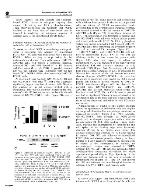
Figure 5 Expression <strong>of</strong> dominant negative DFGFR1 in GM<br />
7373 cells. GM 7373 cells were transfected with a retroviral<br />
expression vector harboring the full length TK + FGFR1 cDNA<br />
or the truncated TK 7 FGFR1 cDNA. Stable transfectants were<br />
isolated, generating GM7373-FGFR1 and GM7373-DFGFR1<br />
cells, respectively. Then, binding <strong>of</strong> 125 I-FGF2 to high anity<br />
receptors was evaluated as described in Materials and methods<br />
(A). Also, cell extracts were probed with anti-FGFR1 antibodies<br />
by Western blotting (B). In C, parental, GM7373-FGFR1, and<br />
GM7373-DFGFR1 cells were incubated for 20 min in the<br />
presence <strong>of</strong> the indicated concentrations <strong>of</strong> soluble FGF2. Then,<br />
Western blot analysis <strong>of</strong> the cell extracts was performed using<br />
anti-phospho-ERK 1/2 antibodies. a, parental GM 7373 cells; b,<br />
GM7373-DFGFR1 cells; c, GM7373-FGFR1 cells<br />
Dominant negative TK FGFR1 abolishes the response <strong>of</strong><br />
endothelial cells to immobilized FGF2<br />
To assess the role <strong>of</strong> FGFR in transducing a mitogenic<br />
signal in endothelial cells adherent to immobilized<br />
FGF2, GM 7373 cells were transfected with a mutated<br />
FGFR1 cDNA carrying a stop codon in the<br />
juxtamembrane domain. These cells, named GM7373-<br />
DFGFR1 cells, will express a dominant negative,<br />
truncated TK 7 DFGFR1 devoid <strong>of</strong> its TK domain<br />
and C-terminus (Li et al., 1994). In parallel, distinct<br />
GM7373 cell cultures were transfected with the full<br />
length TK + FGFR1 cDNA, thus generating GM7373-<br />
FGFR1 cells.<br />
As shown in Figure 5A, both GM7373-DFGFR1 and<br />
GM7373-FGFR1 cells binds 125 I-FGF2 with a capacity<br />
signi®cantly higher than that <strong>of</strong> parental cells. Western<br />
blot analysis <strong>of</strong> the cell extracts probed with a<br />
monoclonal anti-FGFR1 antibody evidenced the presence<br />
<strong>of</strong> a Mr 130 000 immunoreactive band in the cell<br />
extract <strong>of</strong> GM7373-FGFR1 cells (Figure 5B), corresponding<br />
to the full length receptor and comigrating<br />
with a fainter band present in the extract <strong>of</strong> parental<br />
cells. An intense Mr 90 000 immunoreative band,<br />
corresponding to the overexpressed truncated receptor,<br />
was instead detected in the cell extract <strong>of</strong> GM7373-<br />
DFGFR1 cells (Figure 5B). A signi®cant increase <strong>of</strong><br />
ERK 1/2 phosphorylation was detectable in parental and<br />
GM7373-FGFR1 cells adherent to tissue culture plastic<br />
and treated with soluble FGF2. No ERK 1/2 phosphorylation<br />
was instead detected in FGF2-treated GM7373-<br />
DFGFR1 cells, thus con®rming the dominant negative<br />
e€ect <strong>of</strong> the truncated TK 7 receptor (Figure 5C).<br />
GM7373-DFGFR1 and GM7373-FGFR1 cells adhere<br />
to immobilized FGF2, FN, or VN with an<br />
eciency similar to that shown by parental cells<br />
(Figure 6A). Also, their capacity to adhere to<br />
immobilized FGF2 was prevented by the highly speci®c<br />
monoclonal LM 609 antibody directed to a v b 3<br />
(Cheresh, 1987) (Figure 6A). Accordingly, all the cell<br />
lines express similar amounts <strong>of</strong> a v b 3 , as evidenced by<br />
Western blot analysis <strong>of</strong> the cell extracts (data not<br />
shown). However, GM7373-DFGFR1 cells have lost<br />
the ability to proliferate when seeded on FGF2-coated<br />
plastic; this ability is instead retained by GM7373-<br />
FGFR1 transfectants (Figure 6B). As observed for<br />
parental cells, GM7373-FGFR1 and GM7373-<br />
DFGFR1 cells do not proliferate when seeded on<br />
heat-inactivated FGF2, FN, or VN. It must be pointed<br />
out that no signi®cant di€erences were observed in the<br />
proliferation rate <strong>of</strong> the three cell lines when seeded on<br />
tissue culture plastic and maintained in 10% FCS (data<br />
not shown).<br />
Administration <strong>of</strong> FGF2 to the culture medium<br />
a€ects the appearance <strong>of</strong> endothelial cells that acquire<br />
an elongated, ®broblast-like morphology associated to<br />
increased cell motility (Tsuboi et al., 1990). Similarly,<br />
GM7373-FGFR1 cells adherent to FGF2-coated<br />
plastic took an elongated appearance with a crisscross<br />
pattern within 24 ± 48 h after seeding (Figure 7a,b).<br />
Also, crawling cells characterized by lamellipodia and<br />
microspikes at the leading edge were frequently<br />
observed (Figure 7c ± e). Similar even though less<br />
dramatic changes were observed 48 ± 72 h after seeding<br />
in parental GM 7373 cells adherent to immobilized<br />
FGF2 (data not shown), possibly re¯ecting the lower<br />
number <strong>of</strong> FGFR receptors expressed by parental cells<br />
in respect to the transfectants. In contrast, no<br />
morphological changes were observed in FGF2-<br />
adherent GM7373-DFGFR1 cells that retained a ¯atter<br />
cobblestone-like appearance throughout the whole<br />
experimental period (Figure 7f,g). Speci®city <strong>of</strong> the<br />
e€ect was demonstrated by the lack <strong>of</strong> <strong>activity</strong> <strong>of</strong><br />
immobilized FN (Figure 7h ± l) and VN (not shown)<br />
that did not in¯uence the morphological features <strong>of</strong><br />
adherent GM7373-FGFR1 and parental cells.<br />
Immobilized FGF2 recruits FGFR1 in cell-substratum<br />
contact sites<br />
The above data suggest that immobilized FGF2 can<br />
interact with FGFR1 at the basal side <strong>of</strong> the adherent<br />
Oncogene