Three - University of Arkansas Physics Department
Three - University of Arkansas Physics Department
Three - University of Arkansas Physics Department

You also want an ePaper? Increase the reach of your titles
YUMPU automatically turns print PDFs into web optimized ePapers that Google loves.
SHOP NOTES<br />
These are "how to do it"papers. They should be written and illustrared so that the reader may eusily follow whatever<br />
instruction or advice is beirig given.<br />
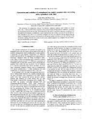
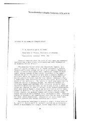
Enabling in situ atomic-scale characterization <strong>of</strong> epitaxial surfaces<br />
and interfaces<br />
J. B. Smathers, D. W. Bullock, Z. Ding, G. J. Salamo, and P. M. ~hibado~)<br />
<strong>Department</strong> <strong>of</strong> <strong>Physics</strong>, Universiy <strong>of</strong> <strong>Arkansas</strong>, Fayetteville, <strong>Arkansas</strong> 72701<br />
B. Gerace<br />
Omicron Associates, Denver, Colorado 80209<br />
W. Wirth<br />
Omicron Vakuumphysik, Taunusstein 65232, Germany<br />
(Received 1 May 1998; accepted 28 August 1998)<br />
A custom designed sample handling system which allows the integration <strong>of</strong> a commercially<br />
available scanning tunneling microscope (STM) facility with a commercially available molecular<br />
beam epitaxy (MBE) facility is described. No customization <strong>of</strong> either the STM imaging stage or the<br />
MBE is required to implement this design. O 1998 American Vacuum Society.<br />
[SO734-2 1 lX(98)01606-01<br />
Molecular beam epitaxy (MBE) has emerged as the technology<br />
pushing the frontiers <strong>of</strong> compound semiconductor device<br />
development. MBE uniquely provides the atomic layer<br />
control <strong>of</strong> the growth process necessary to produce compositionally<br />
abrupt interfaces and exact layer thicknesses required<br />
for band-gap engineering. At present, not all such<br />
novel structures can be routinely fabricated nor utilized in<br />
device applications. Progress in this area will require an improved<br />
understanding <strong>of</strong> the fundamental surface processes<br />
involved in MBE, such as diffusion, nucleation, and growth.<br />
Ideally, one would like to study these processes with spatial<br />
resolution down to the scale <strong>of</strong> individual atoms. In the last<br />
decade, scanning tunneling microscopy (STM) has matured<br />
to become a dominate tool for atomic-scale investigations.<br />
For this reason, it is not surprising that there is growing<br />
interest in integrating MBE and STM technologies into a<br />
single ultrahigh vacuum (IJHV) system. Combining STM<br />
with MBE has been accomplished in only a few<br />
and they have already demonstrated their ability to advance<br />
the fundamental understanding <strong>of</strong> crystal growth<br />
processes.4-6 One limitation <strong>of</strong> existing systems is that they<br />
use custom microscopes, making the successful intcgration<br />
and operation <strong>of</strong> such systems limited to specialized experts<br />
in the STM community. In this Shop Note, we detail the<br />
modifications required to integrate a commercially available<br />
STM~ with a commercially available MBE,~ without customizing<br />
the microscope or deposition system.<br />
Our approach for integrating MBE with STM is to connect<br />
two separate UHV chambers via a third commercially<br />
available ultrahigh vacuum transfer chamber (the MBE and<br />
STM are separated by -2 m). One benefit <strong>of</strong> this approach is<br />
that it allows for the independent operation <strong>of</strong> the MBE and<br />
STM, while avoiding contamination and vibration problems,<br />
- -<br />
a'~lecti-onic mail: thibado@comp.uark.edu<br />
as well as electrical and thermal noise problems inherent to<br />
placing a STM inside a growth chamber.<br />
The customized components required for this integration<br />
are associated with the sample mounts and transfers. We use<br />
both the standard MBE and STM sample mounts as our starting<br />
material, from which we fabricated new sample mounts<br />
that could still be transferred in the conventional way. The<br />
current MBE standard for sample mounting and manipulation<br />
is a circular 3 in. diam molybdenum wafer holder which<br />
will be referred to as a "MBE moly block" throughout this<br />
article. The current STM sample plate standard is a tantalum<br />
plate which is significantly smaller (15 mmXl8 mm) than<br />
the MBE moly block. The MBE moly block is transferred<br />
and mounted using radially oriented pins, while the STM<br />
sample plate is held by an eye hole situated on the top side <strong>of</strong><br />
the mount [see Fig. 1 (b)].<br />
Successful STM studies <strong>of</strong> MBE grown material require<br />
several factors to be simultaneously met. First, the substrate<br />
must be heated to temperatures in excess <strong>of</strong> 600 "C in order<br />
to remove the oxide from the semiconductor substrate. Second,<br />
the substrate surface must have glancing angle access to<br />
perform in situ reflection high-energy electron diffraction<br />
(RHEED). Third, the STM sample plate must be rigidly<br />
mounted to the STM imaging stage.<br />
The core modification made to the standard STM sample<br />
plate which allowed the successful integration is the addition<br />
<strong>of</strong> a 1 mm thick tantalum over plate to the STM sample plate<br />
[see Figs. l(a) and lib)]. The over plate has a square shape<br />
and is sized to match the vertical dimension (15 mm) <strong>of</strong> a<br />
standard sample plate. In addition, a rectangular cutout (7<br />
mmX2 mm) centered on the bottom edge <strong>of</strong> the over plate<br />
allows a compression point contact to be made to the sample<br />
plate when mounted on the STM imaging stage. Once machined<br />
into the proper geometry, the over plate is easily spot<br />
welded to a conventional tantalum sample plate. The shape<br />
3112 J. Vac. Sci. Technol. B 16(6), NovlDec 1998 0734-211W98116(6)1311213/$15.00 01998 American Vacuum Society 3112