Monte Carlo simulations of optical human sinusitis diagnostics
Monte Carlo simulations of optical human sinusitis diagnostics
Monte Carlo simulations of optical human sinusitis diagnostics

Create successful ePaper yourself
Turn your PDF publications into a flip-book with our unique Google optimized e-Paper software.
<strong>Monte</strong> <strong>Carlo</strong> <strong>simulations</strong> <strong>of</strong> <strong>optical</strong> <strong>human</strong> <strong>sinusitis</strong> <strong>diagnostics</strong><br />
Lisa Simonsson,<br />
Department <strong>of</strong> Physics, Lund Institute <strong>of</strong> Technology, P.O. Box 118, SE-221 00 Lund, Sweden<br />
April 2, 2007<br />
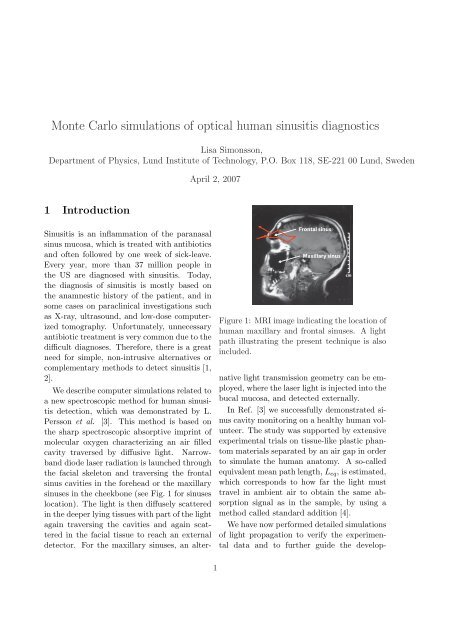
1 Introduction<br />
Sinusitis is an inflammation <strong>of</strong> the paranasal<br />
sinus mucosa, which is treated with antibiotics<br />
and <strong>of</strong>ten followed by one week <strong>of</strong> sick-leave.<br />
Every year, more than 37 million people in<br />
the US are diagnosed with <strong>sinusitis</strong>. Today,<br />
the diagnosis <strong>of</strong> <strong>sinusitis</strong> is mostly based on<br />
the anamnestic history <strong>of</strong> the patient, and in<br />
some cases on paraclinical investigations such<br />
as X-ray, ultrasound, and low-dose computerized<br />
tomography. Unfortunately, unnecessary<br />
antibiotic treatment is very common due to the<br />
difficult diagnoses. Therefore, there is a great<br />
need for simple, non-intrusive alternatives or<br />
complementary methods to detect <strong>sinusitis</strong> [1,<br />
2].<br />
We describe computer <strong>simulations</strong> related to<br />
a new spectroscopic method for <strong>human</strong> <strong>sinusitis</strong><br />
detection, which was demonstrated by L.<br />
Persson et al. [3]. This method is based on<br />
the sharp spectroscopic absorptive imprint <strong>of</strong><br />
molecular oxygen characterizing an air filled<br />
cavity traversed by diffusive light. Narrowband<br />
diode laser radiation is launched through<br />
the facial skeleton and traversing the frontal<br />
sinus cavities in the forehead or the maxillary<br />
sinuses in the cheekbone (see Fig. 1 for sinuses<br />
location). The light is then diffusely scattered<br />
in the deeper lying tissues with part <strong>of</strong> the light<br />
again traversing the cavities and again scattered<br />
in the facial tissue to reach an external<br />
detector. For the maxillary sinuses, an alter-<br />
Frontal sinus<br />
Maxillary sinus<br />
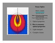
Figure 1: MRI image indicating the location <strong>of</strong><br />
<strong>human</strong> maxillary and frontal sinuses. A light<br />
path illustrating the present technique is also<br />
included.<br />
native light transmission geometry can be employed,<br />
where the laser light is injected into the<br />
bucal mucosa, and detected externally.<br />
In Ref. [3] we successfully demonstrated sinus<br />
cavity monitoring on a healthy <strong>human</strong> volunteer.<br />
The study was supported by extensive<br />
experimental trials on tissue-like plastic phantom<br />
materials separated by an air gap in order<br />
to simulate the <strong>human</strong> anatomy. A so-called<br />
equivalent mean path length, L eq , is estimated,<br />
which corresponds to how far the light must<br />
travel in ambient air to obtain the same absorption<br />
signal as in the sample, by using a<br />
method called standard addition [4].<br />
We have now performed detailed <strong>simulations</strong><br />
<strong>of</strong> light propagation to verify the experimental<br />
data and to further guide the develop-<br />
cm<br />
1
a<br />
Secondary<br />
Scatterer<br />
1 Primary S<br />
l 1<br />
Air gap<br />
S 1<br />
l 1<br />
Scatterer<br />
Mask<br />
Prism<br />
Light<br />
S 2<br />
l 2<br />
Plexiglas<br />
Filter<br />
S 2<br />
Detector<br />
l 2<br />
b<br />
Light<br />
Mask<br />
Filter<br />
Detector<br />
Primary<br />
Scatterer<br />
Air gap<br />
Secondary<br />
Scatterer<br />
Figure 2: Phantom models used in experiments<br />
and <strong>simulations</strong> representing (a) Measurements<br />
on the <strong>human</strong> frontal sinuses with<br />
a backscattering detection geometry. (b) Measurements<br />
on the <strong>human</strong> maxillary sinuses with<br />
a transmission detection geometry.<br />
ment <strong>of</strong> the current technique. For this study<br />
we used the Advanced Systems Analysis Program<br />
(ASAP TM ) s<strong>of</strong>tware which operates on<br />
the <strong>Monte</strong> <strong>Carlo</strong> concept.<br />
2 Results<br />
In Fig.3 and 4 the result from the comparison<br />
with the experimental data is seen. The influence<br />
<strong>of</strong> different <strong>optical</strong> properties is seen in<br />
Fig. 5 A study <strong>of</strong> different detection apertures<br />
is seen in Fig. 6.<br />
3 Conclusion<br />
The power <strong>of</strong> <strong>Monte</strong> <strong>Carlo</strong> <strong>simulations</strong> in<br />
understanding and optimizing the experimental<br />
conditions for <strong>human</strong> sinus monitoring is<br />
demonstrated. The possibility <strong>of</strong> working with<br />
arbitrary geometries in the ASAP TM approach<br />
is particularly valuable. The <strong>simulations</strong> agree<br />
well with experimental data for the limited<br />
setup conditions studied so far. The influence<br />
<strong>of</strong> different <strong>optical</strong> properties is also shown to<br />
a<br />
L eq<br />
[mm]<br />
b<br />
L eq<br />
[mm]<br />
20<br />
15<br />
10<br />
5<br />
Fixed primary<br />
scatterer thickness<br />
0<br />
0 20 40 60<br />
Air distance [mm]<br />
20<br />
15<br />
10<br />
5<br />
Fixed secondary<br />
scatterer thickness<br />
l<br />
2<br />
> 30 mm<br />
l<br />
2<br />
= 10 mm<br />
l<br />
2<br />
= 3 mm<br />
l<br />
1<br />
= 3 mm<br />
l<br />
1<br />
= 10 mm<br />
0<br />
0 20 40 60<br />
Air distance [mm]<br />
Exp.<br />
Sim.<br />
Exp.<br />
Sim.<br />
Exp.<br />
Sim.<br />
Exp.<br />
Sim.<br />
Exp.<br />
Sim.<br />
Figure 3: Phantom experiments and <strong>simulations</strong><br />
for the backscattering model, representing<br />
measurements on the <strong>human</strong> frontal sinuses.<br />
be significant. The <strong>simulations</strong> show that the<br />
outer radius should be as large as possible.<br />
They also show that an inner radius as large as<br />
possible should be used with an upper limit set<br />
by the experimental signal-to-noise ratio. This<br />
gives us confidence in using the <strong>simulations</strong><br />
presented in the process <strong>of</strong> developing a new<br />
and clinically relevant modality for <strong>human</strong><br />
sinus <strong>diagnostics</strong>.<br />
References<br />
[1] Health Matters, Sinusitis, Nat. Inst. <strong>of</strong><br />
Allergy and Infectious Diseases, US Dept. <strong>of</strong><br />
Health and Human Services, Bethesda (2005).<br />
[2] P. Stierna, G. Karlsson, I. Melén, and<br />
M. Jannert, “Aspect on Sinusitis - Diagnosis<br />
and threatment in adults,” Proceedings from<br />
meeting <strong>of</strong> the Swedish Association <strong>of</strong> Otorhinolaryngol,<br />
HNS, Stockholm (1995).<br />
2
a<br />
L eq<br />
[mm]<br />
b<br />
L eq<br />
[mm]<br />
60 Fixed primary<br />
scatterer thickness<br />
40<br />
20<br />
0<br />
0 10 20 30 40<br />
Air distance [mm]<br />
60<br />
40<br />
20<br />
Fixed secondary<br />
scatterer thickness<br />
l<br />
2<br />
= 10 mm<br />
l<br />
2<br />
= 6 mm<br />
l<br />
1<br />
= 10 mm<br />
l<br />
1<br />
= 6 mm<br />
0<br />
0 10 20 30 40<br />
Air distance [mm]<br />
Exp.<br />
Sim.<br />
Exp.<br />
Sim.<br />
Exp.<br />
Sim.<br />
Exp.<br />
Sim.<br />
L eq<br />
[mm]<br />
4<br />
3<br />
2<br />
1<br />
0<br />
Delrin<br />
Adult in vitro<br />
Adult in vivo<br />
Neonate in vivo<br />
10 20 30 40 50 60 70<br />
Air distance [mm]<br />
Figure 5: Different <strong>optical</strong> properties <strong>of</strong> the<br />
primary and secondary scatterer were used in<br />
<strong>simulations</strong> <strong>of</strong> the backscattering geometry.<br />
Figure 4: Phantom experiments and <strong>simulations</strong><br />
for the transmission detection model, representing<br />
measurements on the <strong>human</strong> maxillary<br />
sinuses.<br />
[3] L. Persson, K. Svanberg, and S. Svanberg,<br />
“On the potential <strong>of</strong> <strong>human</strong> sinus<br />
cavity <strong>diagnostics</strong> using diode laser gas spectroscopy,”<br />
Appl. Phys. B 82, 313–317 (2006).<br />
[4] S. Svanberg, Atomic and Molecular<br />
Spectroscopy, 4th edition, Chapter 8,<br />
Springer-Verlag, Berlin Heidelberg (2004)<br />
[5] L. Persson, E. Kristensson, L. Simonsson<br />
and S. Svanberg, “<strong>Monte</strong> <strong>Carlo</strong> Simulations <strong>of</strong><br />
<strong>optical</strong> <strong>human</strong> <strong>sinusitis</strong> <strong>diagnostics</strong>,” Journal<br />
<strong>of</strong> Biomedical Optics Submitted (2006).<br />
a<br />
L eq<br />
[mm]<br />
b<br />
L eq<br />
[mm]<br />
12<br />
10<br />
8<br />
6<br />
4<br />
2<br />
Constant inner radius<br />
0 r o = 5 mm<br />
0 10 20 30 40 50 60 70<br />
Air distance [mm]<br />
12<br />
10<br />
8<br />
6<br />
4<br />
2<br />
r = 10 mm<br />
o<br />
r = 9 mm i<br />
r = 4 mm i<br />
Constant outer radius<br />
0<br />
0 10 20 30 40 50 60 70<br />
Air distance [mm]<br />
Figure 6: Simulations for different annular detection<br />
apertures in the case <strong>of</strong> backscattering<br />
detection geometry. (a) The inner radius <strong>of</strong> the<br />
annular detection aperture, r i , is kept constant<br />
while the outer radius, r o , is varied. (b) The<br />
outer radius <strong>of</strong> the annular detection aperture,<br />
r o , is kept constant while the inner radius, r i ,<br />
is varied.<br />
3