AGES XXIII Annual Scientific Meeting 2013 Abstracts & Program
AGES XXIII Annual Scientific Meeting 2013 Abstracts & Program
AGES XXIII Annual Scientific Meeting 2013 Abstracts & Program
Create successful ePaper yourself
Turn your PDF publications into a flip-book with our unique Google optimized e-Paper software.
<strong>AGES</strong><br />
<strong>XXIII</strong> <strong>Annual</strong> <strong>Scientific</strong> <strong>Meeting</strong><br />
<strong>2013</strong><br />
The Pelvis in Pain<br />
Endometriosis and Beyond<br />
International Guest Speakers:<br />
Prof. Mauricio Abrão Brazil<br />
Prof. Dr. Thomas D’Hooghe Belgium<br />
Asst. Prof. Krisztina Bajzak Canada<br />
<strong>Abstracts</strong> & <strong>Program</strong><br />
Sofitel Brisbane Central<br />
7 to 9 MARCH <strong>2013</strong><br />
Platinum Sponsor of <strong>AGES</strong><br />
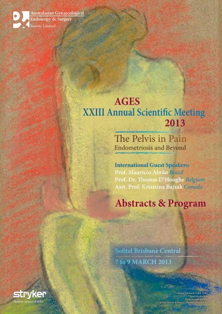
Vuillard Edouard (1868-1940)<br />
Figure de douleur<br />
Paris, musée d'Orsay<br />
© RMN (Musée d'Orsay) / Hervé Lewandowski
The Pelvis in Pain<br />
Endometriosis and Beyond<br />
<strong>AGES</strong><br />
Sponsors & Exhibitors<br />
<strong>AGES</strong> gratefully acknowledges the following sponsors and exhibitors.<br />
Platinum Sponsor of <strong>AGES</strong><br />
Major Sponsors of the <strong>AGES</strong> <strong>Annual</strong> <strong>Scientific</strong> <strong>Meeting</strong> <strong>XXIII</strong> <strong>2013</strong><br />
‘The Pelvis in Pain: Endometriosis and Beyond’<br />
Exhibitors<br />
Applied Medical<br />
Covidien<br />
Device Technologies<br />
Gate Healthcare<br />
Hologic Australia<br />
Avnet Technology Solutions<br />
B Braun Australia<br />
Baxter Healthcare<br />
Boston <strong>Scientific</strong><br />
ConMed Linvatec<br />
Cook Medical<br />
Gytech<br />
InSight Oceania<br />
Investec Medical Finance<br />
Lifehealthcare<br />
Medfin<br />
Sonologic
<strong>AGES</strong> <strong>XXIII</strong> <strong>Annual</strong> <strong>Scientific</strong> <strong>Meeting</strong> <strong>2013</strong><br />
CONTENTS<br />
Sponsors & Exhibitors <br />
Inside Front Cover<br />
Welcome Message2<br />
Conference Faculty, Conference Committee & 3<br />
<strong>AGES</strong> Board Members<br />
CPD and PR&CRM Points 3<br />
Conference <strong>Program</strong><br />
Thursday 7 March 4<br />
Friday 8 March 6<br />
Saturday 9 March 9<br />
<strong>AGES</strong> Awards9<br />
<strong>Program</strong> <strong>Abstracts</strong><br />
Thursday 7 March 10<br />
Friday 8 March 14<br />
Saturday 9 March 17<br />
Free Communications<br />
Thursday 7 March - I 22<br />
Thursday 7 March - II 26<br />
Friday 8 March - Chairmen’s Choice 30<br />
Digital Communications 36<br />
Sponsor Information 41<br />
Conference Information & Conditions48<br />
Future <strong>AGES</strong> <strong>Meeting</strong>s<br />
Back Cover<br />
1
The Pelvis in Pain<br />
Endometriosis and Beyond<br />
Welcome<br />
South-East Queensland is known for its golden beaches,<br />
glorious sunshine and rolling hinterlands. South-East<br />
Queensland’s pearl has to be Brisbane - a world-class<br />
city straddling the Brisbane River, who has grown into a<br />
wondrous woman of beauty, culture and style.<br />
The Board of <strong>AGES</strong> welcomes you to Brisbane for the <strong>Annual</strong><br />
<strong>Scientific</strong> <strong>Meeting</strong>, March 7-9 <strong>2013</strong>, where our international<br />
and local faculty will dissect pelvic pain in its myriad of<br />
manifestations.<br />
Guest speakers include surgical expert Mauricio Abrão<br />
from Sao Paulo Brazil, AAGL Research director and pelvic<br />
pain luminary Krisztina Bajzak from Memorial University,<br />
Newfoundland Canada, and fertility and scientific doyen<br />
Thomas D’Hoogue from Leuven Belgium.<br />
Joining them is a stellar cast of Australians and New<br />
Zealanders who will cover pregnancy, myomas, hysterectomy,<br />
adhesions and every facet of our specialty connected with pain.<br />
Live surgery from St Andrews War Memorial and<br />
Greenslopes Hospitals will showcase laparoscopic<br />
myomectomy, laparoscopic hysterectomy and laparoscopic<br />
resection of endometriosis.<br />
An international panel will be led through a series of<br />
disasters and damage, hypothetical style for their input on<br />
management and the debate entitled “Surgeons should retire<br />
at 50” is one not to be missed.<br />
Throw in a new look Free Communications program<br />
including the introduction of the Digital Communications<br />
Session, cutting edge technology in the <strong>AGES</strong> trade<br />
exhibition, pre-conference workshops on hysteroscopy and<br />
laparoscopic suturing … and you have another outstanding<br />
<strong>AGES</strong> event.<br />
We welcome you to this beautiful city where both Australia<br />
and <strong>AGES</strong> continue to shine.<br />
Assoc. Prof. Anusch Yazdani Dr Jim Tsaltas Assoc. Prof. Jason Abbott<br />
<strong>AGES</strong> Vice President <strong>AGES</strong> President <strong>AGES</strong> Director<br />
Conference Chair <strong>Scientific</strong> Co-Chair <strong>Scientific</strong> Co-Chair<br />
South Bank Brisbane
<strong>AGES</strong> <strong>XXIII</strong> <strong>Annual</strong> <strong>Scientific</strong> <strong>Meeting</strong> <strong>2013</strong><br />
CONFERENCE COMMITTEE<br />
Assoc. Prof. Anusch Yazdani<br />
Dr Jim Tsaltas<br />
Assoc. Prof. Jason Abbott<br />
<strong>AGES</strong> BOARD<br />
Dr Jim Tsaltas<br />
Assoc. Prof. Anusch Yazdani<br />
Assoc. Prof. Harry Merkur<br />
Dr Michael McEvoy<br />
Assoc. Prof. Jason Abbott<br />
Dr Keith Harrison<br />
Dr Kym Jansen<br />
Prof. Ajay Rane<br />
Dr Anna Rosamilia<br />
Dr Stuart Salfinger<br />
Ms Michele Bender<br />
International FACulty<br />
Prof. Mauricio Abrão<br />
Asst. Prof. Krisztina Bajzak<br />
Prof. Dr Thomas D’Hooghe<br />
CPD and PR&CRM POINTS<br />
Conference Chair<br />
<strong>Scientific</strong> Co-Chair<br />
<strong>Scientific</strong> Co-Chair<br />
President<br />
Vice President<br />
Honorary Secretary<br />
Treasurer<br />
Executive Director<br />
Brazil<br />
Canada<br />
Belgium<br />
Full attendance Thursday 6 March, Friday 7 March and<br />
Saturday 8 March<br />
21 CPD points<br />
Thursday 6 March only<br />
8 CPD points<br />
Friday 7 March only<br />
9 CPD points<br />
Saturday 8 March only<br />
5 CPD points<br />
FACulty<br />
Assoc. Prof. Jason Abbott<br />
Dr Reza Adib<br />
Dr Fariba Behnia-Willison<br />
Dr Jason Chow<br />
Assoc. Prof. Michael Cooper<br />
Dr Marilla Druitt<br />
Ms Taryn Hallam<br />
Dr Geogia Hume<br />
Dr Tal Jacobson<br />
Dr Kym Jansen<br />
Dr Neil Johnson<br />
Dr Juliette Koch<br />
Assoc. Prof. Alan Lam<br />
Dr Kenneth Law<br />
Prof. Peter Maher<br />
Dr Michael McEvoy<br />
Dr David Molloy<br />
Dr Grant Montgomery<br />
Dr Erin Nesbitt-Hawes<br />
Prof. Andreas Obermair<br />
Dr Rob O’Shea<br />
Dr Jodie Painter<br />
Assoc. Prof. Luk Rombauts<br />
Dr Anna Rosamilia<br />
Dr Stuart Salfinger<br />
Dr Christopher Smith<br />
Dr Jim Tsaltas<br />
Prof. Thierry Vancaillie<br />
Assoc. Prof. Anusch Yazdani<br />
Dr Michael Wynn-Williams<br />
Membership of <strong>AGES</strong><br />
New South Wales<br />
Queensland<br />
South Australia<br />
New South Wales<br />
New South Wales<br />
Victoria<br />
New South Wales<br />
Queensland<br />
Queensland<br />
Victoria<br />
New Zealand<br />
New South Wales<br />
New South Wales<br />
Queensland<br />
Victoria<br />
South Australia<br />
Queensland<br />
Queensland<br />
New South Wales<br />
Queensland<br />
South Australia<br />
Queensland<br />
Victoria<br />
Victoria<br />
Western Austrlia<br />
New South Wales<br />
Victoria<br />
New South Wales<br />
Queensland<br />
Queensland<br />
Pre-Conference Workshops 6 March<br />
Karl Storz Endoscopy - Hysteroscopic and Resection<br />
<br />
3 CPD points<br />
Olympus - Reload Your Suturing Skills 3 CPD points<br />
Attendance by eligible RANZCOG Members will only be<br />
acknowledged following signature of the attendance roll each<br />
day of the Conference, and for each workshop.<br />
The RANZCOG Clinical Risk Management Activity<br />
Reflection Worksheet (provided in the Conference satchel)<br />
can be used by Fellows who wish to follow up on a meeting<br />
or workshop that they have attended to obtain PR&CRM<br />
points. This worksheet enables you to demonstrate that you<br />
have reflected on and reviewed your practice as a result<br />
of attending a particular workshop or meeting. It also<br />
provides you with the opportunity to outline any follow-up<br />
work undertaken and to comment on plans to re-evaluate<br />
any changes made. Fellows of this College who attend the<br />
<strong>Meeting</strong> and complete the Clinical Risk Management Activity<br />
Reflection Worksheet in accordance with the instructions<br />
thereon can claim for an additional 5 PR&CRM points for<br />
the <strong>Meeting</strong> and for each of the Workshops. For further<br />
information, please contact the College.<br />
Membership application forms are available from the <strong>AGES</strong><br />
website: www.ages.com.au<br />
<strong>AGES</strong> SECRETARIAT<br />
Conference Connection<br />
282 Edinburgh Road<br />
Castlecrag<br />
SYDNEY NSW 2068 AUSTRALIA<br />
Ph: +61 2 9967 2928<br />
Fax: +61 2 9967 2627<br />
Email: secretariat@ages.com.au<br />
3
The Pelvis in Pain<br />
Endometriosis and Beyond<br />
<strong>Program</strong><br />
Sofitel Brisbane Central<br />
0730-0800 Conference Registration<br />
Ballroom Le Grande<br />
Day 1<br />
Thursday<br />
7 March<br />
<strong>2013</strong><br />
0800-0820 Conference Opening and Welcome J Tsaltas, A Yazdani<br />
0820-1030 SESSION 1<br />
When the Lining is Not Always Silver: Problems of the Peritoneum<br />
Chairs: J Tsaltas, A Yazdani<br />
Sponsored by Stryker<br />
0820-0840 Genetics and epidemiology of endometriosis J Painter<br />
0840-0900 Images of pain – the library of lesions M Cooper<br />
0900-0920 Macro to micro – the host of heinous histology K Law<br />
0920-0940 The first cut is the deepest P Maher<br />
0940-1000 Questions<br />
1000-1030 Keynote Lecture<br />
Chair: J Abbott<br />
Imaging and endometriosis – a match made in surgical heaven<br />
M Abrão<br />
1030-1100 Morning Tea and Trade Exhibition<br />
1100-1230 SESSION 2<br />
Piecing Together the Pain Puzzle<br />
Chairs: H Merkur, K Jansen<br />
Sponsored by Karl Storz Endoscopy<br />
1100-1120 Mechanisms of pain K Bajzak<br />
1120-1135 The evidence for endometriosis causing and not causing pain M Druitt<br />
1135-1150 Don’t dys…. me J Abbott<br />
1150-1200 Questions<br />
1200-1230 Keynote Lecture<br />
Chair: L Rombauts<br />
Non-invasive diagnosis of endometriosis<br />
T D’Hooghe<br />
1230-1330 Lunch and Trade Exhibition<br />
1330-1500 SESSION 3<br />
Free Communications Session I<br />
Sponsored by Olympus<br />
Ballroom Le Grande 1<br />
Chairs: P Maher, M Abrão<br />
1330-1340 The effect of Trendelenburg Tilt on cognitive function Lee S, Tan A, Griffiths J, Ang C<br />
1340-1350 Laparoscopic management of an adnexal mass during the second trimester of pregnancy<br />
<br />
Georgiou C<br />
1350-1400 3D ultrasound of the pelvic floor - a reproducibility study Nesbitt-Hawes EM,<br />
<br />
Dietz HP, Abbott JA<br />
1400-1410 Should bilateral salpingectomy be a routine part of hysterectomy Manley T,<br />
<br />
Tsaltas J, Najjar H<br />
1410-1420 Travelling fellowship report Smith C<br />
1420-1430 Predictors of prolapse recurrence following laparoscopic sacrocolpopexy Wong V,<br />
<br />
Guzman-Rojas R, Shek KL, Chou D, Moore K, Dietz H
<strong>AGES</strong> <strong>XXIII</strong> <strong>Annual</strong> <strong>Scientific</strong> <strong>Meeting</strong> <strong>2013</strong><br />
1430-1440 Not a fibroid - application of Myosure to non-myoma pathology Nesbitt-Hawes EM,<br />
<br />
Abbott JA<br />
1440-1450 XCEL Bladeless Trocar versus Veress Needle: A randomised controlled trial comparing<br />
these two entry techniques in gynaecological laparoscopic surgery Manley T,<br />
Wright P, Vollenhoven B, Tsaltas J, Lawrence A, Najjar H,<br />
<br />
Pearce S, Tan J, Chan K-W, Wang L, Amir M, Fernandes<br />
<br />
H, Hyde S, Grant P, McIlwaine K, Cameron M<br />
1450-1500 Rectovaginal endometriosis in obese women. Is surgery more complicated<br />
<br />
Edmonds S, Barclay D, Van der Merwe A,Israel L, Peng S-L<br />
Free Communication Session II<br />
Sponsored by Johnson & Johnson Medical<br />
Ballroom Le Grande 2<br />
Chairs: A Rosamilia, M Wynn-Williams<br />
1330-1340 Pregnancy following laparoscopic radical trachelectomy Yao S-E, Lee S, Tan J<br />
1340-1350 Clinical analysis of 17 cases undergoing laparoscopic pelvic lymphadenectomy for<br />
gynecology malignant tumor<br />
Xu H, Zhang B<br />
Day 1<br />
Thursday<br />
7 March<br />
<strong>2013</strong><br />
1350-1400 Laparoscopic excision of full-thickness bladder endometriotic nodule, partial<br />
cystectomy and bilateral uretericiImplantation in a young lady with long-standing<br />
obstructive nephropathy caused by severe pelvic endometriosis Choi S, Aslan P,<br />
Cario G, Rosen D, Reyftmann L, de Rosnay P, Chou D<br />
1400-1410 Travelling fellowship report Campbell N<br />
1410-1420 Laparoscopic removal and repair of Caesarean scar ectopic pregnancy Titiz, H<br />
1420-1430 The association between pouch of douglas obliteration and surgical findings at<br />
laparoscopy in women with suspected endometriosis Reid S, Lu C, Casikar I, Reid G,<br />
<br />
Abbott J, Cario G, Chou D, Kowalski D, Cooper M, Condous G<br />
1430-1440 Evaluation of Code Critical Cesarean sections at Westmead Hospital Kapurubandara S,<br />
<br />
Tse T, Anpalagan A, McGee T<br />
1440-1450 Can we develop a model to predict Pouch of Douglas obliteration in women with<br />
suspected endometriosis<br />
Reid S, Lu C, Casikar I, Condous G<br />
1450-1500 A retrospective analysis looking at Case-mix and complications in an established<br />
tertiary-level Centre de Rosnay P, Cario G, Rosen D, Chou D,<br />
Cooper M, Reid G, Reyftmann L, Choi S<br />
1500-1530 Afternoon Tea and Trade Exhibition<br />
1530-1715 SESSION 4<br />
managing Messes<br />
Sponsored by Karl Storz Endoscopy<br />
Chairs: M McEvoy, T Jacobson<br />
1530-1550 Single surgeon surgery: perils and pitfalls A Lam<br />
1550-1610 Co-operation creates cohesion for complexity J Tsaltas<br />
1610-1630 Bringing it all together – guidelines for management N Johnson<br />
1630-1715 Damage and Disasters<br />
Panel discussion<br />
Moderators: A Yazdani, J Abbott<br />
Panel: J Tsaltas, A Lam, N Johnson, K Bajzak, T D’Hooghe, M Abrão<br />
1715-1815 Welcome Cocktail Reception Sofitel Brisbane Central<br />
5
The Pelvis in Pain<br />
Endometriosis and Beyond<br />
<strong>Program</strong><br />
Sofitel Brisbane Central<br />
Ballroom Le Grande<br />
Day 2<br />
Friday<br />
8 March<br />
<strong>2013</strong><br />
0830-1030 SESSION 5<br />
live Surgery<br />
Sponsored by Stryker<br />
Moderators: S Salfinger, M Abrão, M Wynn-Williams<br />
0830-1030 Live surgery transmission<br />
Site 1: St Andrews Hospital<br />
Evidence Surgeon Surgery<br />
C Smith D Molloy Total laparoscopic hysterectomy, laparoscopic<br />
Myomectomy<br />
J Chow A Yazdani Laparoscopic resection of endometriosis<br />
Site 2: Greenslopes Hospital<br />
Evidence Surgeon Surgery<br />
E Nesbitt-Hawes A Obermair Laparoscopic hysterectomy<br />
1030-1100 Morning Tea and Trade Exhibition<br />
Digital Presentation Session<br />
1030 Submucous fibroids - a series of hysteroscopic morcellation<br />
<br />
Nesbitt-Hawes EM, Sgroi J, Abbott JA<br />
1035 Thinking outside the box - women’s experience of living with endometriosis: a qualitative study<br />
<br />
Moradi M, Parker M, Lopez V, Sneddon A, Phillips C, Ellwood D<br />
1040 Bladder and bowel dysfunction after excision of deep infiltrating endometriosis<br />
<br />
Chow JSW, Cooper MJW, Korda A, Benness C, Krishnan S<br />
1045 Case report: Cervico-peritoneal fistula following hysteroscopic resection of caesarean scar<br />
ectopic pregnancy<br />
Maley P, Law K.Bourke M, Abbott J<br />
1100-1230 SESSION 6<br />
Pain and Pregnancy: An Unholy Alliance<br />
Chairs: K Harrison, A Rosamilia<br />
Sponsored by Johnson & Johnson Medical<br />
1100-1115 Endometriomas, AMH and fertility – the devil’s triangle J Koch<br />
1115-1130 Pain’s a drain in pregnancy K Jansen<br />
1130-1200 Keynote Lecture<br />
Chair: J Tsaltas<br />
Pain starts with a P not an E<br />
K Bajzak<br />
1200-1300 Lunch and Trade Exhibition<br />
Digital Presentation Session<br />
1200 Luteal phase defect and ectopic pregnancy Miller B, McLinon L, Beckman M<br />
1205 Subsequent management of unsuccessful Fulshi Clip tubal occlusion. Georgiou C<br />
1210 Thrombus-like structures seen in the infundibular-pelvic ligament during the laparoscopic<br />
management of ovarian torsion<br />
Georgiou C<br />
1215 Laparoscopic myomectomy of large intramural fibroid in a Jehovah’s Witness Choi S, Caska P,<br />
<br />
Cario G, Reftmann L, de Rosnay P, Chou D<br />
1220 Can we predict posterior compartment Deeply Infiltrating Endometriosis (DIE) using Office<br />
Sonovaginography (SVG) in women undergoing laparoscopy for chronic pelvic pain Reid S,<br />
<br />
Casikar I, Reid G, Abbott J, Cario G, Chou D, Kowalski D, Cooper M, Condous G<br />
1230 Small bowel injury during laparoscopy to drainiInfected pelvic collections Chohan K, Anpalagan A<br />
1235 Laparoscopic excision of bladder endometriotic nodule Lanziz H, Swift G<br />
1240 Robotic surgery and the Da Vinci Surgical System – pathway to accreditation as experienced by a<br />
tertiary level benign endo-gynaecological unit de Rosnay P, Cario G, Rosen D, Chou D,<br />
<br />
Cooper M, Reid G, Reyftmann L, Choi S
<strong>AGES</strong> <strong>XXIII</strong> <strong>Annual</strong> <strong>Scientific</strong> <strong>Meeting</strong> <strong>2013</strong><br />
1300-1500 SESSION 7<br />
Free Communications - Chairmen’s Choice<br />
Ballroom Le Grande 1<br />
Chairs: J Tsaltas, A Yazdani, J Abbott<br />
Sponsored by Stryker<br />
1300-1310 The use of a multimedia module to aid the informed consent process for gynaecological<br />
laparoscopy for pelvic pain. A Randomized Control Trial. Ellett L, Villegas R,<br />
<br />
Jagasia N, Beischer A, Readman E, Maher P<br />
1310-1320 Laparoscopic repair of caesarean scar defect and pregnancy outcome Kong KY,<br />
<br />
Angstetra D, Reid G<br />
1320-1330 Establishment of a robotic surgical programme for benign gynaecology in an advanced<br />
laparoscopic centre – Proctorship and Beyond Choi S, Rosen D, Chou D,<br />
<br />
Reyftmann L, de Rosnay P, Cario G<br />
1330-1340 Laparoscopic myomectomy of an 1.8kg pedunculated fibroid causing uterine torsion<br />
<br />
Cebola M, Cario G, Rosen D, Reyftmann L, De Rosnay P, Choi S, Chou D<br />
Day 2<br />
Friday<br />
8 March<br />
<strong>2013</strong><br />
1340-1350 To excise or ablate Prospective randomized double blinded trial comparing surgical<br />
treatment of endometriosis over 5 Years<br />
Healey M, Kaur H, Cheng C<br />
1350-1400 The role of laparoscopy in the surgical management of a 4.2kg uterine fibroid – a video<br />
presentation de Rosnay P, Cario G, Rosen D, Cooper M,<br />
Reid G, Reyftmann L, Choi S, Chou D.<br />
1400-1410 The value of MRI in the investigation of pudendal nerve entrapment Chow JSW,<br />
<br />
Sachinwalla T, Jarvis SK, Vancaillie TG<br />
1410-1420 Combined Levonorgestrel intrauterine system and Etonogestrel subdermal implant for<br />
refractory endometriosis-associated pelvic pain: An effective new dual therapy<br />
<br />
Marren AJ, Fraser IS, Al-Jefout MI, Pardey A, Pardey J, Ng CHM<br />
1420-1430 The effect of patient body mass index on surgical difficulty in gynaecological<br />
laparoscopy. A prospective observational study McIlwaine K, Ellett L, Villegas R,<br />
Cameron M, Jagasia N, Readman E, Maher P<br />
1430-1440 Laparoscopy and gynaecological diaphragmatic disease Yao S-E, Lee S, Tan J<br />
1440-1450 Depot Medroxyprogesterone Acetate (DMPA) in the treatment ofeEndometriosis<br />
<br />
Vollenhoven B, Dennerstein G, Fernando S, Fraser I, Polyakov A, Vu P, Wark JD<br />
1450-1500 Retrospective clinical audit reviewing the use of Magnetic Resonance guided Focused<br />
Ultrasound (MRgFUS) in the treatment of submucosal uterine fibroids at the Royal<br />
Women’s Hospital Melbourne<br />
Rajadevan N, Szabo R, Dobrotwir A, Ang WC<br />
1500-1530 Afternoon Tea and Trade Exhibition<br />
Digital Presentation Session<br />
1500 Laparoscopic repair of intrapartum uterine rupture and uterovaginal detachment Lee S, Tan J<br />
1505 Barbed sutures: Are they safe for vault closure at laparoscopic hysterectomy Is there any evidence<br />
<br />
Manley T, Tsaltas J, Najjar H<br />
1510 he prediction of Pouch of Douglas obliteration using off-line analysis of the TVS ‘Sliding Sign’:<br />
Inter- and Intra- Observer Agreement<br />
<br />
Reid S, Lu C, Casikar I, Mein B, Magotti R, Ludlow J, Benzie R, Condous G<br />
1515 Diagnostic accuracy for the prediction of Pouch of Douglas obliteration using off-line analysis of<br />
the TVS ‘Sliding Sign’. Reid S, Lu C, Casikar I, Mein B, Magotti R,<br />
<br />
Ludlow J, Benzie R, Condous G<br />
7
The Pelvis in Pain<br />
Endometriosis and Beyond<br />
<strong>Program</strong><br />
Sofitel Brisbane Central<br />
Day 2<br />
Friday<br />
8 March<br />
<strong>2013</strong><br />
1530-1720 SESSION 8<br />
Future Frontiers<br />
Chairs: H Merkur, R O’Shea<br />
Sponsored by Olympus<br />
1530-1600 The Perpetual Dan O’Connor Lecture<br />
Chair: P Maher<br />
From benchside to bedside – translating endometriosis research<br />
G Montgomery<br />
1600-1615 SILS – Single Incision Laparoscopic Surgery F Behnia-Willison<br />
1615-1630 NOTES – Natural Orifice Translumenal Endoscopic Surgery R Adib<br />
1630-1645 Medical directions L Rombauts<br />
1645-1705 Robotics and endometriosis M Abrão<br />
1705-1720 Questions<br />
1730-1830 <strong>AGES</strong> <strong>Annual</strong> General <strong>Meeting</strong><br />
1930 for 2030 Gala CONFERENCE Dinner<br />
Roof Terrace, Gallery of Modern Art (GOMA),<br />
Stanley Place, Brisbane<br />
Private Tour of the 7th Asia Pacific Triennial of Contemporary Art<br />
Complimentary coach transfers provided. Please assemble in the hotel foyer at 1900.<br />
GOMA Brisbane
<strong>AGES</strong> <strong>XXIII</strong> <strong>Annual</strong> <strong>Scientific</strong> <strong>Meeting</strong> <strong>2013</strong><br />
Ballroom Le Grande<br />
0815-1030 SESSION 9<br />
Thinking Outside the Box: Non-gynaecological Causes of Pelvic Pain<br />
Chairs: A Lam, N Johnson<br />
Sponsored by Karl Storz Endoscopy<br />
0815-0845 Why is classification of endometriosis such a struggle M Abrão<br />
0845-0905 Interstitial cystitis A Rosamilia<br />
0905-0925 Musculoskeletal issues T Hallam<br />
0925-0945 Functional bowel disorders G Hume<br />
0945-1015 Imaging and managing in the canal of pain T Vancaillie<br />
1015-1030 Questions<br />
1030-1100 Morning Tea and Trade Exhibition<br />
Day 3<br />
Saturday<br />
9 March<br />
<strong>2013</strong><br />
1100-1300 SESSION 10<br />
how Far Does Pain Reach<br />
Chairs: J Tsaltas, A Yazdani<br />
Sponsored by Stryker<br />
1100-1130 Hysterectomy and chronic pelvic pain - a forgone conclusion K Bajzak<br />
1130-1200 The price of pain T D’Hooghe<br />
1200-1215 Questions<br />
1215-1245 Presidential Debate:<br />
Surgeons should retire at 50<br />
For: J Abbott, T Jacobson<br />
Against: P Maher, M McEvoy<br />
1245-1300 Awards and close J Tsaltas, A Yazdani<br />
AWARDS<br />
Best Free Communication $1000 Sponsored by Covidien & <strong>AGES</strong><br />
All presenters of free communications, regardless of Fellow or trainee status, with the emphasis on<br />
scientific merit<br />
Outstanding New Presenter<br />
$750 Sponsored by Johnson & Johnson Medical<br />
Open to all first time presenters at an <strong>AGES</strong> meeting. This award is to encourage new trainees and<br />
Fellows who have never previously presented.<br />
Outstanding Video Presentation $750 Sponsored by <strong>AGES</strong><br />
All video presentations<br />
Outstanding Trainee Presentation – $750 Sponsored by Stryker and <strong>AGES</strong><br />
the Platinum Laparoscopic Award<br />
All trainees and Fellows in the <strong>AGES</strong> training program, or Fellows in their first year post-training to<br />
reflect work completed during training<br />
Best Digital Communications Presentation $750 Sponsored by <strong>AGES</strong><br />
All digital free communication presentations<br />
9
The Pelvis in Pain<br />
<strong>Program</strong> <strong>Abstracts</strong> - Thursday 7 March<br />
Endometriosis and Beyond<br />
SESSION 1 / 0840-0900<br />
Images of pain - the library of lesions<br />
Cooper M<br />
This presentation will attempt to display the myriad range of<br />
appearances that endometriosis can appear in.<br />
AUTHOR AFFILIATION: Clinical Associate Professor<br />
Michael Cooper; Department of Obstetrics and Gynaecology<br />
at Sydney University, Royal Prince Alfred Hospital, St Luke’s<br />
Hospital, St Vincent’s Private Hospital, Sydney IVF Sydney<br />
New South Wales, Australia.<br />
SESSION 1 / 0900-0920<br />
Macro to micro – The host of heinous histology<br />
Law K<br />
Endometriosis is defined histologically as the presence<br />
of endometrial glands and stroma at extrauterine sites.<br />
It has a myriad of gross appearances, and can vary from<br />
‘powder burn’ lesions, white or yellow lesions/nodules,<br />
clear vesicles to flame-like red lesions. To the trained eye,<br />
endometriosis can usually be macroscopically recognised,<br />
but some studies have reported the correlation between<br />
laparoscopic diagnosis and histological diagnosis can be<br />
as low as 65%, and may be affected by operator experience<br />
and sampling problems.<br />
Despite ongoing research, it is unclear as to the exact<br />
mechanisms by which pain is generated by endometriotic<br />
lesions. It has been reported that deep endometriotic lesions<br />
may be neurotrophic, with higher expression of nerve growth<br />
factor in comparison with peritoneal and ovarian implants.<br />
In particular, histological studies have shown a proliferation<br />
of nerve fibres associated with rectovaginal nodules.<br />
However a correlation between the type of lesion and the<br />
severity of pain has not been consistently demonstrated.<br />
Ultimately the location of disease may be a more important<br />
predictor of the nature and degree of symptoms.<br />
AUTHOR AFFILIATION; Dr Kenneth Law; Greenslopes<br />
Private Hospital, Brisbane, Queensland, Australia.<br />
Another issue which needs to addressed at the highest<br />
level, i.e. College is who should do this surgery. Too many<br />
inexperienced gynaecologists are setting themselves up in<br />
practice and trumpeting to the unsuspecting public their<br />
“expertise” in this and many other branches of gynaecology.<br />
The approach to each group of patients will be discussed<br />
taking into account age, symptoms and lifestyle impact.<br />
The effect of chronic pelvic pain on treatment will be<br />
discussed according to three age groups of patient that the<br />
author has arbitrarily selected.<br />
Empiric, drug and surgical treatment will be discussed also<br />
according to these groups and the impact of the “first cut”<br />
will be discussed.<br />
At the completion of the presentation the author will revisit<br />
who should do what and to whom.<br />
The first cut is not necessarily the deepest!!<br />
AUTHOR AFFILIATION: Professor Peter Maher; Head,<br />
Department of Endosurger, Mercy Hospital for Women,<br />
Melbourne, Victoria, Australia.<br />
SESSION 1 – Keynote lecture / 1000-1030<br />
Imaging and endometriosis: A match made in<br />
surgical heaven<br />
Abrão M<br />
Endometriosis poses a challenging clinical and surgical<br />
dilemma for many gynecologists. Realizing the depth<br />
and extent of disease prior to surgery can be the key to<br />
pre-operative surgical planning and patient counselling.<br />
This presentation provides a discussion of the role of preoperative<br />
imaging (with a defined protocol in ultrasound)<br />
and a description of the technique. Imaging findings will be<br />
correlated to those in surgery.<br />
AUTHOR AFFILIATION: Professor Mauricio Abrão;<br />
Director of Endometriosis Division, Ob/Gyn, Department,<br />
Sao Paulo University, Brazil. President, SBE - the Brazilian<br />
Endometriosis and Minimally Invasive Gynecology<br />
Association. Director, Reproductive Clinic, Sirio Libanes<br />
Hospital, Sao Paulo, Brazil.<br />
SESSION 1 / 0920-0940<br />
The first cut is the deepest<br />
Maher P<br />
An interesting title but what does it mean. As we know<br />
endometriosis strikes all ages even the pre-menarchal patient.<br />
There is naturally a reluctance to operate on the very<br />
young patient.<br />
SESSION 2 / 1100-1120<br />
Mechanisms of pain<br />
Bajzak K<br />
Chronic pelvic pain (CPP) is responsible for one in ten<br />
gynaecologic outpatient visits and the indication for 15-50%<br />
of gynaecologic laparoscopies and 12% of hysterectomies.<br />
Limited data is available on the prevalence of CPP it is
<strong>AGES</strong> <strong>XXIII</strong> <strong>Annual</strong> <strong>Scientific</strong> <strong>Meeting</strong> <strong>2013</strong><br />
<strong>Program</strong> <strong>Abstracts</strong> - Thursday 7 March<br />
estimated that direct and indirect costs in the US exceed 2<br />
billion per year.<br />
Peripheral activation: Pain sensation begins when a noxious<br />
stimulus activates a peripheral nociceptor (“nerve ending”).<br />
These stimuli activate specific receptors on a neuron which<br />
depolarizes due to an influx of positive ions (Na and Ca2+<br />
usually). Once a threshold is reached the neuron carries the<br />
message up to the spinal cord.<br />
CNS activation: In the spinal cord the neuron releases<br />
transmitters (glutamate, sub-P) which stimulate the next<br />
neuron which can be either excitatory or inhibitory. These in<br />
turn relay the message to brain via the thalamus which acts as<br />
a relay station to other areas of the brain (amygdala, insula &<br />
cortex). Functionally, pain processing in the CNS occurs in 3<br />
domains:<br />
1. Sensory: location and severity of pain<br />
2. Affective: emotional valence of pain<br />
3. Cognitive: what we think and do about pain.<br />
Descending feedback from the brain in response to pain<br />
signal is subject to modulation and can inhibit or enhance<br />
the pain signal. So, the way individuals think about their pain<br />
(stress, anxiety, CBT) can affect both sensory and affective<br />
processing of pain.<br />
Chronic pain is very different from acute pain. The unique<br />
neurobiology of chronic pain leads to several consequences<br />
including neuroplasticity, central sensitization, convergence,<br />
antidromic transmission, neurogenic inflammation and<br />
peripheral sensitization. Once these changes occur pain<br />
becomes neuropathic in nature and independent of the<br />
inciting event. Pain itself becomes a disease.<br />
REFERENCES:<br />
1. Pharmacological Mgt of Chronic Neuropathic pain-<br />
Consensus Statement and Guidelines from the Canadian<br />
Pain Society. J Pain Res Manage. 2007;12:13-21<br />
2. Neurobiology of Chronic Pain: Lessons Learned from<br />
FM and Related Conditions. Clauw DJ. University of<br />
Michigan. Lecture at17th <strong>Annual</strong> <strong>Scientific</strong> <strong>Meeting</strong> on<br />
Chronic Pelvic Pain, 2009<br />
3. Neural Mechanisms of Pelvic Organ Cross-Sensitization.<br />
Malykhina AP. Neuroscience. 2007;149:660-672<br />
4. Course overview of Chronic Pelvic Pain and Pain Theory.<br />
Howard FM. Lecture at AAGL 36th <strong>Annual</strong> Global<br />
Congress, PG Course 3 Chronic Pelvic Pain: Diagnosis<br />
and Treatment, 2007<br />
5. The anatomy and neurophysiology of pelvic pain. Lamvu<br />
G, Steege JF. JMIG. 2006;13:516-522<br />
6. How, Who and When of Opioid Treatment. Dogra S.<br />
University of N Carolina Chapel Hill. Lecture at 14th<br />
<strong>Annual</strong> <strong>Scientific</strong> <strong>Meeting</strong> on Chronic Pelvic Pain, 2006<br />
7. Consensus Guidelines for the Management of CPP. SOGC<br />
Clinical Practice Guideline No. 164, August 2005<br />
8. Pelvic Pain, Diagnosis and Management. Howard FM,<br />
Perry PC, Carter JE, El-Minawi AM. Lippincot Wiliams<br />
and Wilkins. Philadelphia. 2000<br />
9. Neuropathic pain. Bennett M. Oxford University Press. 2007<br />
10. Managing Pain. The Canadian Healthcare Professional’s<br />
Reference. Jovey RD. Healthcare and Financial Publishing,<br />
Rogers Media. 2002<br />
11. Current Therapy in Pain. Smith Hs. Saunders Elsevier. 2009<br />
AUTHOR AFFILIATION: Dr Krisztina Bajzak; Assistant<br />
Professor & Research Director, Discipline of Obstetrics and<br />
Gynecology, Memorial University, Newfoundland, Canada.<br />
SESSION 2 / 1120-1135<br />
The evidence for endometriosis causing and not<br />
causing pain<br />
Druitt M<br />
Endometriosis is a heterogenous disorder with a spectrum of<br />
associated pains: dysmenorrhoea, dyspareunia, non menstrual<br />
chronic pelvic pain, dyschezia, dysuria, musculoskeletal pain<br />
(Stratton and Berkley 2011). The experience of pain is affected<br />
by many variables. Pain can be acute or chronic, somatic<br />
or visceral and can be classified according to mechanism:<br />
nociceptive, inflammatory, neuropathic, psychogenic, mixed or<br />
idiopathic (Howard 2009).<br />
This topic will be considered from the point of view of a<br />
neurological diagram.<br />
Pain in endometriosis could be generated from substances<br />
produced in or found near the endometriotic lesion (eg<br />
TNF and nerve growth factors), these could stimulate<br />
nociceptors or cause inflammation mediated pain. A<br />
neuropathic mechanism (from damage or dysfunction of<br />
neurons in the peripheral or central nervous system) could<br />
contribute with evidence in the periphery of nerve fibres in<br />
lesions (Tokushige 2006) and centrally – evidence for central<br />
sensitisation in women with endometriosis and pain (Bajaj<br />
2003) and grey matter changes on MRI (As Sanie 2012).<br />
The principle of intervention studies can be to remove or<br />
suppress endometriosis. Only 3 surgical RCTs have been<br />
performed to examine if lasering or excising endometriosis<br />
(compared to diagnostic laparoscopy) improves pain<br />
(Sutton 1994, Abbott 2004, Jarrell 2005). These had positive<br />
findings but had significant limitations. Cochrane reviews<br />
of medical treatment for pain in endometriosis include<br />
one RCT of an anti inflammatory (Kauppila 1985) and the<br />
following hormonal treatments: continuous progestagens and<br />
antiprogestagens and danazol which have level 1a evidence<br />
supporting their effect in pain, however, only one RCT of an<br />
cOCP has met the criteria for inclusion (Davis 2007) - cOCP<br />
as good as GnRH analogue in treating pain and one RCT of<br />
LNG-IUs post surgery which found a beneficial effect (Abou-<br />
Setta 2006).<br />
The case for endometriosis not causing pain must mention<br />
those patients with recurrent pain and no recurrent<br />
endometriosis at second surgery (Vercellini 2009),<br />
asymptomatic patients, the poor correlation of stage of<br />
disease and pain and those who derive no benefit from<br />
surgery or medicine.<br />
11
The Pelvis in Pain<br />
<strong>Program</strong> <strong>Abstracts</strong> - Thursday 7 March<br />
Endometriosis and Beyond<br />
REFERENCES:<br />
1. Fred M Howard. Endometriosis and Mechanisms<br />
of Pelvic Pain. J Minim Invasive Gynecol. 2009 Sep-<br />
Oct;16(5):540-50<br />
2. Pamela Stratton and Karen J Berkley. Chronic pelvic<br />
pain and endometriosis: translational evidence of the<br />
relationship and implications. Hum Reprod Update. 2011<br />
May-Jun;17(3):327-46<br />
3. Endometriosis: Science and Practice. Edited by Linda<br />
C.Giudice, Johannes L.H.Evers, David L.Healy. Wiley<br />
Blackwell 2012<br />
AUTHOR AFFILIATION: Dr M Druitt; Fellow in<br />
Laparoscopic Gynaecology, Monash Medical Centre,<br />
Victoria, Australia.<br />
SESSION 2 – Keynote lecture / 1200-1230<br />
Non-invasive methods for diagnosing<br />
endometriosis<br />
D’Hooghe TM<br />
At present, the only way to conclusively diagnose<br />
endometriosis is laparoscopic inspection, preferably with<br />
histological confirmation. This contributes to the delay in<br />
the diagnosis of endometriosis which is 6-11 years. So far<br />
non-invasive diagnostic approaches such as ultrasound,<br />
MRI or blood tests do not have sufficient diagnostic power.<br />
In a clinical practice dealing with women with subfertility<br />
with or without pain, a non-invasive test of endometriosis<br />
with high sensitivity would allow the identification of<br />
those women with endometriosis who could benefit from<br />
laparoscopic surgery reported to improve these symptoms,<br />
ie increase fertility and decrease pain (Kennedy et al., 2005;<br />
D’Hooghe et al., 2006).<br />
As endometriosis can be progressive in up to 50% of<br />
women (D’Hooghe and Debrock, 2002), early noninvasive<br />
diagnosis has the potential to offer early treatment and<br />
prevent progression. Ideally, decreased levels of such a test<br />
during/after treatment would also correlate with decreased<br />
pelvic pain and increased fertility. Such a test would be<br />
useful especially in women with endometriosis which is<br />
not diagnosed by TVU. In a recent paper (Vodolazkaia et<br />
al, 2012), multivariate analysis of 4 biomarkers (Annexin V,<br />
VEGF, CA-125, sICAM-1/ or glycodelin) in plasma samples<br />
obtained during menstruation, enabled the diagnosis of<br />
endometriosis undetectable by ultrasound with sensitivity of<br />
81-90% and specificity of 63-81% in independent trainingand<br />
test data set.<br />
The next step is to apply these models for preoperative<br />
prediction of endometriosis in an independent set of patients<br />
with infertility and /or pain without ultrasound evidence of<br />
endometriosis, scheduled for laparoscopy. Similar results<br />
have been achieved after proteomic analysis of blood samples<br />
(Fassbender et al, 2012a). Additionally, endometrial analysis<br />
for the presence of nerve fibers or for proteomic differences<br />
has also been studied as part of the development of a semiinvasive<br />
test for endometriosis (Bokor et al, 2009; Kyama et<br />
al, 2010; Fassbender et al, 2012b) During this presentation,<br />
we will review the state of the art on noninvasive or semiinvasive<br />
diagnosis of endometriosis based on analysis of<br />
peripheral blood or endometrium, respectively.<br />
REFERENCES:<br />
1. Vodolazkaia A. El-Aalamat Y, Popovic D, Mihalyi A,<br />
Bossuyt X, Kyama CM, Fassbender A, Bokor A, Schols<br />
D, Huskens D, Meuleman C, Peeraer K, Tomassetti C,<br />
Gevaert O, Waelkens E, Kasran A, De Moor B, D’Hooghe<br />
TM. Evaluation of a panel of 28 biomarkers for a noninvasive<br />
diagnosis of endometriosis. Hum Reprod<br />
Advance Acess, published June 26, 2012<br />
2. Fassbender A, Waelkens e, Verbeeck N, Kyama CM, Bokor<br />
A, Vodolazkaia A, Van de Plas R, Meuleman C, Peeraer K,<br />
Tomassetti C, Gevaert O, Ojeda F, De Moor B, D’Hooghe<br />
T. Proteomics analysis of plasma for early diagnosis<br />
of endometriosis. Obstet Gynecol 2012a Feb;119(2,<br />
Part 1):276-285. (impact factor 4.392) 10.1097/AOG.<br />
Ob013e31823fda8d [doi]<br />
3. Fassbender A., Verbeeck N, Börnigen D, Kyama CM,<br />
Bokor A, Vodolazkaia A, Peeraer K, Tomassetti C,<br />
Meuleman C, Gevaert O, Van de Plas R, Ojeda F, De Moor<br />
B, Moreau Y, Waelkens E, D’Hooghe TM. Combined<br />
mRNA microarray and proteomic analysis of eutopic<br />
endometrium of women with and without endometriosis.<br />
Hum Reprod 2012b Jul;27(7):2020-9. Epub 2012 May 3<br />
(impact factor 4.357) 10.1093/humrep/des127[doi]<br />
4. Bokor A, Kyama CM, Vercruysse L, Fassbender A,<br />
Gevaert O, Vodolazkaia A, De Moor B, Fülöp V,<br />
D’Hooghe TM. Density of Small Diameter Sensory Nerve<br />
Fibres in Endometrium: a Semi–Invasive Diagnostic Test<br />
for Minimal to Mild Endometriosis. Hum Reprod 2009<br />
Dec: 24(12): 3025-32. Published 18 August 2009, (impact<br />
factor: 3.543) 10.1093/humrep/dep283 [doi]<br />
AUTHOR AFFILIATION: Prof. Dr Thomas M. D’Hooghe,<br />
MD, PhD; Coordinator Leuven University Fertility Center,<br />
Leuven, Belgium. Professor, Faculty of Medicine, Leuven<br />
University, Belgium. Adjunct Professor, Yale University, New<br />
Haven, USA. President, College Physicians Reproductive<br />
Medicine, Brussels, Belgium. Chair International Advisory<br />
Board, Institute of Primate Research, Nairobi, Kenya. Editorin-Chief,<br />
Gynecologic and Obstetric Investigation, Basel,<br />
Switzerland.<br />
SESSION 4 / 1610-1630<br />
Bringing it all together – guidelines for<br />
management<br />
Johnson N<br />
BACKGROUND: A variety of guidelines on management<br />
of endometriosis have been issued by various authorities.<br />
Recently two consensus processes, one by the ACCEPT<br />
Group of RANZCOG and the other by the World<br />
Endometriosis Society, have been undertaken.<br />
METHODS: The ACCEPT Group of RANZCOG employed<br />
a consensus process and made a consensus statement in 2012<br />
on endometriosis and infertility1 The World Endometriosis
<strong>AGES</strong> <strong>XXIII</strong> <strong>Annual</strong> <strong>Scientific</strong> <strong>Meeting</strong> <strong>2013</strong><br />
<strong>Program</strong> <strong>Abstracts</strong> - Thursday 7 March<br />
Society employed a consensus process and the consensus<br />
group is in the process of finalising a consensus statement on<br />
all aspects of management of endometriosis.<br />
RESULTS: It is remarkable how little complete consensus<br />
can be attained when ‘experts’ get together! From 69 World<br />
Endometriosis Society consensus statements, none of<br />
the statements made achieved 100% agreement without<br />
expression of a caveat about either the statement or the<br />
strength of the statement; only seven of our 65 consensus<br />
statements were associated with a 0% disagreement rate<br />
from the survey respondents. Many statements from both<br />
consensus processes were based on weak evidence or no<br />
research evidence (however such statements could still<br />
be associated with a strong consensus). Some key issues,<br />
where research evidence to inform practice remains sparse,<br />
are: management of adolescents who have, or might have,<br />
endometriosis as well as intervention strategies in the<br />
younger age group designed to prevent endometriosis;<br />
lifestyle and dietary interventions; standardization of long<br />
term strategies for prevention of recurrent endometriosis;<br />
clarification of management strategies, both surgical and<br />
medical, for women with deep infiltrating endometriosis;<br />
development of standards of experience and expertise<br />
required for surgeons undertaking advanced laparoscopic<br />
endometriosis surgery; standardization of centers/networks<br />
of expertise with regard to definition, accreditation and<br />
longevity; development of models of care in low resource<br />
settings. Nonetheless, a summary will be presented to ‘bring<br />
it all together’.<br />
CONCLUSION: Consensus processes, as well as being<br />
enjoyable and educational for those involved, hopefully allow<br />
sensible statements that can be used in a variety of settings to<br />
guide clinical practice.<br />
REFERENCE:<br />
1. Koch J, Rowan K, Rombauts L, Yazdani A, Chapman M,<br />
Johnson N. Endometriosis and infertility - a consensus<br />
statement from ACCEPT (Australasian CREI Consensus<br />
Expert Panel on Trial evidence). Aust N Z J Obstet<br />
Gynaecol 2012; 52:513-22.<br />
AUTHOR AFFILIATION: Dr Neil Johnson; University of<br />
Auckland and Repromed Auckland, New Zealand.<br />
GYN 37-1 a5-quer-australia-04-2009:GYN37-a5landscape-aus 06.04.2009 11:57 Seite 1<br />
ROTOCUT TM G1 –<br />
The New Morcellator Generation<br />
Extremely simple handling combined<br />
with maximum power<br />
GYN 37.1/E/6/07/A<br />
KARL STORZ GmbH & Co. KG, Mittelstraße 8, D-78532 Tuttlingen/Germany, Phone: +49 (0)7461 708-0, Fax: +49 (0)7461 708-105, E-Mail: info@karlstorz.de<br />
KARL STORZ Endoscopy Australia Pty. Ltd., 15 Orion Road, Lane Cove NSW 2066, Phone +61 (0)2 94906700, Fax +61 (0)2 94200695, karlstorz@karlstorz.com.au 13<br />
www.karlstorz.com
The Pelvis in Pain<br />
<strong>Program</strong> <strong>Abstracts</strong> - Friday 8 March<br />
Endometriosis and Beyond<br />
session 5 – Live SURGERY / 0815-1030<br />
Laparoscopic hysterectomy - evidence based<br />
guidelines<br />
Nesbitt-Hawes E<br />
Laparoscopic hysterectomy has gained in popularity since its<br />
introduction in the early 1990s. This technique is superior to<br />
abdominal hysterectomy with respect to blood loss, wound<br />
infection, hospital stay and recovery period.<br />
During this live surgery session, current evidence will be<br />
reviewed including methods, complications, skill acquisition<br />
and learning curves, and factors that affect the success of the<br />
procedure.<br />
AUTHOR AFFILIATION: Dr Erin Nesbitt-Hawes;<br />
Endogynaecology Fellow, Royal Hospital for Women,<br />
Randwick, New South Wales, Australia.<br />
session 5 – Live SURGERY / 0815-1030<br />
Laparoscopic excision of endometriosis - evidence<br />
based guidelines<br />
Chow J<br />
A condensed, evidence-based overview of the surgical<br />
treatment of endometriosis is presented. A summary of<br />
endometriosis surgery in the clinical settings of pain,<br />
infertility and endometriomas is outlined. Areas of ongoing<br />
research are discussed.<br />
AUTHOR AFFILIATION: Dr Jason Chow; Sydney West<br />
Advanced Pelvic Surgery Unit, Lecturer at the University of<br />
Western Sydney, New South Wales, Australia.-<br />
SESSION 6 / 1100-1115<br />
Endometriomas, AMH and fertility – the<br />
devil’s triangle<br />
Koch J<br />
Juliette will discuss the clinical relevance of AMH, the<br />
levels found in patients with endometriosis and the impact<br />
of ovarian reserve on fertility. A review of recent literature<br />
on the impact of endometrioma surgery on AMH will be<br />
presented and the pros and cons of surgery in the setting of<br />
fertility will be outlined. Reference will be made to the recent<br />
CREI Consensus Statement on Endometriosis and Infertility.<br />
AUTHOR AFFILIATION: Dr Juliette Koch; IVF Australia,<br />
Royal Hospital for Women, South Wales, Australia. Conjoint<br />
lecturer, University of New South Wales, Kensington, New<br />
South Wales, Australia.<br />
SESSION 6 – Keynote lecture / 1130-1200<br />
Pain starts with a P not an E<br />
Bajzak K<br />
Chronic pelvic pain (CPP) is defined as non-menstrual/<br />
non-cyclic pelvic pain of at least 6 months duration. Chronic<br />
Pain Syndrome includes the following clinical characteristics:<br />
incomplete relief with most treatments, significantly impaired<br />
function at home and/or work, signs of depression (sleep,<br />
weight) and altered family roles. Treatment most often<br />
becomes focused on management rather than cure, restoring<br />
normal function and improving quality of life.<br />
In the setting of chronic pain physiologic changes<br />
affecting the reproductive tract, surrounding viscera and<br />
musculoskeletal system can and most often do coexist.<br />
Assessment must be thorough and all components of pain<br />
must be treated concurrently. Organ systems involved include<br />
the GI tract (37%), Urinary (31%), Reproductive (20%) and<br />
MSK (12%).<br />
Interstitial cystitis is poorly understood and few treatment<br />
regimens have proven efficacy. CPP is accompanied<br />
by irritative voiding symptoms, dyspareunia and often<br />
associated with a menstrual flare. Initially referred to as the<br />
“Evil Twins”, endometriosis and interstitial cystitis often<br />
coexist. There is no consensus on diagnostic criteria or<br />
treatment.<br />
Irritable bowel syndrome (IBS) is characterized by abdominal<br />
pain/discomfort associated with a change in bowel habits.<br />
Female patients often experience a worsening of symptoms<br />
during menses. IBS symptoms are present in 50-80% of<br />
female patients with CPP. ROME III criteria are used for<br />
diagnosis. Dietary management is the mainstay of therapy.<br />
85% of patients with CPP have MSK dysfunction, including<br />
altered posture, abdominal wall involvement and pelvic<br />
muscle hypertonus. Sustained muscle contraction leads<br />
to microtrauma with release of vasoactive substances and<br />
nociceptor activation and ultimately to localized fibrosis or<br />
“trigger points”.<br />
REFERENCES:<br />
1. Consensus Guidelines for the Management of CPP. SOGC<br />
Clinical Practice Guideline No. 164, August 2005<br />
2. Pelvic Pain, Diagnosis and Management. Howard FM,<br />
Perry PC, Carter JE, El-Minawi AM. Lippincot Wiliams<br />
and Wilkins. Philadelphia. 2000<br />
3. Breaking the cycle of pain in interstitial cystitis/painful<br />
bladder syndrome. Consensus Panel Recommendations.<br />
Forrest JB, Mishell Dr J of Reproductive Medicine. 2009;<br />
54:3-14<br />
4. Patients with CPP:Endometriosis or IC/PBS Butrick CW.<br />
JSLS. 2007;11:182-189
<strong>AGES</strong> <strong>XXIII</strong> <strong>Annual</strong> <strong>Scientific</strong> <strong>Meeting</strong> <strong>2013</strong><br />
<strong>Program</strong> <strong>Abstracts</strong> - Friday 8 March<br />
5. From IC to CPP. Persu C et al. J Med Life. 2010;1-8<br />
6. The Diagnosis of IC revisited: Lessons learned from the<br />
National Institutes of health interstitial cystitis database<br />
study. Hanno PM et al. J Urology. 1999;161:553-557<br />
7. Biopsy Features are associated with primary symptoms in<br />
IC: results from the IC database study. Tomaszewski JE et<br />
al. Urology. 2001;57:67-92<br />
8. IBS and Functional Badominal Pain Syndrome: New<br />
Classification and Treatments. Drossman DA. Chapel Hill.<br />
Lecture presented at IPPS 14th <strong>Annual</strong> <strong>Scientific</strong> <strong>Meeting</strong><br />
on CPP, 2006<br />
9. IBS: clinical evaluation and management. Peura DA.<br />
Lecture at IPPS 17th annual scientific meeting on CPP.<br />
2009. U of Virginia HSC<br />
10. The Role of Pelvic Floor PT in the treatment of Pelvic and<br />
Genital Pain related Sexual Dysfuntion. Rosenbaum TY,<br />
Owens A. J Sex Med 2008;5:513-523<br />
11. PT in the management of women with CPP. Montenegro<br />
MLLS et al. Int J Clin Pract 2008;62:263-269<br />
AUTHOR AFFILIATION: Dr Krisztina Bajzak; Assistant<br />
Professor & Research Director, Discipline of Obstetrics and<br />
Gynecology, Memorial University, Newfoundland, Canada.<br />
SESSION 8 – The PERPETUAL Dan<br />
O’CONNOR Lecture / 1530-1600<br />
From benchside to bedside – translating<br />
endometriosis research<br />
Montgomery GW<br />
Endometriosis is a chronic condition with significant health<br />
costs and effects on quality of life. Disease risk is influenced<br />
by both genetic and environmental factors, but mechanisms<br />
remain poorly understood. Fifteen years ago Dr Dan<br />
O’Connor supported a research program at the Queensland<br />
Institute of Medical Research by Dr Susan Treloar and Prof<br />
Nick Martin to recruit women with endometriosis cases to<br />
identify genes contributing to disease. This long term project<br />
has recently made important gene discoveries through the<br />
advances in genomics. Together with our collaborators in the<br />
International EndoGene Consortium and in Japan, we have<br />
identified the first gene regions associated with disease risk.<br />
We have also shown that there is a stronger genetic<br />
contribution to severe disease and that the genes underlying<br />
risk are the same in both European and Japanese populations.<br />
The individual markers all have small effects and are not<br />
useful as tests to predict individual disease risk. The next<br />
step is to identify the specific genes and the pathways that<br />
contribute to disease and to translate these scientific advances<br />
into the clinic through development of more effective<br />
prevention, diagnosis or treatments.<br />
AUTHOR AFFILIATION: Dr Grant W. Montgomery;<br />
Queensland Institute of Medical Research, Molecular<br />
Epidemiology Laboratory, Brisbane, Queensland, Australia.<br />
SESSION 8 / 1600-1615<br />
SILS – Single Incision Laparoscopic Surgery<br />
Behnia-Willison F<br />
Single Incision Laparoscopic Surgery (SILS) represents the<br />
latest advancement in minimal invasive surgery combining<br />
the benefits of conventional laparoscopic surgery, such<br />
as less pain and faster recovery, with improved cosmesis.<br />
Although the successful use of this technique is well reported<br />
in general surgery and urology, there is a lack of studies on<br />
SILS in gynaecology. This procedure was first described<br />
in gynaecology by Wheeless for tubal ligation in 1969. In<br />
a recent literature review, a large variety of laparoscopic<br />
surgeries were performed through a single incision. The<br />
learning curve for laparoscopic gynaecologic operations<br />
and robotic gynaecologic surgeries has been reported in the<br />
literature, however it seems to be more challenging in SILS<br />
which requires more highly skilled surgeons and theatre staff.<br />
Operative time in SILS has been shown to decrease when<br />
experience has been gained by the surgeon, however there is<br />
also a learning curve for the theatre staff. The development of<br />
virtual reality models and animal labs have helped make the<br />
learning process easier for all novice users.<br />
Most reported single incision surgeries are hysterectomies<br />
and ovarian cystectomies, however in our study the most<br />
common surgeries were series excision of endometriosis<br />
and adhesiolysis. We also performed SILS technique for<br />
pelvic floor reconstruction surgery. In this presentation we<br />
will discuss these procedures as well as the learning curve<br />
for SILS technique, addressing the questions ‘who is skilled<br />
enough to teach this surgery’; and ‘who is potentially ready<br />
to learn the skills’<br />
AUTHOR AFFILIATION: Dr Fariba Behnia-Willison;<br />
Flinders University, Flinders Medical Centre, Adelaide, South<br />
Australia, Australia.<br />
SESSION 8 / 1630-1645<br />
Medical directions<br />
Rombauts L<br />
Endometriosis is a complex disease in which estrogens,<br />
progesterone and its receptors play an important role in<br />
the pathogenesis. The development and maintenance of<br />
endometriotic lesions is also controlled by factors that<br />
regulate cell proliferation, apoptosis, immune function,<br />
angiogenesis and invasion.<br />
The optimal management of endometriosis often relies<br />
on index surgery performed under ideal circumstances<br />
followed by individually-tailored medical treatment to<br />
manage residual symptoms and to reduce the risk of<br />
disease recurrence.<br />
Unfortunately, both surgical and medical treatment often<br />
fall short in delivering acceptable outcomes for patients.<br />
Given the prevalence, the physical and psychological impact<br />
and the health-economic burden of endometriosis, it is<br />
vital that new treatments are developed which are better<br />
tolerated and more effective.<br />
15
The Pelvis in Pain<br />
Endometriosis and Beyond<br />
<strong>Program</strong> <strong>Abstracts</strong> - Friday 8 March<br />
A overview of the current pharmacological drugs in<br />
development will be provided in this lecture. There is an<br />
exciting variety of new agents in the pipeline, including<br />
new GnRH-antagonists, immune-modulators, angiogenesis<br />
inhibitors, selective progesterone receptor modulators, antioxidants<br />
and new classes of pain-killers.<br />
The challenge for many of these drugs will be to show that<br />
the promising results in animal models translate to the clinic.<br />
Many have already failed this test in the past and many<br />
more will follow, but even one new effective drug would be a<br />
welcome addition to our arsenal.<br />
AUTHOR AFFILIATION: Associate Professor Luk<br />
Rombauts; Department of Obstetrics & Gynaecology,<br />
Monash University, Melbourne, Victoria, Australia. Director<br />
of Clinical Research, Monash IVF, Hawthorn, Victoria,<br />
Australia.<br />
SESSION 8 / 1645-1705<br />
Robotics and endometriosis<br />
Abrão M<br />
Complete excision of deep infiltrating endometriosis poses<br />
a surgical challenge. Robotics provides features such 3D<br />
imaging and articulation of instruments that may overcome<br />
many of the limitations of conventional laparoscopy.<br />
The role of robotics in benign gynecology particularly in<br />
endometriosis as evidenced in the current literature will be<br />
discussed. Surgical videos will be presented.<br />
AUTHOR AFFILIATION: Professor Mauricio Abrão;<br />
Director of Endometriosis Division, Ob/Gyn, Department,<br />
Sao Paulo University, Brazil. President, SBE - the Brazilian<br />
Endometriosis and Minimally Invasive Gynecology<br />
Association. Director, Reproductive Clinic, Sirio Libanes<br />
Hospital, Sao Paulo, Brazil.<br />
THUNDERBEAT<br />
Thunderbeat<br />
THUNDERBEAT<br />
THUNDERBEAT<br />
THUNDERBEAT<br />
SYNERGISTIC TECHNOLOGY. ADVANCEMENT BEYOND JUST TWO<br />
Visit the Olympus stand at <strong>AGES</strong> ASM to find out more!<br />
Customer Service 1300 132 992<br />
www.olympusaustralia.com.au
<strong>AGES</strong> <strong>XXIII</strong> <strong>Annual</strong> <strong>Scientific</strong> <strong>Meeting</strong> <strong>2013</strong><br />
<strong>Program</strong> <strong>Abstracts</strong> - Saturday 9 March<br />
SESSION 9 / 0815-0845<br />
Why is classification of endometriosis such<br />
a struggle<br />
Abrão M<br />
Endometriosis is a prevalent disease that is one of the most<br />
important causes of pelvic pain. This enigmatic disease can<br />
behave like a malignant tumor, causing adhesions, distortion<br />
of pelvic anatomy and invasion of adjacent organs. Deep<br />
endometriosis was first described by Cornillie in 1990.<br />
Since then, it has been shown to be highly associated with<br />
significant pain and infertility. The current classification<br />
systems, of which the ASRM (1997) is the most commonly<br />
utilized, do not, however, include the concept of deep<br />
endometriosis and its significant implications. In order to<br />
address deep endometriosis and other clinically relevant<br />
aspects of this disease, the AAGL SIG on Reproductive<br />
Surgery and Endometriosis has determined that it is<br />
imperative and timely to develop a new classification system.<br />
Recently, AAGL created a new endometriosis classification<br />
system. Thirty internationally-recognized experts in<br />
endometriosis surgery have been approached to contribute to<br />
the initial phase of this project. Ultimately, our goal would be<br />
that this new classification system be utilized to further our<br />
understanding of this still perplexing disease, including the<br />
frequent symptom of pain.<br />
AUTHOR AFFILIATION: Professor Mauricio Abrão;<br />
Director of Endometriosis Division, Ob/Gyn, Department,<br />
Sao Paulo University, Brazil. President, SBE - the Brazilian<br />
Endometriosis and Minimally Invasive Gynecology<br />
Association. Director, Reproductive Clinic, Sirio Libanes<br />
Hospital, Sao Paulo, Brazil.<br />
SESSION 9 / 0845-0905<br />
Interstitial cystitis<br />
Rosamilia A<br />
Your doctor may have made the diagnosis of interstitial<br />
cystitis; this leaflet may help you to understand your<br />
condition<br />
INTERSTITIAL CYSTITIS/PAINFUL BLADDER<br />
SYNDROME/BLADDER PAIN SYNDROME<br />
• What is interstitial cystitis <br />
• What causes interstitial cystitis<br />
• How does my doctor know that I have interstitial cystitis <br />
• How interstitial cystitis is treated <br />
What is interstitial cystitis<br />
Interstitial cystitis (IC) is a term used to describe the<br />
condition of bladder pain or discomfort, with a frequent<br />
and often urgent need to pass urine. Nine in ten people with<br />
IC are women. People with interstitial cystitis may have a<br />
bladder wall that is inflamed and irritated (red and painful).<br />
In severe cases, the bladder wall inflammation may make the<br />
bladder wall less flexible and stiffer inflammation can scar<br />
and stiffen the bladder so it can’t easily expand as urine fills<br />
it that may lead to pain and frequency and urgency. Some<br />
women get pin point bleeding of the bladder wall when it<br />
is stretched or have ulcers or fissures of the bladder lining.<br />
Painful Bladder Syndrome (PBS) is another name for the<br />
condition when there is no abnormality visible in the bladder.<br />
Bladder Pain Syndrome is the name suggested to include<br />
both IC and PBS.<br />
People with interstitial cystitis may have many of the<br />
following symptoms:<br />
• An urgent need to urinate, both in the daytime and during<br />
the night<br />
• Frequent need to urinate. Women with severe cases of IC<br />
may urinate as many as 20 times a day or more.<br />
• Pressure, pain and tenderness around the bladder, pelvis<br />
and perineum (the area between the anus and vagina).<br />
This pain and pressure may increase as the bladder fills<br />
and decrease as the bladder empties.<br />
• A bladder that won’t hold as much urine as it did before<br />
• Pain during sexual intercourse<br />
The symptoms of IC vary greatly from person to person<br />
and even in the same individual. Treatment depends on the<br />
severity of the symptoms.<br />
In many women, the symptoms get worse before their<br />
menstrual period. Stress may also make the symptoms worse,<br />
but it doesn’t cause them.<br />
What causes interstitial cystitis<br />
We don’t yet know what causes interstitial cystitis. We do<br />
know that infections with bacteria or viruses don’t cause it.<br />
It might be caused by a defect in the lining of the bladder.<br />
Normally, the lining protects the bladder wall from the<br />
toxic effects of urine. In about 70 percent of people with<br />
interstitial cystitis, the protective layer lining the bladder is<br />
“leaky.” This may let urine irritate the bladder wall, causing<br />
interstitial cystitis.<br />
Other changes seen include an increase of histamine<br />
(produced due to the inflammation process)-producing<br />
cells and increased nerve cells in the bladder wall. An<br />
autoimmune response (when antibodies are made that act<br />
against a part of the body such as rheumatoid arthritis) may<br />
also be the cause in some people.<br />
How does my doctor know I have interstitial cystitis<br />
You may have interstitial cystitis if any of the following occur:<br />
• You have to urinate often or urgently<br />
• You have persistent pelvic or bladder pain<br />
17
The Pelvis in Pain<br />
<strong>Program</strong> <strong>Abstracts</strong> - Saturday 9 March<br />
Endometriosis and Beyond<br />
• A doctor finds bladder wall inflammation, pinpoint<br />
bleeding or ulcers during an exam with a special telescope<br />
(called a cystoscope) that looks inside your bladder<br />
• Your doctor has ruled out other diseases such as urinary<br />
tract infections, vaginal infections, bladder cancer, sexually<br />
transmitted diseases. Diagnostic tests that help identify<br />
other conditions include urinalysis, urine culture, urine<br />
cytology, cystoscopy with distension of the bladder under<br />
anaesthesia, and occasionally biopsy of the bladder wall.<br />
How is interstitial cystitis treated<br />
Because the causes of IC are unknown, current treatments<br />
are aimed at relieving symptoms. One or a combination<br />
of treatments helps most people for variable periods.<br />
As researchers learn more about IC, the list of potential<br />
treatments will change, so patients should discuss their<br />
options with a doctor.<br />
Most people feel better after trying one or more of the<br />
following treatments:<br />
• Diet. You may need to avoid alcohol, spicy, acidic foods<br />
and tobacco.<br />
• Bladder distension. About one third of people feel better<br />
after having a bladder distension. Under anaesthesia, a<br />
doctor overfills your bladder with fluid. This stretches the<br />
walls of the bladder. Doctors don’t know why distension<br />
helps. It may interfere with pain signals sent by nerves in<br />
the bladder. In some people they feel their pain is worse<br />
after this procedure<br />
• Medication. Your doctor may have you take an oral<br />
medicine called pentosan polysulfate (brand name:<br />
Elmiron). This medicine helps to protect the lining of the<br />
bladder wall from the toxic parts of urine. Elmiron may be<br />
expensive in some countries. It helps about 40% of patients.<br />
Another medicine that may help is amitriptyline (brand name:<br />
Endep). It blocks pain and reduces bladder spasms. This<br />
medicine can make you sleepy, so it’s usually taken at bedtime.<br />
Oral antihistamines or cimetidine have helped some patients<br />
with an allergic tendency.<br />
Physical therapies and trigger point therapy can help the<br />
pelvic floor muscle spasm that accompanies bladder pain<br />
syndrome.<br />
Bladder instillation. During a bladder instillation, a catheter<br />
(a thin tube) is used to fill your bladder with a liquid<br />
medicine. You hold the medicine inside your bladder for<br />
a few seconds to 15 minutes. Then the liquid drains out<br />
through the catheter. Treatments are given every one to two<br />
weeks for six to eight weeks. The treatment can be repeated as<br />
needed. The substances used can include<br />
• Heparin alone or in combination<br />
• Steroids alone or in combination<br />
• Local anaesthetic with or without sodium bicarbonate<br />
• Dimethyl sulfoxide or DMSO (Rimso-50), a chemical<br />
solvent from wood pulp which has several uses in<br />
medicine. It is known to reduce inflammation and block<br />
pain in about 3/4 of IC patients. It has the side-effect of<br />
leaving patients smelling of garlic that may last up to 72<br />
hours after treatment.<br />
Difficult Cases<br />
Some treatments which are being currently assessed for<br />
people who do not respond to the above options include<br />
botulinum toxin bladder injection and sacral nerve<br />
stimulation. Some people need stronger pain medication<br />
such as morphine like drugs or gabapentin (nerve<br />
modulating drugs). Referral to a multidisciplinary pain<br />
clinic may be of benefit in severe cases. An extremely<br />
small minority of patients are not helped with regular<br />
treatments and require surgery. Many approaches and<br />
techniques are used, each of which has its own advantages<br />
and complications that should be discussed with a surgeon.<br />
Surgery should be considered only if all available treatments<br />
have failed and the pain is disabling.<br />
What else can I do to help my symptoms<br />
• Diet - Alcohol, tomatoes, spices, chocolate, caffeine, citrus<br />
drinks, artificial sweeteners and acidic foods may irritate<br />
your bladder. Try eliminating these foods from your diet<br />
for a couple of weeks. Then try eating one food at a time<br />
to see if it makes your symptoms worse.<br />
• Smoking - Many people with interstitial cystitis find<br />
that smoking makes their symptoms worse. Because<br />
smoking is also a main cause of bladder cancer, people<br />
with interstitial cystitis have another good reason to quit<br />
smoking.<br />
• Bladder training – If you have no pain, many people<br />
can train their bladder to hold more. You can train your<br />
bladder by going to the bathroom at scheduled times and<br />
using relaxation techniques. After a while, you try to make<br />
the time you can wait longer. Your physiotherapist can<br />
help you with bladder training and relaxation techniques.<br />
• Physical therapy and biofeedback - People with interstitial<br />
cystitis may have painful spasms of the pelvic floor<br />
muscles. If you have muscle spasms, you can learn<br />
exercises to help strengthen and relax your pelvic floor<br />
muscles.<br />
• TENS (“transcutaneous electrical nerve stimulation”).<br />
You can use a TENS machine to put mild electrical pulses<br />
often over the bladderdo this at least two times a day.<br />
You might do it for a few minutes, or you might do it<br />
for a longer time. Ideally, this is commenced under the<br />
supervision of a physical therapist.<br />
• Relaxation therapy, stress reduction, hypnotherapy and<br />
acupuncture can have a beneficial role in some people.<br />
Where can I get more information about interstitial cystitis<br />
The support of family, friends and other people with<br />
interstitial cystitis is very important to help you cope with<br />
this problem. People who learn about interstitial cystitis and<br />
participate in their own care do better than people who don’t.<br />
People with interstitial cystitis can get more information on<br />
this disease from these groups:<br />
• Interstitial Cystitis Support Group of Australia<br />
Chairman: Alice Terry<br />
P.O. Box 767, Kingswood<br />
2747 New South Wales Australia<br />
http://www.users.bigpond.net.au/ICSG/
<strong>AGES</strong> <strong>XXIII</strong> <strong>Annual</strong> <strong>Scientific</strong> <strong>Meeting</strong> <strong>2013</strong><br />
<strong>Program</strong> <strong>Abstracts</strong> - Saturday 9 March<br />
• Interstitial Cystitis Support Group<br />
Mercy Hospital for Women<br />
Clarendon Street,<br />
East Melbourne, 3002 Victoria Australia<br />
Phone : (03) 8458 4444<br />
Contact: Christine Murray<br />
Websites for further information:<br />
• Interstitial Cystitis Network (Australia)<br />
http://www.icnaustralia.com/<br />
• Interstitial Cystitis Association<br />
http://www.ichelp.org/<br />
This handout provides a general overview on this topic and<br />
may not apply to everyone. To find out if this handout applies<br />
to you and to get more information on this subject, talk to<br />
your doctor.<br />
AUTHOR AFFILIATION: Dr Anna Rosamilia;<br />
Urogynaecologist, Monash Medical Centre, Clayton,<br />
Victoria, Australia.<br />
SESSION 9 / 0905-0925<br />
Musculoskeletal issues<br />
Hallam T<br />
Pelvic pain of greater than 6months duration has commonly<br />
been sub-categorised into one of two broad, yet distinct<br />
clinical entities. The term CPP or ‘chronic pelvic pain’, has<br />
tended to be allocated when the clinical assumption has<br />
been one of underlying visceral pathology, whilst ‘chronic<br />
pelvic girdle pain’ has been used when the assumed<br />
pathophysiology has been one of orthopaedic/myofascial<br />
dysfunction of the sacroiliac joint, pubic symphysis and/or<br />
external pelvic musculature.<br />
There is no doubt that this historical demarcation of<br />
pelvic pain into either visceral (chronic pelvic pain) or<br />
somatic (chronic pelvic girdle pain) has enabled a clear,<br />
simple distinction between the role of the physiotherapist<br />
and gynaecologist for the patient presenting with pelvic<br />
pain. However, recent advances in understanding the<br />
pathophysiological mechanisms behind chronic pelvic pain<br />
have now led to questions surrounding a model that attempts<br />
to demarcate chronic pelvic pain as either visceral OR<br />
somatic. This presentation will therefore have two main foci.<br />
PART One<br />
The presentation will begin by exploring the anatomical<br />
research now implicating myofascial dysfunction as a<br />
possible factor in chronic gynaecological pelvic pain. Direct<br />
musculoskeletal pain mechanisms including myofascial<br />
trigger point referral patterns, as well as muscular<br />
hypertonicity and associated myalgia will be discussed. In<br />
addition, recent research surrounding theories of viscerosomatic<br />
and somato-visceral convergence, antidromic<br />
propagation and the possible role of myofascial dysfunction in<br />
neurogenic inflammation within the viscera will be discussed.<br />
Part TWO<br />
This possibility that myofascial dysfunction may contribute<br />
to chronic “gynaecological” pelvic pain has now led to<br />
challenges for both the gynaecologist, whose expertise has<br />
traditionally been focused on visceral pathology, and the<br />
physiotherapist whose expertise has tended to focus on<br />
myofascial dysfunction. For the physiotherapist, whose<br />
traditional area of expertise has been the anatomy and<br />
physiology of striated skeletal muscle, the challenge is to<br />
now develop the necessary knowledge to assess and monitor<br />
changes in visceral symptomatology that may be impacted by a<br />
myofascial approach. In contrast, for the gynaecologist, whose<br />
area of expertise has traditionally been visceral pathology, the<br />
challenge is to now identify the subgroup of patients in which<br />
myofascial dysfunction may be of relevance.<br />
Many assessments of myofascial dysfunction utilise<br />
specialised equipment including SEMG, manometry<br />
and 3D ultrasound. However, baseline screening of<br />
myofascial dysfunction can occur during a routine<br />
vaginal examination involving observation, palpation and<br />
functional assessment of both the levator ani and obturator<br />
internus. The second half of this presentation will therefore<br />
provide instruction on the observation of function and use<br />
of palpation in the screening of pelvic floor and obturator<br />
internus myofascial dysfunction which may be related to<br />
CPP. Cadaver images will be utilised to provide a detailed<br />
understanding of anatomical landmarks that can be utilised<br />
when undertaking a vaginal examination to assess the<br />
internal musculature.<br />
REFERENCES:<br />
1. Abbott JA, Jarvis SK, Lyons SD, Thomson A and Vancaillie<br />
TG 2006, Botulinum toxin type A for chronic pain and<br />
pelvic floor spasm in women: a randomized controlled<br />
trial, Obstet Gynecol, vol 108, no.4, 915-923<br />
2. Fitzgerald M, Payne C, Lukacz E, Yang C, Peters K, Chair<br />
T et al 2012, Randomized multicentre clinical trial of<br />
myofascial physical therapy in women with interstitial<br />
cystitis/painful bladder syndrome and pelvic floor<br />
tenderness, The Journal of Urology, vol 187, issue 6, pp<br />
2113-2118<br />
3 George S, Clinton S and Borello-France D <strong>2013</strong>, Physical<br />
Therapy Management of Female Chronic Pelvic Pain:<br />
Anatomical Considerations, Clinical Anatomy, vol 26,<br />
pp77-88<br />
4. Hoffman D 2011, Understanding multisymptom<br />
presentations in chronic pelvic pain: the interrelationships<br />
between the viscera and myofascial pelvic<br />
floor dysfunction, Curr Pain Headache Rep, vol 15, pp<br />
343-346<br />
5. Jantos M 2007, Understanding Chronic Pelvic Pain,<br />
Pelviperineology, vol 26, pp66-69<br />
6. Kavvadias T, Pelikan S, Roth P, Baessler K, Scheussler B<br />
<strong>2013</strong>, Pelvic floor muscle tenderness in asymptomatic,<br />
nulliparous women: topographical distribution and<br />
reliability of a visual analogue scale, International<br />
Urogynaecology Journal, vol 24, issue 2, pp. 281-286.<br />
7. Khachikyan I, Sinaii N, Shah J et al 2010, CNS<br />
sensitisation and myofascial dysfunction in patients with<br />
endometriosis and chronic pelvic pain, 66th <strong>Annual</strong><br />
19
The Pelvis in Pain<br />
<strong>Program</strong> <strong>Abstracts</strong> - Saturday 9 March<br />
Endometriosis and Beyond<br />
<strong>Meeting</strong> of the American Society for Reproductive<br />
Medicine (ASRM) Denver<br />
8. Kotarinos R 2012, Myofascial pelvic pain, Current<br />
Pain and Headache Reports, vol 16, issue 5, pp<br />
433-438Langford C, Nagy S and Ghoniem G 2007,<br />
Levator Ani trigger point injections: an underutilized<br />
treatment for chronic pelvic pain, vol 26, issue 1, pp 59-62<br />
9. Montenegro M, Gomide L, Mateus-Vasconcelos E, Rosse-Silva<br />
J, Candido-dos-Reis F, Nogueira A and Poli-Neto<br />
O 2009, Abdominal myofascial pain syndrome must be<br />
considered in the differential diagnosis of chronic pelvic<br />
pain, European Journal of Obstetrics and Gynecology and<br />
Reproductive Biology, vol 137, issue 1, pp 21-24<br />
10. Montenegro M, Mateus-Vasconcelos E, do Reis F, Silva<br />
J, Novueira A and Neto O 2010, Thiele massage as a<br />
therapeutic option for women with chronic pelvic pain<br />
caused by tenderness of pelvic floor muscles, Journal of<br />
Evaluation in Clinical Practice, vol 16, 981-982<br />
11. Pastore E and Katzman W 2012, Recognizing myofascial<br />
pain in the female patient with chronic pelvic pain,<br />
Journal of Obstetric, Gynecologic and Neonatal Nursing,<br />
vol 41, issue 5, no. 680-691<br />
12. Rogers R 1999, Basic Neuroanatomy for Understanding<br />
Pelvic Pain, The Journal of the American Association of<br />
Gynaecologic Laparoscopists, vol 6, no. 1, pp15-29<br />
13. Tu F, As-Sanie S, Steege J 2006 Prevalence of pelvic<br />
musculoskeletal disorders in a female chronic pain clinic,<br />
Journal of Reproductive Medicine, vol 5, pp185-189<br />
14. Tu F, Fitzgerald C, Kuiken T, Farell T, Norman H 2007,<br />
Comparative measurement of pelvic floor pain sensitivity<br />
in chronic pelvic pain, Obstet Gynecol, vol 110, pp1244-<br />
1248<br />
15. Weiss P, Rich J and Swisher E 2012, Pelvic Floor spasm:<br />
the missing link in chronic pelvic pain, Contemporary<br />
Ob/Gyn, online published Oct 01, 2012<br />
16. Won H and Abbott J 2010, Optimal management of<br />
chronic cyclical pelvic pain: an evidence-based and<br />
pragmatic approach, International Journal of Women’s<br />
Health, vol 2, pp263-277<br />
AUTHOR AFFILIATION: Ms Taryn Hallam;<br />
Physiotherapist, Alana Health Care for Women, Sydney, New<br />
South Wales, Australia. Lecturer, Women’s Health Training<br />
Associates, Australia.<br />
SESSION 10 / 1130-1200<br />
The price of pain<br />
D’Hooghe TM<br />
The price of endometriosis-associated pain will be discussed<br />
based on a systematic review and on 2 studies sponsored by<br />
the World Endometriosis Research Foundation: the Endocost<br />
study and the Global Study on Women’s Health (GSWH).<br />
The GSWH study (Nnoaham et al, 2012) assessed the<br />
impact of endometriosis on health-related quality of life<br />
(HRQoL) and work productivity prospectively in 16 clinical<br />
centers in 10 countries, including 1,418 premenopausal<br />
women without a previous surgical diagnosis of<br />
endometriosis, having laparoscopy to investigate symptoms<br />
or to be sterilized. There was a delay of 6.7 years, principally<br />
in primary care, between onset of symptoms and a surgical<br />
diagnosis of endometriosis, which was longer in centers<br />
where women received predominantly state-funded health<br />
care (8.3 vs. 5.5 years). Delay was positively associated<br />
with the number of pelvic symptoms (chronic pelvic pain,<br />
dysmenorrhoea, dyspareunia, and heavy periods) and a<br />
higher body mass index. Physical HRQoL was significantly<br />
reduced in affected women compared with those with<br />
similar symptoms and no endometriosis. Each affected<br />
woman lost on average 10.8 hours (SD 12.2) of work weekly,<br />
mainly owing to reduced effectiveness while working. Loss<br />
of work productivity translated into significant costs per<br />
woman/week, from US$4 in Nigeria to US$456 in Italy<br />
(Nnoaham et al, 2012).<br />
In a systematic review of cost-of-illness analyses<br />
quantifying the economic impact of endometriosis and<br />
cost analyses calculating diagnostic and treatment costs<br />
of endometriosis, annual healthcare costs and costs of<br />
productivity loss associated with endometriosis were<br />
estimated at $2,801 and $1,023 per patient, respectively<br />
(Simoens et al, 2007). Extrapolating these findings to the<br />
US population, this study calculated that annual costs of<br />
endometriosis attained $22 billion in 2002 assuming a<br />
10% prevalence rate among women of reproductive age.<br />
These costs are considerably higher than those related to<br />
Crohn’s disease or to migraine. Based on this review, it was<br />
not possible to determine whether a medical approach is<br />
less expensive than a surgical approach in the treatment of<br />
patients with endometriosis presenting with chronic pelvic<br />
pain (Simoens et al, 2007).<br />
The ENDOCOST study was a prospective, international,<br />
multi-centre questionnaire-based survey which measured<br />
costs and quality of life of women with endometriosisassociated<br />
symptoms in ambulatory care and in 12 tertiary<br />
care centres in ten countries (Simoens et al, 2012). Data<br />
analysis of 909 women demonstrated that the average annual<br />
total cost per woman was €9,579 (95% CI €8,559-€10,599).<br />
Costs of productivity loss of €6,298 per woman were double<br />
the health care costs (€3,113 per woman). Health care<br />
costs were mainly due to surgery (29%), monitoring tests<br />
(19%) and hospitalization (18%). Endometriosis-associated<br />
symptoms generated 0.809 quality-adjusted life years per<br />
woman. Decreased quality of life was the most important<br />
predictor of direct health care and total costs. Costs were<br />
greater with increasing severity of endometriosis, presence<br />
of pelvic pain, presence of infertility, and higher number<br />
of years since diagnosis. These data demonstrate that the<br />
economic burden associated with endometriosis treated<br />
in referral centres is high and is similar to other chronic<br />
diseases (diabetes, Crohn’s disease, rheumatoid arthritis). It<br />
arises predominantly from productivity loss, and is predicted<br />
by decreased quality of life.<br />
In conclusion, endometriosis impairs HRQoL and work<br />
productivity across countries and ethnicities, yet women<br />
continue to experience diagnostic delays in primary care.<br />
Decreased quality of life is the most important predictor of<br />
direct health care and total costs, that increase with increasing<br />
severity of endometriosis, presence of pelvic pain, presence of<br />
infertility, and higher number of years since diagnosis.
<strong>AGES</strong> <strong>XXIII</strong> <strong>Annual</strong> <strong>Scientific</strong> <strong>Meeting</strong> <strong>2013</strong><br />
<strong>Program</strong> <strong>Abstracts</strong> - Saturday 9 March<br />
References:<br />
1. Simoens S, Hummelshoj L, D’Hooghe TM. Endometriosis:<br />
cost estimates and methodological perspective. Hum<br />
Reprod Update 2007;13:395-404<br />
2. Simoens S, Hummelshoj L, Dunselman G, Brandes<br />
I, Dirksen C, EndoCost Consortium, D’Hooghe T.<br />
Endometriosis cost assessment (the EndoCost study):<br />
a cost-of-illness study protocol. Gynecol Obstet Inves<br />
2011;71(3):170-6<br />
3. Nnoaham KE, Hummelshoj LH, Webster P, D’Hooghe<br />
T, De Cicco Nardone F, De Cicco Nardone C, Jenkinson<br />
C, Kennedy SH, Zondervan KT on behalf of the WERF<br />
GSWH consortium. Impact of endometriosis on quality of<br />
life and work productivity: A multi-centre study across 10<br />
countries. Fertil Steril 2011;96(2):366-373<br />
4. Simoens S, Meuleman C, D’Hooghe T. Non-health<br />
care costs associated with endometriosis. Hum Reprod<br />
2011;26(9):2363-2367<br />
5. Simoens S, Dunselman G, Dirksen C, Hummelshoj L,<br />
Bokor, A, Brandes I, Brodszky V, Canis M, Colombo<br />
GL, DeLeire T, Falcone T, Graham B, Halis G, Horne<br />
A, Kanj O, Kjer JJ, Kristensen J, Lebovic D, Mueller M,<br />
Vigano P, Wullschleger M, D’Hooghe T. The burden of<br />
endometriosis: costs and quality of life of women with<br />
endometriosis treated in referral centres. Hum Reprod<br />
2012;27(5):1292-9<br />
AUTHOR AFFILIATION: Prof. Dr Thomas M. D’Hooghe,<br />
MD, PhD; Coordinator Leuven University Fertility Center,<br />
Leuven, Belgium. Professor, Faculty of Medicine, Leuven<br />
University, Belgium. Adjunct Professor, Yale University, New<br />
Haven, USA. President, College Physicians Reproductive<br />
Medicine, Brussels, Belgium. Chair International Advisory<br />
Board, Institute of Primate Research, Nairobi, Kenya. Editorin-Chief,<br />
Gynecologic and Obstetric Investigation, Basel,<br />
Switzerland.<br />
Enhancing tissue protection<br />
And your capabilities<br />
Now you can go with your surgical choice, either ultrasonic or advanced<br />
bipolar technology, and be sure of having a device that’s gentle on surrounding<br />
tissue — letting you operate confidently around delicate structures.<br />
HARMONIC® devices combine precision and multifunctionality: with one device you<br />
can dissect, cut, grasp and coagulate in a range of procedures.<br />
ENSEAL® advanced bipolar devices unite a high-compression jaw design with patented temperatureregulating<br />
technology to yield strong seals with minimal sticking in large vessels up to 7mm.<br />
To experience this performance directly, contact your Ethicon product specialist.<br />
Harmonic<br />
Enseal<br />
For more information on our products please call customer service in<br />
Australia on 02 9815 4278 or 1800 252 194 and in New Zealand on 0800 803 988<br />
AUSTRALIA: Johnson & Johnson Medical Pty Ltd., 1–5 Khartoum Road, North Ryde, NSW 2113<br />
NEW ZEALAND: 507 Mt Wellington Highway, Mt Wellington, Auckland 1060<br />
©<strong>2013</strong> Ethicon, Inc. All Rights Reserved. FR4165/02/13. E<strong>2013</strong>02_16<br />
21
The Pelvis in Pain<br />
Endometriosis and Beyond<br />
Free Communications 1 - Thursday 7 March<br />
SESSION 3 - Free COMMUNICATIONS I /<br />
1330-1340<br />
The effect of Trendelenburg tilt on cognitive<br />
function<br />
Lee S, Tan A, Griffiths J, Ang C<br />
A Trendelenburg tilt is used almost ubiquitously in<br />
gynaecological laparoscopic surgery. When laparoscopy<br />
was used only for diagnosis, the duration of this procedure<br />
would last for only half an hour. As operative procedures<br />
have increased in complexity, so has the length of procedures.<br />
For example, the duration of operative procedures such as<br />
total laparoscopic hysterectomies can last for three hours,<br />
especially during the learning curve.<br />
Some studies suggest that there is an increase in cerebral<br />
oedema from the use of Trendelenburg tilt with an<br />
established carbon dioxide pneumoperitoneum. There have<br />
also been studies demonstrating deterioration in cognitive<br />
testing in elderly patients following general anaesthetic.<br />
One study has described no difference in cerebral oedema<br />
after Trendelenburg tilt in gynaecological laparoscopic<br />
procedures. However, the literature is sparse and studies<br />
differ in methodology. These results make it difficult to know<br />
the effect, if any, of Trendelenberg tilt on patients’ cognitive<br />
function.<br />
The researchers performed paired analysis of 10 participants’<br />
cognitive function immediately before, immediately after,<br />
and 4 weeks after Trendelenburg tilt. By removing the main<br />
confounding factors such as general anaesthesia and its<br />
associated drugs, and carbon dioxide pneumoperitoneum,<br />
the intention of this project is to establish if Trendelenberg<br />
tilt in isolation has any immediate or lasting effect on<br />
cognitive function.<br />
The results from this research project are presented and the<br />
future direction of our investigation into the cognitive effects<br />
of Trendelenberg tilt is outlined.<br />
AUTHOR AFFILIATION: S. Lee 1 , A. Tan 2 , J. Griffiths 3 , C.<br />
Ang 3 ; 1.King Edward Memorial Hospital, Subiaco, Western<br />
Australia, Australia. 2.Royal Hospital for Women, Parkville,<br />
Victoria, Australia. 3.Royal Women’s Hospital, Parkville,<br />
Victoria, Australia.<br />
SESSION 3 - Free COMMUNICATIONS I /<br />
1340-1350<br />
Laparoscopic management of an adnexal mass<br />
during the second trimester of pregnancy<br />
Georgiou C<br />
The incidence of pregnant women requiring non-obstetric<br />
related abdominal surgery is not a rare clinical presentation.<br />
The reported incidence is approximately 1:500.<br />
One of the gynaecological indications for this type of surgery<br />
is the presence of an ovarian mass with malignant potential<br />
in an otherwise asymptomatic pregnant patient.<br />
Elective surgery during pregnancy is usually performed<br />
during the second trimester. If possible, deferring surgery<br />
to this trimester allows for the period of relatively high<br />
miscarriage rate to pass.<br />
During the second trimester, in the absence of a definitive<br />
diagnosis and the possibility of non-benign pathology, the<br />
decision whether to perform a laparotomy or a laparoscopic<br />
procedure is controversial.<br />
However, if laparoscopy is contemplated the enlarging<br />
uterine mass may pose significant risks to the patient and the<br />
developing fetus.<br />
Prior to embarking on the procedure consultation with gynae<br />
oncology is advisable and discussion with the patient with<br />
respect to the definitive approach, possible complications and<br />
diagnosis should be mandatory.<br />
This presentation describes the management of such a case<br />
in a 24 yr old woman in her second pregnancy with an<br />
adnexal mass identified during the first trimester dating<br />
scan, Following consultation with the gynae oncologists<br />
a salpingoophrectomy was scheduled during the second<br />
trimester. The included video demonstrates the laparoscopic<br />
procedure resulting in the removal of the enlarged ovary<br />
located within the Pouch of Douglas.<br />
In addition, a review of the literature with respect to<br />
laparoscopy during pregnancy is included.<br />
AUTHOR AFFILIATION: C. Georgiou; Illawarra Health and<br />
Medical Research Institute / University of Wollongong /<br />
Wollongong Hospital, Wollongong, New South Wales,<br />
Australia.<br />
SESSION 3 - Free COMMUNICATIONS I /<br />
1350-1400<br />
3D ultrasound of the pelvic floor – a<br />
reproducibility study<br />
Nesbitt-Hawes EM, Dietz HP, Abbott JA<br />
OBJECTIVE: To confirm the reproducibility of threedimensional<br />
ultrasound (3D US) images of the pelvic floor<br />
performed in a general gynaecology clinic setting.<br />
METHODS: A prospective observational study was<br />
performed. Thirty women attending a general gynaecology<br />
clinic for any reason were approached to participate. A<br />
translabial 3D US of the pelvic floor was performed in<br />
the supine position after voiding. Biometric indices of<br />
the pubovisceral muscle as well as the levator hiatus were<br />
taken at rest, with pelvic floor contraction and on Valsalva<br />
manoeuvre. The measurements were sent for test-retest<br />
analysis by an expert in the field. Interclass correlation
<strong>AGES</strong> <strong>XXIII</strong> <strong>Annual</strong> <strong>Scientific</strong> <strong>Meeting</strong> <strong>2013</strong><br />
Free Communications 1 - Thursday 7 March<br />
coefficients were performed to assess the reproducibility of<br />
the measurements.<br />
RESULTS: Thirty six patients had 3D US of the pelvic floor.<br />
The women had a mean age of 39 and BMI of 26. 26/36<br />
women were nulliparous. The data are currently undergoing<br />
analysis and will be complete by the end of February.<br />
CONCLUSIONS: Pelvic pain is a common gynaecological<br />
presentation that affects up to 15% of reproductive aged<br />
women. The pelvic floor contributes to pain in many of<br />
these women, however there are few options for screening or<br />
objectively assessing the muscles of the pelvic floor. In this<br />
novel study pelvic floor muscles were assessed in a static and<br />
dynamic manner to consider if 3D ultrasound could be used<br />
as a tool to assess pelvic pain. Images and measurements were<br />
found to be reproducible and data collection for a nomogram<br />
is underway.<br />
AUTHOR AFFILIATION: E. M. Nesbitt-Hawes 1 , H. P. Dietz 2 ,<br />
J. A. Abbott 1 ; 1.Royal Hospital for Women, Randwick, New<br />
South Wales, Australia. 2.University of Sydney, Sydney, New<br />
South Wales, Australia.<br />
SESSION 3 - Free COMMUNICATIONS I /<br />
1400-1410<br />
Should bilateral salpingectomy be a routine part<br />
of hysterectomy<br />
Manley T, Tsaltas J, Najjar H<br />
This presentation will include an introduction and<br />
background to the fallopian tube as a primary source of<br />
serous carcinomas, a discussion of the proposed benefits of<br />
salpingectomy and a proposed guideline for clinical practice.<br />
CONFLICT OF INTEREST: There is no known conflict of<br />
interest.<br />
OUTLINE: Since 2004 evidence suggests that serous<br />
carcinomas originate from the fallopian tube. Removal of<br />
the fallopian tubes at the time of hysterectomy may be one<br />
way of reducing the lifetime ovarian cancer risk in women<br />
not otherwise at increased risk. As there currently is no<br />
evidence that implementing this strategy will reduce the<br />
ovarian cancer risk the RANZCOG statement aligns with the<br />
Society of Gynecologic Oncology of Canada (GOC) which<br />
encourages discussion regarding the benefits and risks of<br />
salpingectomy at the time of hysterectomy with each patient.<br />
There is no reported morbidity associated with post<br />
reproductive salpingectomy, which raises the question of<br />
whether this should be used as the default method of female<br />
sterilization. This in turn may also reduce the lifetime ovarian<br />
cancer risk and prevent future benign tubal pathology.<br />
Salpingectomy as a form of female sterilization may be<br />
particularly useful in women requesting the procedure who<br />
are >35 years old given that this cohort of women are unlikely<br />
to regret the decision in the future.<br />
REFERENCES:<br />
1. Dietl J, Wischhusen J, Häusler SF. The post-reproductive<br />
Fallopian tube: better removed Hum Reprod 2011; 26<br />
(11): 2918-24. Epub 2011 Aug 16<br />
2. The Society of Gynecologic Oncology of Canada. Position<br />
statement regarding salpingectomy and ovarian cancer<br />
prevention. Sept 15, 2011. Accessed January 2012<br />
3. The Royal Australian and New Zealand College of<br />
Obstetricians and Gynaecologists. College Statement<br />
C-Gyn 25. Managing the adnexae at the time of<br />
hysterectomy for benign gynaecological disease.<br />
November 2012. Accessed January <strong>2013</strong><br />
AUTHOR AFFILIATION: T. Manley, J. Tsaltas, H. Najjar;<br />
Southern Health, Monash Medical Centre, Clayton, Victoria,<br />
Australia.<br />
SESSION 3 - Free COMMUNICATIONS I /<br />
1420-1430<br />
Predictors of prolapse recurrence following<br />
laparoscopic sacrocolpopexy<br />
Wong V, Guzman-Rojas R, Shek KL, Chou D,K.<br />
Moore K, Dietz HP<br />
BACKGROUND: This was a retrospective observational<br />
study to determine effectiveness of laparoscopic sacro-/<br />
hysterocolpopexy and to identify predictors for prolapse<br />
recurrence.<br />
METHODS: This was an external audit of 209 patients<br />
after laparoscopic sacro-/hysterocolpopexy at a tertiary<br />
unit between Jan 2005 and June 2012. All participants were<br />
invited for a standardised interview, clinical examination<br />
(ICS POP-Q) at maximal valsalva and 4D transperineal<br />
ultrasound (US) as previously described 2 . Offline analysis<br />
of US volumes for pelvic organ descent and levator<br />
morphobiometry was performed with proprietary software,<br />
blinded against all clinical data. Main outcome measures<br />
were recurrent prolapse symptoms (lump/drag), recurrence<br />
on clinical examination (ICS POP-Q ≥ Stage 2) and prolapse<br />
recurrence on US (defined as cystocele ≥10mm below<br />
the symphysis pubis (SP) or uterine prolapse/enterocele<br />
at or below the SP and/or descent of the rectal ampulla<br />
≥15mm below the SP) 3 . Potential predictors of recurrence<br />
were identified using univariate analysis and multivariate<br />
regression. The study was approved by the local human<br />
research ethics committee.<br />
RESULTS: One hundred patients (48% of those originally<br />
operated) attended a follow-up. Mean follow-up was 3.03<br />
yrs [0.13-6.87]), mean age 59.8 (28.1-77.4), median parity<br />
2 (0-8) and mean BMI 27 (19-40). Pre-operatively, all<br />
patients had a ≥ Stage 2 prolapse. 97 had a laparoscopic<br />
sacrocolpopexy, 3 a laparoscopic sacrohysteropexy. At<br />
follow-up, 88 patients considered themselves cured/<br />
improved and 32 had prolapse symptoms. On clinical<br />
exam, there were 61 cystoceles, 43 rectoceles but only 1<br />
vault recurrences. None of the patients were re-operated<br />
for prolapse. Sonographic recurrence of the anterior<br />
compartment was seen in 53, uterine/enterocele in 12 and<br />
rectocele in 65. Forty had a levator avulsion (18 unilateral,<br />
22 bilateral), mean hiatal area was 32.5 (15.2 – 60.1) cm 2 .<br />
Risk factors for prolapse recurrence were tested on univariate<br />
analysis with the state of the patient’s pelvic floor the only<br />
consistent predictor. On multivariate analysis, hiatal area (P<br />
23
The Pelvis in Pain<br />
Endometriosis and Beyond<br />
Free Communications 1 - Thursday 7 March<br />
<strong>AGES</strong> <strong>XXIII</strong> <strong>Annual</strong> <strong>Scientific</strong> <strong>Meeting</strong> <strong>2013</strong><br />
Free Communications 1 - Thursday 7 March<br />
complications recorded in the study population. Time to<br />
create a pneumoperitoneum was significantly faster in the<br />
XCEL bladeless trocar arm (mean difference 175 sec, p=<br />
40Kg/ m 2 ). The remaining<br />
37% (12) had a BMI < 25 Kg/m 2 ).<br />
Surgery was completed laparoscopically in 21 (66%) women,<br />
of whom 19 had pure excision of disease and a further 2<br />
included hysterectomy. Laparotomy was performed in the<br />
remaining 11 (34%), including 2 women who had pure<br />
excision alone, 1 myomectomy, 7 hysterectomies (including<br />
1 Crohns stoma refashioning and 1 de novo caecal resection)<br />
and 1 anterior resection (3% of all cases).<br />
A total of 23 women (72%) had fertility sparing surgery, in<br />
whom 19 (83%) were performed laparoscopically.<br />
There was a higher mean BMI in the laparoscopic group (30<br />
Kg/m 2 ) when compared with the laparotomy group (26 Kg/<br />
m 2 ). Only 1 out of the 11 in the laparotomy group had a BMI<br />
> 30.<br />
Three major complications occurred in the laparotomy<br />
group, including 2 wound infections requiring surgical<br />
debridement and 1 return to theatre for a pelvic/wound<br />
abscess.<br />
CONCLUSIONS: Obesity did not reduce the chances of<br />
successfully completing the surgery laparoscopically, whereas<br />
the addition of hysterectomy or bowel resection increased the<br />
risk of laparotomy.<br />
Complications occurred in the group undergoing laparotomy<br />
and this group had the lower BMI when compared with the<br />
laparoscopy group.<br />
Laparoscopic rectal shaving of the disease in the majority<br />
of cases is possible, with a subsequent very low anterior<br />
resection rate and low complication rates, particularly in<br />
the fertility sparing group, as reported in other published<br />
studies 2 .<br />
REFERENCES:<br />
1. Association of Endometriosis with body size and figure.<br />
Hediger ML, Hartnett HJ, Louis GM. Fertil Steril 2005<br />
Nov; 84 (5): 1366-74<br />
2. Complications, Pregnancy and Recurrence in a<br />
Prospective Series of 500 patients operated on by the<br />
shaving technique for deep rectovaginal endometriotic<br />
nodules. Donnez J, Squifflet J. Hum Reprod 2010:25(8):<br />
1949-1958<br />
AUTHOR AFFILIATION: S. Edmonds 1 , D. Barclay 1 , A.<br />
Van der Merwe 1 , L. Israel 2 , S.-L. Peng 2 ; 1. Department of<br />
Gynaecology, Middlemore Hospital, Auckland, New Zealand.<br />
2. Department of Colorectal Surgery, Middle more Hospital,<br />
Auckland New Zealand.<br />
25
The Pelvis in Pain<br />
Endometriosis and Beyond<br />
Free Communications 2 - Thursday 7 March<br />
SESSION 3 - Free COMMUNICATIONS II /<br />
1330-1340<br />
Pregnancy following laparoscopic radical<br />
trachelectomy<br />
Yao S-E, Lee S, Tan J<br />
This video presentation will demonstrate a novel laparoscopic<br />
approach to early stage cervical cancer in a 31 year-old<br />
nulliparous woman.<br />
Following diagnosis of cervical SCC on cone biopsy with<br />
lymphovascular space invasion and with the strong wish<br />
for fertility preservation decision was made to proceed<br />
with a laparoscopic radical trachelectomy and pelvic<br />
lymphadenectomy.<br />
The procedure was uncomplicated with an operating time of<br />
140 minutes. LB was discharged 2 days after surgery.<br />
Pathological examination revealed no residual malignancy in<br />
the tracehelectomy specimen and lymph nodes were negative<br />
for malignancy.<br />
Follow-up to date has been relatively unremarkable with<br />
minor vaginal spotting from ectopic endometrial tissue noted<br />
4 months post-operatively requiring diathermy and negative<br />
colposcopic examination at 6 and 12 months. The patient<br />
achieved pregnancy spontaneously and is 18 weeks pregnant<br />
at time of the writing of this abstract.<br />
The procedure was recorded in high definition video and we<br />
present our surgical techniques and invite discussion about<br />
the approach and techniques used.<br />
AUTHOR AFFILIATION: S-E. Yao, S. Lee, J. Tan; King Edward<br />
Memorial Hospital, Subiaco, Western Australia, Australia.<br />
SESSION 3 - Free COMMUNICATIONS<br />
SESSION II / 1340-1350<br />
Clinical analysis of 17 cases undergoing<br />
laparoscopic pelvic lymphadenectomy for<br />
gynecology malignant tumor<br />
Xu H, Zhang B<br />
OBJECTIVE: To summarize the experience of<br />
lymphadenectomy under laparoscopic for Gynecology<br />
malignant tumor, for the practical price takes, exploration,<br />
advantages and points for attention.<br />
METHODS: Retrospectively analyze the 17 cases that<br />
received laparoscopic pelvic lymphadenectomy to pelvic<br />
malignancies after follow-up serves.<br />
RESULTS: All the 17 cases were successfully performed by<br />
laparoscopic pelvic lymphadenectomy with no transferred<br />
to opening appendectomy. Blood loss in operation was<br />
(200-510) ml with the mean loss of 310ml. The median<br />
number of harvested lymph nodes was 18(range15-31).<br />
Postoperative pelvic drainage tubes keep 4-7 days, average<br />
5 days. Only a case of 17 cases happened lymphocyst with a<br />
follow up of 3 moths.<br />
CONCLUSIONS: Lymphadenectomy under laparoscopy<br />
has clear vision, no more bleeding, keeping postoperative<br />
pelvic drainage tube retention time shorter, less lymphocyst<br />
formation, it is advantage of microtrauma laparocopic in the<br />
area of gynecology.<br />
KEY WORDS: Laparoscope; Pelvic lymphadenectomy;<br />
Malignant tumor<br />
AUTHOR AFFILIATION: H. Xu, B. Zhang; Department of<br />
Gynecology, the Central Hospital of XuZhou, XuZhou, China.<br />
SESSION 3 - Free COMMUNICATIONS II /<br />
1350-1400<br />
Laparoscopic excision of full-thickness bladder<br />
endometriotic nodule, partial cystectomy and<br />
bilateral ureteric implantation in a young lady<br />
with long-standing obstructive nephropathy<br />
caused by severe pelvic endometriosis<br />
Choi S, Aslan P, Cario G, Rosen D, Reyftmann L,<br />
De Rosnay P, Chou D<br />
This is a video presentation of laparoscopic bladder<br />
endometriotic nodule excision, partial cystectomy and<br />
bilateral ureteric implantation in a young lady with longstanding<br />
obstructive nephropathy caused by severe pelvic<br />
endometriosis.<br />
This 27-year-old lady presented to us with recurrent severe<br />
dysmenorrhea. Ultrasound and CT scans showed right<br />
hydroureteronephrosis and a shrunken right kidney, secondary<br />
to obstruction by a 4cm bladder mass at the right vesicoureteric<br />
junction. Cystoscopic examination showed a large exophytic<br />
bladder endometriotic nodule in the midline, just above the<br />
trigone and in close proximity to the ureteric orifices. The right<br />
ureteric orifice was obstructed and dragged over to left side<br />
with distortion from scarring. The left ureter was still functional<br />
but its orifice was encased in endometriosis. A diagnostic<br />
laparoscopy confirmed the presence of right hydroureter, and a<br />
bladder nodule that drew in and down the round ligaments and<br />
fallopian tubes from both sides. Preoperative creatinine level<br />
suggested borderline renal function.<br />
In liaison with urologist, bilateral ureteric stents were firstly<br />
inserted cystoscopically. The laparoscopic procedures then<br />
started with right ureterolysis. Next, the bladder nodule was<br />
freed from the lower body of uterus, cervix and vagina. The<br />
bladder peritoneum and paravesical fat were stripped from<br />
the underlying bladder mascularis. This was followed by<br />
partial cystectomy under cystoscopy guidance. The disease<br />
free margin was outlined with laparoscopic monopolar<br />
diathermy under cystoscopic control. After that, the right
<strong>AGES</strong> <strong>XXIII</strong> <strong>Annual</strong> <strong>Scientific</strong> <strong>Meeting</strong> <strong>2013</strong><br />
Free Communications 2 - Thursday 7 March<br />
ureter was divided at approximately 1.5cm proximal from its<br />
bladder insertion, so that its distal end, which was encased<br />
with fibrotic disease, was removed together with the bladder<br />
wall. Left ureteric implantation was also decided in view of<br />
significant resistance noted upon left ureteric stent insertion,<br />
as well as the close proximity between the left ureteric orifice<br />
and bladder excision margin. Bilateral ureteric implantation<br />
was performed through small lateral cystotomy in a tensionfree<br />
manner. Meanwhile, the right ureter was lateralized<br />
and re-implanted away from the cervix and vagina in order<br />
to protect it from potential recurrent disease at these areas.<br />
Finally, the bladder was closed under no tension after being<br />
adequately mobilized from the cave of Retzius. Ureteric<br />
stent patency and bladder integrity were confirmed with<br />
cystoscopy at the end of the procedure.<br />
AUTHOR AFFILIATION: S. Choi 1 , P. Aslan 2 , G. Cario 1 , D.<br />
Rosen 1 , L. Reyftmann 1 , P. De Rosnay 1 , D. Chou 1 ; 1. Sydney<br />
Women’s Endosurgery Centre (SWEC), St. George Private<br />
Hospital, Kogarah, New South Wales, Australia. 2. Urology<br />
Sydney, St. George Private Hospital, Kogarah, New South<br />
Wales, Australia.<br />
SESSION 3 - Free COMMUNICATIONS II /<br />
1410-1420<br />
Laparoscopic removal and repair of caesarean scar<br />
ectopic pregnancy<br />
Titiz, H<br />
Caesarean scar pregnancy (CSP) is a form of ectopic<br />
pregnancy implanted within a fibrous tissue surrounding<br />
the previous caesarean section scar. It is one of the rare<br />
form of ectopic pregnancy (1:1800 ,prevalence in the early<br />
pregnancy assessment unit) 1 .Its natural history, exact cause<br />
and mechanism is not well known. CSP may present from<br />
as early as 5–6 weeks to as late as 16 weeks. It may present<br />
with vaginal bleeding (39%),pain (24%). Also it can be an<br />
incidental finding in an asymptomatic woman (37%) 2 .<br />
Diagnosis is usually made with transvaginal ultrasound and<br />
color flow Doppler. The following are diagnostic criteria:<br />
1. Empty uterine cavity<br />
2. Gestational sac located anteriorly at the level of the<br />
internal os covering the visible or presumed site of the<br />
previous lower uterine segment Cesarean section scar<br />
3. Evidence of functional trophoblastic/placental circulation<br />
on Doppler examination<br />
4. Negative ‘sliding organs sign’ 1 .<br />
Early diagnosis can offer treatment options of avoiding<br />
uterine rupture and haemorrhage, thus preserving the<br />
uterus and future fertility. A delay in diagnosis and/or<br />
treatment can lead to uterine rupture, major haemorrhage,<br />
hysterectomy and serious maternal morbidity and<br />
mortality 2 . No universal treatment guidelines for the<br />
management of CSP have been published. Expectant<br />
management should not be recommended due to poor<br />
prognosis 3 . Medical treatment options include systemic<br />
methotrexate and local embryocides e.g. methotrexate,<br />
potassium chloride.<br />
Surgical treatment options includes hysteroscopy,<br />
laparoscopy, laparotomy (hysterotomy, hysterectomy),<br />
transvaginal removal, uterine artery embolization. This is a<br />
video demonstration of laparoscopic technique to remove<br />
and repair the Caesarean scar ectopic (early) pregnancy.<br />
REFENCES:<br />
1. Jurcovic D, Hillaby K, Woelfer B et al, First-trimester<br />
diagnosis and management of pregnancies implanted<br />
into the lower uterine segment Cesarean section scar.<br />
Ultrasound Obstet Gynecol 2003; 21: 220–227<br />
2. Ash A, Smith A, Maxwell D Caesarean scar pregnancy. British<br />
Journal Obstetrics and Gynaecology 2007;114:253–263<br />
3. Litwicka K and Greco E Caesarean scar pregnancy: a<br />
review of management options. Current Opinion in<br />
Obstetrics and Gynecology 2011, 23:415–421<br />
AUTHOR AFFILIATION: H. Titiz; Everton Park,<br />
Queensland, Australia.<br />
SESSION 3 - Free COMMUNICATIONS II /<br />
1420-1430<br />
The association between Pouch of Douglas<br />
obliteration and surgical findings at laparoscopy<br />
in women with suspected endometriosis<br />
Reid S, Lu C, Casikar I, Reid G, Abbott J, Cario G,<br />
Chou D, Kowalski D, Cooper M, Condous G<br />
OBJECTIVE: To determine the association between the<br />
presence of pouch of Douglas (POD) obliteration and surgical<br />
findings at laparoscopy in women with endometriosis.<br />
METHODS: Multi-centre prospective observational study<br />
undertaken from January 2009 to November 2011. This study<br />
included women with symptoms of chronic pelvic pain +/-<br />
history of endometriosis, who were planned for laparoscopic<br />
endometriosis surgery. The following surgical features were<br />
recorded at the time of laparoscopy: uterine version (i.e.<br />
anteversion/retroversion), ovarian mobility, ovarian cyst,<br />
deep infiltrating endometriotic (DIE) nodules affecting the<br />
anterior rectum, rectosigmoid colon, rectovaginal septum,<br />
vagina and/or uterosacral ligaments. The association between<br />
surgical findings and the finding of POD obliteration at<br />
laparoscopy was then analyzed.<br />
RESULTS: 100 consecutive women with laparoscopic<br />
outcomes were included in the final analysis. Mean age was<br />
32.8 years and mean age for diagnosis of endometriosis was<br />
27.4 years. At laparoscopy, 30/100 (30%) women had an<br />
obliterated POD. The association between surgical findings<br />
and POD obliteration at laparoscopy was significant for the<br />
following features: uterine retroversion (p= 0.004), bilateral<br />
ovarian fixation (p < 0.001), bilateral ovarian endometrioma<br />
(p = 0.02), and DIE nodules involving the anterior rectum,<br />
rectosigmoid colon, vagina and rectovaginal septum (p<br />
=
The Pelvis in Pain<br />
Endometriosis and Beyond<br />
Free Communications 2 - Thursday 7 March<br />
bowel surgery and the need for bowel surgery was<br />
significantly associated with POD obliteration (p < 0.01).<br />
CONCLUSION: This study helps to define the surgical<br />
findings that may be encountered with POD obliteration<br />
associated with endometriosis at laparoscopy. The presence<br />
of uterine retroversion, endometriomata, and DIE involving<br />
the rectosigmoid colon, anterior rectum, vagina and<br />
rectovaginal septum were all significantly associated with<br />
POD obliteration, as was the need for bowel surgery. The<br />
ability to predict these markers for endometriosis preoperatively<br />
may alert the clinician to the possibility of<br />
difficult endometriosis surgery, and therefore further aid in<br />
the surgical planning.<br />
AUTHOR AFFILIATION: S. Reid 1,2 , C. Lu 3 , I. Casikar 4 , G.<br />
Reid 5 , J. Abbott 6,7 , G. Cario 8 , D. Chou 8 , D. Kowalski 9 , M.<br />
Cooper 9 , G. Condous 4 ; 1. University of Sydney, Sydney, New<br />
South Wales, Australia. 2. Nepean Hospital, Penrith, New<br />
South Wales, Australia. 3. Department of Computer Sciences,<br />
University of Aberystwyth, United Kingdom. 4. Acute<br />
Gynaecology, Early Pregnancy and Advanced Endosurgery<br />
Unit, Nepean Medical School, Nepean Hospital, Penrith,<br />
New South Wales, Australia. 5. Liverpool Public Hospital,<br />
Liverpool, New South Wales, Australia. 6. University of<br />
New South Wales, Kensington, New South Wales, Australia.<br />
7. Prince of Wales Private Hospital, Randwick, New South<br />
Wales, Australia. 8. St George Private Hospital, Kogarah,<br />
New South Wales, Australia. 9. Royal Prince Alfred Hospital,<br />
Department of Obstetrics and Gynaecology, University of<br />
Sydney, Sydney, New South Wales, Australia.<br />
SESSION 3 - Free COMMUNICATIONS II /<br />
1430-1440<br />
Evaluation of Code Critical Caesarean Sections at<br />
Westmead Hospital<br />
Kapurubandara S, Tse T, Anpalagan A, McGee T<br />
INTRODUCTION: A code critical caesarean section<br />
is a rapid response system developed to facilitate the<br />
performance of an urgent caesarean section in situations that<br />
threaten maternal or fetal wellbeing.<br />
AIM: This clinical audit examined the code critical caesarean<br />
sections in a 24 month period at a tertiary referral center. The<br />
aim was to investigate the indications and appropriateness<br />
of code criticals called, the decision to delivery time,<br />
the type of anaesthesia used and the maternal and fetal<br />
outcomes. Interim results were presented after 6 months at a<br />
multidisciplinary meeting where guidelines were developed<br />
for calling the code critical and an evaluation of the mode of<br />
anaesthesia was performed.<br />
RESULTS: 233 code critical caesarean sections were<br />
conducted in the audit period, of which 169 (72%) cases<br />
met criteria for an appropriate code critical. The most<br />
common indications were non-reassuring CTG (34%) and<br />
fetal scalp lactate >4.9mmol/L (22%). 150 (64%) cases were<br />
performed under general anaesthetic with 73% resulting<br />
in maternal complications. 136 (56%) neonates returned<br />
to the ward and 63 (26%) required admission to neonatal<br />
ICU with majority delivered under general anaesthetics.<br />
Despite these admissions, there was no significant<br />
difference between general and regional anaesthesia when<br />
examining lactate, arterial pH, base excess and Apgars<br />
at 5minutes as markers of neonatal outcome. The overall<br />
decision to delivery time was 19.7 minutes with a difference<br />
of 3 minutes between general anaesthetic (18.3mins) and<br />
regional anaesthetics (21.7mins).<br />
CONCLUSION: This clinical audit confirmed that a decision<br />
to delivery time of less than thirty minutes, a widely accepted<br />
audit standard 1 , is achievable in a tertiary level hospital with<br />
either general or regional anaesthesia. The neonatal outcomes<br />
between each anaesthetic were insignificant; however<br />
maternal morbidity was significantly increased with general<br />
anaesthesia. The implementation of guidelines at 6 months<br />
led to a significant decrease of inappropriate code critical<br />
caesarean sections, likely secondary to increased awareness<br />
of criteria. There was also a decrease in rates of code critical<br />
caesareans performed under general anaesthesia post<br />
presentation of interim results. This decrease in inappropriate<br />
calls potentially reduces inappropriate caesareans and<br />
exposure to general anaesthetics.<br />
REFERENCE:<br />
1. NHS.Caesarean Section Clinical Guidelines.<br />
NHS National Institute for Health and Clinical<br />
Excellence,2004. http://www.nice.org.uk/nicemedia/pdf/<br />
CG013NICEguideline.pdf<br />
AUTHOR AFFILIATION: S. Kapurubandara, T. Tse, A.<br />
Anpalagan, T. McGee; Westmead Hospital, Westemead, New<br />
South Wales, Australia.<br />
SESSION 3 - Free COMMUNICATIONS II /<br />
1440-1450<br />
Can we develop a model to predict Pouch of<br />
Douglas obliteration in women with suspected<br />
endometriosis<br />
Reid S, Lu C, Casikar I, Condous G<br />
OBJECTIVES: The aim was to develop a preliminary<br />
model to predict pouch of Douglas (POD) obliteration in<br />
women with suspected endometriosis who are scheduled for<br />
laparoscopic surgery.<br />
METHODS: Multi-centre prospective observational study<br />
undertaken from January 2009 to November 2011. This study<br />
included women with symptoms of chronic pelvic pain +/-<br />
history of endometriosis, who were planned for laparoscopic<br />
endometriosis surgery. All women underwent a standardized<br />
history and detailed transvaginal ultrasound (TVS), including<br />
assessment of the POD for obliteration using the TVS ‘sliding<br />
sign’, prior to laparoscopy. More than 50 historical, clinical and<br />
TVS end points were recorded for analysis. Variables for the<br />
development of the preliminary model were determined using<br />
stepwise logistic regression analysis.<br />
RESULTS: 100 consecutive women with a history of<br />
chronic pelvic pain underwent TVS and laparoscopy for<br />
suspected endometriosis. POD obliteration was confirmed<br />
in 30/100 (30%) of women at laparoscopy. Two models<br />
for the prediction of POD obliteration at laparoscopy
<strong>AGES</strong> <strong>XXIII</strong> <strong>Annual</strong> <strong>Scientific</strong> <strong>Meeting</strong> <strong>2013</strong><br />
Free Communications 2 - Thursday 7 March<br />
were constructed based on the following variables: Model<br />
1(M1): the presence of an endometriotic nodule, right<br />
ovary fixation, and POD obliteration (predicted using the<br />
‘sliding sign’ technique) and Model 2 (M2): the presence<br />
of an endometriotic nodule, right ovary fixation and bowel<br />
infiltration. The predictive ability of the preliminary models<br />
(with 95% confidence intervals) gave the following results<br />
for M1 vs. M2, respectively: area under the curve (AUC)<br />
= 0.97 vs. 0.97, accuracy = 96% vs. 95%, sensitivity = 97%<br />
vs. 97%, specificity = 93% vs. 90%, positive predictive<br />
value = 97% vs. 96%, negative predictive value = 93% vs.<br />
93%, positive likelihood ratio = 14.6 vs. 9.7, and negative<br />
likelihood ratio 0.03 vs. 0.03. When the ultrasound finding<br />
of “POD obliteration” was used as a single variable for the<br />
prediction of POD obliteration at surgery, the AUC was<br />
0.90, and was significantly different from the AUC for M1<br />
(p = 0.02) and M2 (p = 0.04).<br />
AUTHOR AFFILIATION: P. de Rosnay, G. Cario, D. Rosen,<br />
D. Chou, M. Cooper, G. Reid, L. Reyftmann, S. Choi; Sydney<br />
Women’s Endosurgery Centre (SWEC), St. George Private<br />
Hospital, Kogarah, New South Wales, Australia.<br />
CONCLUSIONS: We have developed two new models to<br />
predict the likelihood of POD obliteration in women with<br />
chronic pelvic pain who are scheduled to undergo endometriosis<br />
surgery. Both of these models outperformed the TVS “sliding<br />
sign” alone as a predictor of POD obliteration. We aim to test<br />
these models prospectively to evaluate their performance in the<br />
pre-operative prediction of POD obliteration.<br />
AUTHOR AFFILIATION: S. Reid 1,2 , C. Lu 3 , I. Casikar 4 , G.<br />
Condous 4 ; 1. University of Sydney, Sydney, New South Wales,<br />
Australia. 2. Nepean Hospital, Penrith, New South Wales,<br />
Australia. 3. Department of Computer Sciences, University of<br />
Aberystwyth, United Kingdom. 4. Acute Gynaecology, Early<br />
Pregnancy and Advanced Endosurgery Unit, Nepean Medical<br />
School, Nepean Hospital, Penrith, New South Wales, Australia.<br />
SESSION 3 - Free COMMUNICATIONS II /<br />
1450-1500<br />
A retrospective analysis looking at case-mix and<br />
complications in an established tertiary-level centre<br />
de Rosnay P, Cario G, Rosen D, Chou D, Cooper M,<br />
Reid G, Reyftmann L, Choi S<br />
Town Hall Brisbane<br />
We present cumulative data covering the full spectrum of<br />
benign endo-gynaecological surgery at the Sydney Women’s<br />
Endosurgery Centre (SWEC).<br />
This tertiary-level unit comprises a team of advanced<br />
laparoscopic surgeons, working at a number of different centres,<br />
operating on complex cases with significant risk of intra- and<br />
postoperative complications. These include laparoscopic<br />
hysterectomy, myomectomy, pelvic floor repair, resection of<br />
endometriosis as well as a range of hysteroscopic interventions.<br />
Robotic surgery has recently been introduced at SWEC and<br />
will form an integral part of our annual audit.<br />
Patient outcomes including ‘Quality of Life’ as well as<br />
surgery-related morbidity are constantly being evaluated with<br />
the view to improving standards of care.<br />
This annual review provides valuable insight into our current<br />
practise but also enables us to use the cumulative data to look<br />
at how our caseload and complication rates have evolved over<br />
the years.<br />
29
The Pelvis in Pain<br />
Endometriosis and Beyond<br />
Free Communications Chairmen’s Choice - Friday 8 March<br />
SESSION 7 - Free COMMUNICATIONS<br />
Chairmen’s Choice / 1300-1310<br />
The use of a multimedia module to aid the informed<br />
consent process for gynaecological laparoscopy<br />
for pelvic pain. A randomised control trial<br />
Ellett L, Villegas R, Jagasia N, Beischer A, Readman<br />
E, Maher P<br />
Patients should be actively involved in decision making<br />
about their health care. Information is a key issue and<br />
communication difficulties have been linked to both patient<br />
anxiety and medico-legal sequelae. Multimedia modules<br />
claim to improve the informed consent process by holding<br />
a patient’s interest longer and aiding their understanding.<br />
Our group was involved in the development of a 15 minute<br />
multimedia module which explains the indications, benefits<br />
and complications associated with operative laparoscopy.<br />
OBJECTIVE: To examine whether watching a multimedia<br />
module will improve patient understanding about their<br />
operation and what role this might play on patient anxiety<br />
levels.<br />
DESIGN: Randomised control trial (Canadian task Force I)<br />
in a tertiary public gynaecology unit and Epworth private<br />
hospital.<br />
METHODS: 48 patients booked for operative laparoscopy<br />
for pelvic pain were approached. 41 agreed to participate. All<br />
patients had routine informed consent with their surgeon, who<br />
was blinded to the group the patient was assigned. The patients<br />
were then randomized to watch the multimedia module<br />
(intervention group) or not (control group). All patients<br />
then completed a knowledge questionnaire, the Spielberger<br />
short form STAI (state trait anxiety inventory). 6 weeks after<br />
recruitment patients completed the knowledge questionnaire<br />
again to look at retention of the information and the STAI.<br />
RESULTS: The intervention group had improved knowledge<br />
scores. Mean score in the control arm 7.9 (SD 2.63) vs 11.3<br />
(SD 1.79) p
<strong>AGES</strong> <strong>XXIII</strong> <strong>Annual</strong> <strong>Scientific</strong> <strong>Meeting</strong> <strong>2013</strong><br />
Free Communications Chairmen’s Choice - Friday 8 March<br />
All patients had post-operative ultrasound between 6 to<br />
10 weeks, which show intact uterine wall with no evidence<br />
of scar defect and resolution of hydrometra. Six patients<br />
underwent IVF cycle with a mean of 2.17 cycles (1-3 cycles).<br />
There were 5 clinical pregnany and 4 live births recorded. The<br />
time interval from repair of scar defect to delivery was 15<br />
months (12-19 months). 1 patient was lost to follow-up.<br />
CONCLUSION: Caesarean scar defect is an underrecognized<br />
contributing factor to secondary infertility 1 .<br />
Repair of caesarean scar defect laparoscopically may improve<br />
pregnancy outcome in these patients 2 . Laparoscopic repair of<br />
caesarean scar defect has the advantage of being minimally<br />
invasive while allowing optimal view during dissection of<br />
vesico-vaginal space and restoring of integrity of the uterine<br />
wall, without major cormobidty to the patients 3 .<br />
REFERENCES:<br />
1. Gubbini G, Centini G, Nascetti D, Marra E, Moncini<br />
I, Bruni L, Petraglia F, Florio P. Surgical hysteroscopic<br />
treatment of cesarean-induced isthmocele in restoring<br />
fertility: prospective study. J Minim Invasive Gynecol.<br />
2011 Mar-Apr;18(2):234-7<br />
2. Donnez O, Jadoul P, Squifflet J, Donnez J. Laparoscopic<br />
repair of wide and deep uterine scar dehiscence after cesarean<br />
section. Fertil Steril. 2008 Apr;89(4):974-80. Epub 2007 Jul 10<br />
3. Klemm P, Koehler C, Mangler M, Schneider U, Schneider<br />
A. Laparoscopic and vaginal repair of uterine scar<br />
dehiscence following cesarean section as detected by<br />
ultrasound. J Perinat Med. 2005;33(4):324-31<br />
AUTHOR AFFILIATION: K. Y. Kong 1 , D. Angstetra 2 ,<br />
G. Reid 3 ; 1. Women’s & Children’s Health, St George<br />
Hospital, Sydney, Australia. 2. Department of Obstetrics &<br />
Gynaecology, Gold Coast Hospital, Southport, Queensland,<br />
Australia. 3. Department of Obstetrics & Gynaecology,<br />
Liverpool Hospital, Sydney, Australia.<br />
SESSION 7 - Free COMMUNICATIONS<br />
Chairmen’s Choice / 1320-1330<br />
Establishment of robotic surgical programme for<br />
benign gynaecology in an advanced laparoscopic<br />
centre – proctorship and beyond<br />
Choi S, Rosen D, Chou D, Reyftmann L, De Rosnay<br />
P, Greg Cario G<br />
BACKGROUND: In this oral presentation, we share our<br />
experience in establishing a robotic surgery programme<br />
dedicated to treatment of benign gynaecological conditions<br />
in an advanced laparoscopic centre. Our early data, including<br />
those in the proctorship period, are discussed.<br />
METHODS: Prospective data were collected from July 2012<br />
to January <strong>2013</strong> for all women undergoing robot-assisted<br />
procedures performed by the three advanced laparoscopic<br />
surgeons within Sydney Women’s Endosurgery Centre<br />
(SWEC). Patient demographic, intraoperative, perioperative<br />
and postoperative data were collected.<br />
RESULTS: Twenty robot-assisted cases were performed within<br />
SWEC. Seven cases of total robot-assisted hysterectomy were<br />
performed during proctorship. Only one patient required<br />
conversion to conventional laparoscopic procedure. No major<br />
complications were recorded. The mean hospital stay was 1.3<br />
+/- 0.2 days. A trend of reduction in docking time, surgeon<br />
console time, total operation time and anaesthetic time was<br />
observed with gaining experience and technical refinement.<br />
CONCLUSION: Our experience with robotic-assisted<br />
hysterectomy appears promising even during the initial<br />
learning curve. The technique, docking time and operative<br />
time gradually improved with experience.<br />
AUTHOR AFFILIATION: S. Choi, D. Rosen, D. Chou,<br />
L. Reyftmann, P. De Rosnay, G. Cario; Sydney Women’s<br />
Endosurgery Centre (SWEC), St. George Private Hospital,<br />
Kogarah, New South Wales, Australia.<br />
SESSION 7 - Free COMMUNICATIONS<br />
Chairmen’s Choice / 1330-1340<br />
Laparoscopic myomectomy of an 1.8kg<br />
pedunculated fibroid causing uterine torsion<br />
Cebola M, Cario G, Rosen D, Reyftmann L, De<br />
Rosnay P, Choi S, D<br />
This is a surgical video presentation of a laparoscopic<br />
myomectomy of a 17cm pedunculated fibroid in a 47 years old<br />
nulliparous lady. The patient primarily had pressure symptoms<br />
including urinary frequency, bloating and pelvic discomfort.<br />
Preoperative MRI confirmed pedunculated nature of the<br />
fibroid. Intraoperatively, the uterus was noted to be in 270 deg<br />
torsion in clockwise direction, such that the left cornua was<br />
located posteriorly. Left adnexal vasculatures were congested<br />
but blood supply to the fibroid was unaffected. Following<br />
detortion of the uterus and injection of dilute Vasopressin, the<br />
thick pedicle was suture ligated with Roeder, extracorporeal<br />
slip knot. The long arm of the sutures were not cut so that the<br />
knot could be easily tightened further with knot pusher as they<br />
inevitable loosen as one works towards dividing the fibroid<br />
from it’s pedicle. The pedicle was further coagulated and<br />
sutured for additional haemostatic measure. The fibroid was<br />
extracted with a 15mm electrical morcellator.<br />
AUTHOR AFFIILIATION: M. Cebola, G. Cario, D. Rosen, L.<br />
Reyftmann, P. De Rosnay, S. Choi, D. Chou; Sydney Women’s<br />
Endosurgery Centre (SWEC), St. George Private Hospital,<br />
Kogarah, New South Wales, Australia.<br />
SESSION 7 - Free COMMUNICATIONS-<br />
Chairmen’s Choice / 1340-1350<br />
To excise or ablate endometriosis A prospective<br />
randomized double blinded trial after 5 years<br />
follow-up<br />
Healey M, Kaur H, Cheng C<br />
BACKGROUND: At present, gynaecologists performing<br />
laparoscopic treatment of endometriosis choose to excise<br />
or to ablate lesions. The current medical literature does not<br />
provide evidence to support one method over the other (1).<br />
31
The Pelvis in Pain<br />
Endometriosis and Beyond<br />
Free Communications Chairmen’s Choice - Friday 8 March<br />
This study aims to compare the reduction of pain following<br />
laparoscopy after ablation or excision of endometriosis. The<br />
published analysis at 12 months follow-up did not show any<br />
significant overall pain reduction between the two groups (2).<br />
Here is a further analysis of the long term five year follow up<br />
of these women.<br />
METHODS: This study was a prospective randomized double<br />
blind study conducted in the endometriosis and pelvic pain<br />
clinic at Royal Women’s Hospital Melbourne. Recruitment<br />
occurred from July 2001 to September 2007 and follow up<br />
from July 2001 to September 2012. Subjects were women of<br />
reproductive age presenting with pelvic pain and visually<br />
proven endometriosis on laparoscopy that provided informed<br />
consent, spoke English, over 18 years of age and were not<br />
on hormonal therapy. Preoperatively, subjects completed<br />
a questionnaire rating their various pain symptoms using<br />
Visual Analogue Scales (VASs). Intra-operatively, subjects<br />
were randomly assigned to excision or ablation surgical<br />
treatment after American Fertility Society system scoring and<br />
visual exclusion of obvious endometriosis involving muscle<br />
levels of bowel, bladder or ureter. Follow up questionnaires at<br />
3 monthly intervals in the first year and 6 monthly intervals<br />
for the following 4 years documented pain levels. Analysis<br />
of data was performed using SPSS version 11.5. The study<br />
was approved by the Research and Ethics Committees of the<br />
Royal Women’s Hospital, Melbourne.<br />
RESULTS: A total of 335 subjects were recruited and<br />
underwent laparoscopy. Of these, 178 subjects were assigned<br />
randomly to ablation (N=89) and excision (N=89). Of the 157<br />
subjects not randomized, 101 did not have endometriosis, 20<br />
had endometriosis involving rectum, bladder or ureter muscle<br />
and 36 had possible endometriosis confirmed on histology.<br />
Median revised AFS scores (95% confidence interval) were 10<br />
for the excision group and 7 for the ablation group. Response<br />
rates for questionnaires at 6 months, 12 months and 5 years<br />
post laparoscopy were 80%, 71% and 46% respectively.<br />
There was a reduction in all pain scores over the five year<br />
follow up in both treatment groups but there remained no<br />
significant difference in reduction of overall pain between the<br />
excision and the ablation groups. There was a trend towards<br />
a higher reduction in sex pain in the excision group which<br />
became significant at 3 years 6 months (p=0.015); 4 years 6<br />
months (p=0.047) and at 5 years (p=0.031).<br />
There was significantly more medical treatment used within<br />
the ablation group over five years (p
<strong>AGES</strong> <strong>XXIII</strong> <strong>Annual</strong> <strong>Scientific</strong> <strong>Meeting</strong> <strong>2013</strong><br />
Free Communications Chairmen’s Choice - Friday 8 March<br />
3mm sequence and a volume THRIVE T1 weighted<br />
sequence acquired in the coronal plane. Supplementary<br />
coronal T2 fat supressed and sagittal T2 weighted images<br />
were also obtained.<br />
MRI studies were evaluated for dimensions of the pudendal<br />
nerve canal above and below the level of the ischial<br />
spine. Positive studies demonstrated narrowing of the<br />
interligamentous space less than 3mm when superior to<br />
the ischial spine and focal reduction in perineural fat when<br />
below the ischial spine. Positive studies distal to Alcock’s<br />
canal demonstrated focal changes consistent with scarring<br />
around distal pudendal nerve branches.<br />
Patients also underwent a pudendal nerve block as a<br />
diagnostic test for pudendal nerve entrapment according to<br />
the criteria of Nantes. The block was performed under image<br />
intensifier control with Urografin 30 as contrast medium. A<br />
standard amount of 5cc of Chirocaine 0.5% was injected. A<br />
positive block is defined as the occurrence of numbness in<br />
the distribution of the pudendal nerve and resolution of the<br />
perineal pain. A block is considered negative if the pain is not<br />
or only partly resolved.<br />
RESULTS: 124 patients underwent an MRI between January<br />
and November 2012 and 59 patients underwent a pudendal<br />
nerve block with the above protocols. 64.4% patients had a<br />
positive block. Of the patients with a positive block, 68.4%<br />
had MRI indicating possible pudendal nerve compression.<br />
Of the patients with a negative block, 47.6% had MRI<br />
demonstrating possible pudendal nerve compression.<br />
CONCLUSION: Patients with a positive pudendal nerve<br />
block did not have findings on MRI different from patients<br />
with a negative pudendal nerve block.<br />
AUTHOR AFFILIATION: J. S. W. Chow 1 , T. Sachinwalla 1,2 ,<br />
S. K. Jarvis1, T. G. Vancaillie 1 1. Women’s Health & Research<br />
Institute of Australia (WHRIA), Sydney, Australia.<br />
2. Northside Medical Imaging, Sydney, Australia.<br />
SESSION 7 - Free COMMUNICATIONS<br />
Chairmen’s Choice / 1410-1420<br />
Combined levonorgestrel intrauterine system and<br />
etonogestrel subdermal implant for refractory<br />
endometriosis-associated pelvic pain: An effective<br />
new dual therapy<br />
Marren AJ, Fraser IS, Al-Jefout MI, Pardey A,<br />
Pardey J, Ng CHM<br />
BACKGROUND: Endometriosis is an oestrogen-dependant<br />
condition where endometrial-like tissue is present outside<br />
the endometrial cavity. Endometriosis usually presents as<br />
chronic pelvic pain and/or infertility. Its true prevalence<br />
is uncertain. However, endometriosis is found in 50% of<br />
women undergoing laparoscopy for infertility, 47% for pelvic<br />
pain (Shah and Missmer, 2011), and 18% in asymptomatic<br />
women (McLeod and Retzloff, 2010). Conservative surgery is<br />
often combined with first-line medical management, which<br />
involves non-steroidal anti-inflammatory drugs (NSAIDs),<br />
progestogens, and/or the combined oral contraceptive pill<br />
(COCP). Where these fail to adequately control the patient’s<br />
symptoms, advanced medical treatment and/or conservative/<br />
non-conservative surgical treatment are usually offered.<br />
This study details the very successful simultaneous use of<br />
a levonorgestrel-releasing intrauterine system (LNG-IUS)<br />
combined with an etonogestrel subdermal implant (ESI) in<br />
women with debilitating and refractory endometriosis.<br />
MATERIALS AND METHODS: Ethics approval was<br />
obtained from the appropriate institutions. Medical records<br />
(n = 40) of two clinicians who have utilised both slowreleasing<br />
progestogen-only systems were searched. Data<br />
were collected and analysed.. Data items included: age at<br />
first review, family history of endometriosis, pre-treatment<br />
symptoms and duration, prior therapy, method of diagnosis,<br />
duration of dual treatment, post-treatment symptomatology,<br />
and the presence of side effects.<br />
RESULTS: 40 women used the dual therapy, following failure<br />
of various first-line and/or advanced medical therapies<br />
(i.e. COCP [n = 30], progestogens [n = 14], NSAIDs [n =<br />
12], GnRH analogues [n = 12] and Danazol [n = 4]) and<br />
conservative surgery (n = 31). Overall, 60 % (n = 24) of these<br />
women experienced dramatic or marked improvement in<br />
all symptoms, with minimal side effects (3 needing longer<br />
follow up). The responses were particularly favourable in a<br />
difficult group of adolescents (age 20 or less) with familial<br />
endometriosis (n = 12), of whom 5 had ‘dramatic’ response,<br />
5 markedly improved and 2 borderline. Of the 40 women,<br />
either the LNG-IUS or ESI or both were removed in 7<br />
women, as a result of some persistent pain or side-effects and<br />
in one of these patients, a new primary diagnosis of pelvic<br />
floor myalgia (for Botox).<br />
CONCLUSION: Dual therapy with LNG-IUS and ESI is an<br />
effective new treatment option in women with persistent<br />
endometriosis-associated symptoms who have failed<br />
multiple advanced medical therapies and/or conservative<br />
surgery. A randomised controlled trial comparing the dual<br />
therapy versus LNG-IUS alone is warranted to determine<br />
broader application of this novel therapy in management of<br />
endometriosis symptoms.<br />
REFERENCES:<br />
1. McLeod BS, Retzloff MG. Epidemiology of endometriosis:<br />
An assessment of risk factors. Clinical Obstetrics and<br />
Gynecology. 2010; 2:389-396<br />
2. Shah DK, Missmer SA. <strong>Scientific</strong> investigation of<br />
endometriosis among adolescents. Journal of Pediatric<br />
and Adolescent Gynecology. 2011; 5:S18-S19<br />
AUTHOR AFFILIATION: A. J. Marren 1,2 , I. S. Fraser 1,2 , M.<br />
I. Al-Jefout 3 , A. Pardey 4 , J. Pardey 4 , and C. H. M. Ng 1,2 ; 1.<br />
Department of Reproductive Endocrinology and Infertility,<br />
RPA Women and Babies, Royal Prince Alfred Hospital,<br />
Camperdown, New South Wales, Australia. 2. Department of<br />
Obstetrics, Gynaecology and Neonatology, Queen Elizabeth<br />
II Research Institute for Mothers and Infants, The University<br />
of Sydney, Sydney, New South Wales, Australia.<br />
3. Department of Obstetrics and Gynaecology, Mutah<br />
Medical Faculty, Mutah University, Karak, Jordan.<br />
4. Department of Obstetrics, Gynaecology and Neonatology,<br />
Nepean Clinical School, The University of Sydney, Sydney,<br />
New South Wales, Australia.<br />
33
The Pelvis in Pain<br />
Endometriosis and Beyond<br />
Free Communications Chairmen’s Choice - Friday 8 March<br />
SESSION 7 - Free COMMUNICATIONS<br />
Chairmen’s Choice / 1420-1430<br />
The effect of patient body mass index on surgical<br />
difficulty in gynaecological laparoscopy. A<br />
prospective observational study<br />
McIlwaine K, Ellett L, Villegas R, Cameron M,<br />
Jagasia N, Readman E, Maher P<br />
BACKGROUND: In the developing world rates of obesity are<br />
increasing at an alarming rate. Open surgery is associated with<br />
increased morbidity and mortality in the obese population.<br />
Obese women are the patients most likely to benefit from<br />
laparoscopic surgery, because such surgery is associated with<br />
less postoperative pain, earlier ambulation, shorter hospital<br />
stays and earlier recovery. However, there is limited data<br />
available to aid the gynaecologist with regards to the effect of<br />
body mass index on laparoscopic surgical difficulty.<br />
Objective: This prospective trial looked at surgical<br />
outcomes and the impact of body mass index.<br />
DESIGN: prospective observational study (Canadian Task<br />
Force Classification II-2)<br />
SETTING: tertiary referral hospital in Melbourne, Australia.<br />
METHODS: 307 women undergoing laparoscopic<br />
gynaecological surgery for benign pathology were recruited.<br />
Preoperative measurements included: weight, height, waist<br />
and hip circumference and intended outcomes of surgery.<br />
Intra-operative variables included: ease of identification of<br />
anatomical landmarks (ureters + inferior epigastric arteries),<br />
entry technique and number of attempts, conversion to<br />
laparotomy, difficulty of surgery and any complications<br />
encountered. At the 6 week review post-operative<br />
complications were recorded.<br />
RESULTS: From January 2009 until October 2012 316<br />
women scheduled to undergo gynaecological laparoscopic<br />
surgery were approached and invited to participate. 307<br />
women were included in the study. 143 women were of<br />
normal BMI, 91 women overweight (BMI 25 – 29.9) 45<br />
women obese (BMI 30 – 34.9) 28 women morbidly obese<br />
(BMI ≥35). As BMI increased the ease of identification of<br />
important anatomical landmarks significantly decreased.<br />
Although there were perceived increased number of entry<br />
attempts and complication rates these figures did not reach<br />
statistical significance. The rate of conversion to laparotomy<br />
was overall quite small (2.9%) Conversion rates in each BMI<br />
group were as follows: BMI
<strong>AGES</strong> <strong>XXIII</strong> <strong>Annual</strong> <strong>Scientific</strong> <strong>Meeting</strong> <strong>2013</strong><br />
Free Communications Chairmen’s Choice - Friday 8 March<br />
SESSION 7 - Free COMMUNICATIONS<br />
Chairmen’s Choice / 1440-1450<br />
Depot medroxyprogesterone acetate (DMPA) in<br />
the treatment of endometriosis<br />
Vollenhoven B, Dennerstein G, Fernando S, Fraser<br />
I, Polyakov A, Vu P, Wark JD<br />
This is an account of 141 consecutive women diagnosed with<br />
endometriosis and treated with DMPA injections between<br />
1973 and 2011 in one gynaecological private practice.<br />
The data were collected prospectively and entered in a<br />
purpose-made database. Many individuals were treated over<br />
prolonged periods of time. Dosages were given according to<br />
recommended schedules for endometriosis, usually 150mg<br />
every 3 months.<br />
The data have been analyzed with regard to duration and<br />
success of the treatment and side effects, including the effect<br />
on bone mineral density.<br />
Pelvic pain was relieved with a high degree of success and<br />
limited side-effects (in line with published data). Bone<br />
mineral density measurements show no significant difference<br />
from a normal female population collected in the same city<br />
using the same hospital-based BMD practice.<br />
It is concluded that DMPA is a safe and effective treatment<br />
that should be offered as an alternative long-term medical<br />
option to all women diagnosed with endometriosis who do<br />
not wish to conceive within 12 to 18 months. Published data<br />
on this option are very limited, but anecdotally it is used<br />
widely around the world for endometriosis.<br />
AUTHOR AFFILIATION: B. Vollenhoven 1 , G. Dennerstein 2 ,<br />
S. Fernando 1 , I. Fraser 3 , A. Polyakov 4 , P. Vu 1 , J. D. Wark 5 ;<br />
1. Monash Medical Centre, Clayton, Victoria, Australia.<br />
2. Melbourne University, Melbourne, Victoria, Australia.<br />
3. Sydney University, Sydney, New South Wales, Australia.<br />
4. Melbourne IVF, East Melbourne, Victoria, Australia.<br />
5. Royal Melbourne Hospital, Parkville, Victoria, Australia.<br />
SESSION 7 - Free COMMUNICATIONS<br />
Chairmen’s Choice / 1450-1500<br />
Retrospective clinical audit reviewing the use of<br />
Magnetic Resonance guided Focused Ultrasound<br />
(MRgFUS) in the treatment of submucosal uterine<br />
fibroids at the Royal Women’s Hospital Melbourne<br />
Rajadevan N, Szabo R, Dobrotwir A, Ang WC<br />
of fibroids is increasing, effective treatment options which<br />
relieve symptoms whilst also preserving fertility are<br />
becoming increasingly more important. MRgFUS provides<br />
one such treatment alternative.<br />
AIM: To review all cases of submucosal uterine fibroids<br />
treated by MRgFUS at the Royal Women’s Hospital<br />
Melbourne between 2009-2012 over the first 30 months of<br />
MRgFUS related experience. Specific outcomes assessed<br />
included treatment success as defined by a reduction in<br />
symptom severity score measured at 4 and 12 months posttreatment,<br />
fibroid volume shrinkage and features predictive<br />
of treatment failure.<br />
METHODS: A retrospective clinical audit was undertaken<br />
of 62 women who underwent treatment with MRgFUS for<br />
at least one submucosal fibroid over the initial 30 month<br />
treatment period at the Royal Women’s Hospital Melbourne.<br />
Patients were identified through a database maintained by<br />
the Radiology department and data regarding demographics,<br />
clinical characteristics and treatment outcomes was obtained<br />
through the clinical results database and individual patient<br />
hospital records.<br />
RESULTS: Of the 62 patients identified, 39 (62.9%) of<br />
patients were successfully treated by MRgFUS with a<br />
decrease in symptom severity score at 4 months whilst at 12<br />
months 32 (59.3%) of patients were considered to have been<br />
successfully treated. Ten patients (16.1%) required further<br />
intervention despite treatment with MRgFUS including<br />
either hysterectomy or open, laparoscopic or hysteroscopic<br />
myomectomy. Patients who failed MRgFUS treatment were<br />
more likely to be of a younger age (40.4 vs 42.7yrs), decreased<br />
parity (0.6 vs 1.1) and with an increased overall uterine and/<br />
or fibroid volume (mean uterine volume 520.8 vs 490.8mL).<br />
Overall reduction in treated fibroid volume also varied<br />
between those who were treated successfully in comprison to<br />
those who failed treatment.<br />
CONCLUSION: In selected cases for the treatment of<br />
submucosal fibroids MRgFUS provides a safe and suitable<br />
treatment alternative. Whilst further studies are required<br />
to more fully determine long-term outcomes and optimal<br />
patient characteristics, the benefits of MRgFUS over other<br />
treatment modalities include its non-invasive nature,<br />
relatively low complication rate, quicker recovery and<br />
preservation of fertility.<br />
AUTHOR AFFILIATION: N. Rajadevan, R. Szabo, A.<br />
Dobrotwir, W. C. Ang; Royal Women’s Hospital Parkville,<br />
Victoria, Australia.<br />
BACKGROUND: Uterine fibroids are hormonally dependent<br />
with a maximal incidence during the reproductive years<br />
and are reported to occur in 20-40% of reproductive aged<br />
women and are clinically apparent in approximately 12-25%.<br />
Symptoms attributable to uterine fibroids are generally<br />
classified into three categories: abnormal uterine bleeding,<br />
pelvic pressure and pain and reproductive dysfunction. A<br />
number of treatment alternative are available. However,<br />
given the tendency for women to start a family at an age<br />
where natural female fertility is in decline and the incidence<br />
35
The Pelvis in Pain<br />
Digital Communications<br />
Endometriosis and Beyond<br />
Free COMMUNICATIONS / DIGITAL<br />
COMMUNICATIONS SESSION<br />
Submucous fibroids – a series of hysteroscopic<br />
morcellation<br />
Nesbitt-Hawes EM, Sgroi J, Abbott JA<br />
OBJECTIVE: To report surgical outcomes for women having<br />
submucous myomas treated with the Myosure hysteroscopic<br />
morcellator.<br />
METHODS: A prospective, observational study was<br />
performed. Nineteen women who were scheduled for<br />
hysteroscopic resection of submucous myomas were<br />
identified as candidates to be treated with the Myosure<br />
device. Demographic and pre-operative data were obtained<br />
including ultrasound characteristics of the myomas. The<br />
morcellator was used according to the manufacturer’s<br />
instructions, including continuous, electronic fluid<br />
monitoring. Intraoperative data were collected and analysed.<br />
RESULTS: Between July and December 2012, there were<br />
22 cases of myoma morcellation at the Royal Hospital for<br />
Women and Prince of Wales Private Hospital. There was<br />
agreement between the ultrasound and hysteroscopic<br />
assessment of the myoma in 9/22 (41%) cases. 10/22 (45%)<br />
were type 0, 10/22 (45%) were type 1 and 2/22 (9%) were<br />
type 2 myomas, with a mean diameter of 26mm (SD 12-<br />
39mm). The mean cutting time was 5 minutes and 3 seconds<br />
and the saline deficit 1222mL. The pathology was removed<br />
completely in 16/22 (73%) of the cases, with 2/22 (9%)<br />
requiring further treatment at the time of the first surgery<br />
and 3/22 (14%) requiring a second surgery. There were no<br />
intra-operative or post-operative complications.<br />
CONCLUSIONS: The Myosure hysteroscopic morcellator is<br />
effective in the treatment of submucous myomas including<br />
up to 50mm diameter. Difficulties with the morcellator<br />
seemed to be associated with calcified myomas and may<br />
relate to the calibre of the device.<br />
AUTHOR AFFILIATION: E. M. Nesbitt-Hawes, J. Sgroi, J. A.<br />
Abbott; Royal Hospital for Women, Randwick, New South<br />
Wales, Australia.<br />
Free COMMUNICATIONS / DIGITAL<br />
COMMUNICATIONS SESSION<br />
Thinking outside the box. Women’s experience of<br />
living with endometriosis: a qualitative study<br />
Moradi M, Parker M, Lopez V, Sneddon A, Phillips<br />
C, Ellwood D<br />
OBJECTIVES: Endometriosis is a chronic disease defined<br />
as the presence of endometrial tissue outside the uterus and<br />
is characterised by period and pelvic pain for many women.<br />
However QoL research has suggested that the impact and<br />
experience of living with endometriosis extends beyond the<br />
pelvis. 1 The aim of this study was to explore women’s long<br />
term experiences of living with endometriosis, the impact of<br />
disease on all aspects of their lives and whether impact varies<br />
across age groups.<br />
METHOD: A qualitative descriptive design with a<br />
phenomenological framework was used. Ten Semistructured,<br />
in-depth, face to face focus groups were run for<br />
three age groups (16-24, 25-34, 35 plus years). A sample<br />
of 35 Australian women with a laparoscopic diagnosis of<br />
endometriosis was purposefully recruited, including 23<br />
women from the Endometriosis Centre at The Canberra<br />
Hospital and 12 women who had not attended the Centre.<br />
Thematic analysis was used to analyze the data transcribed<br />
using NVivo 9 software.<br />
RESULTS: 10 focus group discussions of 2-2.5 hours were<br />
conducted. The age of participants ranged from 17-53 years.<br />
Average delayed diagnosis was 8.1 years and women had<br />
experienced symptoms for a range of 2 to 40 years.<br />
Common symptoms experienced were: period pain,<br />
dyspareunia, heavy bleeding, irregular bleeding, and infertility.<br />
20 final themes emerged from data analysis; six themes<br />
with regard to experiences of living with endometriosis and<br />
fourteen themes with regard to the impact of endometriosis<br />
on women’s lives. Most participants had experienced<br />
prolonged and severe pain, delayed diagnosis, various<br />
treatments, several surgeries and recurrence of disease.<br />
The majority of women reported that endometriosis had<br />
a high impact on their life with the four most commonly<br />
highlighted areas for all age groups being marital/sexual<br />
relationship, social life, physical and psychological although<br />
order of priority varied. However some differences in<br />
the next most highlighted areas were noted, education in<br />
the young women (16-24 years), life opportunities and<br />
employment for the 25-34 year olds; and financial impact for<br />
the 35 plus age group.<br />
CONCLUSION: Greater awareness of the negative<br />
impact of endometriosis highlights the importance health<br />
professionals play in best practice disease management that<br />
extends beyond the pelvis to reduce the negative impact<br />
of disease across the menstrual lifespan. For example,<br />
achieving a fulfilling sex life despite endometriosis may<br />
improve relationships and ensuring that young women with<br />
endometriosis are supported to complete their education may<br />
help maximize future employment and life opportunities.<br />
REFERENCE:<br />
1. Gao X. et al. Health-related quality of life burden of<br />
women with endometriosis: a literature review. Current<br />
Medical Research and Opinion 2006; 22(9): 1787- 1797<br />
KEYWORDS: Endometriosis, qualitative research, quality of<br />
life, impact<br />
AUTHOR AFFILIATIONS: M. Moradi 1 , M. Parker 2 , V.<br />
Lopez 3 , A. Sneddon 4 , C. Phillips 3 , D. Ellwood 3 ; 1. Australian
<strong>AGES</strong> <strong>XXIII</strong> <strong>Annual</strong> <strong>Scientific</strong> <strong>Meeting</strong> <strong>2013</strong><br />
Digital Communications<br />
National University, Canberra, Australian Capital Territory,<br />
Australia. Scholarship from Health Ministry of Iran.<br />
2. Canberra Endometriosis Centre, ACT Government -<br />
Health Directorate, Australian Capital Territory, Australia.<br />
3. The Australian National University, Medical School,<br />
Canberra, Australian Capital Territory, Australia. 4. Griffith<br />
University School of Medicine, Queensland, Australia.<br />
Free COMMUNICATIONS / DIGITAL<br />
COMMUNICATIONS SESSION<br />
Bladder and bowel dysfunction after excision of<br />
deep infiltrating endometriosis<br />
Chow JSW, Cooper MJW, Korda A, Benness C,<br />
Krishnan S<br />
BACKGROUND: Laparoscopic excision of endometriosis<br />
reduces pain and improves quality of life 1 . Excision of deep<br />
infiltrating endometriosis (DIE) involving the uterosacral<br />
ligaments and rectovaginal septum may however cause<br />
injury to the pelvic autonomic supply and result in bladder<br />
and bowel dysfunction. Nerve sparing surgery for DIE has<br />
been proposed as a possible means of reducing bladder<br />
dysfunction postoperatively 2,3 .<br />
AIM: This study aims to compare outcomes in bladder and<br />
bowel function in women undergoing non-nerve sparing<br />
excision of DIE with women undergoing excision of<br />
endometriosis from other sites.<br />
METHODS: A retrospective cohort study was performed<br />
comparing the two groups of women. Patients underwent<br />
surgery for severe endometriosis by two level 6 laparoscopic<br />
surgeons. Bladder and bowel function were evaluated by<br />
validated questionnaire. Primary outcomes measured were<br />
symptom scores for urinary voiding and stool evacuation.<br />
Secondary outcomes measured were urinary stress and<br />
urge incontinence, and a global score for urinary and bowel<br />
symptoms.<br />
RESULTS: There was no difference in the primary outcomes<br />
of voiding dysfunction or stool evacuation scores between<br />
women undergoing non-nerve sparing excision of DIE with<br />
women undergoing excision of endometriosis from other sites.<br />
CONCLUSION: Bladder and bowel dysfunction occurs after<br />
excision of deep infiltrating endometriosis. The role of nervesparing<br />
surgery for DIE requires ongoing study.<br />
REFERENCES:<br />
1. Abbott J, Hawe J, Hunter D et al. Laparoscopic excision<br />
of endometriosis: a randomised, placebo-controlled trial.<br />
Fertility & Sterility. 82(4):878, 2004 Oct<br />
2. Kavallaris A, Mebes I, Evagyelinos D et al. Follow-up<br />
of dysfunctional bladder and rectum after surgery of a<br />
deep infiltrating rectovaginal endometriosis. Archives of<br />
Gynecology & Obstetrics. 283(5):1021, 2011 May<br />
3. Ceccaroni M, Clarizia R, Bruni F et al. Nerve-sparing<br />
laparoscopic eradication of deep endometriosis with<br />
segmental rectal and parametrial resection: the Negrar<br />
method. A single-center, prospective, clinical trial.<br />
Surgical Endoscopy. 26(7):2029, 2012 Jul<br />
AUTHOR AFFILIATION: J. S. W. Chow1,2, M. W. Cooper3,<br />
A. Korda3, C. Benness3, S. Krishnan2,3,4,5; 1.WHRIA<br />
(Women’s Health and Research Institute of Australia) Sydney,<br />
New South Wales, Australia. 2.Royal Hospital for Women,<br />
Randwick, New South Wales, Australia. 3.Royal Prince<br />
Alfred Hospital, Camperdown, New South Wales, Australia.<br />
4.St Vincent’s Hospital, Darlinghurst, New South Wales,<br />
Australia. 5.Prince of Wales Private, Camperdown, New<br />
South Wales, Australia.<br />
Free COMMUNICATIONS / DIGITAL<br />
COMMUNICATIONS SESSION<br />
Case Report: Cervico-peritoneal fistula following<br />
hysteroscopic resection of caesarean scar ectopic<br />
pregnancy<br />
Maley P, Law K, Bourke M, Abbott J<br />
BACKGROUND: Caesarean scar ectopic pregnancy (CSP)<br />
is rare, with an estimated incidence of 1:1,800 to 1:2,216<br />
pregnancies. With the increase in abdominal deliveries,<br />
more referrals to tertiary centres are being made for this<br />
complication. Management options include systemic or local<br />
delivery of methotrexate, hysteroscopic and laparoscopic<br />
surgery. This case reports the management of a cervicoperitoneal<br />
fistula, a complication after hysteroscopic<br />
resection of a CSP.<br />
Case Description: A woman with a sonographic diagnosis<br />
of CSP underwent hysteroscopic resection of products with<br />
haemostasis by monopolar diathermy and Foley catheter<br />
She had a representation one week later with a haematoma<br />
diagnosed and tracked by pelvic ultrasound. At 7 months<br />
the patient reports persistent daily watery vaginal loss.<br />
Radiological investigations and cystoscopy excludes urinary<br />
tract fistula, however hysteroscopy demonstrates a cervicoperitoneal<br />
fistula.<br />
Laparoscopic resection of the fistulous tract is undertaken<br />
and the patient recovers well and at 6 week follow-up<br />
discharge has resolved. An intra-uterine, fundal pregnancy<br />
was achieved 9 months post fistula repair. With subsequent<br />
successful repeat caesarean delivery at term and no evidence<br />
of the tract at surgery.<br />
Images of the sonographic diagnosis, hysteroscopic and<br />
laparoscopic management will be included in the DCS.<br />
AUTHOR AFFILIATION: P. Maley, K. Law, M. Bourke, J.<br />
Abbott; Royal Hospital for Women, Randwick, New South<br />
Wales, and University of New South Wales, Sydney, New<br />
South Wales, Australia.<br />
37
The Pelvis in Pain<br />
Digital Communications<br />
Endometriosis and Beyond<br />
Free COMMUNICATIONS / DIGITAL<br />
COMMUNICATIONS SESSION<br />
Luteal phase defect and ectopic pregnancy<br />
Miller B, Mclindon L, Beckmann M<br />
Luteal phase deficiency, while commonly observed and<br />
managed in stimulated in-vitro fertilisation cycles, is a more<br />
contentious phenomenon in natural cycles. Difficulty arises<br />
in the definition and diagnosis of luteal phase deficiency 1 .<br />
Jones first described luteal phase defect based on temperature<br />
criteria, urinary pregnanediol studies and histological<br />
appearance of the endometrium in 1949. Difficulties occur in<br />
the timing of the luteal phase from which an evaluation of its<br />
length and hormonal parameters can be made.<br />
The time of ovulation and thereby the length of the luteal<br />
phase can reliably and reproducibly be determined by teaching<br />
women to identify their Peak Symptom Day of cervical mucus<br />
(the last day of any mucus discharge that is clear, stretchy, or<br />
lubricative). There is an abrupt and dramatic change in the<br />
characteristic fertile pattern of pre-ovulatory mucus that is due<br />
to the effects of progesterone post-ovulation. Correlation with<br />
ultrasound and hormonal evaluation indicates that the mucus<br />
observation occurs within 2 days of ovulation 2 . Using such a<br />
fertility awareness method, the American Academy of Fertility<br />
Care Professionals has defined a luteal phase deficiency as a<br />
deficiency in the length of the luteal phase, or a deficiency of<br />
the hormones progesterone and oestradiol that occurs during<br />
the luteal phase.<br />
Using such a definition, the Natural Fertility Services unit<br />
at the Mater Mothers’ (Brisbane, Australia) has to date<br />
demonstrated that a luteal phase deficiency exists in 36%<br />
of infertile couples who presented for infertility treatment,<br />
and consented to be involved in its Pregnancy Achieving<br />
Trials (PATrial). A finding of interest, of 19 women who<br />
had previously experienced a miscarriage and an ectopic<br />
pregnancy, 75% (12 of 16) of those assessed met the criteria<br />
for a luteal phase deficiency.<br />
Previous research has suggested this association between<br />
luteal phase deficiency and ectopic pregnancy in subfertile<br />
couples 3 . Could luteal phase deficiency, a hormonal disorder,<br />
be useful in the definition of ectopic pregnancy risk<br />
REFERENCES:<br />
1. The Practice Committee of the American Society for<br />
Reproductive Medicine. The clinical relevance of luteal<br />
phase deficiency: a committee opinion. Fertility and<br />
Sterility. 1 November 2012 (volume 98 issue 5 Pages 1112-<br />
1117 DOI: 10.1016/j.fertnstert.2012.06.050)<br />
2. Hilgers T. The Medical and Surgical Practice of<br />
NaproTechnology. PPVI Institute Press. 2004; pp196-203<br />
and pp 425-452<br />
3. Guillaume AJ, Benjamin F, Sicuranza B, Deutsch S, Spitzer<br />
M. Luteal phase defects and ectopic pregnancy. Fertility<br />
and sterility. Jan 1995;63(1):30-3<br />
AUTHOR AFFILIATION: B. Miller 1 , L. Mclindon 2 , M.<br />
Beckmann 3 ; 1. Flinders Medical Centre, Bedford Park, South<br />
Australia, Australia. 2. Nambour General Hospital, Nambour,<br />
Queensland, Australia. 3. Mater Mothers Hospital, South<br />
Brisbane, Queensland, Australia.<br />
Free COMMUNICATIONS / DIGITAL<br />
COMMUNICATIONS SESSION<br />
Subsequent management of unsuccessful Fulshi<br />
clip tubal occlusion.<br />
Georgiou C<br />
Laparoscopic tubal occlusion by the application of Fulshi<br />
clips to the fallopian tubes is a common gynaecological<br />
procedure performed throughout the world. It is usually<br />
considered straightforward with minimal laparoscopic<br />
difficulty in comparison to other forms of laparoscopic<br />
adnexal surgery.<br />
As a method of contraception that is usually considered<br />
permanent, it does have a reported failure rate of<br />
approximately 1:200. The patient should be informed of this<br />
and in order to minimize this failure rate, a number of steps<br />
are suggested. These include; obtaining a comprehensive<br />
history particularly with respect to menstrual cycle and<br />
previous surgery, performing a urine pregnancy test on<br />
the day of the procedure and finally, avoidance of clip<br />
application if unprotected sexual intercourse has occurred<br />
during the luteal portion of the current menstrual cycle.<br />
However, despite these measures, a subsequent pregnancy<br />
may ensue.<br />
This presentation describes TWO such cases of failed<br />
laparoscopic Fulshi clip sterilization resulting in subsequent<br />
pregnancies. The accompanying videos demonstrate the<br />
potential cause of the “failure” as well as the intraoperative<br />
management to re-sterilize the patients.<br />
Finally, a brief literature review of such cases is included<br />
together with a suggested management plan.<br />
AUTHOR AFFILIATION: C. Georgiou; Illawarra Health<br />
and Medical Research Institute / University of Wollongong/<br />
Wollongong Hospital, Wollongong, New South Wales,<br />
Australia.<br />
Free COMMUNICATIONS / DIGITAL<br />
COMMUNICATIONS SESSION<br />
Thrombus-like structures seen in the<br />
infundibular-pelvic ligament during the<br />
laparoscopic management of ovarian torsion.<br />
Georgiou C<br />
After obtaining a directed history and performing an<br />
appropriate examination, the management of lower<br />
abdominal pain in the non-pregnant female usually involves<br />
some form of imaging in order to ascertain a possible<br />
gynaecological cause.<br />
Ultrasound scanning is the initial gynaecological imaging<br />
modality of choice, although it may not necessarily be<br />
performed in such cases as the presenting history and<br />
examination may vindicate a diagnostic laparoscopy.<br />
Alternatively, depending on the background of the initial
<strong>AGES</strong> <strong>XXIII</strong> <strong>Annual</strong> <strong>Scientific</strong> <strong>Meeting</strong> <strong>2013</strong><br />
Digital Communications<br />
assessor, a CT scan may be performed prior to discussion<br />
with a gynaecologist.<br />
One of the common, but difficult to diagnose presentations<br />
of lower abdominal pain is ovarian torsion. Although the<br />
above imaging modalities are not necessarily conclusive,<br />
the presence of an associated enlarged ovary is suspicious of<br />
ovarian torsion.<br />
Laparoscopic findings usually include an edematous, and<br />
sometimes haemorrhagic, ovary that may appear non-viable.<br />
Historically, the adnexae were usually removed because<br />
there was a suggestion that untwisting the adnexae could<br />
increase the risk of thromboembolism. More recently<br />
recommendations suggest that the ovary is de-torted and<br />
some form of ovarian reduction is contemplated to avoid<br />
recurrence in large ovarian masses.<br />
This presentation includes the case of an 18yr old woman<br />
in whom a diagnostic laparoscopy was performed based on<br />
the possibility of ovarian torsion. An enlarged 8cm bilobed,<br />
torted ovarian mass was seen that confirmed this diagnosis.<br />
However, during the laparoscopic procedure, structures<br />
suggestive of thrombi were identified within the fascia of the<br />
infundibular-pelvic ligament.<br />
A brief literature review of the evidence of thrombosis and<br />
thromboembolism during ovarian torsion is also included.<br />
AUTHOR AFFILIATION: C. Georgiou; Illawarra Health<br />
and Medical Research Institute / University of Wollongong<br />
/ Wollongong Hospital, Wollongong, New South Wales,<br />
Australia.<br />
Free COMMUNICATIONS / DIGITAL<br />
COMMUNICATIONS SESSION<br />
Laparoscopic myomectomy of large intramural<br />
fibroid in a Jehovah’s Witness<br />
Choi S, Caska P, Cario G, Rosen D, Reyftmann L,<br />
De Rosnay P, Chou D<br />
This is a surgical video presentation of a laparoscopic<br />
myomectomy for an 11-cm intramural fibroid in a Jehovah’s<br />
Witness. Besides the considerable size of the fibroid,<br />
the fact that blood transfusion was unacceptable to this<br />
lady made haemostatic control a major challenge. This<br />
video demonstrated multiple surgical strategies to reduce<br />
intraoperative bleeding prophylactically.<br />
This 32-year-old nulliparous lady presented with<br />
significant pressure symptoms and a 22-week-sized<br />
uterus. Preoperative ultrasound and MRI scans showed a<br />
fundal fibroid occupying the majority of the pelvic cavity,<br />
extending up to the level of S1.<br />
Under laparoscopy, firstly, the uterine arteries on both<br />
sides were dissected retroperitoneally under anterior<br />
approach and then ligated with LigaClip. Secondly, bilateral<br />
infundibulopelvic ligaments were skeletonized and loosely<br />
ligated with sutures, in order to further reduce uterine<br />
blood supply during the operation. These sutures were later<br />
released upon the completion of myomectomy. Thirdly,<br />
diluted vasopressin was infiltrated to the myometrium along<br />
the incision line. Nevertheless, in spite of these prophylactic<br />
measures, the uterine wall still proved to be remarkably<br />
vascular. Several active bleeders were encountered during<br />
incision over the myometrial capsule. Fortunately, with<br />
additional haemostatic stitches, the bleeding was under<br />
control and the fibroid was successfully enucleated.<br />
The operation was finished with closure of the myometrial<br />
defect using V-Loc and electrical morcellation of the fibroid.<br />
Haemostasis was satisfactory at the end of the procedure.<br />
She recovered well without significant anaemia in the<br />
postoperative period.<br />
AUTHOR AFFIIIATION: S. Choi, P. Caska, G. Cario,<br />
D. Rosen, L. Reyftmann, P. De Rosnay, D. Chou; Sydney<br />
Women’s Endosurgery Centre (SWEC), St. George Private<br />
Hospital, Kogarah, New South Wales, Australia.<br />
Free COMMUNICATIONS / DIGITAL<br />
COMMUNICATIONS SESSION<br />
Can we predict posterior compartment Deeply<br />
Infiltrating Endometriosis (DIE) using office<br />
Sonovaginography (SVG) in women undergoing<br />
laparoscopy for chronic pelvic pain<br />
Reid S, Casikar S, Reid G, Abbott G, Cario G, Chou<br />
D, Kowalski D,Cooper M, Condous G<br />
OBJECTIVE: To use sonovaginography (SVG) to predict<br />
endometriosis location and severity, in women planned for<br />
laparoscopic endometriosis surgery and in turn challenge the<br />
conventional ultrasound reporting of a “normal” pelvis.<br />
METHODS: Ongoing, multi-centre prospective observational<br />
study (June 2009 – November 2012). All women included in<br />
this study were of reproductive age, had a history of chronic<br />
pelvic pain, and had a plan for laparoscopic endometriosis<br />
surgery. A history was obtained and an ultrasonographic<br />
evaluation with office SVG was performed on all women<br />
prior to laparoscopy. During SVG, 20 mL of ultrasound gel<br />
was inserted into the posterior fornix of the vagina, followed<br />
by the insertion of a transvaginal (TV) ultrasound probe.<br />
The gel created an acoustic window between the TV probe<br />
and the surrounding structures of the vagina, allowing<br />
for visualization of the posterior compartment. SVG was<br />
used to predict posterior compartment deeply infiltratiing<br />
endometriosis (DIE) prior to laparoscopy. The correlation<br />
between SVG findings and laparoscopic findings was<br />
analyzed to assess the ability of SVG to predict posterior<br />
compartment DIE.<br />
RESULTS: 178 consecutive women with pre-operative<br />
SVG and laparoscopic outcomes were included in the<br />
final analysis. At laparoscopy, 137/1178 (77%) women had<br />
endometriosis (35.4% isolated peritoneal endometriosis,<br />
27% ovarian endometrioma/s, 32.6% deep infiltrating<br />
endometriosis). At laparoscopy, 44/178 (24.7%) had POD<br />
obliteration and 40/178 (22.5%) had evidence of bowel<br />
endometriosis. The sensitivity, specificity, PPV and NPV for<br />
SVG in the prediction of midline posterior compartment<br />
DIE (rectovaginal, retrocervical and rectosigmoid nodules)<br />
was 90.2%, 92.7%, 78.7% and 96.9%, respectively; for bowel<br />
endometriosis 87.5%, 92.8%, 77.8%, 96.2%, respectively.<br />
39
The Pelvis in Pain<br />
Digital Communications<br />
Endometriosis and Beyond<br />
The sensitivity, specificity, PPV and NPV for SVG in the<br />
prediction of lateral DIE (uterosacral ligament nodules) was<br />
40.0%, 97.6%, 50.0% and 97.6%, respectively.<br />
CONCLUSION: SVG demonstrated a high specificity/NPV,<br />
i.e. correlates highly with a “normal pelvis”. Office SVG<br />
provides additional diagnostic information to conventional<br />
pelvic sonography, which allows for the planning of specific<br />
endometriosis surgery and the need for colorectal input.<br />
AUTHOR AFFILIATION: S. Reid 1,2 , C. Lu 3 , I. Casikar 3 , G.<br />
Reid 4 , J. Abbott 5,6 , G. Cario 7 , D. Chou 7 , D. Kowalski 8 , M.<br />
Cooper 8 , G. Condous 3 ; 1. University of Sydney, Sydney,<br />
New South Wales, Australia. 2. Nepean Hospital, Penrith,<br />
New South Wales, Australia. 3. Acute Gynaecology, Early<br />
Pregnancy and Advanced Endosurgery Unit, Nepean<br />
Medical School, Nepean Hospital, Penrith, New South Wales,<br />
Australia. 4. Liverpool Public Hospital, Liverpool, New<br />
South Wales, Australia. 5. University of New South Wales,<br />
Kensington, New South Wales, Australia. 6. Prince of Wales<br />
Private Hospital, Randwick, New South Wales, Australia.<br />
7. St George Private Hospital, Kogarah, New South Wales,<br />
Australia. 8. Royal Prince Alfred Hospital, Department of<br />
Obstetrics and Gynaecology, University of Sydney, Sydney,<br />
New South Wales, Australia.<br />
Free COMMUNICATIONS / DIGITAL<br />
COMMUNICATIONS SESSION<br />
Small bowel injury at laparoscopy to drain<br />
infected pelvic collection<br />
Chohan K, Anpalagan A<br />
Infected pelvic collection is not an uncommon<br />
gynaecological problem. Treatment usually consists of<br />
antibiotics, or antibiotics and surgical drainage. Surgical<br />
drainage can be achieved via radiological guidance (CT or<br />
Ultrasound), laparoscopy, or laparotomy. Herein we report<br />
three cases of small bowel injury after attempted laparoscopic<br />
drainage for infected pelvic collections.<br />
In two of them the collections were due to tubo-ovarian<br />
abscesses, and one was a post caesarean section collection.<br />
Case 1, 2 and 3 had Veres needle and radially expanding<br />
port, Veres needle and Optiview port, and palmers point<br />
5mm port, direct entry respectively. All three patients<br />
required midline laparotomy conversion to repair the<br />
injury. The intended procedure was not completed in<br />
any of them. Three different post advanced laparoscopic<br />
fellowship surgeons were involved in each of these cases.<br />
There appears to be no report of similar cases in the<br />
literature. Should one require a surgical drainage, we<br />
may consider image guided drainage as the first option in<br />
our department in the future. If that fails an open entry<br />
technique laparoscopy or laparotomy would be considered<br />
rather than closed entry laparoscopy. A laparoscopy or<br />
laparotomy is best done after at least 4-6 weeks of antibiotic<br />
treatment to reduce the risk of injury due to acute<br />
inflammation.<br />
REFERNCES:<br />
1. Journal of Pediatric Surgery 2011;46:1385-1389<br />
2. Int J Colorectal Dis 2012;27:199-206<br />
3. Infect Dis Obstet Gynecol 2003;11:45-51<br />
AUTHOR AFFILIATION: K. Chohan, A. Anpalagan;<br />
Department of Obstetrics and Gynaecology, Westmead<br />
Hospital, Westmead, New South Wales, Australia.<br />
Free COMMUNICATIONS / DIGITAL<br />
COMMUNICATIONS SESSION<br />
Laparoscopic excision of bladder endometriotic<br />
nodule<br />
Lanziz H, Swift G<br />
The incidence of bladder endometriosis in the general<br />
population is considered to be 1%. The lesion can involve<br />
solely the peritoneum, or sometimes the full thickness of<br />
the bladder. We present a video of the surgery performed<br />
on a 30-year-old woman with bladder endometriosis. She<br />
was referred with a history of menorrhagia, dysmenorrhoea,<br />
and cyclical dysuria. Her pelvic computed tomography<br />
and ultrasound revealed the presence of a mass within the<br />
bladder wall. After discussion with the urological team, the<br />
mass was thought to be an endometriotic nodule.<br />
Joint surgery was organised involving the urologist for the<br />
cystoscopic work and the gynaecologist for the laparoscopic<br />
elements. 4 ports were used as the standard laparoscopic<br />
approach, inserted after Hassan entry. The surgery<br />
commenced with defining the lesion laparoscopically and<br />
cystoscopically. After identifying the lateral margins, and<br />
opening the vesico-vaginal fold, the lesion was then finally<br />
mobilized. Once the lesion was mobile, the bladder was<br />
entered and the lesion excised from the bladder, whilst<br />
preserving as much healthy tissue as possible. The bladder<br />
was then closed in vertical fashion from posterior to anterior.<br />
2-0 PDS was used to close the bladder and the tightness<br />
was checked at the end of procedure by filling the bladder<br />
with 600ml of fluid. An in-dwelling urinary catheter was<br />
left in situ for 10 days. The patient had an excellent recovery<br />
and bladder function was normal at 6 weeks post-operative<br />
follow-up.<br />
Unique aspects of this case are the use of monopolar spatula<br />
energy, the large size of the lesion requiring excision of a<br />
large part of the bladder, and the vertical closure (rather than<br />
the usual horizontal closure).<br />
AUTHOR AFFILIATION: H. Lanziz, G. Swift; The Gold<br />
Coast Hospital, Southport, Queensland, Australia.
<strong>AGES</strong> <strong>XXIII</strong> <strong>Annual</strong> <strong>Scientific</strong> <strong>Meeting</strong> <strong>2013</strong><br />
Digital Communications<br />
Free COMMUNICATIONS / DIGITAL<br />
COMMUNICATIONS SESSION<br />
Robotic surgery and the da Vinci Surgical System<br />
– pathway to accreditation as experienced by a<br />
tertiary level benign endo-gynaecological unit<br />
de Rosnay P, Cario G, Rosen D, Chou D, Cooper M,<br />
Reid G, Reyftmann L, Choi S<br />
Intuitive Surgical introduced the da Vinci® Surgical System in<br />
1999. Since then there have been a number of modifications<br />
culminating in the third generation ‘da Vinci Si’ launched in<br />
April 2009.<br />
The da Vinci Surgical System comprises three components:<br />
a surgeon’s console, a patient-side robotic cart with 4 arms<br />
manipulated by the surgeon (one to control the camera and<br />
three to manipulate instruments), and a high-definition 3D<br />
vision system.<br />
Today the ‘robot’ is used across a range of surgical<br />
specialties including urology, colorectal, head and neck,<br />
cardiothoracics and general surgery. However, it is in the field<br />
of gynaecological surgery that the robot has shown rapid<br />
expansion in recent years.<br />
Intuitive Surgical has introduced a training program to<br />
‘optimise safety, efficacy and utilisation’ of the robot. This<br />
involves progression through a number of phases including<br />
completion of on-line training modules, participation in an<br />
animal workshop, observation of live surgery, culminating in<br />
proctoring by an accredited robotic surgeon.<br />
We present our experience of the accreditation process, which<br />
involved a combination of local training as well as instruction<br />
in an approved training centre in the United States.<br />
AUTHOR AFFILIATION: P. de Rosnay, G. Cario, D. Rosen,<br />
D. Chou, M. Cooper, G. Reid, L. Reyftmann, S. Choi; Sydney<br />
Women’s Endosurgery Centre (SWEC), St. George Private<br />
Hospital, Kogarah, New South Wales, Australia.<br />
Free COMMUNICATIONS / DIGITAL<br />
COMMUNICATIONS SESSION<br />
Laparoscopic repair of intrapartum uterine<br />
rupture and uterovaginal detachment<br />
Lee S, Tan JJ-S<br />
Urgent ventouse extraction was performed on a 30 year-old<br />
patient for prolonged fetal bradycardia in second stage of<br />
labour. Post delivery, omentum was found extruding from<br />
the patient’s vagina.<br />
On speculum examination, an obvious vaginal evisceration<br />
was not evident, and a deep tear causing exposure of<br />
ischiorectal fat (instead of omentum) was thought to be the<br />
diagnosis. However, on bimanual examination, peritoneal<br />
contents including the liver and gall bladder could be<br />
palpated and a large anterior full thickness uterovaginal tear<br />
was assumed. A decision was made to perform a diagnostic<br />
laparoscopy to assess the injury.<br />
In addition to the abovementioned tear leading to<br />
detachment of the uterus from the vagina with narrow intact<br />
vagina posteriorly, a longitudinal midline uterine rupture<br />
extending toward the uterine fundus was also observed.<br />
This video presentation demonstrates the ensuing surgical<br />
technique employed to reattach the uterus and cervix<br />
to the vagina followed by the repair of the significant<br />
uterine rupture via a laparoscopic approach with the novel<br />
application of everyday surgical tools.<br />
Due to the minimally invasive surgical approach, the patient<br />
underwent an uncomplicated postoperative recovery and was<br />
able to be discharged home on day 3 postoperatively.<br />
AUTHOR AFFILIATION: S. Lee 1 , J. J.-S. Tan 1,2 1. King<br />
Edward Memorial Hospital, Subiaco, Western Australia,<br />
Australia. 2. St John Of God, Subiaco, Western Australia,<br />
Australia.<br />
Free COMMUNICATIONS / DIGITAL<br />
COMMUNICATIONS SESSION<br />
Barbed sutures: Are they safe for vault closure at<br />
laparoscopic hysterectomy Is there any evidence<br />
Manley T, Tsaltas J, Najjar H<br />
Unidirectional and bidirectional barbed sutures are<br />
commonly employed in gynaecological surgery as a method<br />
of vault closure. The major benefit of these sutures is their<br />
ease of use, requiring no knot tying, which in turn results<br />
in a reduction in operating time. Available evidence would<br />
suggest that barbed sutures oppose tissue with at least equal<br />
strength as conventional sutures and may even disperse<br />
the forces more equally along the wound. There are no<br />
randomized prospective studies comparing barbed sutures<br />
and conventional sutures used for vault closure at the time<br />
of hysterectomy. There are two retrospective cohort studies<br />
comparing conventional sutures to barbed sutures for vaginal<br />
vault closure Siedhoff etal(1) and Neubauer etal(2). The<br />
larger of the two studies (Siedhoff etal) included 387 patients<br />
and found a decreased incidence of vault dehiscence in the<br />
barbed suture group. The other included 134 patients and<br />
had no dehiscence in either group. They concluded from this<br />
that barbed sutures were safe and well tolerated.<br />
Some local and international experts have tried barbed<br />
sutures and have had vault dehiscence which they believe<br />
may be related to the suture (3). Given the scarcity of robust<br />
safety data relating to vault closure, should barbed sutures be<br />
used for this purpose<br />
AUTHOR AFFILIATION: T. Manley, J. Tsaltas, H. Najjar;<br />
Southern Health, Monash Medical Centre, Clayton, Victoria,<br />
Australia.<br />
41
The Pelvis in Pain<br />
Digital Communications<br />
Endometriosis and Beyond<br />
Free COMMUNICATIONS / DIGITAL<br />
COMMUNICATIONS SESSION<br />
The prediction of pouch of doglas obliteration<br />
using off-line analysis of the TVS ‘sliding sign’:<br />
inter- and intra- observer agreement<br />
Reid S, Lu C, Casikar I, Mein B, Magotti R, Ludlow<br />
J, Benzie R, Condous G<br />
OBJECTIVE: The aim of this study was to determine the<br />
inter-/intra-observer agreement in predicting pouch of<br />
Douglas (POD) obliteration (secondary to endometriosis)<br />
at off-line analysis of two-dimensional (2D) videos using<br />
the dynamic real-time TVS ‘sliding sign’ technique.<br />
METHODS: 2-D videos of 30 women presenting with<br />
chronic pelvic pain were assessed “off-line” by six observers.<br />
Four of the observers specialised in gynaecological<br />
ultrasound, while the other two observers specialised in<br />
fetal medicine. The sonologists viewed the TVS ‘sliding<br />
sign’ technique in 2 anatomical locations (retro-cervix<br />
and posterior uterine fundus). The POD was deemed<br />
not obliterated, if ‘sliding sign’ was positive in both<br />
anatomical locations (i.e. anterior rectum/recto-sigmoid<br />
glided smoothly across the retro-cervix/posterior fundus,<br />
respectively). If the ‘sliding sign’ was negative (i.e. anterior<br />
rectum/rectosigmoid did not glide smoothly over retrocervix/posterior<br />
fundal region, respectively), the POD was<br />
deemed obliterated. Inter-/intra-observer agreement among<br />
the six sonologists was evaluated.<br />
RESULTS: Presence/absence of POD obliteration was<br />
confirmed at laparoscopy in 24/30 (80.0%) women.<br />
Agreement (Cohen’s kappa with 95% confidence intervals)<br />
between any 2 examiners in the assessment of the ‘sliding<br />
sign’ in both anatomical regions (anterior rectum/ retrocervix<br />
and recto-sigmoid/posterior fundal region) ranged<br />
from 0.35 - 0.93 for all six observers, compared to 0.63 –<br />
0.93 when the two fetal medicine observers were excluded.<br />
The multiple rater agreement for the interpretation of<br />
the ‘sliding sign’ was higher for the retro-cervical region<br />
compared to the fundal region (Fleiss’ kappa 0.54 vs. 0.37,<br />
p-value < 0.01). When comparing all six observers for<br />
the multiple rater agreement on the prediction of POD<br />
obliteration, overall Fleiss’ kappa was 0.43, compared to<br />
0.69 for the four gynaecological ultrasound specialists.<br />
Intra-observer agreement among the six examiners for the<br />
interpretation of the ‘sliding sign’ and prediction of POD<br />
obliteration ranged from kappa values of 0.60 – 0.95 and<br />
0.46 – 1.0 (p-value < 0.01), respectively, compared with<br />
the four gynaecological sonologists with kappa values<br />
ranging from 0.71 – 0.95 and 0.67 – 1.0 (p-value < 0.01),<br />
respectively, when the two fetal medicine specialists<br />
were excluded.<br />
CONCLUSION: The high inter- and intra-observer<br />
correlation between gynaecological sonologists in<br />
evaluating the “sliding sign” for prediction of POD<br />
obliteration ranged from substantial to almost perfect<br />
agreement. This study validates the dynamic real-time<br />
TVS “sliding sign” technique for the prediction of POD<br />
obliteration, which has major implications for the surgical<br />
planning and counselling of women with chronic pelvic<br />
pain / suspected endometriosis prior to their surgery.<br />
AUTHOR AFFILIATION: S. Reid 1,2 , C. Lu 3 , I. Casikar 4 , B.<br />
Mein 5 , R. Magotti 5 , J. Ludlow 6 , R. Benzie 5 , G. Condous 4 ; 1.<br />
University of Sydney, Sydney, New South Wales, Australia.<br />
2. Nepean Hospital, Penrith, New South Wales, Australia.<br />
3. Department of Computer Sciences, University of<br />
Aberystwyth, United Kingdom. 4. Acute Gynaecology,<br />
Early Pregnancy and Advanced Endosurgery Unit, Nepean<br />
Medical School, Nepean Hospital, Penrith, New South<br />
Wales, Australia. 5. Perinatal Ultrasound, Nepean Hospital,<br />
Penrith, New South Wales, Australia. 6. Royal Prince Alfred<br />
Hospital, Camperdown, New South Wales, Australia.<br />
Free COMMUNICATIONS / DIGITAL<br />
COMMUNICATIONS SESSION<br />
Diagnostic accuracy for the prediction of Pouch<br />
of Douglas obliteration using off-line analysis of<br />
the TVS ‘sliding sign’<br />
Reid S, Lu C, Casikar I, Mein B, Magotti R, Ludlow<br />
J, Benzie R, Condous G<br />
OBJECTIVE: The aim of this study was to determine<br />
the diagnostic accuracy in predicting pouch of Douglas<br />
(POD) obliteration (secondary to endometriosis) at off-line<br />
analysis of two-dimensional (2D) videos using the dynamic<br />
real-time TVS ‘sliding sign’ technique.<br />
METHODS: 2-D videos of 30 women presenting with<br />
chronic pelvic pain were assessed “off-line” by six observers.<br />
Four of the observers specialised in gynaecological<br />
ultrasound, while the other two observers specialised in<br />
fetal medicine. The sonologists viewed the TVS ‘sliding<br />
sign’ technique in 2 anatomical locations (retro-cervix<br />
and posterior uterine fundus). The POD was deemed<br />
not obliterated, if ‘sliding sign’ was positive in both<br />
anatomical locations (i.e. anterior rectum/recto-sigmoid<br />
glided smoothly across the retro-cervix/posterior fundus,<br />
respectively). If the ‘sliding sign’ was negative (i.e. anterior<br />
rectum/rectosigmoid did not glide smoothly over retrocervix/posterior<br />
fundal region, respectively), the POD was<br />
deemed obliterated. Diagnostic accuracy among the six<br />
sonologists was evaluated.<br />
RESULTS: “Gold standard” laparoscopy findings were<br />
known for 24/30 (80.0%) women, whereas the POD<br />
obliteration outcome for the remaining 6/30 (20%)<br />
cases was determined by an experienced clinician in the<br />
interpretation of the “sliding sign”. For the prediction of<br />
POD obliteration for all six observers, the TVS “sliding<br />
sign” test gave an accuracy, sensitivity, specificity, positive<br />
and negative predictive value of 70.2 – 100%, 33.3 – 100%,<br />
57.6 – 100%, 50.0 – 100%, 85.2 – 100%, respectively. When<br />
comparing the four gynaecological observers, the results<br />
for accuracy, sensitivity, specificity, positive and negative<br />
predictive value were 93.1 - 100%, 92.9 - 100%, 90.9 - 100%,<br />
77.8 - 100%, and 97.7 - 100%, respectively. Unsure cases<br />
were excluded from the accuracy analysis and ranged from<br />
0% - 11.7% for the gynaecological ultrasound specialists vs.<br />
3.33% – 21.7% for the two fetal medicine specialists.<br />
CONCLUSION: The TVS “sliding sign” technique for<br />
the prediction of POD obliteration appears to have very<br />
acceptable diagnostic accuracy, particularly amongst
<strong>AGES</strong> <strong>XXIII</strong> <strong>Annual</strong> <strong>Scientific</strong> <strong>Meeting</strong> <strong>2013</strong><br />
Digital Communications<br />
sonologists specialising in gynaecological ultrasound, and<br />
should be considered in the pre-operative imaging work-up<br />
for all women with suspected endometriosis.<br />
AUTHOR AFFILIATION: S. Reid 1,2 , C. Lu 3 , I. Casikar 4 , B.<br />
Mein 5 , R. Magotti 5 , J. Ludlow 6 , R. Benzie 5 , G. Condous 4 ; 1.<br />
University of Sydney, Sydney, New South Wales, Australia.<br />
2. Nepean Hospital, Penrith, New South Wales, Australia.<br />
3. Department of Computer Sciences, University of<br />
Aberystwyth, United Kingdom. 4. Acute Gynaecology,<br />
Early Pregnancy and Advanced Endosurgery Unit, Nepean<br />
Medical School, Nepean Hospital, Penrith, New South<br />
Wales, Australia. 5. Perinatal Ultrasound, Nepean Hospital,<br />
Penrith, New South Wales, Australia. 6. Royal Prince Alfred<br />
Hospital, Camperdown, New South Wales, Australia.<br />
ANZAC Memorial<br />
43
The Pelvis in Pain<br />
Sponsors & Exhibitors<br />
Endometriosis and Beyond<br />
Stryker Corporation is a world leader in medical technology<br />
that serves the healthcare professionals who help millions<br />
of people around the world lead more active and more<br />
satisfying lives. We pride ourselves on helping healthcare<br />
professionals do their jobs more effectively and efficiently,<br />
each and every day.<br />
Since the Company’s founding in 1941, our commitment to<br />
quality and innovation has made us a leader in the worldwide<br />
medical technology market and placed us at the forefront<br />
of medicine’s most promising solutions. Stryker delivers<br />
results through a wide range of capabilities including joint<br />
replacements, trauma, spine and micro implant systems,<br />
biologics, powered surgical instruments, surgical navigation<br />
systems and endoscopic products, patient handling and<br />
emergency medical equipment, as well as medical device<br />
reprocessing and remanufacturing.<br />
Headquartered in Kalamazoo, Michigan, Stryker operates<br />
through 12 manufacturing and distribution divisions that<br />
employ approximately 18,000 people around the world. We<br />
push the frontiers of medical research with uncompromising<br />
clinical integrity. We care deeply about our work – and the<br />
work of our customers. We measure our success by our<br />
ability to partner with respected medical professionals – to<br />
help millions of people, around the world, lead more active<br />
and more satisfying lives.<br />
Since its beginnings in 1945, KARL STORZ has established<br />
itself worldwide as an international and highly regarded<br />
company in the production and sale of medical instruments<br />
and devices. KARL STORZ are leaders in matters that count:<br />
creativity, versatility and competence.<br />
The range of endoscopic equipment for human medicine<br />
now encompasses over 8,000 products. New developments<br />
such as the OR1 fully integrated operating room supplement<br />
the comprehensive range of endoscopic equipment. The aim<br />
of KARL STORZ OR1 is to provide the ideal operating<br />
room layout for performing minimally invasive procedures.<br />
KARL STORZ sets new standards for safety, ergonomics and<br />
efficiency in the OR.<br />
In times when planning is becoming increasingly constrained<br />
by tight financial resources, greater emphasis is being placed<br />
on preserving the value of existing instruments and planning<br />
future investments efficiently. The EndoProtect1 Service<br />
<strong>Program</strong>me is the ideal solution for maintaining the quality<br />
ofyour endoscopic equipment.<br />
KARL STORZ will maintain and extend even further its<br />
leading position in technological development. The familyowned<br />
company applies tradition and experience to think<br />
about tomorrow, today. The future has tradition, but tradition<br />
too has a future!
<strong>AGES</strong> <strong>XXIII</strong> <strong>Annual</strong> <strong>Scientific</strong> <strong>Meeting</strong> <strong>2013</strong><br />
Sponsors & Exhibitors<br />
In July 1997, Japan confirmed its commitment to the<br />
Australian marketplace by establishing OLYMPUS<br />
AUSTRALIA Pty. Ltd.<br />
Olympus is an international leader in medical technology,<br />
manufacturing a range of high quality products used for<br />
diagnostic and therapeutic procedures.<br />
In February 2012 Olympus released a next generation High<br />
Definition imaging platform called the VISERA ELITE.<br />
Compatible with surgical video-scopes, video-laparoscopes<br />
and surgical camera heads Olympus answers both your<br />
endoscopic and surgical needs.<br />
In April 2012, Olympus launched THUNDERBEAT into the<br />
Australian and New Zealand market. THUNDERBEAT is<br />
the world’s FIRST integration of both advanced bipolar and<br />
ultrasonic energies delivered simultaneously from a single,<br />
versatile instrument. This integration delivers the widely<br />
recognised benefits of each individual energy; the ability to<br />
rapidly cut tissue with ultrasonic energy, and the ability to<br />
create reliable vessel seals with bipolar energy.<br />
Ethicon is part of Johnson & Johnson Medical Pty Ltd (JJM),<br />
the leading provider of medical devices to the Australian and<br />
New Zealand health care systems. The company is part of the<br />
Johnson & Johnson Family of Companies, one of the world’s<br />
most comprehensive health care organizations, and our<br />
origins date back to 1955.<br />
JJM has a longstanding commitment to the Gynaecological<br />
Community through our range of products that specialise<br />
in wound closure; women’s health and urology; minimally<br />
invasive technology including laparoscopic instruments<br />
and mechanical staplers; sterilization; hand hygiene and<br />
decontamination.<br />
We live by our vision of ‘Caring for the world, one person at a<br />
time...’ Our Credo is at the heart of what we do every day and<br />
reminds us of our responsibilities to our patients; doctors and<br />
nurses; our employees; our community and the environment<br />
and our shareholders.<br />
We were founded on a purpose beyond profit and have<br />
a meaningful Corporate Social Responsibility <strong>Program</strong>.<br />
We support programs that improve health and well-being<br />
through our community partners and proudly sponsor<br />
surgical missions throughout the Asia-Pacific region.<br />
45
The Pelvis in Pain<br />
NOTES<br />
Endometriosis and Beyond
<strong>AGES</strong> <strong>XXIII</strong> <strong>Annual</strong> <strong>Scientific</strong> <strong>Meeting</strong> <strong>2013</strong><br />
NOTES<br />
47
The Pelvis in Pain<br />
Endometriosis and Beyond<br />
ConFERENCE INFORMAtion AND ConDitionS<br />
Deposits and final payments:<br />
All Conference costs are payable in advance, If, for any reason,<br />
your entire payment has not been received by the due date, we<br />
reserve the right to treat your booking as cancelled and will apply<br />
the appropriate cancellation fees.<br />
Faxed or posted registration forms will only be processed/<br />
confirmed if valid credit card details or cheque payment<br />
accompany the forms. You may not pay your fees by Electronic<br />
Funds Transfer.<br />
Cancellation and Refund Policy:<br />
Should you or a member of your party be forced to cancel, you<br />
should advise the Conference Organisers in writing addressed<br />
to ‘<strong>AGES</strong> c/- Conference Connection, 282 Edinburgh Road<br />
Castlecrag NSW Australia 2068.’<br />
Single <strong>Meeting</strong> Registrations: the Conference cancellation policy<br />
allows a cancellation fee of AU$250.00 of registration fees for<br />
cancellations received up to 8 weeks prior to the first day of the<br />
Conference, and of 50% of registration fees for cancellations up<br />
to 4 weeks prior to the first day of the Conference. No refund will<br />
be made after this time.<br />
Multiple meeting registrants: no refunds apply.<br />
Hotels and other suppliers of services, depending on date<br />
of cancellation, may also impose cancellation charges.<br />
Accommodation payments will be forfeited if the room is not<br />
occupied on the requested check-in date. Please note that a claim<br />
for reimbursement of cancellation charges may fall within the<br />
terms of travel insurance you effect.<br />
The Conference Organisers reserve the right to cancel any<br />
workshop or course if there are insufficient registrations. Also,<br />
at any time, without notice and without giving reasons, the<br />
Conference Organisers may cancel or postpone the Conference,<br />
change the venue or any published timetables, activities,<br />
presenters or particulars without being liable for any loss,<br />
damage or expense incurred or suffered by any person.<br />
Refunds of the whole or any part of the fees and payments<br />
received by the Conference Organisers will only be made if<br />
the Conference Organisers in the exercise of their absolute<br />
discretion, determine that persons have been unfairly prejudiced<br />
by any cancellation, postponement or change.<br />
Insurance:<br />
Registration fees do not include insurance of any kind. It is<br />
strongly recommended that at the time you register for the<br />
Conference and book your travel you take out an insurance<br />
policy of your choice. The policy should include loss of fees/<br />
deposit through cancellation of your participation in the<br />
Conference, or through cancellation of the Conference, loss of<br />
international/domestic air fares through cancellation for any<br />
reason, loss of tour monies through cancellation for any reason<br />
including airline or related services strikes within and/or outside<br />
Australia, failure to utilise tours or pre-booked arrangements<br />
due to airline delay, force majeure or any other reason, medical<br />
expenses (including sickness and accident cover), loss or damage<br />
to personal property, additional expenses and repatriation<br />
should travel arrangements have to be altered. The Conference<br />
Organisers cannot take any responsibility for any participant<br />
failing to arrange his/her own insurance. This insurance is to be<br />
purchased in your country of origin.<br />
Pricing policy:<br />
It is impossible to predict increases to cost elements such as<br />
government taxes and other service provider tariffs. In the event of<br />
such fluctuations or increases affecting the price of the Conference,<br />
we reserve the right to adjust our prices as may be necessary at<br />
any time up to and including the first date of the Conference, even<br />
though the balance payment may have been made.<br />
If we are forced to change your booking or any part of it for any<br />
reason beyond our control – for instance, if an airline changes<br />
its schedule – we reserve the right to vary your itinerary and will<br />
give you, or cause to be given to you, prompt notice thereof.<br />
Conference Costs do not include: Insurance, telephone calls,<br />
laundry, food and beverage except as itemised in the brochure,<br />
and items of a personal nature.<br />
Travel and Accommodation:<br />
The Conference Organisers are not themselves carriers or<br />
hoteliers nor do we own aircraft, hotels, or coaches. The flights,<br />
coach journeys, other travel and hotel accommodation herein<br />
are provided by reputable carriers and hoteliers on their own<br />
conditions. It is important to note, therefore, that all bookings<br />
with the Conference Organisers are subject to terms and<br />
conditions and limitations of liability imposed by hoteliers and<br />
other service providers whose services we utilise, some of which<br />
limit or exclude liability in respect of death, personal injury,<br />
delay and loss or damage to baggage.<br />
Our responsibility:<br />
The Conference Organisers cannot accept any liability of<br />
whatever nature for the acts, omissions or default, whether<br />
negligent or otherwise of those airlines, coach operators,<br />
shipping companies, hoteliers, or other persons providing<br />
services in connection with the Conference pursuant to a<br />
contract between themselves and yourself (which may be<br />
evidenced in writing by the issue of a ticket, voucher, coupon or<br />
the like) and over whom we have no direct and exclusive control.<br />
The Conference Organisers do not accept any liability in contract<br />
or in tort (actionable wrong) for any injury, damage, loss, delay,<br />
additional expense or inconvenience caused directly or indirectly<br />
by force majeure or other events which are beyond our control,<br />
or which are not preventable by reasonable diligence on our part<br />
including but not limited to war, civil disturbance, fire, floods,<br />
unusually severe weather, acts of God, act of government or any<br />
authorities, accidents to or failure of machinery or equipment<br />
or industrial action (whether or not involving our employees<br />
and even though such action may be settled by acceding to the<br />
demands of a labour group). Please note that add prices quoted<br />
are subject to change without notice.<br />
Privacy:<br />
Collection, maintenance and disclosure of certain personal<br />
information are governed by Australian legislation. Please note<br />
that your details may be disclosed to the parties mentioned in<br />
this brochure and your details may be included in the list of<br />
delegates.<br />
Entry to Australia:<br />
All participants from countries outside Australia are responsible<br />
for complying with Australian visa and entry requirements and<br />
re-entry permits to their own countries. Letters to support visa<br />
applications will be sent upon request, but only after receipt of<br />
registration forms and fees.<br />
Conference Badges:<br />
Official name badges must be worn or produced on demand at<br />
all times during the Conference to obtain entry to all Conference<br />
sessions and to social functions. Proof of identity will be required<br />
for the issue of replacement badges.<br />
The Conference Organisers:<br />
References to ‘the Conference Organisers’ in the above<br />
Conference Information and Conditions mean Australasian<br />
Gynaecological Endoscopy and Surgery Society Limited ACN<br />
075 573 367 and Michele Bender Pty Limited ACN 003 402 328<br />
trading as Conference Connection, and if the context requires,<br />
each of them severally.
<strong>AGES</strong> <strong>XXIII</strong> <strong>Annual</strong> <strong>Scientific</strong> <strong>Meeting</strong> <strong>2013</strong><br />
Endoscopy<br />
Visionary.<br />
1488 HD Camera<br />
Better Outcomes. Healthier Hospitals.<br />
5
Future <strong>AGES</strong> <strong>Meeting</strong>s<br />
<strong>AGES</strong> Pelvic Floor Symposium & Workshop XIV <strong>2013</strong><br />
The Pelvic Floor From Every Angle<br />
Hilton Sydney 16 & 17 August <strong>2013</strong><br />
Trainee<br />
Workshop IX<br />
30 & 31<br />
AUGUST<br />
<strong>2013</strong><br />
<strong>AGES</strong> & RANZCOG<br />
Trainee Workshop IX<br />
Melbourne 30 & 31 August <strong>2013</strong><br />
<strong>AGES</strong> Focus <strong>Meeting</strong><br />
Hilton Auckland New Zealand<br />
1 & 2 November <strong>2013</strong>


