A manual of rice seed health testing - IRRI books - International Rice ...
A manual of rice seed health testing - IRRI books - International Rice ...
A manual of rice seed health testing - IRRI books - International Rice ...
- No tags were found...

Create successful ePaper yourself
Turn your PDF publications into a flip-book with our unique Google optimized e-Paper software.
General colony appearance <strong>of</strong> the<br />
described Pseudomonas species on<br />
nutrient agar is as follows (see also<br />
Figs. 15.la., 15.2a, 15.3a, and 15.4a.<br />
P. avenae colonies are round,<br />
smooth, raised, chalk white, and<br />
glistening. Old colonies are sticky<br />
and adhere to the agar.<br />
P. fuscovaginae colonies are round,<br />
smooth, raised, white to light brown,<br />
glistening, translucent, and 3-5 mm<br />
in diameter.<br />
P. glumae colonies are round,<br />
smooth, raised, grayish white, and<br />
viscid.<br />
P. syringae pv. syringae colonies<br />
are small, round, smooth, raised,<br />
whitish with a translucent margin<br />
which becomes undulate in older<br />
colonies.<br />
Colony morphology <strong>of</strong> the described<br />
Xanthomonas oryzae<br />
pathovars on nutrient agar is as follows<br />
(see also Figs. 15.5a and l5.6a).<br />
Xoo colonies are round, smooth,<br />
convex, butyrous, whitish yellow to<br />
straw yellow later, and opaque<br />
against transmitted light. The colonies<br />
appear as small dots on the 3rd<br />
or 4th d and reach 1-2 mm diameter<br />
on the 5th to 7th d—they grow<br />
slowly.<br />
Xcola colonies arc round, smooth,<br />
convex, viscid, whitish to pale yellow<br />
later with maturity. The colonies<br />
reach 1 mm diameter in 3 d—they<br />
grow much more rapidly than Xoo.<br />
PIGMENTATION<br />
The two main groups <strong>of</strong><br />
phytopathogcnic pseudomonads are<br />
the fluorescent group and the<br />
nonfluorescent group.<br />
Some Pseudomonas species produce<br />
diffusible yellow-green pigments<br />
that are sometimes mistaken<br />
for fluorescent pigments. These can<br />
be distinguished by examining the<br />
cultures on solid media with ultraviolet<br />
light <strong>of</strong> short wavelength (254-<br />
360 nm) under which only the fluorescent<br />
pigments will fluoresce. The<br />
most widely used medium for fluorescent<br />
pigment production is King’s<br />
medium B.<br />
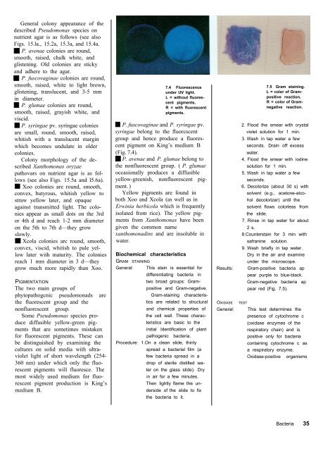
7.4 Fluorescence<br />
under UV light.<br />
L = without fluorescent<br />
pigments,<br />
R = with fluorescent<br />
pigments.<br />
P. fuscovaginae and P. syringae pv.<br />
syringae belong to the fluorcscent<br />
group and hence produce a fluorescent<br />
pigment on King’s medium B<br />
(Fig. 7.4).<br />
P. avenae and P. glumae belong to<br />
the nonfluorescent group. ( P. glumae<br />
occasionally produces a diffusible<br />
yellow-greenish, nonfluorescent pigment.<br />
)<br />
Yellow pigments are found in<br />
both Xoo and Xcola (as well as in<br />
Erwinia herbicola which is frequently<br />
isolated from <strong>rice</strong>). The yellow pigments<br />
from Xanthomonas have been<br />
given the common name<br />
xanthomonadins and are insoluble in<br />
water.<br />
Biochemical characteristics<br />
GRAM STAINING<br />
General: This stain is essential for<br />
differentiating bacteria in<br />
two broad groups: Grampositive<br />
and Gram-negative.<br />
Gram-staining characteristics<br />
are related to structural<br />
and chemical properties <strong>of</strong><br />
the cell wall. These characteristics<br />
are basic to the<br />
initial Identification <strong>of</strong> plant<br />
pathogenic bacteria.<br />
Procedure: 1.On a clean slide, thinly<br />
spread a bacterial film (a<br />
few bacteria spread in a<br />
drop <strong>of</strong> sterile distilled water<br />
on the glass slide). Dry<br />
in air for a few minutes.<br />
Then lightly flame the underside<br />
<strong>of</strong> the slide to fix<br />
the bacteria to it.<br />
Results:<br />
OXIDASE<br />
General:<br />
7.5 Gram staining.<br />
L = color <strong>of</strong> Grampositive<br />
reaction,<br />
R = color <strong>of</strong> Gramnegative<br />
reaction.<br />
2. Flood the smear with crystal<br />
violet solution for 1 min.<br />
3. Wash in tap water a few<br />
seconds. Drain <strong>of</strong>f excess<br />
water.<br />
4, Flood the smear with iodine<br />
solution for 1 min.<br />
5. Wash in tap water a few<br />
seconds.<br />
6. Decolorize (about 30 s) with<br />
solvent (e.g., acetone-alcohol<br />
decolorizer) until the<br />
solvent flows colorless from<br />
the slide.<br />
7. Rinse in tap water for about<br />
2 s.<br />
8.Counterstain for 3 min with<br />
safranine solution.<br />
9. Wash briefly in tap water.<br />
Dry in the air and examine<br />
under the microscope.<br />
Gram-positive bacteria ap<br />
pear purple to blue-black.<br />
Gram-negative bacteria ap<br />
pear red (Fig. 7.5).<br />
TEST<br />
This test determines the<br />
presence <strong>of</strong> cytochrome c<br />
(oxidase enzymes <strong>of</strong> the<br />
respiratory chain) and is<br />
positive only for bacteria<br />
containing cytochrome c as<br />
a respiratory enzyme.<br />
Oxidase-positive organisms<br />
Bacteria 35