

ULTIMATE COMPUTING - Quantum Consciousness Studies
ULTIMATE COMPUTING - Quantum Consciousness Studies
ULTIMATE COMPUTING - Quantum Consciousness Studies
- No tags were found...

Create successful ePaper yourself
Turn your PDF publications into a flip-book with our unique Google optimized e-Paper software.
<strong>ULTIMATE</strong> <strong>COMPUTING</strong> 1<br />
<strong>ULTIMATE</strong> <strong>COMPUTING</strong><br />
Biomolecular <strong>Consciousness</strong><br />
and NanoTechnology<br />
Billionth scale activities in biomolecular assemblies<br />
could define life itself, and provide<br />
a frontier for the evolution of technology.<br />
Stuart R. Hameroff<br />
Department of Anesthesiology<br />
College of Medicine<br />
University of Arizona<br />
Tucson, Arizona, U.S.A<br />
© Elsevier Science Publishers B.V., 1987<br />
All Rights Reserved.<br />
ISBN: 0 444 70283 0<br />
In 2003, this electronic edition was derived from the original 1987 print<br />
edition for the use of Stuart R. Hameroff, who retains electronic distribution<br />
rights. Apart from some mostly software-related formatting changes (or any<br />
uncaught scanning glitches), the main body of this text should be essentially<br />
identical to the original.
2 Contents<br />
Contents<br />
<strong>ULTIMATE</strong> <strong>COMPUTING</strong> ............................................................................ 1<br />
Contents ........................................................................................................... 2<br />
0 Prelude....................................................................................................... 5<br />
0.1 Acknowledgements .......................................................................... 6<br />
0.2 Dedication......................................................................................... 6<br />
1 Toward Ultimate Computing..................................................................... 7<br />
1.1 Mind/Tech: Merger in the Nanoscale ............................................... 7<br />
1.2 Evolution of Technology .................................................................. 8<br />
1.3 Collective Intelligence.................................................................... 10<br />
1.3.1 Parallelism ............................................................................. 12<br />
1.3.2 Connectionism ....................................................................... 15<br />
1.3.3 Cooperativity and Coherence................................................. 17<br />
1.4 Molecular Computing..................................................................... 20<br />
1.5 Dynamic Pattern Representation .................................................... 22<br />
1.5.1 Reaction Diffusion Systems................................................... 22<br />
1.5.2 Holograms.............................................................................. 24<br />
1.5.3 Macrons ................................................................................. 25<br />
1.5.4 Cellular Automata.................................................................. 26<br />
2 Brain/Mind/Computer ............................................................................. 33<br />
2.1 Metaphors of <strong>Consciousness</strong> .......................................................... 33<br />
2.2 Historical Perspectives—<strong>Consciousness</strong> as ................................... 35<br />
2.2.1 <strong>Consciousness</strong> as Particle/Wave Physics............................... 35<br />
2.2.2 <strong>Consciousness</strong> as a Property of Protoplasm........................... 36<br />
2.2.3 <strong>Consciousness</strong> as Learning .................................................... 36<br />
2.2.4 <strong>Consciousness</strong> as a Metaphysical Imposition ........................ 37<br />
2.2.5 The Helpless Spectator Theory.............................................. 37<br />
2.2.6 Emergent Evolution ............................................................... 38<br />
2.2.7 Behaviorism ........................................................................... 38<br />
2.2.8 <strong>Consciousness</strong> as Dynamic Activities of the Brain’s Reticular<br />
Activating System ........................................................................................ 39<br />
2.2.9 Neural Net Connectionism..................................................... 41<br />
2.2.10 Holography ............................................................................ 42<br />
2.2.11 Cytoskeletal Basis of <strong>Consciousness</strong> ..................................... 44<br />
3 Origin and Evolution of Life................................................................... 47<br />
3.1 Soup vs Mud, Chicken vs Egg........................................................ 47<br />
3.2 Prokaryote to Eukaryote—Symbiotic Jump ................................... 50<br />
3.3 Centrioles—Evolution’s Hijackers................................................. 53<br />
3.4 Biotech Evolution—The Next Symbiosis ...................................... 57<br />
4 From Brain to Cytoskeleton .................................................................... 59<br />
4.1 Nervous System Evolution ............................................................. 59<br />
4.2 Nervous System Organization ........................................................ 59<br />
4.2.1 Architecture............................................................................ 60<br />
4.2.2 Neuronal Signaling ................................................................ 61<br />
4.2.3 Interneuronal Synapses .......................................................... 61<br />
4.3 Representation of Information........................................................ 64<br />
4.3.1 Integration—Sherrington’s Reflex Centers ........................... 65<br />
4.3.2 Pulse Logic ............................................................................ 66<br />
4.3.3 Connectionism and Neural Networks .................................... 67<br />
4.3.4 Distributedness....................................................................... 71<br />
4.3.5 Synaptic Mechanisms of Learning and Memory ................... 73<br />
4.3.6 Axoplasmic Transport............................................................ 76
Contents 3<br />
4.3.7 Parallelism, Collective Cooperativity, and the Grain of the<br />
Engram 77<br />
4.4 Toward Molecular <strong>Consciousness</strong>.................................................. 79<br />
5 Cytoskeleton/Cytocomputer.................................................................... 80<br />
5.1 The Nature of Cytoplasm................................................................ 80<br />
5.2 Microtubules................................................................................... 84<br />
5.2.1 Microtubule Structure and Function ...................................... 84<br />
5.2.2 Microtubule Assembly and the Generation of Form.............. 88<br />
5.2.3 Microtubule Organizing Centers (MTOC) and Centrioles .... 93<br />
5.2.4 Microtubule Associated Proteins (MAPs) ............................. 97<br />
5.3 Intermediate Filaments ................................................................. 103<br />
5.4 The Cytoplasmic Ground Substance ............................................ 105<br />
5.4.1 The Microtrabecular Lattice (MTL) .................................... 105<br />
5.4.2 The Cytomatrix .................................................................... 108<br />
5.4.3 Cytoplasmic Solid State....................................................... 109<br />
5.5 Cytoskeletal Motility .................................................................... 111<br />
5.5.1 Cytoplasmic Probing............................................................ 112<br />
5.5.2 Bending Sidearms ................................................................ 116<br />
5.5.3 Ciliary and Collective Movement........................................ 118<br />
5.5.4 Geodesic Tensegrity Gels .................................................... 119<br />
5.6 The Cytoskeleton and Development............................................. 121<br />
5.7 The Cytoskeleton and Medicine ................................................... 124<br />
5.8 Intelligence in the Cytoskeleton ................................................... 126<br />
6 Protein Conformational Dynamics........................................................ 129<br />
6.1 Protein Structure........................................................................... 129<br />
6.2 Protein Conformation ................................................................... 131<br />
6.3 Proteins and Energy...................................................................... 134<br />
6.4 Protein Cooperativity—Historical View ...................................... 135<br />
6.5 Living Water and Hydrophobic Interactions ................................ 136<br />
6.6 Electret, Piezo, and Pyroelectric Effects....................................... 139<br />
6.7 Solitons/Davydov ......................................................................... 140<br />
6.8 Coherent Excitations /Fröhlich..................................................... 143<br />
6.9 Massless Bosons, Cytoskeletal Self-Focusing.............................. 146<br />
7 Anesthesia: Another Side of <strong>Consciousness</strong> ......................................... 148<br />
7.1 Levels of Anesthesia/<strong>Consciousness</strong> ............................................ 148<br />
7.2 Memory ........................................................................................ 151<br />
7.3 Mechanisms of Anesthesia ........................................................... 153<br />
8 Models of Cytoskeletal Computing....................................................... 157<br />
8.1 Energy and Information in Microtubules ..................................... 157<br />
8.2 Cytoskeletal Information Processing............................................ 158<br />
8.2.1 MT Sensory Transduction/Atema........................................ 158<br />
8.2.2 MT Mechano-lonic Transducers/Moran and Varela............ 159<br />
8.2.3 Cytomolecular Computing/Conrad and Liberman............... 160<br />
8.2.4 MT Signal Processing/DeBrabander.................................... 160<br />
8.2.5 Cytoskeletal String Processors/Barnett................................ 163<br />
8.2.6 Microtubule “Gradions”/Roth, Pihlaja, Shigenaka .............. 164<br />
8.2.7 Gyroscopic Centrioles/Bornens ........................................... 166<br />
8.2.8 Centriole-MT Signaling/Albrecht-Buehler .......................... 167<br />
8.2.9 Dynamic Tensegrity/Heidemann and Jarosch...................... 170<br />
8.2.10 Dynamic MT Probing/Kirschner and Mitchison ................. 171<br />
8.2.11 Sphere Packing Screw Symmetry/Koruga........................... 172<br />
8.2.12 Cytoskeletal Self-Focusing/Del Giudice.............................. 174<br />
8.2.13 MT Automata, Holography/Hameroff, Watt, Smith............ 174
4 Contents<br />
8.3 The Cytoskeletal Connection........................................................ 181<br />
9 Viruses/Ambiguous Life Forms ............................................................ 184<br />
9.1 What Is the Essence of Living Matter......................................... 184<br />
9.2 Virus (Mis)Behavior..................................................................... 184<br />
9.3 Virus Structure and Collective Oscillations ................................. 186<br />
9.4 Nature and Origin of Viruses........................................................ 187<br />
9.5 Domesticated Viruses ................................................................... 188<br />
10 NanoTechnology............................................................................... 190<br />
10.1 Early NanoTechnologists ......................................................... 190<br />
10.2 Scanning Tunneling Microscopes (STMs)............................... 192<br />
10.3 STM/Feynman Machines (FMs) .............................................. 203<br />
10.4 Micro/Nano STM Contest........................................................ 205<br />
10.5 STM/FMs and Molecular Computing ...................................... 205<br />
10.6 STM/FMs and Biomedical Applications.................................. 211<br />
10.7 Replicating Automata............................................................... 215<br />
11 The Future of <strong>Consciousness</strong> ............................................................ 217<br />
12 Bibliography ..................................................................................... 219<br />
12.1 STM References....................................................................... 248<br />
12.2 Near-Field Scanning Optical Microscopes............................... 256<br />
12.3 Other STM-Related Instruments .............................................. 256<br />
12.4 Replicating Systems References .............................................. 257<br />
12.5 Collective Computing References............................................ 259<br />
12.6 <strong>Quantum</strong> Computing References ............................................. 261<br />
Index ............................................................................................................ 264
Prelude 5<br />
0 Prelude<br />
What is this book, and why has it been written by an anesthesiologist This<br />
book is a view of the co-evolution of consciousness and technology-past, present<br />
and future.<br />
This book has been written by an anesthesiologist because of a confluence of<br />
two fascinations. The first is the nature of consciousness, which anesthesiologists<br />
routinely erase and restore in their patients. The second is a fifteen year trail of<br />
notions that would not go away. While a third year medical student in 1972, I<br />
spent a summer research elective in a cancer laboratory. For some reason I<br />
became fascinated and fixated by one particular question. When cells divided, the<br />
chromosomes were separated and daughter cell architecture established by wispy<br />
strands called mitotic spindles (“microtubules”) and cylindrical organelles called<br />
centrioles. Somehow, the centrioles and spindles “knew” when to move, where to<br />
go, and what to do. The uncanny guidance and orientation mechanism of these<br />
tiny biomolecular structures seemed to require some kind of motorized<br />
intelligence. At about the same time, electron microscopy techniques were<br />
revealing the interior of all living cells to be densely filled with wispy strands,<br />
some of which were identical to mitotic spindles. Interconnected in dynamic<br />
parallel networks, these structures were thought to serve a purely supportive, or<br />
mechanical structural role and were collectively termed the “cytoskeleton.”<br />
But several factors suggested that the cytoskeleton was more than the<br />
structural “bones” of the cell: they manipulated dynamic activities, orchestrating<br />
complex and highly efficient processes such as cell growth, mitosis and transport.<br />
Another factor was a lack of any other candidate for “real time” dynamic<br />
organization within cells. Long term blueprints and genetic information clearly<br />
resided in DNA and RNA, and membranes performed dynamic functions at cell<br />
surfaces. However, a mechanism for the moment to moment execution,<br />
organization, and activities within cells remained unknown. Where was the<br />
nervous system within the cell Was there a biological controller This book is<br />
based on the premise that the cytoskeleton is the cell’s nervous system, the<br />
biological controller/computer. In the brain this implies that the basic levels of<br />
cognition are within nerve cells, that cytoskeletal filaments are the roots of<br />
consciousness. The small size and rapid conformational activities of cytoskeletal<br />
proteins are just beyond the resolution of current technologies, so their potential<br />
dynamics remain unexplored and a cytoskeletal controlling capability untested.<br />
Near future technologies will be able to function in the nanoscale (nano = 10 -9 ;<br />
nanometer = one billionth meter, nanosecond = one billionth second and will<br />
hopefully resolve these questions. If indeed cytoskeletal dynamics are the texture<br />
of intracellular information processing, these same “nanotechnologies” should<br />
enable direct monitoring, decoding and interfacing between biological and<br />
technological information devices. This in turn could result in important<br />
biomedical applications and perhaps a merger of mind and machine: Ultimate<br />
Computing.<br />
A thorough consideration of these ideas involves a number of disciplines, all<br />
of which are at least tangentially related to anesthesiology. These include<br />
biochemistry, cognitive science, computer science, engineering, mathematics,<br />
microbiology, molecular biology, pharmacology, philosophy, physics,<br />
physiology, and psychology. As an expert in none, but a dabbler in all, I hope true<br />
experts in these fields will find my efforts never-the-less interesting.<br />
Starting from a cytoskeletal perspective, this book flings metaphors at the<br />
truth. Perhaps one or more will land on target, or at least come close.
6 Prelude<br />
0.1 Acknowledgements<br />
This book is a collective effect of the following people: Ralph Abraham, Ross<br />
Adey, Sigma Alpha, Fred Anderson, Amy Barnes, Joanne Barnes, Michel<br />
Bornens, Burnell Brown, George Carlson, Forrest Carter, Peter Christiansen, Jim<br />
Clegg, John Condeelis, Leonor Cruzeiro, Marc DeBrabander, Yves Engelborghs,<br />
Lawrence Fried, Herbert Fröhlich, Kit Grantham, Jamie, Harrison, Lillian, Amy,<br />
and the entire Hameroff/Kaplan/Bowman family, Max Headroom, Emery Hetrick,<br />
Dixie Holmes, Paul Jablonka, Martha Juarez, Jan Julianus, Ben Kahn, Charles<br />
Kiselyak, Djuro Koruga, Julio Kuperman, Teruo Matsumoto, Leo Martin,<br />
Kathleen McAuliffe, Claris Nelson, Michio Okuma, Karl Pribram, Mary Quimby,<br />
Steen Rasmussen, Conrad Schneiker, Alwyn Scott, Steven Smith, Branko Soucek,<br />
Arthur Villa, Rich Watt, Juli Weiss and Arthur Winfree.<br />
Most of the artwork was thoughtfully done by scientist/systems<br />
engineer/artist Paul Jablonka. Conrad Schneiker supplied most of the material on<br />
nanotechnology and replicators for Chapter 10, and compiled the appendices. I<br />
am indebted to the Laboratory for Advanced Mathematics and Physics at the<br />
Technical University of Denmark and the Danish Camping Union who hosted me,<br />
Jamie and Harrison during our sabbatical. I sincerely appreciate the efforts of my<br />
colleagues in the Department of Anesthesiology and the College of Medicine,<br />
University of Arizona who permitted me time and mental latitude. Thanks to<br />
Personal TeX’s port of Don Knuth’s TeX, plus Leslie Lamport’s LaTeX,<br />
Textset’s DVILASER/PS, Boreland’s Turbo Lightning, Adobe Systems’<br />
PostScript, Apple’s Macintosh and LaserWriter, IBM’s PC/AT and its clones,<br />
QMS’s PS800 laser printer and Xerox’s machines and their clones. The ever<br />
friendly and competent technical support from Textset, Personal TeX and HDS<br />
Systems is also greatly appreciated. [2003 Note: For various reasons, the<br />
electronic version of this book was formatted by the often wonderfully convenient<br />
but also sometimes obnoxiously troublesome Microsoft Word 2002. While Word<br />
2002 was a great time-saver overall compared to the previous tools, some pretty<br />
basic things that previously worked fine out of the box would have required too<br />
much additional work to reasonably replicate here. Hence the above-mentioned<br />
people, products, and companies should not be held responsible for the ironically<br />
sometimes less satisfactory typographical results some 16 years later.]<br />
Finally, Jan Julianus and Elsevier North-Holland deserve credit for their<br />
instigation and patience.<br />
0.2 Dedication<br />
To H. H., who loved to gamble.
Toward Ultimate Computing 7<br />
1 Toward Ultimate Computing<br />
1.1 Mind/Tech: Merger in the Nanoscale<br />
Biology and technology are both evolving toward more efficient methods of<br />
information processing. With a head start of a billion years, biology has evolved<br />
human consciousness; technology appears to be catching up rapidly.<br />
Ultimate Computing is the common destination for the evolution of<br />
information processing systems in both biology and technology. At this point it is<br />
an extrapolation of converging trajectories, but Ultimate Computing may soon<br />
exist in the nanoscale. Nano = 10 -9 , one nanometer is a billionth of a meter, and<br />
one nanosecond is a billionth of a second. Subunits within biological protein<br />
assemblies (cytoskeletal polymers, organelles, membrane proteins, virus coats)<br />
are of nanometer size scale and undergo conformational oscillations in the<br />
nanosecond time scale. Nanoscale excitations, which may be coherent and<br />
coupled to intraprotein dipole shifts, can generate communicative “collective<br />
modes” within protein assemblies and provide a substrate for biological<br />
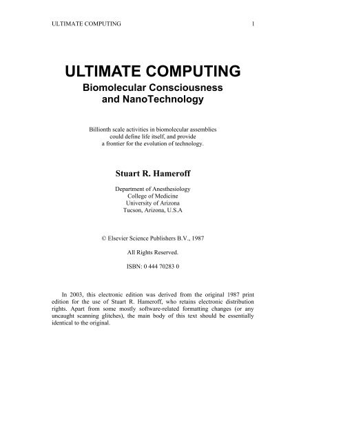
information processing. Thus the “nanoscale” (Figure 1.1) may be where living<br />
intelligence has evolved. Coincidentally, nanoscale devices including molecular<br />
computers, Feynman machines and von Neumann replicators are becoming<br />
feasible through technologies such as scanning tunneling microscopy. A<br />
nanoscale marriage of biomolecules and nanotech devices, providing direct<br />
communication and information transfer, could have profound benefits for<br />
biomedicine and our culture in general.<br />
Figure 1.1: Sizing the Nanoworld. The diameter of each circle is given in<br />
nanometers (nm). A) 0.30 nm—a carbon atom, 0.15 nm in diameter. B) 0.50<br />
nm—alanine, an amino acid with 13 atoms including 3 carbons, is about .33 nm in<br />
diameter. C) 12 nm—a tubulin dimer protein, the subunit of microtubules, is 8 nm<br />
long. It is composed of 2 similar monomers (alpha and beta tubulin), each made<br />
of about 440 amino acids. Cross hatching suggests the approximate amount of<br />
space available for each amino acid. D) 50 nm—a microtubule, 13 sided tube with<br />
an outside cross-sectional diameter of 25 nm. E) 1900 nm—a small 1000 nm<br />
diameter nerve axon might contain 100 microtubules (shown) and 1000 smaller<br />
filaments (not shown). Microtubules associate in informal clumps of 1 to 5<br />
microtubules each, represented by dots. F) 40,000 nm—a nerve cell grown on the<br />
surface of a Motorola 68000 computer chip. The wire thickness is 15,000 nm<br />
wide. G) 170,000 nm—a nematode is a small worm of less than 1000 cells, 300 of<br />
which are neurons. Nematodes have a brain, teeth, muscles, gut, and sex lives.
8 Toward Ultimate Computing<br />
This one is about 450,000 nm long. H) 5,000,000 nm—one quarter of a human<br />
thumbnail with 50 nematodes represented to scale. By Paul Jablonka.<br />
Comingling of consciousness and computer technology is a prevalent dream.<br />
Artificial intelligence based on brain/mind organization is a tentative step in this<br />
direction, as is the proposed use of self assembling protein arrays as switching<br />
circuits or “biochips.” The Japanese effort towards the “Sixth Generation<br />
Computer” aims to “integrate biology and technology” by merging research in<br />
artificial intelligence and the functions of living organisms (Corcoran, 1987). By<br />
attempting to understand the conditions required to maintain biological<br />
“homeostasis”, the Japanese are hoping to embark on a symbiosis between<br />
intelligent biological structures and technological devices, and even predict an<br />
“artificial brain”! One missing ingredient for such a Mind/Tech merger is an<br />
understanding of the mechanism of consciousness. Most models of brain<br />
organization consider nerve cells and their connections to be the brain’s<br />
fundamental units of information processing. However, profoundly complex and<br />
intelligent activities occur within nerve cells. Further, simple organisms like<br />
single cell amoeba and paramecium perform complex tasks without benefit of<br />
brain or nervous system. In this book we view the cytoskeleton—networks of<br />
protein polymers which occupy and organize the interiors of all living cells<br />
(Figure 1.2)—as a highly evolved information processing system operating at<br />
nanoscale levels. Collective nanoscale activities of the cytoskeleton and related<br />
structures can explain biological organization, information processing, and<br />
consciousness, and be the target for the future evolution of technology.<br />
Figure 1.2: Cytoskeleton within cells who have just divided. Intracellular<br />
microtubules are visualized by immunostaining. Spherical areas are cell nuclei,<br />
adjacent to which are the dense microtubule organizing centers (MTOC). With<br />
permission from DeBrabander, Geuens, DeMey and Joniav (1986), courtesy of<br />
Marc DeBrabander.<br />
1.2 Evolution of Technology<br />
Technological emulation of life since the 13th century has been reviewed by<br />
author Claris Nelson (1985). Albertus Magnus is said to have create a life-like<br />
mechanical servant out of metal, wood, glass, leather and wax that could open<br />
doors and greet visitors. It was considered blasphemous work of the devil by<br />
Magnus’ student Saint Thomas Aquinas who destroyed it. Science fiction writers<br />
predicted computers and robots long before they existed. In 1879, Edward Page<br />
Mitchell’s The Ablest Man in the World featured a mechanical brain and in
Toward Ultimate Computing 9<br />
Edmund Hamilton’s 1928 The Metal Giants an artificial brain turned against its<br />
creators.<br />
Computers descended from calculating machines, the earliest of which was<br />
the abacus. In 1642 French mathematician and philosopher Pascal made a<br />
mechanical calculator that used the decimal system to add and subtract. In 1694,<br />
German mathematician/philosopher Leibniz created a “Stepped Reckoner,” which<br />
was supposed to multiply, divide and take square roots. It didn’t work, but<br />
utilized principles later essential to modern computers. Tasks were broken down<br />
into a great many simple mathematical steps using binary numbers and were<br />
performed sequentially. When computers later came to be operated by electricity,<br />
binary zero and one became represented by off and on. In the early 1800’s George<br />
Boole developed “Boolean algebra,” the mathematical logic by which computer<br />
circuits are designed. Charles Babbage and Ada Lovelace—Lord Byron’s eldest<br />
daughter—designed an “analytical engine” using punched cards. Their<br />
contemporary technology could not construct the machine accurately enough, but<br />
it was built and functioned in the twentieth century.<br />
The first electronic computer was apparently constructed and operated in<br />
1939 by John Vincent Atanasoff, a theoretical physicist at Iowa State University<br />
(Mackintosh, 1987). Shortly thereafter, Alan Turing and colleagues in Bletchley,<br />
England designed a computer to perform all possible mathematical calculations. It<br />
was based on Turing’s work proving the logical limits of computability and was<br />
used to decipher the German “Enigma” code during World War II. In a masterful<br />
presentation of key ideas previously developed by other pioneers, John von<br />
Neumann further advanced computer design by separating the machine from its<br />
problems. Prior to von Neumann, a computer would have to be rewired for each<br />
new task. With enough time, memory and software, computers could solve the<br />
problems that could be broken down into finite sequences of logical steps. Most<br />
current computers use “serial” processing based on von Neumann’s design. In the<br />
1940’s, the University of Pennsylvania developed the first electronic computer,<br />
the Electronic Numerical Integrator and Calculator or “ENIAC.” It weighed 30<br />
tons, took up 3,000 cubic feet of space, and contained 18,000 vacuum tubes, one<br />
of which failed every seven minutes. It could calculate nuclear physics problems<br />
in two hours that would have taken 100 engineers a year to complete. Today, the<br />
same capacity is available on one chip. In 1950 Remington Rand marketed<br />
UNIVAC, which dealt with words and numbers stored by their binary equivalent.<br />
Since that time, roughly four generations of computers have evolved due to<br />
increased demand and advances in design, chip size, materials and other factors.<br />
For the same reasons further advances seem inevitable.<br />
Von Neumann and Turing hoped that computers could duplicate our ability to<br />
think, so that our minds could be amplified just as our muscles had been by<br />
industrial machines. However further evolution of computers using serial<br />
processing seems limited. Computers and artificial intelligence are now evolving<br />
to parallel systems based on brain architecture and neural net models; a future<br />
step may be nanoscale, self organizing intelligence.<br />
Von Neumann is one of several “fathers of the computer.” In the “serial”<br />
processing which he skillfully formalized, information flows in one dimension. In<br />
the 1950’s and 1960’s, von Neumann (1966) and Stanislav Ulam developed the<br />
mathematics of computing in multiple dimensions. They considered two<br />
dimensional information spaces with discrete subunits (“cells”) whose states<br />
could vary depending on the states of neighboring cells. Each cell and its<br />
neighbor relations were identical. Relatively simple rules among neighbors and<br />
discrete time intervals (“generations”) led to evolving patterns and selforganization<br />
which were exquisitely sensitive to initial conditions. They called
10 Toward Ultimate Computing<br />
these systems “cellular automata.” Von Neumann described a “universal<br />
computer” automaton which could solve any problem if given sufficient area and<br />
time. Today, computer technologists are considering the profound advantages of<br />
implementing molecular scale automata (Milch, 1986).<br />
Edward Fredkin of Massachusetts Institute of Technology has considered<br />
multidimensional automata and the discreteness of time and matter. He argues<br />
that the universe is a cellular automaton whose “cells” are atomic and subatomic<br />
particles (Wright, 1985). The universe is made of information, Fredkin reasons.<br />
Cellular automata may be generalized “primordial computers” of which all other<br />
computers and complex systems are particular examples. Cellular automata in<br />
conformational states of cytoskeletal subunits could process biological<br />
information and be the substrate of consciousness.<br />
The current trend in computer design and artificial intelligence or “AI” is<br />
parallel connectedness, emulating the brain. Many types of problems can be<br />
solved by breaking them down into serial mathematical steps. Today’s electronic<br />
computers serially process very rapidly and can solve complex mathematical<br />
problems far faster than can humans alone. However qualitative functions which<br />
the brain performs naturally-recognizing patterns, or making judgments-are<br />
extremely difficult for computers. Consider the letter “a.” We recognize it<br />
automatically, in any typeface, in all but the worst handwriting. To our brains it’s<br />
simple, quick, obvious even if it’s missing. If we see, “Sally ‘red’ a newspaper,”<br />
we mentally insert the absent “a.” Computer/Al scientist Jerome Feldman (1985)<br />
cites the example of interpreting the statement “John threw a ball for charity.”<br />
The inherent ambiguities of this type of statement can be resolved in a highly<br />
parallel system in which multiple simultaneous interpretations are processed and<br />
evaluated. Hurling a sphere versus hosting a dance can be resolved by the<br />
qualifier “for charity” which is much more consistent with a dance than with a<br />
sphere. Human brains commonly resolve conflicts among differing drives or<br />
input, although failure to do so may cause psychiatric or emotional problems. At<br />
least according to science fiction, computers can suffer similar disturbances. In<br />
Arthur C. Clarke’s and Stanley Kubrick’s 2001: Space Odyssey and its sequel<br />
2010, the computer “Hal 9000” becomes psychotic because of conflicting<br />
instructions and reacts by killing the space voyagers because their mission was<br />
too important to be entrusted to them. The brain/mind can perform “cognitive”<br />
functions including resolution of conflict by “subcognitive” processes such as<br />
recognizing patterns, making assumptions and performing imaginative leaps. The<br />
net effect is consciousness: a collective effect of simpler processes.<br />
1.3 Collective Intelligence<br />
A collective phenomenon is more the product of, rather than the sum of, its<br />
parts, and has been explained by Cal Tech biophysicist John Hopfield (1982)<br />
whose “neural net” models are collective.<br />
Suppose you put two molecules in a box, every once in a while they<br />
collide and that’s an exciting event. ... If we’d put ten or even a<br />
thousand more molecules in the box all we’d get is more collisions.<br />
But if we put a billion billion molecules in the box, there is a new<br />
phenomenon-sound waves.
Toward Ultimate Computing 11<br />
Figure 1.3: Axoplasmic transport occurs by coordinated activities of microtubule<br />
attached sidearm, contractile proteins (“dynein”) which cooperatively pass<br />
material in a “bucket brigade.” The orchestration mechanism is unknown, but<br />
shown here as the consequence of signaling by “soliton” waves of microtubule<br />
subunit conformational states. By Fred Anderson.<br />
Observation of two, or ten, or thousands of those molecules would not<br />
suggest the Mozart or Madonna that can arise in a collection of more than a<br />
trillion trillion molecules. Other examples of collective phenomena may be seen<br />
in beehives, ant colonies, football teams, governments and various types of<br />
material phase transitions. For example, superconductivity and magnetism are<br />
collective effects which occur in certain metals as their individual atoms come<br />
into alignment. By cooling these metals, thermal fluctuations cease, atoms<br />
become highly aligned, and below a critical temperature totally different<br />
qualitative properties of superconductivity or magnetism emerge.<br />
How might collective phenomena be tied to consciousness Brain neuron<br />
synaptic transmissions are relatively slow at several milliseconds per<br />
computation-they are about 100,000 times slower than a typical computer switch.<br />
Nevertheless vision and language problems can be solved in a few hundred<br />
milliseconds or what would appear to be about 100 serial steps. Artificial<br />
Intelligence (AI) researchers conclude that this computational richness is<br />
accounted for by collective effects of parallelism and rich interconnectedness.<br />
With billions of neurons, and with each neuron connected to up to hundreds of<br />
thousands of other neurons, Al “connectionists” view the brain as a collective<br />
phenomenon of individually stupid neurons. Groups of highly connected neurons<br />
are thought to attain intelligent behavior through properties of feedback and<br />
reverberation. Walter Freeman (1972, 1975, 1983) of the University of California<br />
at Berkeley contends that a “critical mass” of about 100,000 neurons yields<br />
intelligent behavior. However, intelligent behavior occurs within nematode<br />
worms of 1000 cells and 300 neurons, within cytoplasm in single cell organisms<br />
and within single neurons. Individual neurons with tens to hundreds of thousands<br />
of connections cannot be stupid and fulfill their multiple functions, integrate<br />
input/output and modulate synaptic connection strength. Each nerve cell is a<br />
sophisticated information processing system in and of itself! The cytoskeleton<br />
within neurons and all living cells is a parallel connected network which can<br />
utilize its own collective phenomena to organize and process subcellular
12 Toward Ultimate Computing<br />
information (Figure 1.3). The cytoskeleton can convey analog patterns which may<br />
be connected symbols (Chapter 8). Although overlooked by AI researchers, the<br />
cytoskeleton may take advantage of the same attributes used to describe neural<br />
level networks. Properties of networks which can lead to collective effects among<br />
both neurons and cytoskeletal subunits include parallelism, connectionism, and<br />
coherent cooperativity.<br />
1.3.1 Parallelism<br />
The previous generations of computer architecture have been based on the<br />
von Neumann concept of sequential, serial processing. In serial processing,<br />
computing steps are done consecutively which is time consuming. One false bit of<br />
information can cascade to chaotic output. The brain with its highly parallel nerve<br />
tracks shines as a possible alternative. In parallel computing, information enters a<br />
large number of computer pathways which process the data simultaneously. In<br />
parallel computers information processors may be independent of each other and<br />
proceed at individual tempos. Separate processors, or groups of processors, can<br />
address different aspects of a given problem asynchronously. As an example,<br />
Reeke and Edelman (1984) have described a computer model of a parallel pair of<br />
recognition automata which use complementary features (Chapter 4). Parallel<br />
processing requires reconciliation of multiple outputs which may differ due to<br />
individual processors being biased differently than their counterparts, performing<br />
different functions, or because of random error. Voting or reconciliation must<br />
occur by lateral connection, which may also function as associative memory.<br />
Output from a parallel array is a collective effect of the input and processing, and<br />
is generally a consensus which depends on multiple features of the original data<br />
input and how it is processed. Parallel and laterally connected tracks of nerve<br />
fibers inspired AI researchers to appreciate and embrace parallelism. Cytoskeletal<br />
networks within nerve cells are highly parallel and interconnected, a thousand<br />
times smaller, and contain millions to billions of cytoskeletal subunits per nerve<br />
cell!<br />
Present day evolution of computers toward parallelism has engendered the<br />
“Connection Machine” (Thinking Machines, Inc.) which is a parallel assembly of<br />
64,000 microprocessors. Early computer scientists would have been impressed<br />
with an assembly of 64,000 switches without realizing that each one was a<br />
microprocessor. Similarly, present day cognitive scientists are impressed with the<br />
billions of neurons within each human brain without considering that each neuron<br />
is itself complex.<br />
Another stage of computer evolution appears as multidimensional network<br />
parallelism, or “hypercubes.” Hypercubes are processor networks whose<br />
interconnection topology is seen as an “n-dimensional” cube. The “vertices” or<br />
“nodes” are the processors and the “edges” are the interconnections. Parallelism<br />
in “n-dimensions” leads to hypercubes which can maximize available computing<br />
potential and, with optimal programming, lead to collective effects. Complex<br />
interconnectedness observed among brain neurons and among cytoskeletal<br />
structures may be more accurately described as hypercube architecture rather than<br />
simple parallelism. Hypercubes are exemplified in Figures 1.4, 1.5, and 1.6.<br />
Al/Roboticist Hans Moravec (1986) of Carnegie-Mellon University has<br />
attempted to calculate the “computing power” of a computer, and of the human<br />
brain. Considering the number of “next states” available per time in binary digits,<br />
or bits, Moravec arrives at the following conclusions. A microcomputer has a<br />
capacity of about 10 6 bits per second. Moravec calculates the brain “computing”<br />
power by assuming 40 billion neurons which can change states hundreds of times<br />
per second, resulting in 40 x 10 11 bits per second. Including the cytoskeleton
Toward Ultimate Computing 13<br />
increases the potential capacity for information processing immensely.<br />
Microtubules are the most visible cytoskeletal structures. Making some rough<br />
assumptions about cytoskeletal density (i.e. microtubules spaced about 1000<br />
nanometers apart) and the volume of brain which is neuronal cytoplasm leads to<br />
about 10 14 microtubule subunits in a human brain (ignoring neurofilaments and<br />
other cytoskeletal elements). As described in Chapters 5 and 6, the frequency of<br />
cytoskeletal subunit state changes may be greater than billions per second! The<br />
cytoskeleton is capable not only of immense information capacity, but appears to<br />
be designed such that interacting conformational state patterns may perform<br />
computing functions. Several theories which propose such mechanisms will be<br />
described in Chapter 8.<br />
Figure 1.4: Six dimensional hypercube with 64 nodes, and 6 connections per<br />
node. Computer generation by Conrad Schneiker.<br />
The brain is a continuous system. Classical computers have operated on<br />
recursive repetitive functions to process information in batches and the output is<br />
obtained as the final product. Similarly, most parallel processing designs have<br />
discrete input and output points. Carl Hewitt (1985) has described open systems<br />
within computers in which processing may never halt, which can provide output<br />
while computing is still in operation, and can accept input from sources not<br />
anticipated when the computation began. Like the human brain/mind, open<br />
continuous systems can interact with the environment and adapt to new situations.<br />
Hewitt describes an asynchronous parallel computer system which can make use<br />
of multiple inputs and outputs and whose parallel elements are connected by<br />
“arbiters” which “weigh” and reconcile differing content, and can provide<br />
continuous input and output. Among brain neurons, “arbiters” would appear to be
14 Toward Ultimate Computing<br />
synaptic connections among laterally connected parallel neurons. Within the<br />
cytoskeleton, laterally connecting filaments and microtubule associated proteins<br />
(“MAPs”) could serve as logical arbiters.<br />
Figure 1.5: Eight dimensional hypercube with 256 nodes, and 8 connections per<br />
node. Computer generation by Conrad Schneiker.<br />
Hewitt argues that parallel, open systems are “non-hierarchical” because input<br />
and output are continuously processed throughout the system. Early views of<br />
brain/mind organization assumed a hierarchical arrangement of processing units.<br />
Sensory input was thought to be processed and relayed to higher and higher levels<br />
of cognition until reaching a single “Grandfather neuron” or “Mind’s Eye” which<br />
comprehended the input’s “essence.” Classical brain research by Lashley (1929,<br />
1950) and others (Chapter 4) strongly suggest that memory and information are<br />
distributed throughout the brain and that specific anatomical hierarchical<br />
arrangements leading to “Grandfather neurons” do not exist. The “Mind’s Eye” is<br />
not localized to a given site but is mobile over wide volumes of brain. Assuming<br />
that humans actually do comprehend the essence of at least some things, who or<br />
what is comprehending The site and nature of attention, “self,” consciousness or<br />
the Mind’s Eye remains a philosophical issue and barrier to Mind/Tech merger.<br />
Neuroanatomical structure and the distributed storage of brain information point<br />
toward highly parallel, open brain/mind computing systems which may occur<br />
both at the neural level, and within neurons in the cytoskeleton. The perception<br />
component of consciousness, the “Mind’s Eye” may be a mobile hierarchy<br />
determined by collective dynamics.
Toward Ultimate Computing 15<br />
1.3.2 Connectionism<br />
The Mind’s Eye may be the apex of a collective hierarchy of parallel systems<br />
in which the cytoskeleton and related structures are the ground floor. Parallel<br />
systems in both computers and biological systems rely on lateral connections and<br />
networks to provide the richness and complexity required for sophisticated<br />
information processing. Computer simulations of parallel connected networks of<br />
relatively simple switches (“neural nets”) develop “cognitive-like functions” at<br />
sufficient levels of connectedness complexity-a “collective phenomenon”<br />
(Huberman and Hogg, 1985). Philosopher John Searle (Pagels, 1984), who has an<br />
understandable bias against the notion that computer systems can attain human<br />
consciousness equivalence, points out that computers can do enormously complex<br />
tasks without appreciating the essence of their situation. Searle likens this to an<br />
individual sorting out Chinese characters into specific categories without<br />
understanding their meaning, being unable to speak Chinese. He likens the<br />
computer to the individual sorting out information without comprehending its<br />
essence.<br />
It would be difficult to prove that human beings comprehend the essence of<br />
anything. Nevertheless, even the simulation of cognitive-like events is interesting.<br />
Neural net models and connectionist networks (described further in Chapter 4)<br />
have been characterized mathematically by Cal Tech’s John Hopfield (1982) and<br />
others. His work suggests that solutions to a problem can be understood in terms<br />
of minimizing an associated energy function and that isolated errors or incomplete<br />
data can, within limits, be tolerated. Hopfield describes neural net energy<br />
functions as having contours like hills and valleys in a landscape. By minimizing<br />
energy functions, information (metaphorically) flows like rain falling on the<br />
landscape, forming streams and rivers until stable states (“lakes”) occur. A new<br />
concept in connectionist neural net theory has emerged with the use of multilevel<br />
networks. Geoffrey Hinton (1985) of Carnegie-Mellon University and Terry<br />
Sejnowski of Johns Hopkins University have worked on allowing neural nets to<br />
find optimal solutions, like finding the lowest particular lake in an entire<br />
landscape. According to Sejnowski (Allman, 1986; Hinton, Sejnowski and<br />
Ackley, 1984) the trick is to avoid getting stuck in a tiny depression between two<br />
mountains:<br />
Imagine you have a model of a landscape in a big box and you want<br />
to find a lowest point on the terrain. If you drop a marble into the<br />
box, it will roll around for a while and come to a stop. But it may not<br />
be the lowest point, so you shake the box. After enough shaking you<br />
usually find it.
16 Toward Ultimate Computing<br />
Figure 1.6: Ten dimensional hypercube with 1,024 nodes, and 10 connections<br />
per node. Computer generation by Conrad Schneiker.<br />
Hinton and Sejnowski have used this concept of mathematically shaking their<br />
neural net simulations to find optimal solutions. It requires a multilevel hierarchy<br />
of parallel systems so that one level can “shake” or tune a lower level. Such an<br />
arrangement can perhaps explain the relationship between hierarchical layers of<br />
parallel systems within the brain. For example, neural networks based on synaptic<br />
connection may regulate (and be regulated by) smaller, faster, more<br />
comprehensive networks in the intracellular cytoskeleton.<br />
Extensive comparisons between information processing in the brain and<br />
artificial intelligence have been reviewed by A. M. Decallatay (1986) who feels<br />
the laws of thought described in philosophy have been rediscovered by AI: “The<br />
mental world of Plato is reproduced in the physical symbols of Newell and<br />
Simon.” DeCallatay observes that Al represents data by virtual pointers which<br />
connect symbols. In computers these virtual relations are actual wires with<br />
potential gate connection; in the brain they appear to be neuronal synaptic<br />
connections. Within neurons they may be cross-bridge filaments connecting<br />
cytoskeletal microtubules. As a computer expert evaluating the brain, DeCallatay<br />
states that the brain learns by opening gates to build new connections between<br />
elements simultaneously activated. He sees the presence or absence of dendritic<br />
spines playing the role of an “all or none” switch at the neural level. Dendritic<br />
spines are knobby projections of membrane covered cytoplasm on neuronal<br />
dendrites which are generated and maintained by the cytoskeleton and form<br />
synapses with other neurons. The most accepted theory for learning and memory
Toward Ultimate Computing 17<br />
in the brain is that of strengthening of specific synapses within neural circuits, an<br />
idea generated by Donald Hebb (1949). As will be described in Chapters 4 and 5,<br />
dynamic structural activities of the cytoskeleton are responsible for all<br />
cytoplasmic rearrangements including formation and regulation of dendritic<br />
spines and synapses. The spines are branchings of dendrites which themselves are<br />
branchings of neurons. A further dimension of complexity, these cytoskeletal<br />
appendages are prime candidates for “synaptic plasticity,” the cornerstone for<br />
prevalent models of brain learning and memory.<br />
1.3.3 Cooperativity and Coherence<br />
Collective effects manifest as diffuse reverberation, sustained oscillation,<br />
phase transitions, and deterministic chaos have been observed in computer<br />
simulation of parallel networks (Choi and Huberman, 1984). Collective<br />
mechanisms can exert long-range cooperativity and an executive level of<br />
organization within parallel arrays. Collective phase transitions in brain parallel<br />
arrays could be a fabric of consciousness, an “idea” emerging like the property of<br />
superconductivity from a large number of simple, “aligned” subunits. In most<br />
views the neuronal synapse is the brain’s fundamental subunit, however synaptic<br />
activities are the net result of dynamic processes orchestrated by the cytoskeleton.<br />
Layers of cytoskeletal organization are evident within neurons, and their<br />
participation in cognitive functions appears unavoidable. Thus the highly<br />
branched cytoskeleton may be another dimension of brain organization, perhaps<br />
related to neuronal networks as a “fractal.” Many natural processes manifest<br />
fractals, growth patterns in which local areas are scaled down images of the entire<br />
pattern. This occurs through some form of long range correlation in the pattern:<br />
components “know about each other over distances far in excess of the range of<br />
the forces between them” (Sander, 1986). Fractal relationships are one type of<br />
long range cooperativity (Figures 1.7 and 1.8). Densely parallel interconnected<br />
networks of cytoskeletal structures resemble larger scale networks of neurons, and<br />
may be viewed as fractal subdimensions of neural networks.<br />
Long range cooperativity and collective mechanisms are favored by the<br />
property of coherence which means peak energy excitations within an area occur<br />
“in phase,” or simultaneously as in a laser. How may coherence arise in<br />
distributed processes DeCallatay (1986) proposes that coherence in the brain and<br />
AI need to be imparted from the top of a hierarchy downward, like the chief<br />
executive of a corporation setting goals and intentions. A different view is that of<br />
an underlying rhythm or beat to which all elements are tuned. Rhythmic coupling<br />
among neurons may be important, and some interpreters of brain electrical<br />
activity (EEG) believe regional brain wave entrainment leads to functional<br />
regions of mental representation. A more fundamental coherence at the level of<br />
protein assemblies may be universally important for biological cooperativity and<br />
communication.
18 Toward Ultimate Computing<br />
Figure 1.7: Tree fractal in which branching patterns are the same at every scale,<br />
or dimension. Long range order is present. Computer generation by Conrad<br />
Schneiker.
Toward Ultimate Computing 19<br />
Figure 1.8: Branching box fractal in which patterns are identical at every scale, or<br />
dimension. Long range order is present. Computer generation by Conrad<br />
Schneiker.<br />
Proteins and their components oscillate among specific conformational states<br />
which exist transiently for durations ranging from femtoseconds (10 -15 sec) to<br />
minutes or longer. As will be described in Chapter 6, functional conformational<br />
states appear coupled to nanosecond (10 -9 sec) oscillations and more prolonged<br />
“metastable states.” Herbert Fröhlich, an eminent physicist who helped develop<br />
the theory of superconductivity in the 1950’s, has devoted recent efforts to the<br />
question of cooperativity in biological systems. Fröhlich (1970, 1975, 1984) argues<br />
that biochemical energy supplied to biomolecular assemblies can result in coherent<br />
elastic vibrations of individual subunits in the sub-nanosecond time range. The effect<br />
presupposes a voltage effect in the biomolecule (i.e. an “electret”) and an organized
20 Toward Ultimate Computing<br />
spatial structure whose geometry favors coupling among subunits. Coherent<br />
oscillations in an appropriate medium like the cytoskeleton can lead to collective<br />
phenomena such as long range cooperativity, communication, and holography.<br />
Another model can help explain long range cooperativity in biomolecules. Soviet<br />
biophysicist A. S. Davydov has considered almost lossless energy transfer in<br />
biomolecular chains or lattices as wave-like propagations of coupled conformational<br />
and electronic disturbances: “solitons.” Davydov used the soliton concept to explain<br />
molecular level events in muscle contraction, however solitons in the cytoskeleton<br />
may do what electrons do in computers.<br />
The Fröhlich and Davydov approaches may be seen as complementary<br />
(Tuszynski, Paul, Chatterjee, and Sreenivasan, 1984). Fröhlich’s coherency model<br />
focuses on time-independent effects (stable states) leading to order whereas<br />
Davydov’s model looks at time-dependent effects which propagate order through the<br />
system. These and other theories of collective effects applied to information<br />
processing in cytoskeletal lattices will be described in Chapters 6 and 8.<br />
1.4 Molecular Computing<br />
To approach the cognitive capabilities of the human brain, Al must emulate<br />
brain structure at the nanoscale. Computer hardware is indeed evolving to smaller<br />
switching components, and advantages of proteins themselves are being<br />
considered. The smallward evolution of technological computing elements<br />
embraces a number of concepts and material collectively known as “molecular<br />
computing.”<br />
The potential advantages of molecular computers have been described by D.<br />
Waltz (1982) of Thinking Machines Corporation. 1) Current “planar” computer<br />
design is limited in overall density and use of three dimensional space. 2) Further<br />
miniaturization is limited with silicon and gallium arsenide technologies. Chips<br />
and wires cannot be made much smaller without becoming vulnerable to stray<br />
cosmic radiation or semiconductor impurities. 3) Biomolecular based devices may<br />
offer possibilities for self-repair or self-regeneration. 4) Certain types of analog,<br />
patterned computation may be particularly suited to molecular computers.<br />
Forrest L. Carter (1984) of the Naval Research Laboratory has catalyzed the<br />
molecular computing movement through his own contributions and by sponsoring<br />
a series of meetings on Molecular Electronic Devices (in 1981, 1983, 1986).<br />
Strategies described by Carter and others at his meetings have been aimed at<br />
implementing nanoscale computing through switching in material arrays of<br />
polyacetylenes, Langmuir-Blodgett films, electro-optical molecules, proteins and<br />
a number of other materials. Interfacing between nanoscale devices and<br />
macroscale technologies is an obstacle with several possible solutions: 1)<br />
engineering upward, self assembling components, 2) optical communication, 3)<br />
molecular wires, 4) don’t interface; build systems that are totally nanoscale<br />
(though they’d have to be somehow developed and tested), and 5) a sensitive<br />
bridge between macroscale and nanoscale. Technologies which may fulfill this<br />
latter possibility include ion beam nanolithography, molecular spectroscopy,<br />
quantum well devices, and scanning tunneling microscopy (STM). In STM,<br />
piezoceramic positioners control an ultra sharp conductor with a monoatomic tip<br />
which can probe and image material surfaces with atomic level resolution. STM<br />
related nanotools may soon be capable of ultraminiature fabrication and<br />
interfacing: “nanotechnology” (Chapter 10).<br />
The medium of information flow in conventional computers is electronic<br />
current flow, but electron transfer may be too energetically expensive and<br />
unnecessary at the molecular nanoscale. Many of the projected modes of<br />
molecular computing rely on propagation of nonlinear coupling waves called<br />
“solitons” similar to what Davydov proposed for linear biomolecules. Carter
Toward Ultimate Computing 21<br />
(1981) proposed that solitons could propagate through switching circuits made of<br />
branched polyacetylene chains. He has also considered molecular computing in<br />
periodic arrays using electron tunneling, soliton “valving” and photo-activated<br />
conformational changes in lattice materials. He envisions three dimensional<br />
molecular scale memory and switching densities of 10 15 to 10 18 elements per<br />
cubic centimeter, near the theoretical limit for charge separation. A number of<br />
materials may be suitable for soliton switching and biological propagation of<br />
solitons in proteins has been suggested. Several authors have argued for<br />
cytoskeletal solitons mediating information processing (Chapter 8).<br />
Wayne State University’s Michael Conrad has defined his vision of a<br />
molecular computer in which proteins integrate multiple input modes to perform a<br />
functional output (Conrad, 1986). In addition to smaller size scale, protein based<br />
molecular computing offers different architectures and computing dimensions.<br />
Conrad suggests that “non-von Neumann, nonserial and non-silicon” computers<br />
will be “context dependent,” with input processed as dynamical physical<br />
structures, patterns, or analog symbols. Multidimensional conditions determine<br />
the conformational state of any one protein: temperature, pH, ionic<br />
concentrations, voltage, dipole moment, electroacoustical vibration,<br />
phosphorylation or hydrolysis state, conformational state of bound neighbor<br />
proteins, etc. Proteins integrate all this information to determine output. Thus<br />
each protein is a rudimentary computer and converts a complex analog input to<br />
an output state or conformation.<br />
Conrad and Liberman (1982) have defined an “extremal computer” as one<br />
which uses physical resources as effectively as possible for computation. They<br />
suggest that an extremal computer should be a molecular computer, with<br />
individual switches or information representation subunits composed of<br />
molecules. The state of each information. subunit should be coupled to an energy<br />
event near the quantum limit. Protein conformational states leveraged to dipole<br />
oscillations in the nanoscale may be that limit. Conrad and Liberman conclude<br />
that, within biological systems, macromolecular computing occurs by<br />
conformational changes generating “reaction diffusion patterns” of concentrations<br />
of biochemical energy molecules (cyclic AMP).<br />
A 1984 conference (Yates, 1984) considered Chemically Based Computer<br />
Designs (Yates, 1984) and attempted to answer 6 relevant questions. 1) Are there<br />
fundamental, quantum mechanical limitations on computation This question<br />
deals with energy loss due to friction or other factors in computation. The work of<br />
Benioff (1980, 1982), Landauer (1982) and Feynman (1986) lead to the<br />
conclusion that, in principle, computation can be achieved by a frictionless,<br />
energy conserving system. Thus there appear to be no quantum mechanical<br />
limitations on computation. 2) Are there fundamental, thermodynamic limitations<br />
on computation Although there are some computing operations that are<br />
irreversible and dissipative, the work of Landauer (1982) and Bennett (1982)<br />
show that there are no fundamental thermodynamic limitations on computation<br />
per se. 3) Are there fundamental limits to serial processing on digital computers<br />
based on binary switches This question has philosophical implications (does the<br />
universe function through continuous or discrete processes) and so cannot be<br />
answered assuredly. The consensus of the conference was that there are probably<br />
limits on serial, digital computing. 4) What are the practical physical limitations<br />
on computer design There are several practical limitations to the further<br />
miniaturization of digital switching circuits. However those limits probably won’t<br />
be reached for decades. 5) What are the potential contributions of molecular<br />
electronics to digital computer design The conference considered molecular<br />
conformational changes, solitons, charge flow and other approaches. Molecular
22 Toward Ultimate Computing<br />
gates, wires and switches may be worth trying to build, although redundancy and<br />
parallelism may be necessary. 6) Do biochemical systems inspire technological<br />
imitations for the purpose of computer design Many biological systems (DNA,<br />
antibodies, receptors, enzymes) were reviewed and a major conclusion was that,<br />
None of these materials is as rich in chemoelectric physical<br />
phenomena as are (cytoskeletal) microscopic biological objects.<br />
Microtubules offer the most possibilities for inspiring chemically<br />
based computation! (Yates, 1984)<br />
1.5 Dynamic Pattern Representation<br />
Processing of patterns or symbols is conducive to optimal computing.<br />
Patterns can be dynamically represented by a number of descriptive mechanisms<br />
which would be useful in both AI and biological systems. These include reactiondiffusion<br />
systems, holograms, macrons, and cellular automata.<br />
1.5.1 Reaction Diffusion Systems<br />
Reaction diffusion systems are evolving patterns which result from various<br />
types of reactions and product diffusion within a dynamic medium. Biological<br />
reaction diffusion systems within the submembrane cytoplasm have been<br />
suggested by Conrad and Liberman (1982) as a mechanism of biological<br />
information representation. In their model, reaction diffusion patterns of the<br />
energy rich nucleotide, cyclic AMP, which are regulated by the membrane are the<br />
texture of cytoplasmic information. Propagation and interaction of chemical,<br />
nonlinear waves lead to pattern formation in a number of chemical and biological<br />
media (Winfree and Strogatz, 1984). In the well studied “Belousov-Zhabotinsky<br />
reaction,” spiral chemical reaction waves propagate at uniform speed and interact<br />
with other waves to produce complex patterns. Waves radiate from spiral centers<br />
at a rate of a few millimeters per minute as the spirals turn in about one minute.<br />
Several chemical reactions with suitable diffusion rates and visible color changes<br />
of reaction products show these characteristic patterns, as do cultured amoeba<br />
cells responding to pulses of cyclic AMP (Figure 1.9). Similar phenomena have<br />
also been reported in retinal and cortical nerve nets and in heart muscle. Smaller<br />
scale reaction diffusion patterns are accordingly faster.
Toward Ultimate Computing 23<br />
Figure 1.9: Self organizing spatial and temporal patterns described by the<br />
chemical reaction-diffusion system known as the Belousov-Zhabotinsky reaction<br />
and emulated by biological systems. With permission from Arthur Winfree.<br />
Winfree and Strogatz (1984) have studied the 3 dimensional behavior of<br />
reaction diffusion systems. They find that reaction diffusion waves commonly<br />
appear as involute spirals or scrolls radiating from tiny rotating activity patterns<br />
called “organizing centers.” The scrolls emanate from their central organizing<br />
axis which typically forms a closed ring or toroidal vortex. The origin of the<br />
waves is defined as a phase singularity whose immediate neighborhood is a<br />
rotating pattern of chemical activities, the pivot of the rotating spiral wave from<br />
which it radiates. The ostensibly flat spiral is actually a cross section of a three<br />
dimensional wave shaped like a scroll which emerges from a filament of<br />
singularity in 3 dimensions (Figure 1.10).
24 Toward Ultimate Computing<br />
Figure 1.10: Three dimensional computer simulation of a reaction-diffusion<br />
system. A filamentous organizing center emanates “scroll ring” patterns. With<br />
permission from Arthur Winfree.<br />
Cytoplasmic microtubules and centrioles are organizing centers which could<br />
behave like the singularities described by Winfree and Strogatz. Dynamic<br />
activities of the cytoskeleton may release diffusing waves of calcium ions which<br />
can alter the nature of surrounding cytoplasm by sol-gel transformations (Chapter<br />
5). Coding by microtubule associated proteins (MAPs) and other factors could<br />
result in reaction-diffusion patterns specific to the dynamic state of the organizing<br />
center. Such patterns could suffice as short term memory in cells ranging from<br />
simple protozoa to human brain neurons. Another type of interactive, 3<br />
dimensional pattern with interesting properties is the hologram.<br />
1.5.2 Holograms<br />
The brain stores image files in a “distributed” manner which is resistant to<br />
local damage and allows for correct retrieval even when variable cues or<br />
addresses are presented. One explanation is that memory, learning and real time<br />
cognitive functions are represented in the brain by interference patterns which are<br />
the convergence of two or more wave trains: signal and reference information<br />
sources (Hudspeth and Jones, 1975). Interference patterns can be dynamic,<br />
expressive, ordered or chaotic; one example is the ocean surf as an interface and<br />
monitor of the collective effects of wind, current, beach, tides, water bonds, etc.<br />
Interference patterns are used in information and imaging technologies such as<br />
interferometry, coherent processing, autocorrelation filtering, pattern recognition<br />
and many others whose capabilities are limited by their coupling medium<br />
(Dolgoff, 1975). All space in the universe is, as 17th century German philosopher<br />
Leibniz said, “the result of harmonious coexistence of forces.” <strong>Consciousness</strong> as<br />
well may be described as the dynamic coexistence of forces within the brain,<br />
although the harmony may vary over time.
Toward Ultimate Computing 25<br />
A method of recording and reconstructing wavefronts associated with<br />
interference patterns is call “holography,” a technology whose mechanism has<br />
inspired numerous speculations of “holographic” brain function and<br />
consciousness. Holography is a method of information storage employing<br />
coherent beams of electromagnetic radiation. It was invented in the late 1940’s by<br />
Denis Gabor (1948) who won the Nobel prize, and achieved technical importance<br />
with the arrival of the laser as a convenient source of coherent light in the 1950’s.<br />
A hologram is a permanent record of the pattern of interference between two<br />
sources of coherent light (or any coherent waveforms) in localized regions of<br />
space, usually a photographic film plate. Subsequent reference waves unlock the<br />
patterns from storage. The record of both the original interfering waves are stored<br />
and the relevant information used as an address to retrieve patterns. Each portion<br />
of the hologram contains information about each part of both original interfering<br />
waves. Consequently reillumination of any small fragment of a hologram will<br />
recreate the entire image stored there, losing only focus or clarity. Holograms thus<br />
store image files in a “distributed” manner, much like the brain is thought to<br />
function, and are also “fractal,” in that small portions are scaled down versions of<br />
the whole. By exposing a hologram to time varying sets of interfering waves, it<br />
can function as a distributed memory. These properties led to a flurry of<br />
holographic brain models (Westlake, 1970; Longuet-Higgins, 1968; Pribram,<br />
1971). Among these, van Heerdon (1968) discussed methods of optical<br />
information storage in solids using coherent light. Van Heerdon pointed out that<br />
such systems can store large amounts of information although they require a<br />
calibrating system to maintain exact phase relations between waves.<br />
Requirements for well tuned filters or coherent resonators to maintain phase<br />
relations between patterns in the spatial domain remain a major question<br />
regarding holographic models of brain function and memory. Consequently the<br />
biological existence of holograms has been questioned, based on the assumption<br />
that the coherence and phase relation would have to be provided at the cellular or<br />
neural level. However, nanoscale coherence may have the required spatial and<br />
temporal periodicity to generate cytoplasmic holograms. Photo-refractive crystals<br />
can produce dynamic, real time holography (Gower, 1985). Conformational<br />
dynamics of the cytoskeleton could tune and generate coherent standing waves<br />
and interference patterns of calcium gradient fields, sol-gel states, and structure of<br />
the cytoskeletal microtrabecular lattice (Chapters 6 and 8). Dynamic and<br />
deterministic intracellular patterns would be useful in biological activities of all<br />
sorts. Holographic models of consciousness including a cytoskeletal approach<br />
will be described further in later chapters.<br />
1.5.3 Macrons<br />
The evolution of form and information from chaos has been termed<br />
“morphogenesis” and related to philosophical literature from many cultures.<br />
Mathematician Ralph Abraham (1976) has compared mathematical descriptions<br />
of the dynamic evolution of biological form to the Rigveda, I-Ching, Kabala, and<br />
Heraclitus. Using the catastrophe theory of Rene Thom (1973) and an<br />
observational device, the macroscope of Hans Jenny, Abraham has studied<br />
collective vibrational patterns which occur widely in nature and which he calls<br />
“macrons.” Abraham describes physical, chemical, and electrical categories of<br />
macrons which may be further subdivided according to the material state of the<br />
macron medium. For example, physical macrons may occur within a solid,<br />
isotropic liquid, liquid crystal, or gas. Abraham cites one example of a solid<br />
macron: if a flat metal plate is vibrated transversely by an external force such as<br />
coupled electromechanical transducers, a vibrational pattern may be observed as a
26 Toward Ultimate Computing<br />
“spider-web” of motionless curves (the “Chladni” nodal lines). Originally<br />
observed by sprinkling sand on a vibrating plate, these patterns more recently<br />
have been observed by laser interferometry. The pattern is the “macron” and<br />
depends upon intrinsic dimensions and elasticity of the medium, and extrinsic<br />
frequency and amplitude of the driving force. The plate is a two dimensional<br />
example, however a simple rubber ball may be visualized with stable vibrational<br />
modes characterized by symmetric distortions of shape separated by motionless<br />
nodal surfaces. Another macron example is a round dish filled with a thin layer of<br />
isotropic liquid. If the bottom of the dish is heated, the liquid will soon begin to<br />
simmer; careful observation reveals nodal lines and packed hexagons called<br />
Benard cells within which the liquid convects toroidally. This Benard<br />
phenomenon, also seen as wind induced patterns in the sands of the Sahara and<br />
other deserts, is also considered by Abraham as a macron. These macrons or<br />
stable modes also depend on intrinsic controls such as shape, compressibility and<br />
viscosity, and external controls such as frequency and amplitude of the driving<br />
force.<br />
Other forms of macrons described by Abraham include smoke rings,<br />
opalescences like abalone shell, and the aurora borealis or Northern Lights.<br />
Turning to the brain, Abraham conjectures: “a thought is a macron of the brain<br />
bioplasma.” He proposes that spatial patterns of EEG are electrical macrons at<br />
dendritic surfaces or that macrons occur within nerve cells. He suggests that<br />
repetitive reinforcement of specific macrons “hardens” them into a structural form<br />
in a learning mechanism. Abraham’s macrons may be compared to standing<br />
waves, reaction diffusion systems, and holograms which can all manifest 3<br />
dimensional analog patterns of interactive information suitable to the<br />
cytoskeleton. Another “digital” system of interactive patterns in dynamic lattices<br />
is the “cellular automaton.”<br />
1.5.4 Cellular Automata<br />
Complex behavior resulting from collective activities of simple subunits<br />
occurs in “cellular automata.” Von Neumann’s (1966) original cellular automaton<br />
consisted of a large number of identical “cells” connected in a uniform pattern.<br />
The term “cell” was chosen by Von Neumann and others as the indivisible<br />
subunit in “cellular automata” based on biological “cells” as indivisible subunits<br />
of life. Much like atoms once indivisible, are now recognized to be composed of<br />
electrons, protons, neutrons, quarks, etc., it is now apparent that biological cells<br />
are complex entities whose actions depend on collective functions of intracellular<br />
structures including the cytoskeleton. Nevertheless, “cellular” in cellular<br />
automaton jargon means an indivisible grain, a discrete subunit with a finite<br />
number of states. The essential features of cellular automata are 1) at a given time,<br />
each cell is in one of a number of states. 2) The cells are organized according to a<br />
fixed geometry. 3) Each cell communicates only with other cells in its<br />
neighborhood; the size and shape of the neighborhood are the same for all cells.<br />
Depending on geometry, the number of neighbors may be 4 (rectangular), 6<br />
(hexagonal), 8 (rectangular with corners) or more neighbors per subunit or cell. 4)<br />
There is a universal clock. Each cell may change to a new state at each tick of the<br />
clock depending on its present state, and the present states of its neighbors. The<br />
rules for changing state are called the transition rules of the cellular automata. At<br />
each clock tick (or “generation”) the behavior of each cell depends only on the<br />
states of its neighbors and its own state. In cellular automaton, simple neighbor<br />
rules can lead to complex, dynamic patterns.<br />
Cellular automaton may be considered similar to lattice models such as a two<br />
dimensional Ising generator. Based on magnetic spin states of components within
P<br />
Toward Ultimate Computing 27<br />
a lattice, Ising generators evolve to stable patterns in which states of opposite spin<br />
align in one direction, and like spins align in another direction. A generalized two<br />
dimensional Ising generator is shown in Figure 1.11. Cellular automaton models<br />
in microtubules (Chapter 8) evolve to similar states in which opposite states align<br />
in one direction, and similar states align in another direction (Figure 1.12).<br />
Von Neumann studied how cellular automata could perform useful<br />
computations. He assumed a large number of cells start in a quiescent, or inactive<br />
state and that input was encoded by placing a number of contiguous cells in a<br />
specific pattern. By then running the clock through a sequence of generations, an<br />
output can be obtained by the patterns of states at a later time. A cellular<br />
automaton is said to be universally computing if, for any solvable problem, there<br />
is an initial configuration of the cellular automaton which evolves to a<br />
configuration containing the solution. As far as implementing such computing<br />
capabilities, access to every cell must be established to set its initial state and read<br />
its final state. Von Neumann discovered a universally constructing cellular<br />
automaton in which an initial configuration of a small number of cells (the<br />
“constructor”) can set initial states of distant cells to the pattern required to solve<br />
any problem. The constructor communicates with distant cells through<br />
intermediate cells according to transition rules. If a cellular automaton is<br />
universally constructing, it can be “programmed” to solve any problem, even if<br />
only a few cells can communicate with the outside world. Several universally<br />
constructing cellular automata have been devised in simulation; “constructors” as<br />
patterns of cytoskeletal subunit conformation would be useful mechanisms for<br />
biological computation.
28 Toward Ultimate Computing<br />
Figure 1.11Two dimensional Ising generator evolves to stable state of opposite<br />
spin states aligned horizontally, and like spin states aligned diagonally. This<br />
stable configuration is similar to MT automaton simulation (Figure 1.12).<br />
Computer generation by Conrad Schneiker.<br />
Cellular automata are frequent topics of Scientific American’s Mathematical<br />
Games columns. Written by Martin Gardner and, more recently, A. K. Dewdney<br />
these columns have intermittently focused on the game of “Life,” a cellular<br />
automaton invented by Cambridge mathematician John Conway in 1968<br />
(Gardner, 1970). “Life” is played on a large two-dimensional grid of square cells.<br />
Each cell has eight neighbors, four at the edges and fourat the corners, and exists<br />
in one of two states: “dead” or “alive.” At each generation, cells may die or come<br />
alive, their fate determined by the number of living neighbors. For example a<br />
living cell with fewer than two living neighbors, or more than three, will not<br />
survive (due to lack of sustenance or overcrowding, respectively). A dead cell
Toward Ultimate Computing 29<br />
will be born in a subsequent generation if it has exactly three living neighbors (or<br />
“parents”). Conway’s game was named “Life” because the cells could be either<br />
dead or alive, however the behavior of the patterns of “living” cells included<br />
some “life-like” behaviors. These included movement through the grid and<br />
oscillatory patterns which came to be called blinkers, beacons, gliders, and<br />
beehives. Repeating von Neumann, though in a much simpler format, Dewdney<br />
(1985) showed that a computer could exist within the game of Life.<br />
Figure 1.12: Cellular automaton model in microtubules (Chapter 8) reaches<br />
stable state in which opposite states are aligned along long axis of MT, and like<br />
states aligned along rows. A “kink-like” pattern is seen moving through the<br />
structure. By Paul Jablonka.<br />
d<br />
Carter Bays has extended the game of “Life” to three dimensions (Dewdney,<br />
1987). In his version, each cell is a cube with 26 neighbors, but the neighbor rules<br />
are essentially the same as in Conway’s two-dimensional “Life.” A variety of<br />
interesting behaviors ensue in Bay’s “Life,” dependent on initial patterns. For<br />
example he observed a 10 cube “glider” traveling through space like a “sofa in<br />
free fall.” Another 7 cube form (a “greeter”) dies unless it is in the presence of<br />
another structure. Gliders which pass near greeters are grabbed and held until<br />
rescued by a second glider which collides with the repressive greeter. Other stable<br />
patterns emerge which Bays has called arcades, stairs, helices, and space-time<br />
barriers. Life and other cellular automata have enraptured computer buffs who can<br />
now create their own realms. Beyond that, cellular automata have serious<br />
scientific and mathematical implications.<br />
Stephen Wolfram (1984) has viewed cellular automata as systems of simple<br />
components capable of complex collective effects such as the simulation of partial<br />
differential equations and deterministic chaos. He has described four general<br />
behaviors for cellular automata patterns. 1) They disappear with time, 2) they
30 Toward Ultimate Computing<br />
evolve to a fixed finite size, 3) they grow indefinitely at a fixed speed, 4) they<br />
grow and contract irregularly. Type three, which grow indefinitely at a fixed<br />
speed, are often found to be self similar in scale; parts of such patterns when<br />
magnified are indistinguishable from the whole. Thus these cellular automata<br />
patterns are characterized by a fractal dimension.<br />
Wolfram notes that the mechanisms for information processing in natural<br />
systems appear more similar to those in cellular automata which are highly<br />
parallel than to conventional serial processing computers. The “results” are given<br />
by the configuration obtained; the “medium is the message.” Further, “it is<br />
common in nature to find systems whose complexity is generated by the<br />
cooperative effect of many simple identical components.” Cellular automata are<br />
sensitive to initial conditions and their behavior is characterized by the stability or<br />
predictability of their behavior under small perturbations in initial configurations.<br />
With a given set of rules, changes in a single initial site value can lead to<br />
markedly different patterns. Such perturbations have characteristic effects on<br />
Wolfram’s four classes of cellular automata: 1) no change in final state, 2)<br />
changes only in a finite region, 3) changes over an ever increasing region, 4)<br />
irregular change. Thus at least some cellular automata patterns are nonlinear and<br />
deterministic.<br />
Figure 1.13: Self replicating automata described by Edward Fredkin (Dewdney,<br />
1985). “Off” states are shown as all black. Computer generation by Conrad<br />
Schneiker.
Toward Ultimate Computing 31<br />
Figure 1.14: Self replicating automata described by Edward Fredkin (Dewdney,<br />
1985). Computer generation by Conrad Schneiker.<br />
Cellular automata can be ascribed to exist within a variety of environments.<br />
Perhaps the most extreme view is that the universe is a cellular automaton. MIT’s<br />
Edward Fredkin has been contending that the universe may work according to the<br />
same principles as a cellular automaton (Wright, 1985). He believes the basic<br />
material of which everything is made of can be considered as information rather<br />
than mass and energy. Working at the interface of physics and computer science,<br />
Fredkin has become intrigued with the relations between cellular automata and<br />
nature. With the right rules, a cellular automaton can simulate the formation of a<br />
snowflake, mollusc shell, or galaxy. Fredkin’s view is to apply cellular automata<br />
to fundamental levels of physics and the rules needed to model the motion of<br />
molecules, atoms, electrons, and quarks. With sufficient information to model<br />
these particles, an automaton may be designed that describes the physical world<br />
with perfect precision. At that level, says Fredkin, the universe is a cellular<br />
automatonin three dimensions: a lattice of interacting logic units, each one<br />
deciding billions of times per second whether it will be “off or on” at the next<br />
instant. Fredkin sees this information as the fabric of reality, the stuff from which<br />
matter and energy are made. He argues that cellular automata can represent the<br />
universe as usefully as can differential equations, the prevalent mathematical<br />
alternative. The cellular automaton view is by far the simpler. A child can<br />
understand the rules governing a cellular automaton and with pencil, paper and<br />
enough time can predict the course of an automaton including charting the growth
32 Toward Ultimate Computing<br />
of a snowflake, the ripples of a pond or a sound wave. Cellular automata are the<br />
language of pure information and may be involved in biological information<br />
processing as well as future computer devices. Forrest Carter (1984) of the Naval<br />
Research Lab and James Milch (1986) of Eastman Kodak have both proposed the<br />
construction of molecular automata, extolling the virtues of the cellular automaton<br />
concept applied to problems of interfacing to molecular scale devices. With a<br />
large cellular automaton molecular computer, communication to the “macro”<br />
world need only interface with a “constructor,” a small portion which can take<br />
advantage of the entire cellular automaton capacity.<br />
Subunits of cytoskeletal microtubules may be particularly suitable for<br />
“cellular” automaton behavior and information processing in the nanoscale.<br />
Cytoskeletal automata and their dynamic consequences may be an important<br />
substrate of biological computing ranging from the actions of single cells to<br />
brain/mind consciousness. Will they pave the way to Ultimate Computing
Brain/Mind/Computer 33<br />
2 Brain/Mind/Computer<br />
2.1 Metaphors of <strong>Consciousness</strong><br />
Systems for information processing are evolving within both biological life<br />
forms and computer technologies. The most highly evolved information<br />
processing system currently appears to be human consciousness which resides in<br />
the human brain. The scientific relationships between consciousness and<br />
structural brain activities remain obscure and are often referred to as the<br />
brain/mind “duality.” To explain this duality, humans have historically perceived<br />
their minds in the context of predominant cultural themes, particularly<br />
information technologies. Author Julian Jaynes (1976) has chronicled how the<br />
metaphors of the mind are the world it perceives. The trail of brain/mind<br />
metaphors perhaps began during the Greeks’ Golden Age. According to Plato,<br />
Socrates said:<br />
Imagine ... that our minds contain a block of wax ... and say that<br />
whenever we wish to remember something we hear or conceive in<br />
our own minds, we hold this wax under the perceptions or ideas and<br />
imprint them on it as we might stamp the impression of a seal ring.<br />
The Greeks traveled about in freedom (while their slaves did the work) and<br />
consciousness was perceived by free men as a free entity. Heraclitus described<br />
consciousness as an “enormous space whose boundaries could never be found<br />
out.” Later, Augustine of Carthage described “the mountains and hills of my high<br />
imagination,” “the plains and caves and caverns of my memory” with “spacious<br />
chambers wonderfully furnished with innumerable stores.”<br />
The geological discoveries in the 19th century revealed a record of the past<br />
written in layers of the earth’s crust. <strong>Consciousness</strong> became viewed as layers<br />
recording an individual’s past in deeper and deeper layers until the original record<br />
could no longer be read. This emphasis on the unconscious mind grew until the<br />
late 19th century when most psychologists thought that’ consciousness was but a<br />
small part of the mind. As chemistry superceded geology in scientific esteem,<br />
consciousness became viewed as a compound structure that could be analyzed in<br />
a laboratory into precise elements of sensations and feelings. When steam engines<br />
became commonplace, the subconscious was perceived as a boiler of straining<br />
energy demanding release, and when repressed, pushing up and out into neurotic<br />
behavior. In the early part of the 20th century, mind metaphors continued to<br />
encompass technologies for information processing such as telephone switching<br />
circuits, tape recorders, clocks, holograms, and computers.<br />
The computer is the most recent brain/mind metaphor and has evolved<br />
qualitatively beyond its predecessors (as has human consciousness). Computer<br />
technology has approached, and in some cases surpassed, some aspects of human<br />
brain function such as “brute force” calculations. Computers may also be used to<br />
simulate dynamical systems (including the brain), thus providing a metaphorical<br />
medium. In efforts to construct computing machines capable of independent logic<br />
and decision making, artificial intelligence (AI) researchers have examined what<br />
is known about the workings of the brain and mind. Accordingly, they have been<br />
led away from classical “serial” computers towards massively parallel systems<br />
with high degrees of lateral interconnection. Because the brain at first glance is a<br />
parallel aggregation of billions of neurons with tens of thousands of connections<br />
per neuron, AI researchers of the connectionist school have viewed and modeled<br />
the brain as “neural networks” which may be simulated on conventional
34 Brain/Mind/Computer<br />
computers. These neural net models, to be discussed later in this book, are based<br />
on relatively simple assumptions regarding interneuronal synapses as switches<br />
between neurons. Dynamic patterns of neural net activity can simulate systems<br />
capable of learning, independent recognition, different “mental” states, and with<br />
some imagination, rudimentary consciousness. The general architecture of<br />
parallel computers is similar to neurons within the brain, and can take advantage<br />
of simultaneous processing with lateral resolution of conflicting concepts. Despite<br />
these apparent similarities, the brain’s complexity and the dynamic vastness of<br />
human consciousness remain unassailable by current technology. The mind<br />
remains enigmatic to brain and computer.<br />
Figure 2.1: The Brain/Mind/Computer metaphorical triangle. Is the cytoskeleton<br />
the key to understanding<br />
Brain, mind, and computer are mutually metaphorical; each is related to the<br />
other in ways that are not clearly understood. This impasse, the<br />
“brain/mind/computer triangle,” is based on an incorrect assumption. The<br />
irreducible substrate of information processing within the brain has been assumed<br />
to be the notoriously slow interneuronal synapse. Consequently, synapses have<br />
been compared to simple switches, and the brain has been compared to a<br />
computer composed of a collection of synaptic switches. Because each neuron<br />
within the brain has up to several hundred thousand synapses, it must “integrate”<br />
information from among these synapses to regulate its output. Neurons utilize a<br />
variety of analog functions including dendritic morphology, slow wave membrane<br />
properties, and cytoskeletal activities which determine their responses within<br />
neural networks, and which alter synaptic efficacy as apparent mechanisms of<br />
learning. Thus each of the billions of neurons in the brain is a computer.<br />
Similarly, single cell organisms which have no synapses and are independent<br />
agents perform complex tasks involving rudimentary decision making, behavior,
Brain/Mind/Computer 35<br />
and organization. Thus, the basic irreducible substrate of information should<br />
reside within biological cells, and the brain may then be viewed as an organized<br />
assembly of billions of computers in which collective emergent properties may be<br />
specifically related to consciousness. The hierarchy of brain organization may<br />
thus have a secret basement-a new “dimension.” Advances in intracellular<br />
imaging and molecular biology have illustrated the complex dynamic<br />
organization of intracellular cytoplasm. Specifically a dense, parallel, highly<br />
interconnected solid state network of dynamic protein polymers, the<br />
“cytoskeleton,” is a medium which appears to be ideally suited for information<br />
processing, and which is actively involved in virtually all cell functions.<br />
Appreciation of this “cytoskeletal dimension” may be the key to the<br />
brain/mind/computer triangle (Figure 2.1).<br />
2.2 Historical Perspectives—<strong>Consciousness</strong> as ...<br />
Many disciplines have concerned themselves with attempts to understand<br />
consciousness. Like the proverbial group of blind men trying to describe an<br />
elephant, each discipline’s perception is highly dependent on its orientation and<br />
particular elephant part it happens to contact. The blind men succeed, largely<br />
because they have the elephant surrounded.<br />
Some feel the mind is too complicated to be described by the human brain.<br />
Perhaps the mystery of the mind is a necessary barrier to man’s “roboticization”<br />
As philosopher Richard Rorty has said: “the ineffability of the mental serves the<br />
same cultural function as the ineffability of the divine-it vaguely suggests that<br />
science does not have the last word” (Jaynes, 1976). Despite these worries, a<br />
progression of theories and metaphors of the mind have evolved and are reviewed<br />
historically in Julian Jaynes’ book, The Origin of <strong>Consciousness</strong> and the<br />
Breakdown of the Bicameral Mind. Jaynes describes eight solutions to the<br />
brain/mind problem developed through the 20th century. They describe<br />
consciousness as a property of matter, of protoplasm, of learning, as a<br />
metaphysical imposition, a helpless spectator, an emergent property of evolution,<br />
behavior, and as activity within the brain’s reticular activating system. These are<br />
reviewed with modifications and additions relevant to computer technology and<br />
the cytoskeletal dimension.<br />
2.2.1 <strong>Consciousness</strong> as Particle/Wave Physics<br />
Great discoveries in 19th century particle physics dissolved the solidity of<br />
matter into mere mathematical relationships in space. Thoughts, feelings,<br />
introspection, and mind-environment interactions were related to the brain as<br />
waves to electrons. In the 20th century, Nobel biochemist Albert Szent-Gyorgyi<br />
(1960) wrote Introduction to a Submolecular Biology in which he perceived the<br />
essence of life and consciousness to exist in coordinated electron movement<br />
within semiconductive proteins. Others, including Russian physicists Pullman and<br />
Pullman (1963) compared life and consciousness with the mobility of electrons<br />
within resonant bond orbitals. Scottish biologist A. G. Cairns-Smith (1985) has<br />
theorized that life developed from crystals of clay. The molecular lattice structure<br />
in clay allows for shifting neighbor relationships and processing of information<br />
which Cairns-Smith has likened to genetic development. These views equate<br />
life’s basic processes with those of atoms and sub-atomic particles. Information is<br />
represented as dynamic electron patterns within computers, and life and<br />
consciousness are certain to be related to fundamental particle activities. The<br />
questions are how, where and at what level of organization
36 Brain/Mind/Computer<br />
2.2.2 <strong>Consciousness</strong> as a Property of Protoplasm<br />
Believing that consciousness is a fundamental property of all living things,<br />
some 19th century biologists saw its essence in the irritability of the smallest one<br />
cell organisms. Popular books of this era included, The Animal Mind by M. F.<br />
Washburn, and The Psychic Life of Microorganisms by Alfred Bonet.<br />
Observation of an amoeba hunting food or responding to various stimuli, or<br />
paramecium avoiding obstacles or conjugating led to application of human<br />
psychology to such behavior. These concepts were accepted by Charles Darwin<br />
and E. B. Titchner, who saw such rudimentary consciousness related to man<br />
through the course of evolution.<br />
Circumstantial support for this thesis is found in the inhibitory effects .of<br />
general anesthetic gases on protoplasmic streaming in slime molds, and anesthetic<br />
inhibition of amoeboid and paramecium motility. This suggests a common link<br />
between these primitive organism activities and brain activities related to<br />
consciousness in that all are reversibly sensitive to the same anesthetic gas<br />
molecules at comparable concentrations. Protoplasmic streaming,<br />
amoeboid movement and paramecium motility all depend on dynamic<br />
activities of cytoskeletal structures including “computer-like” cytoplasmic<br />
microtubules, actin sol-gel transitions, and ciliary appendages (Chapter 5). The<br />
cytoskeletal link among anesthetic sensitive processes could be a clue to the<br />
brain/mind/computer triangle.<br />
Jaynes objects to protoplasmic consciousness, suggesting that humans may be<br />
projecting their own mind functions onto protozoan behavior which he believes to<br />
reside entirely in physical chemistry rather than introspective psychology.<br />
However, introspective psychology itself is in all probability a function of<br />
physical chemistry at some level. If an amoeba, or slime mold, or paramecium are<br />
not conscious, at what point in the evolutionary hierarchy does consciousness<br />
emerge Stanford University Professor Karl Pribram (1966), known for his<br />
conceptualization of mind functions as “holographic,” recalls being confronted<br />
with this issue during a lecture at the Montreal Neurological Institute in the late<br />
1950’s. Famed neuroscientist Wilder Penfield asked Pribram whether the<br />
difference between man and the non-human primates was quantitative or<br />
qualitative. Pribram replied that the difference was quantitative but to such an<br />
extent that qualitative changes emerged. He cited the relatively new computer<br />
technology as an example: vast increases in the capacity of memory and central<br />
processors had changed computational power not only quantitatively but<br />
qualitatively. Penfield argued for a more fundamental distinction to distinguish<br />
man. Pribram countered by saying that, although the only difference in brain<br />
structure between man and other animals is quantitative, changes in organization,<br />
chemical composition, developmental sequence, and in time and duration of<br />
critical periods had led to collective emergence of qualitatively distinctive<br />
properties. The quantitative common link of consciousness may be the<br />
cytoskeleton within cells ranging from single cell organisms, viruses, (and<br />
perhaps more simple “life” forms such as “prions,” or independent protein<br />
structures) to neurons within the human brain. The qualitative differences appear<br />
to lie in nonlinear collective properties related through evolution to structural<br />
complexity.<br />
2.2.3 <strong>Consciousness</strong> as Learning<br />
Proponents of this view believed that consciousness began at some specific<br />
time after life evolved and was directly related to learning. The rationale was: if<br />
an animal modifies its behavior on the basis of experience, it must be having an<br />
experience and therefore must be conscious. By equating learning, experience and
Brain/Mind/Computer 37<br />
consciousness, this school viewed associative processes as the essential element<br />
of consciousness.<br />
Models of associative memory in neural net computer simulations may be<br />
bolstered by the historical glorification of learning per se, and learning is<br />
important for biological success. Structural correlates of learning in mammalian<br />
brain (discussed in Chapter 4) appear to involve strengthening of specific<br />
synapses brought about by a dynamic reorganization of the neuronal cytoskeleton.<br />
However, as Jaynes observes, learning and consciousness are separate problems.<br />
AI systems can learn, but they clearly are not conscious. Information may be<br />
perceived in human consciousness, exist in short term memory, but fail to be<br />
stored in long term memory-hence no “learning” occurs. Certain drugs, including<br />
some anesthetics and tranquilizers, specifically block long term memory storage<br />
and retrieval in conscious patients. Thus consciousness constitutes more than<br />
learning.<br />
2.2.4 <strong>Consciousness</strong> as a Metaphysical Imposition<br />
Assessment of the evolutionary link, but intellectual chasm, between civilized<br />
man and apes resulted in a metaphysical view: consciousness could not have<br />
evolved merely by natural selection from assemblages of molecules and cells.<br />
Something must have been added from outside of the closed system to account for<br />
an entity so different as human consciousness. This school was founded by Alfred<br />
R. Wallace, co-discoverer of the theory of natural selection with Charles Darwin.<br />
Wallace believed that some metaphysical force had directed evolution at three<br />
different points: the beginning of life, the beginning of consciousness, and the<br />
beginning of civilized culture. Because Wallace sought evidence for a<br />
metaphysical force among vitalists, spiritualists, and seances, he was discredited<br />
and Darwin became known as the discoverer of evolution.<br />
Some so called vitalists and spiritualists attempted to apply particle/wave<br />
physics to what was then known about cell biology in their search for<br />
consciousness. Like Wallace, they were vilified because they had no proof and<br />
the scientific establishment felt that to explain consciousness by metaphysical<br />
imposition was outside the realm of science.<br />
Modern bioelectromagnetic field theories pertaining to embryology and<br />
consciousness have been proposed by many authors but remain undocumented.<br />
Dynamic nanoscale activities within a cytoskeletal information system could<br />
provide such a field yet be beyond detection by current technologies. Future<br />
nanotechnology may permit detection of these fields, if they exist. The<br />
metaphysical imposition theory, its “vitalist” and particle/wave physics<br />
counterparts remain speculation, but the degree to which they irritate the scientific<br />
establishment is noteworthy. Perhaps it is because they blur the distinction<br />
between science, philosophy and religion. This may presage violent opposition to<br />
the future development of artificial consciousness.<br />
2.2.5 The Helpless Spectator Theory<br />
A materialistic view of the origin of consciousness arose in response to the<br />
metaphysical imposition theory. The helpless spectator theory suggests that life is<br />
like a roller coaster ride and that consciousness does nothing at all, being an<br />
epiphenomenon to important biological activities. As a helpless spectator of<br />
cosmic events, consciousness was described as<br />
the heat given off by wires, colors laid on the surface of a mosaic,<br />
the movement of a train going along tracks that have determined its<br />
destiny, the melody that floats from a harp but cannot pluck its
38 Brain/Mind/Computer<br />
strings, the foam raging from a river that cannot change its course,<br />
the shadow of a pedestrian (Jaynes, 1976).<br />
Giving up on free will, T. H. Huxley bleakly summarized “we are conscious<br />
automata.” (The negative connotation of automata as helpless spectators prevails<br />
in the context of robots and machines, however should not be confused with the<br />
notion of cellular automata which may independently process information and<br />
deterministically compute, and which have been likened to biological processes.)<br />
The helpless spectator theory was rejected by William James who found<br />
inconceivable the notion that consciousness should have nothing to do with the<br />
business it so faithfully attends. He asked, “why is consciousness more intense<br />
when action is most hesitant, why are we least conscious when doing something<br />
most habitual”<br />
2.2.6 Emergent Evolution<br />
In this view, consciousness was rescued from the undignified position of a<br />
helpless spectator by reconciling the metaphysical imposition view with collective<br />
emergent properties. One metaphor used was: as the property of wetness cannot<br />
be derived from the properties of hydrogen and oxygen atoms alone, so<br />
consciousness emerged at some point in evolution in a way underivable from its<br />
constituent parts. John Stewart Mill and others suggested that as properties of<br />
matter emerged from an unspecified forerunner, properties of complex<br />
compounds emerged from conjunction of simpler compounds, and properties<br />
distinctive of living things emerged from the conjunction of these complex<br />
compounds, and finally consciousness emerged from these living things (Jaynes,<br />
1976). Thus a scaffolding of new conjunctions were thought to result in<br />
previously unseen relationships bringing new emergent phenomena. Coalescing<br />
as something genuinely new at a critical stage of evolution, consciousness<br />
assumed guidance over the course of events in the brain, and causal efficacy in<br />
bodily behavior. In some ways this view is like the “Indian rope trick” in which<br />
the Fakir tosses a rope into the air where it mysteriously stays, he then climbs up<br />
the rope, pulls it behind him and disappears. Evolutionary processes may have<br />
provided for the development and existence of consciousness which then assumed<br />
control and guidance of biological systems. The conditions leading to the<br />
appearance of consciousness may be viewed as a nonlinear emergence from<br />
evolutionary events.<br />
The emergent evolution theory liberated biologists and neuroscientists from<br />
their burden of needing to base all of their results on known physical properties.<br />
The mind could thus be dealt with in a subjective sense, allowing psychiatry and<br />
Freudian theory to become acceptable without a concrete basis for concepts such<br />
as ego, id, and superego. Significant questions which then arose included: When<br />
did consciousness emerge Where In what species And what was it The<br />
brain/mind duality still existed and in fact the mind was dealt with only in broad<br />
and nebulous generalities.<br />
2.2.7 Behaviorism<br />
The problem of consciousness could be solved by ignoring it. Behaviorists<br />
traced their roots to the so called epicurians of the 18th century and before that to<br />
attempts to generalize plant tropisms to the actions of animals and man.<br />
Behaviorists explained all cognitive processes on reflexes and conditioned<br />
responses which were comparable across wide varieties of organisms. Thus<br />
human behavior, no matter how noble or furtive, could be explained on reflex<br />
responses to given situations or needs to satisfy bodily functions. Behaviorism did
Brain/Mind/Computer 39<br />
lend itself to experimentation very readily and was a boon to the credibility of<br />
scientists studying the mind. Behaviorist laboratories flourished in universities<br />
throughout the world; rat mazes and conditioned responses became the operant<br />
paradigms. Behaviorists were able to attract university positions, grant money,<br />
and behaviorist laboratories dominated neuroscience for a significant period of<br />
time. The failing of behaviorism is that it is a method rather than a theory and is<br />
patently hypocritical in denying or ignoring consciousness. Behaviorism did,<br />
however, purge psychology to place it squarely in the mainstream of academic<br />
science.<br />
2.2.8 <strong>Consciousness</strong> as Dynamic Activities of the Brain’s<br />
Reticular Activating System<br />
As technology accelerated, brain and mind theorists eventually turned back to<br />
the brain-the three and a half pound lump of pinkish-gray “wonder tissue.”<br />
Mathematician-philosopher Rene Descartes, who had defined the brain/mind<br />
duality by his statement “cogito, ergo sum,” (I think, therefore I am) chose the<br />
brain’s pineal gland as the site of consciousness. His choice was partly based on<br />
the fact that the pineal gland is a midline, single structure. Thus, unlike nearly all<br />
other brain regions it had no duplicate, and was therefore thought to be essential.<br />
Descartes’ proposal was readily refuted by neurophysiologists but did start the<br />
search for a single site of brain consciousness. Since many researchers viewed the<br />
brain/mind as a hierarchical arrangement of information processing, the logical<br />
conclusion was that at a certain site or region all information was recognized and<br />
assimilated by the “Mind’s Eye,” the site of consciousness, the Grandfather<br />
neuron, or some manifestation of a hierarchical apex. Recently, most of the brain<br />
has been found to be involved with wide (“distributed”) parallel files of<br />
information, memory, and cognition. Historically, however, many workers<br />
focused on the reticular activating system (RAS) as the neural substrate of<br />
consciousness.<br />
Maintenance of consciousness depends to an important extent upon the RAS,<br />
an organized tangle of tiny interconnecting neurons extending from the top of the<br />
spinal cord up through the brain stem into the thalamus and hypothalamus. The<br />
RAS integrates collaterals from sensory and motor nerves, has direct lines to half<br />
a dozen major areas of the cortex and probably all of the nuclei of the brainstem,<br />
and sends fibers down the spinal cord where it influences the peripheral sensory<br />
and motor systems. Its function is to sensitize or awaken selective neurons and<br />
nervous centers and desensitize others such that it can regulate the activity and<br />
wakefulness of the entire brain. Anesthetic induction drugs such as sodium<br />
thiopental are thought to exert their effects largely on the RAS. Destructive<br />
lesions of this area also produce permanent sleep and coma, and stimulating the<br />
RAS electrically can wake up a sleeping animal. It can also regulate the activity<br />
of most other parts of the brain through its own internal electrical excitability and<br />
neurochemistry.
40 Brain/Mind/Computer<br />
Figure 2.2: A group of associated neurons such as brain cortical pyramidal cells.<br />
The long axons and dendrites extending from cell bodies are on the ,order of a<br />
few microns (thousand nanometers) thick. Some lateral synaptic connections are<br />
evident. By Jamie Bowman Hameroff.<br />
Although the RAS regulates wakefulness, the riddles of consciousness are<br />
unsolved. For example, high level integration and associative functions implicit to<br />
cognition occur predominantly in the cortex, structurally more evolved in<br />
advanced animals such as man. Conversely, the RAS is one of the oldest<br />
evolutionary parts of the nervous system and is relatively unchanged when<br />
compared among lower animals and humans. Jaynes observes that even if we had<br />
a complete wiring diagram, if we were aware of every transmitter in the nervous<br />
system, understood all of the billions or trillions of synapses, we could still not<br />
discern that a specific brain contained a consciousness like our own.
Brain/Mind/Computer 41<br />
2.2.9 Neural Net Connectionism<br />
This movement has been fortified by computer scientists’ efforts to mimic the<br />
brain by constructing artificial intelligence systems. Based on approximation of<br />
cortical neurons as linear threshold units, a large number of “neural net” models<br />
have been constructed and simulated on computers. A key concept in relating<br />
neural network dynamics to the facts of psychology is the cell assembly<br />
introduced by Donald Hebb in 1949. In his view, cell assemblies are specially<br />
organized reverberatory circuits that constitute elements of thought. An individual<br />
neuron may participate in many of them just as an individual member of society<br />
participates in many social assemblies. By allowing strengthening or<br />
reinforcement of repeatedly used connections (“synaptic plasticity”), recognition,<br />
learning and problem solving become manifest in lowered thresholds of specific<br />
loops. By assigning energy levels to various patterns (“landscapes”) within the<br />
net, mathematical solutions can also be imposed. The “intelligence” or<br />
capabilities of a given neural net model depends on the richness of its<br />
interconnections and nonlinear feedback. Neural net connectionist theory may<br />
help to advance robotic and computer systems for artificial intelligence, and may<br />
provide significant insight into brain function. These theories have provided<br />
evidence that dynamic activities within a given network can at least mimic some<br />
aspects of brain activities.<br />
Shortcomings of early neural net models are that they have been based on<br />
hypothetical neurons with huge assumptions about neural function. Each neuron<br />
has been considered an on/off gate or switch, and interneuron synapses viewed as<br />
variable weight interconnections. More recent models incorporate axonal<br />
impulses, synaptic delays, dendritic analog functions and spatial coherence. In<br />
their most elegant form, neural net theories provide possible representations of<br />
mental objects (“consciousness”) in the transient instantaneous patterns of<br />
network activity. “Temporally stable cooperative coupling” among sets of<br />
neurons are suggested to manifest thoughts and images by the work of Hebb,<br />
Kohonen, Edelman, Thom, von der Malsburg, Hopfield, Pellionisz, Llinas,<br />
Changeux, and others. Some of their work suggests the brain forms sets of<br />
“prerepresentations” of what is expected from which sensory input induces<br />
“selection” of its reality candidate. In this context, Changeux (1985) has defined<br />
consciousness as “a kind of global regulatory system dealing with mental objects<br />
and computation using those objects.” Neural net models and associative<br />
memories have significantly advanced understanding of collective neural<br />
capabilities. Their essential features (parallel processors with lateral variable<br />
connections) may also be operant within neurons in the cytoskeleton.
42 Brain/Mind/Computer<br />
Figure 2.3: Closer view of neurons with intraneuronal cytoskeleton visible.<br />
Dendrites, ascending from below into cell bodies, have numerous dendritic<br />
pines. Several synapses are depicted. By Jamie Bowman Hameroff.<br />
2.2.10 Holography<br />
Holograms may be formed from interference patterns generated from two or<br />
more coherent wave sources. Initially a laboratory curiosity, holography became<br />
important with the advent of lasers as coherent light sources in the early 1960’s. A<br />
hologram recorded in a photographic plate appears to be mere garble until<br />
reilluminated with one of the original coherent wave sources and thereupon<br />
projects three dimensional spatial images. Two properties of holograms have<br />
attracted ‘ interest and comparisons with consciousness and mental imagery. One<br />
is that holograms have an enormous capacity for information storage, although
Brain/Mind/Computer 43<br />
coherent reference waves are necessary for recall. The second is that much<br />
information in a given hologram is contained in any one small portion of it,<br />
although with reduced resolution and signal to noise ratio. Recently, dynamic<br />
real-time holography has been developed with the use of photorefractive crystals<br />
(Gower, 1985).<br />
Denis Gabor (1948), who received the 1969 Nobel prize for his invention of<br />
holography, remarked:<br />
for some years now this property of holograms has attracted the<br />
interest of neurophysiologists who were puzzled by the difficulty in<br />
locating the ‘engram’ in the human or animal memory. As is now<br />
well known, especially since the famous experiments of Lashley,<br />
large parts of the brain can sometimes be destroyed without wiping<br />
out a learned pattern of behavior. This has led to speculation that the<br />
brain may contain a holographic mechanism.<br />
Gabor was skeptical, however, about the “existence of waves or tuned<br />
resonators in the brain.” The mantle of holographic brain theory was taken up by<br />
Stanford’s Karl Pribram (1986) who has contended that the brain perceives<br />
sensory information by analysis of the interference of neural firing frequencies.<br />
What results within the brain, according to Pribram, is a holographic domain in<br />
which space and time are enfolded. Consequently transformations into ordinary<br />
domains can be achieved from any part of the encoded records. This is the<br />
property of distributedness of information which characterizes holograms and<br />
brain functions. Holographic brain models have been based on coherent wave<br />
interference at the level of neuronal activities, particularly dendritic-dendritic<br />
interactions. Verification of coherency among neurons has been lacking, and the<br />
neural level hologram remains merely an interesting mathematical model.<br />
However, the cytoskeleton within neurons (and all cells) may be well suited for<br />
holographic mechanisms due its spatial coherence (i.e. 8 nanometer periodicity)<br />
and potential temporal coupling (coherent nanosecond oscillations, see Chapter<br />
6). Thus intracellula cytoplasm surrounding the cytoskeleton may be the substrate<br />
for holographic consciousness.<br />
The holographic concept of consciousness has psycho-physical implications<br />
(Weber, 1975). Physicist David Bohm has remarked that our perceptions of<br />
reality are conditioned on lenses (eyes, cameras, microscopes, etc.) which focus,<br />
objectify, form boundaries, and particularize. Lensless holograms are distributed,<br />
lack boundaries and are “holistic.” Bohm suggests that the reality ‘of the universe<br />
is mathematically similar to a hologram (the “implicate” domain) which deals<br />
with frequency domain, and fluctuating waveform properties as opposed to our<br />
lens conditioned Euclidean-Newtonian impressions. This approach seems<br />
consistent with modern physics views on wave/particle duality and the<br />
uncertainty principle. Experiences reported by mystics, schizophrenics and<br />
hallucinogenic drug experimenters describe loss of spatial and temporal<br />
boundaries, and a holographic (“fractal”) characteristic of the whole being<br />
represented in every part. One may argue whether mystical/schizoid/drug induced<br />
perceptions are aberrations or clarified reality, but certain properties of holograms<br />
(distributedness of information, vast storage capacity, three dimensional spatial<br />
imagery) bear some resemblance to consciousness.
44 Brain/Mind/Computer<br />
2.2.11 Cytoskeletal Basis of <strong>Consciousness</strong><br />
Figure 2.4: Interior of neuron showing cytoskeletal network. Straight cylinders are<br />
microtubules, 25 nanometers in diameter. Branching interconnections are<br />
microtrabecular lattice filaments. Neurofilaments are not shown. By Jamie<br />
Bowman Hameroff.<br />
Classical approaches to understanding the brain/mind have assumed a<br />
hierarchy of organization in which interneuronal synapses are the indivisible<br />
substrates of information transfer (Figure 2.2). However, neurons appear far too<br />
complex to be simple digital switches or gates and must contain intrinsic<br />
information processing systems.<br />
The neuron is often and mistakenly described as a simple device that<br />
compares a weighted sum of dendritic “analog” input signals with some threshold<br />
level above which an output “digital” pulse is transmitted along an axon. The<br />
structure of neurons, in which vast arborizations of dendritic fibers may accept<br />
some 100,000 synaptic inputs per neuron, indicates a very complex system. While<br />
some have viewed the dendritic tree as being a passive transmitter of impulses,<br />
data suggest action potentials in large dendrites, slow depolarization waves in<br />
others, and the possibility of elementary logic operations at branching points<br />
(Scott, 1977). Each dendritic branch point may act as a logical “OR” gate if a<br />
pulse on either daughter branch can supply sufficient charge to excite a pulse on<br />
the parent; otherwise it may act as a logical “AND.” Dendrites may also be
Brain/Mind/Computer 45<br />
transmitting presynaptic information to other neurons by graded potentials at<br />
dendrodendritic synapses, a process called “whispering together” (Adey, 1977).<br />
The extent and significance of “electrotonic” current pathways among dendritic<br />
membrane glycoproteins, membrane patches and dendritic spines are unknown<br />
but could also be important. The net summation of dendritic potentials in cerebral<br />
cortex is recorded at the scalp as electroencephalography (EEG). On the axonal or<br />
output side the parent fibers do not necessarily excite all daughters at each<br />
branching point, and branch points of some axons contain regions where high<br />
frequency filtering, alternate firing and other forms of information processing can<br />
occur (Scott, 1977). Branch point conductance may be influenced by small<br />
changes in local geometry and electrical coupling, thus providing intraneural<br />
regulation of neural transmission. Composition of membranes, spatial distribution<br />
of glycoproteins, ion channels, myelin gaps (“nodes of Ranvier”) and clustering<br />
of receptors have also been viewed as information coding. All potential neural<br />
information processing modes (synaptic plasticity, axon and dendrite<br />
morphology, membrane protein distribution) depend on the dynamic cytoskeletal<br />
functions of axoplasmic transport and’ trophism (Chapter 5).<br />
The neuron’s complexity may indeed be more like a computer than a single<br />
gate. But what are the gates What is the indivisible substrate of neuronal<br />
function Where is the neuron’s neuron How does an amoeba or paramecium<br />
perform complex tasks without benefit of a synapse, neuron, or brain Relatively<br />
recently, with perfection of electron microscopic fixation, immunofluorescence<br />
and other techniques, the interior of living cells has been revealed. It has been<br />
shown to possess complex, highly parallel interconnected networks of<br />
cytoskeletal protein lattices which connect to and regulate membranes and all<br />
other cellular components. The structure of these protein polymers, their dynamic<br />
activities and the lack of a clear alternative understanding of cognition have led to<br />
theoretical consideration of dynamic cytoskeletal activities as functional<br />
information processing modes such as cellular automata and holograms.<br />
This cytoskeletal view, developed in detail later in this book, is consistent<br />
with many of the earlier schools of understanding consciousness. From the<br />
cytoskeletal perspective, consciousness is a property of protoplasm (specifically<br />
related to cytoskeletal proteins) but the vertebrate variety of consciousness is a<br />
nonlinear collective effect-an emergent evolution-of that which exists in simple<br />
organisms. An early nonlinear jump in the evolution of consciousness may have<br />
occurred with the introduction of cytoskeletal centrioles and microtubules, and the<br />
concomitant transformation of prokaryotic cells to eukaryotic cells about one<br />
billion years ago (Chapter 3). Perhaps, as cytoskeletal networks and higher<br />
organizational levels such as neural networks reached sufficient complexity in the<br />
brains of mammals, collective properties emerged nonlinearly due to<br />
cooperativity, resonance, phase transitions, and coherent phenomena allowing for<br />
automata, holography, or some other mechanism of information processing.<br />
Neural network theory, parallelism, connectionism, and the AI approach to<br />
consciousness have provided enlightenment regarding the brain/mind. Many of<br />
these approaches may be applied to the cytoskeleton, a fractal subdimension of<br />
neural networks (Figures 2.3 and 2.4). Levels of neural network connections such<br />
as the reticular activating system or hippocampal circuits may depend on<br />
intracellular cytoskeletal dynamics for their regulation. For example, learning in<br />
neural net theories is based on Donald Hebb’s (1949) suggestion that circuits of<br />
connected neurons develop more conductive synapses which facilitate activation<br />
of that circuit. Firing along given patterns following a specific stimulus is thought<br />
to represent a specific concept, thought, or memory-information. Learning is then<br />
thought to occur by reinforcement or strengthening of synaptic connections, or
46 Brain/Mind/Computer<br />
formation of new synapses along given neural net patterns. Synaptic strength and<br />
neuronal connection depend on intracellular cytoskeletal rearrangements and<br />
axoplasmic transport to maintain synaptic efficacy and to form new synapses and<br />
dendritic spines. Ingredients for synapses, membrane proteins, receptors, ion<br />
channels, enzymes, and apparatus for neurotransmitter releasing mechanisms are<br />
synthesized in cell bodies and transported to synapses along relatively great<br />
lengths by axoplasmic transport, a coordinated effort of cytoskeletal microtubules.<br />
Synaptic modulation may represent a mechanism by which separate dynamic<br />
layers in an information hierarchy communicate and “tune” each other.<br />
A dozen models of cytoskeletal information processing have been published<br />
and will be reviewed in Chapter 8. Nanoscale activities in cytoskeletal lattices<br />
may offer a future bridge between consciousness and emerging nanotechnology.<br />
As complex and highly connected as neuronal branches are (i.e. dendritic trees the<br />
cytoskeleton within all neurons may be a forest within those trees.
Origin and Evolution of Life 47<br />
3 Origin and Evolution of Life<br />
3.1 Soup vs Mud, Chicken vs Egg<br />
What is life Living organisms have certain properties that are nearly<br />
synonymous with the trait of being alive-organization, growth, reproduction,<br />
dynamic purposeful activities and (at least in higher organisms) intelligence and<br />
consciousness. Life forms that we have come to know are all based on the same<br />
type of genetic blueprints (DNA, RNA) and building blocks (proteins), suggesting<br />
a common ancestry. That ancestry, life’s emergence, is generally viewed as a<br />
rearrangement of cosmic matter originally produced in the “Big Bang” which is<br />
presumed to have given birth to the universe some 14 billion years ago. Life’s<br />
molecular emergence can be viewed in the context of two basic questions<br />
concerning place of origin (“soup vs mud”) and molecular cause and effect<br />
(“chicken vs egg”).<br />
In the 1920’s Russian biochemist A. I. Oparin (1938) and British biologist J.<br />
B. S. Haldane (1947) described their concept of a “primordial soup” of organic<br />
molecules existing in the earth’s oceans a mere 4 billion years ago. Their soup<br />
was thought to be a product of geochemical processes and energy sources acting<br />
in an atmosphere of unoxidized gases such as methane, ammonia and hydrogen,<br />
similar to what exists currently on Jupiter. This primordial atmosphere was the<br />
view of eminent chemist Harold Urey (1939), whose graduate student Stanley L.<br />
Miller carried out a key experiment in the early 1950’s. Miller created a closed<br />
environment containing such a primitive atmosphere and passed electric sparks<br />
simulating lightning through it. He detected organic molecules relevant to living<br />
processes. Fifteen percent of the original methane carbon was found in molecules<br />
which included four amino acids, the building blocks of proteins. Miller also<br />
found precursors of DNA and ribose sugars from which RNA is formed. Because<br />
the most central molecules of life are identical in all organisms on earth, Miller’s<br />
primordial soup has been considered to represent the conditions from which life<br />
emerged. Other research has questioned the hydrogen rich atmosphere upon<br />
which Urey and Miller based their experiment and still other work has shown that<br />
at least some organic precursors of life can be generated in many types of<br />
atmospheres.<br />
There are other candidates for the site of life’s origin. Conditions above the<br />
thermal vents recently discovered on the ocean floor are believed conducive to the<br />
formation of organic compounds, leading some to propose these spots, rather than<br />
the traditionally imagined ponds or tidal pools, as the cradle of life (“deep soup”).<br />
Thermal vents are home to strange and exotic life forms such as giant tube worms<br />
which thrive in the great pressures of the deep ocean. Organic compounds have<br />
also been found commonly in intrastellar dust, in comets, and in meteorites that<br />
fall to earth. Many believe the supply of organic precursors to life was augmented<br />
from space while few admit to believing that primitive cells were transplanted to<br />
earth from space.<br />
An alternative explanation has been advanced by A. Graham Cairns-Smith<br />
(1982) of Glasgow University, who suggests that early organisms utilized preexisting<br />
information templates in the form of wet clay crystals (“mud”).<br />
Crystalline inorganic materials appear to have many “life-like” properties such as<br />
the ability to store and replicate information in the form of crystal defects,<br />
dislocations, twin boundaries, and substitutions. Clay minerals like kaolinite<br />
crystallize at ordinary temperatures from aqueous solutions of common rock.<br />
Their catalytic surfaces and complex morphology suggested to Cairns-Smith an
48 Origin and Evolution of Life<br />
environment not unlike living material. He observed that defects in crystals could<br />
supply multiple, stable alternative configurations which can store and process<br />
information much like modern computers. Crystal defects which can move are<br />
very similar to primitive cellular automata, dynamic patterns occurring in lattice<br />
neighborhoods capable of computing. Cairns-Smith reasoned that certain clays<br />
proliferated with their replicating defects (representing information) acting as<br />
primordial genetic information and proving useful in alignment of amino acids<br />
and protein synthesis. As more efficient organic synthesis developed, Cairns-<br />
Smith argues that clay machinery became expendable and was jettisoned in favor<br />
of a new biotechnology—DNA and RNA.<br />
Whether or not Cairns-Smith’s clay theory is correct, he demonstrates the<br />
capacity for information storage in crystal defects. Perfectly ordered crystals<br />
which are repetitive and homogeneous have no capacity for information storage<br />
but are also extremely rare or do not exist at all. Real crystals have defect<br />
structures superimposed. Simply to be finite-to have a shape and size-is a defect,<br />
but many other features are almost invariably present. Units are often missing or<br />
are replaced by others, and sections of the crystal structure may be misaligned in<br />
various ways. While such features can be very small in scale, they provide real<br />
crystals with a large potential capacity for information. Certain classes of crystals<br />
might have defect structures that replicate as the crystal grows by having the right<br />
combination of structural characteristics, growth patterns and cleavage properties.<br />
Cairns-Smith (1982) concludes by posing a challenge to discover crystal genes of<br />
various materials. He asks:<br />
... Imagine doing experiments with crystals that could evolve, setting<br />
them problems-applying selection pressures-and seeing how they<br />
cope. This would be an interesting thing to do any way whatever the<br />
crystals are made of. We would soon find out whether mineral<br />
versions of replicating systems are plausible although we might lose<br />
interest in our ultimate ancestors once we had in our hands the first<br />
organisms of another kind: the first organisms of our own contriving.<br />
The implications of Cairns-Smith’s ideas include the possibility of alternative<br />
life forms from propagating crystalline structures and a suggestion that DNA and<br />
RNA are not necessarily the only carriers of genetic information. This is in<br />
concert with a demystification of life in general. At an international meeting on<br />
the origins of life (Eckholm, 1986), Dr. Cyril Ponnamperuma of the University of<br />
Maryland suggested “the division between life and nonlife is perhaps an artificial<br />
one.” He views the animate and inanimate as lying on a continuum both over<br />
evolutionary time and among currently existing systems. On such a scale prions,<br />
proteinoids, and some viruses would lie near the middle as might some ancient<br />
unknown protocell that became the ancestor of life on earth. To speak of<br />
advanced chemistry rather than divine creation is certain to disturb religious<br />
fundamentalists. Equating life with oscillations in crystals does have an almost<br />
biblical resonance, and narrows the conceptual gap between life molecules and<br />
technological devices.<br />
Regardless of the precise environment in which life-related molecules<br />
emerged, other major questions include whether the carriers of genetic<br />
information, DNA and RNA, preceded proteins whose amino acid sequences they<br />
determine, or whether proteins, including enzymes and structural elements<br />
seemingly necessary for genetic replication, came first. Thus a chicken (DNA,<br />
RNA) vs egg (protein) conundrum regarding life’s origins has developed. A<br />
primary information flow from nucleic acid to protein (chicken before egg) was a<br />
“central dogma” in molecular biology. Fox and Dose (1972) challenged this
Origin and Evolution of Life 49<br />
conviction by proposing an alternative evolutionary continuum from the<br />
beginning of the material cosmos to the first organisms. In their “egg before<br />
chicken” view the sequence of life’s organization was from interstellar gases, to<br />
amino acids, to polymers, to organized microsystems. As evidence they cite the<br />
self organization of amino acids and proteins into “proteinoid” microspheres<br />
which can establish communication links and perform other “life-like” functions.<br />
Evidence for a “chicken before egg” view has been found by chemist Leslie Orgel<br />
and colleagues (Schwarz and Orgel,1985) of the Salk Institute in La Jolla,<br />
California. They recently discovered a 15 nucleotide long DNA-like molecule that<br />
had formed spontaneously from much simpler carbon compounds and zinc in the<br />
absence of living cells or protein enzymes. Other work has suggested that RNA<br />
can function enzymatically to facilitate reactions and the bulk of recent findings<br />
leans towards the primacy of nucleic acids.<br />
Manfred Eigen (1971) views this cause/effect problem as a “closed loop”<br />
whose original starting point is unimportant. What is important, in Eigen’s view,<br />
is how molecular self-organization occurs from random events and feedback<br />
which lead to macroscopic functional organization, self-reproduction, selection,<br />
and evolution: “hypercyles.” Eventually, according to Eigen, such systems can<br />
escape the prerequisites of their origin and change the environment to their own<br />
advantage.<br />
A view of primary nucleic acid (chicken) organization in a primordial<br />
aqueous environment (soup) is summarized and elaborated in the writings of<br />
biologist Lynn Margulis and Dorion Sagan (1986). They view as logical the facts<br />
that RNA and DNA spontaneously formed in the shallow seas of early earth and<br />
also became able to self replicate perfect copies of themselves. They liken RNA<br />
molecules to half of an open zipper. With the proper complementary ingredients,<br />
the missing half forms by using the existing RNA as a template.<br />
Margulis and Sagan (1986) note:<br />
An RNA molecule can do more than copy itself. The sequence of its<br />
nucleotides can also serve as a signal for a neighboring strand of<br />
RNA to attach the amino acids in its environment, thus forming a<br />
portion of a protein which will in turn accelerate the matching of<br />
other RNA molecules producing more RNA, more protein like<br />
fragments, and so on.<br />
This suggests that at a critical level of evolution, nonlinear accelerations<br />
occurred due to the level of associative inter-relationships among evolving<br />
molecules. This can help explain how biological systems can produce “order from<br />
chaos,” and thus apparently violate the second law of thermodynamics which<br />
states that ordered systems must dissipate towards disorder.<br />
Margulis and Sagan describe the following scenario for the development of<br />
life on earth. RNA formation in the primordial soup led to the evolution of double<br />
stranded DNA eons later. This in turn allowed the full variety of life-as manifest<br />
in the richness of structures and functions of proteins and other macromolecules.<br />
Survivability was enhanced by enclosure of dynamic molecules inside<br />
membranes, apparently formed when phospholipid hydrocarbons aligned and,<br />
because they were charged on one end, formed spherical droplets which<br />
sequestered biomolecules. With the advent of ion channels and other membrane<br />
proteins came regulatory voltages and a discrete microcosm: the “prokaryotic”<br />
bacterial cell.
50 Origin and Evolution of Life<br />
3.2 Prokaryote to Eukaryote—Symbiotic Jump<br />
Proliferation of prokaryotes literally changed the face of the earth. According<br />
to the Margulis/Sagan scenario, collective teams of bacteria gathered nutrients,<br />
disposed of toxins, recycled organic matter by turning waste into food and<br />
stabilized the atmosphere. Prokaryotic bacteria produced ammonia which adjusted<br />
the acidity of oceans and lagoons and increased the earth’s temperature through a<br />
“greenhouse” effect similar to that of carbon dioxide (which lets in more solar<br />
radiation than can escape). About two billion years ago purple and green<br />
photosynthetic bacteria began using water to manufacture hydrogen rich<br />
compounds, giving off oxygen which was poisonous to most (“anaerobic”)<br />
prokaryotes. This “toxic waste crisis” pressured adaptations including motility<br />
systems to escape oxygen exposure, detoxification, and eventually oxygen<br />
breathing. The resultant early “aerobic” prokaryotic bacteria flourished for a few<br />
hundred million years, but as atmospheric oxygen increased, aerobic and<br />
anaerobic bacteria begat a new improved form of life, the eukaryotic or nucleated<br />
cell.<br />
We are all eukaryotes, as are all animals and nearly all plants existing on earth<br />
today. Eukaryotic cells differ from their prokaryotic ancestors by having<br />
organized cell interiors (cytoplasm or protoplasm) including separate membrane<br />
enclosed compartments (nuclei) which contain, among other structures,<br />
chromosomes: DNA libraries and their supportive proteins. Eukaryotic cytoplasm<br />
usually contains mitochondria, chemical energy factories which utilize oxygen to<br />
generate ATP to fuel cellular activities (respiration) and, within green plants,<br />
chloroplasts which convert solar energy to chemical energy foodstuffs<br />
(photosynthesis).<br />
Eukaryotic cells are enormously sophisticated compared to their prokaryotic<br />
predecessors. Fossil records indicate that eukaryotes appeared abruptly, with no<br />
apparent intermediate form which would indicate progressive genetic mutation<br />
from prokaryotes. This evolutionary gap, which separates bacteria and blue-green<br />
algae from all other present day cellular life forms, is a mysterious dichotomy, an<br />
evolutionary chasm. Explanations based on symbiosis-a mutually beneficial<br />
association-were advanced by Marishkowski in 1905 and Wallen in 1922<br />
(Margulis and Sagan, 1986). They proposed that eukaryotic cells resulted from a<br />
symbiotic association of two types of prokaryotes-a primitive “monera” and a<br />
more advanced cocci-type bacteria. Ingestion of the cocci by the monera is<br />
thought to have led to a stable symbiosis in which the more evolved cocci became<br />
the nuclear material and the monera became the cytoplasm. Marishkowski<br />
proposed other examples of symbiosis such as the emergence of green plants from<br />
a union of colorless nucleated cells and minute cyanophycae which became<br />
chloroplasts specialized for photosynthesis. This proposed union is similar to the<br />
symbiosis of green algae and fungi to form lichen, and of chloroplasts in metazoa<br />
such as hydra. Wallen proposed that mitochondria originated as symbiotic<br />
bacteria which entered, and became indispensably entrenched within, animal<br />
cells.<br />
A more complete “endosymbiotic” theory of eukaryote origin was introduced<br />
by biologist Lynn Sagan (later Lynn Margulis) in 1967. She suggested that<br />
prokaryotic cells (specifically anaerobic heterotrophic bacteria) underwent a<br />
series of three symbiotic events leading to the first eukaryotes. During the period<br />
of adaptation to oxygen breathing an aerobic heterotroph was engulfed by an<br />
anaerobic heterotroph. The aerobic bacteria became the ancestor of the<br />
mitochondria, converting oxygen to ATP and remained as an intracellular<br />
organelle. The next symbiotic event, according to Sagan-Margulis, was the<br />
ingestion of a spirochete-a motile organism which traveled by whip-like beating
Origin and Evolution of Life 51<br />
of its tail-like flagellum composed of cytoskeletal proteins. Ingestion of flagellae<br />
and their intracellular anchors, basal bodies, are thought to have led to cilia,<br />
centrioles, and microtubules-cytoskeletal structural and organizational elements<br />
which brought the capabilities for cell movement, cytoplasmic organization, and<br />
(apparently) information processing (Figure 3.1). Multiple cilia attached to cell<br />
membranes and extending outward enabled single cell organisms such as<br />
paramecium to swim about in their aqueous medium, greatly expanding their<br />
ability to find food, avoid predators, and increase their horizons. In other<br />
stationary organisms cilia could flow the environmental medium past the<br />
organism, achieving the same results. Within the cytoplasm, cytoskeletal<br />
structures such as centrioles, basal bodies and microtubules organized, oriented,<br />
and transported organelles and materials. The eukaryotic cytoskeleton took on<br />
functions akin to mechanical scaffolding, conveyor lattice, and the cell’s own<br />
nervous system.<br />
Basal bodies, cilia, flagella, and centrioles are assemblies of microtubules,<br />
themselves complex cylindrical assemblies of protein subunits, and are ubiquitous<br />
throughout eukaryotic biology. In these organelles, nine pairs or triplets of<br />
microtubules are arranged in a super-cylinder, which may have an additional<br />
microtubule pair in its center (9+2 or 9+0 arrangements, Figure 3.2). Involvement<br />
of these structures in nearly all instances of dynamic cell activities (mitosis,<br />
growth and differentiation, locomotion, food ingestion or phagocytosis,<br />
cytoplasmic movement etc.) greatly accelerated the capabilities of eukaryotic<br />
cells. Utilizing the chemical energy from mitochondrial ATP, these cytoskeletal<br />
elements appear to have provided not only stable structure and motility, but also a<br />
sophisticated “computer-like” information processing system.<br />
Eukaryotic microbial technology was as different from the basic bacterium as<br />
a main frame computer to an abacus. The eukaryotes flourished, evolved and<br />
solved environmental problems by mixing and merging. Forming new collectives,<br />
they eventually found their way from water to land and air and branched into the<br />
myriad forms of plant and animal life that have since populated the biosphere.<br />
The human brain and nervous system are recent innovations; Homo sapiens<br />
apparently appeared about 50 thousand years ago.<br />
The endosymbiotic theory can explain the nonlinear jump in evolution that<br />
occurred with the advent of eukaryotes, but does have its detractors. For example,<br />
Hyman Hartman (1975) of MIT has noted that, while mitochondria and<br />
chloroplasts are agreed to have originated as free living prokaryotic cells, there is<br />
some question as to the pedigree of basal bodies and centrioles. Sagan-Margulis<br />
(1967) had claimed that:<br />
upon entry into a host, such a symbiot may lose from none to all of<br />
its synthetic capabilities except the ability to replicate its own DNA<br />
and synthesize complementary RNA from that DNA—the sine qua<br />
non of any organism.
52 Origin and Evolution of Life<br />
Figure 3.1: Symbiotic ingestion of motile spirochetes by a primitive bacteria,<br />
resulting in the first “eukaryotic” cells. The spirochete’s filamentous proteins<br />
became, according to Sagan-Margulis (1967), the centrioles and cytoskeleton<br />
providing movement and organization of cytoplasm. By Paul Jablonka<br />
This implies that symbiotic organelles which originated as separate organisms<br />
must retain their nucleotide synthesis capabilities. Mitochondria and chloroplasts<br />
have been shown to have their own DNA, however DNA has not been isolated<br />
with centrioles or basal bodies. These cytoskeletal organelles in fact, routinely<br />
self replicate without DNA, although Hartman has shown that RNA may exist in<br />
association with basal bodies. Perhaps centriolar DNA became lost in the<br />
evolutionary shuffle, or the cytoskeleton possesses other mechanisms of
Origin and Evolution of Life 53<br />
information transfer. Evidence of cytoplasmic information being transmitted over<br />
hundreds of generations of paramecium without genetic involvement<br />
(Aufderheide, Frankel and Williams, 1977) suggests that centrioles and other<br />
cytoskeletal elements may have a degree of independence (Figure 5.27). Real<br />
time information processing is in the cytoskeletal province, so DNA replication<br />
may not be the “sine qua non” of living organisms. Dynamic, collective activities<br />
of centrioles, microtubules, and other cytoskeletal proteins may manifest<br />
biological intelligence and be closer to life’s essence than are genetic<br />
mechanisms. Ambiguous life forms may be particularly important in the future.<br />
3.3 Centrioles—Evolution’s Hijackers<br />
Appearance of centrioles and related cytoskeletal structures as motile<br />
intelligent organizers on the evolutionary scene one billion years ago may have<br />
been the key to success for eukaryotes, and initiation of the lineage that has led to<br />
human consciousness.<br />
Centrioles are the specific apparatus within living cells which trigger and<br />
guide major reorganizations of cellular structure occurring during mitosis, growth<br />
and differentiation. Centrioles are composed of two similar cylinders (each of<br />
which is also referred to as a “centriole”) whose diameters are 0.2 microns (200<br />
nanometers). Each cylinder possesses a nine fold radial symmetry and consists of<br />
microtubule triplets longitudinally fused. A cartwheel filamentous structure (or<br />
“pinwheel”) appears to hold together the end of each centriole cylinder. One<br />
centriole begets another by replication which occurs at right angles to the long<br />
axis of the cylinders. This perpendicular replication which initiates mitosis is<br />
counter-intuitive compared to longitudinal replication or fission, and remains one<br />
of the mysteries surrounding centrioles.
54 Origin and Evolution of Life<br />
Figure 3.2: Centrioles are cylinders of 9 triplets of microtubules, shown here in<br />
cross-section. Pairs of centrioles associate at right angles to each other, shown<br />
here at bottom. By Paul Jablonka.<br />
Centrioles reside within a portion of the cytoplasm known as the centrosome<br />
(or centrosphere) adjacent to the cell nucleus outer membrane. By triggering and<br />
guiding polymerization of microtubules and other cytoskeletal elements,<br />
centrioles temporally and spatially organize cytoplasm (“microtubule organizing<br />
center”—MTOC, Figure 3.3 and Chapter 5). For example centrioles and<br />
microtubules (mitotic spindles) separate chromosomes and establish daughter cell<br />
architecture (Figure 3.4)—features which had enormous advantages in<br />
accelerating eukaryotic evolution.
Origin and Evolution of Life 55<br />
Figure 3.3: Spindle pole centrioles in PtK2 kidney mitotic cell. Perpendicular<br />
centrioles are seen in the dense pericentriolar material from which MT radiate,<br />
dotted by immunogold. A filamentous network is seen to the right of, and above,<br />
the microtubule organizing center (MTOC). Scale: 3.3 millimeters on micrograph =<br />
100 nanometers. With permission from Geuens, Gundersen, Nuydens,<br />
Cornelissen, Bulinski and DeBrabander (1986), courtesy of Marc DeBrabander<br />
and Janssen Pharmaceutica Research Laboratories<br />
Centriole-like basal bodies, acting near cell membranes, induce formation of<br />
cilia as appendages which protrude from outer surfaces of cells. Cilia have<br />
structures virtually identical to centrioles except being membrane covered and, in<br />
the case of motor cilia, having a central microtubule pair and contractile<br />
interconnections which act to bend and wave cilia in a variety of control<br />
functions. These range from propulsion of single celled paramecium to expulsion<br />
of dust and particles from human airways. Similarly, sensory cilia permit<br />
communications with external environments across a wide biological range from<br />
single cell organisms to the inner ears of human beings, transducing mechanical<br />
sound into the nervous system. Generations of motile and sensory cilia allowed<br />
eukaryotic cells like paramecium to roam about vaster quantities of their aqueous<br />
environment, serving to maximize their food supply as well as perceive sensory<br />
information from their environment. This resulted in complex activities involving<br />
logic and information processing which led 19th century scientists to ascribe<br />
rudimentary consciousness to such organisms.<br />
Another mystery surrounding centrioles is their command of orientation in<br />
space and ability to convey that information to other cytoskeletal structures.<br />
Navigation and gravity sensation have been suggested to represent a “gyroscopic”<br />
function of centrioles (Bornens, 1976) which have also been described as<br />
perfectly designed signal detectors (Albrecht-Buehler, 1981). These and other<br />
models of information processing and intelligence in centrioles and the<br />
cytoskeleton will be covered in Chapter 8.
56 Origin and Evolution of Life<br />
Relevant to evolution is that centrioles provided eukaryotes with a<br />
sophisticated cellular information processing and communication system. The<br />
consequences of such a system on biology is perhaps analogous to the potential<br />
impact of computers on societies. The mystery and aesthetic elegance of<br />
centrioles, as well as the fact that in certain instances they appear superfluous,<br />
have created an enigmatic aura about this marvelous organelle. In the forward to<br />
Wheatley’s (1982) book Centrioles: The Central Enigma in Cell Biology,<br />
biochemist B. R. Brinkley states:<br />
Before Gallileo’s telescope challenged their views, early scholars<br />
argued that the earth was the center of the universe around which<br />
revolved the sun. Following their discovery by light microscopists,<br />
centrioles were given an equally permanent role in the cytoplasm of<br />
eukaryotic cells. This minute organelle was thought to be the center<br />
of the cytoplasmic universe ... .<br />
Centrioles’ structural beauty, unfathomable geometry, intricate behavior,<br />
navigational command, and apparent origin as invader from the prokaryotic<br />
kingdom add to their mystique. Writing in Wheatley’s book, Patelca states:<br />
“biologists have long been haunted by the possibility that the primary significance<br />
of centrioles has escaped them.”<br />
A possible conclusion is that centrioles are intelligent nano-engines who<br />
“jumped ship” from a previous species to symbiotically upscale their lifestyle. By<br />
so doing, they have coopted biology and, in concert with other dynamic<br />
cytoskeletal structures, pushed intelligence to its current stage of evolution. The<br />
next symbiotic event may have equally profound implications. Nanoscale<br />
technologies may directly interact with biomolecular intelligence.<br />
Figure 3.4: Centrioles in cell division. 1) Cross-section of centriole microtubule<br />
triplet. 2) Cross section of a centriole with 9 microtubule triplets, 9 satellite bodies,<br />
and central “pinwheel” structure. 3) Centriole pairs near cell nucleus, prior to cell<br />
division. 4) Centriole pairs have separated and migrated; chromosomes ready for<br />
separation. 5) Mitotic spindles, composed of microtubules, have formed from
Origin and Evolution of Life 57<br />
centriole centers (MTOCs) with chromosomes in middle. 6) Centriole anchored<br />
microtubules separating a pair of duplicate chromosomes. By Paul Jablonka.<br />
3.4 Biotech Evolution—The Next Symbiosis<br />
There are several indications that the evolution of technology will force<br />
another nonlinear acceleration in biological evolution which has dealt with crises<br />
such as toxic oxygen two billion years ago, utilized new energy sources, inhabited<br />
new environments, developed new forms, and spawned technologies which<br />
themselves have evolved. Many observers have been alarmed by technological<br />
evolution. Nineteenth century scientist/satirist Samuel Butler (Margulis and<br />
Sagan, 1986) considered the possibility of machines suppressing humans and<br />
assuming supremacy of earth:<br />
man will become to the machine what the horse and the dog are to<br />
man-he may continue to exist, even improve, and will probar bly be<br />
better off in a state of domestication under the beneficent rule of the<br />
machines than he is in his present wild state. After all we treat our<br />
horses, dogs, cattle, and sheep on the whole with great kindness. We<br />
give whatever experience teaches us to be best for them. In like<br />
manner it is reasonable to suppose that machines will treat us kindly<br />
for their existence is as dependent upon ours as ours is upon lower<br />
animals.<br />
Of course we eat some animals, and experiment upon others. It seems every<br />
new technology is a double-edged sword with capacity for good or evil—the<br />
basic “Frankenstein” scenario. But reliance on new technology is probably<br />
necessary and inevitable for adaptation and survival in an ever crowding and<br />
progressively toxic world. Margulis and Sagan (1986) cite general systems<br />
theorist John Platt who is a student of evolutionary acceleration and believes that<br />
life on earth may be nearing an enormously important turning point. The global<br />
computing and communication that has emerged following World War Two has<br />
become, according to Platt: “a collective social nervous system for managing<br />
millions of our problems, and its importance for the long range future may be as<br />
great as that of the first learning nervous system.”<br />
New technologies may help biology to deal directly with current and future<br />
crises. In their book, Microcosmos: 4 Billion Years of Evolution from our<br />
Microbial Ancestors, Margulis and Sagan (1986) describe some surprising<br />
possibilities. They feel that current man is little more than communities of<br />
bacteria, modular manifestations of the nucleated cell, and that new “artificial”<br />
life forms will emerge from symbiotic fusion of biology and technology. They see<br />
this happening along three lines: genetic biotechnology, computer robotics, and<br />
biochips:<br />
... one day soon entire suits of genes, proteins and hormones may be<br />
dovetailed in the laboratory to create new species of microbes. As<br />
we gain a greater understanding of embryology and immunology we<br />
will surely clone cells into progressively larger and more complex<br />
organisms sure to intervene in our own evolution.<br />
As computer robotics evolve smallward to become nanotechnology,<br />
collective interactions with genetic biotechnology and natural biochips could<br />
precipitate the next evolutionary phase transition: mind/tech symbiosis.<br />
Margulis and Sagan (1986):
58 Origin and Evolution of Life<br />
Robotics and bacterium [may become] ... ultimately united in<br />
biochips based not on silicon, but on complex organic compounds. ...<br />
Manufactured molecules would exchange energy with their<br />
surroundings ... to turn it into information [and] open doors to<br />
‘cybersymbiosis,’ the comingling of human and manufactured parts<br />
in new life forms and ultimately enable us to remake our species. ...<br />
Homo sapiens might survive only as a rudimentary organ, a<br />
delicately dissected nervous system attached to electronically driven<br />
plastic arms.<br />
Hans Moravec (1986) of Carnegie-Mellon University’s Robotics Laboratory<br />
and author of Mind Children has his own vision of mind/tech symbiosis in which<br />
ultra-precise robotic brain surgeons transfer the software of human consciousness<br />
to a supercomputer. He describes advantages of existing in silicon or gallium<br />
arsenide with robotic bodies. These include being impervious to harsh<br />
environments, electronic transportation across galaxies and immunity to disease.<br />
Max Headroom is a hypothetical television personality whose consciousness<br />
exists solely within computers and electronic equipment. The mind content of a<br />
head injured motocyclist (“Max. Headroom 2.3m” was his last image before the<br />
crash) is somehow transferred, collected, and actively existing in electronic<br />
circuitry. Somewhat of a video cult figure, Max Headroom may be the first of a<br />
breed of technocognitive entities.<br />
Comingling of mind and technology would be a neat trick, fraught with<br />
potential benefits and dangers! Certainly it would depend on an understanding of<br />
the mechanism of consciousness which is not currently available. Perhaps<br />
imminently available nanosensors will be able to interact dynamically at the level<br />
of cytoskeletal protein lattices within all living cells. This interaction may lead to<br />
the next symbiosis, one which will have as profound effects on biology as did the<br />
conversion from prokaryote to eukaryote. If nanotechnology and biology become<br />
symbiotic, consciousness can be a commodity.
From Brain to Cytoskeleton 59<br />
4 From Brain to Cytoskeleton<br />
4.1 Nervous System Evolution<br />
The German philosopher Nietzsche wrote:<br />
“Then you must be a scientist whose field is the leech” said<br />
Zarathustra, “and you must pursue the leech to its last rock bottom,<br />
you conscientious man!” “Oh Zarathustra!” answered the man, “that<br />
would be an enormity, how could I take up such a huge task What I<br />
am the master and connoisseur of is the brain of the leech: that is my<br />
field and it is a whole universe.”<br />
The human brain appears to have evolved from predecessors of earthworms<br />
and leeches whose development was a milestone in eukaryotic evolution (Somjen,<br />
1983). These organisms’ nervous systems probably consisted of a chain of<br />
organized clumps of nerve cells called ganglia, or perhaps two chains of<br />
symmetrically paired ganglia with an enlarged head ganglion at the front end. The<br />
polarity and preferred axis of orientation which defined these basic nervous<br />
systems are related to polarity and asymmetry within their component nerve cells,<br />
or neurons, each a “universe” of its own. As will be described in the next<br />
chapters, neuronal orientation and asymmetry are determined by the cytoskeleton<br />
which, in many ways, is the nervous system within all higher plant and animal<br />
cells.<br />
Over the course of evolution the primitive leech’s head ganglion began to<br />
dominate other members of its chain, performing “decisions” which required<br />
cooperation of the entire assembly. Each segmental ganglion still retained some<br />
autonomy of action and, when cut into pieces, such a creature may have been able<br />
to regenerate complete new individual organisms like its current descendants.<br />
Pairs of leech ganglion chains resemble sympathetic ganglion chains of<br />
vertebrates which retain a measure of autonomy. For example, man’s autonomic<br />
nervous system can efficiently regulate heart, intestine, blood vessels and other<br />
organ systems even when disconnected from the brain and spinal cord.<br />
Transition from a segmented organism to a nervous system like our own<br />
probably occurred due to fusion of the paired chains of ganglia into a tubelike<br />
structure of nervous tissue. Paired nerve roots then emerged from the primitive<br />
central system similar to the spinal roots of today’s vertebrates. These roots<br />
connected the central nervous system with the peripheral sensory organs, muscles<br />
and glands. Eventually, the head end of the neural tube increased in size and<br />
importance until it dominated most nervous system functions, a process termed<br />
“encephalization” by famed English neurologist Hughlings Jackson (Somjen,<br />
1983). Encephalization, which occurred over eons and may be continuing<br />
presently within man, reflects development of a hierarchical organization in an<br />
otherwise parallel, distributed system. Brain components which are more highly<br />
organized and capable of more complex functions are generally newer on the<br />
evolutionary scale (i.e. “neocortex”). A collective hierarchy of parallel<br />
information processing systems based on functional organization which includes<br />
subcellular elements (cytoskeleton and cytoplasmic ground substance) is shown in<br />
Table 4.1.<br />
4.2 Nervous System Organization<br />
Brain activities have been intensively studied by various disciplines for many<br />
years. In the following sections, essential elements of brain organization are
60 From Brain to Cytoskeleton<br />
presented. The purpose is to provide sufficiently comprehensive background in<br />
order to justify the contention that the cytoskeleton is an underlying medium of<br />
information processing within brain neurons.<br />
4.2.1 Architecture<br />
The central nervous system (CNS) of vertebrates including man is organized<br />
in an ascending hierarchy of parallel structures-spinal cord, brain stem, and brain.<br />
The peripheral nervous system consists of peripheral nerves and the ganglia of the<br />
autonomic nervous system. Human brains contain about a hundred billion<br />
neurons. Evolution has caused a “cephalic” shift of importance, relative size, and<br />
control towards the higher centers or neocortex, which in man is larger and more<br />
complex than in other mammals. There are generalized similarities in structure,<br />
composition and functioning of central nervous systems in all vertebrates.<br />
Neurons within all nervous systems are themselves organized by their component<br />
cytoskeletons.<br />
Brain/Mind<br />
consciousness, “self,” “Mind’s Eye,” attention<br />
Brain Systems, Homunculi, “Centers”<br />
functionally related neurons, anatomical regions,<br />
assemblies of networks, reverberation<br />
Neural Synaptic Networks, Cartels, Modules<br />
cooperativity due to dense interconnectedness, parallelism,<br />
associative memory, learning, synaptic plasticity<br />
Neuron<br />
multiple synaptic inputs and outputs, dendritic processing,<br />
synaptic plasticity, axoplasmic transport<br />
Cytoskeleton<br />
centrioles, microtubules, filaments, synaptic morphology,<br />
spatiotemporal cellular organization,<br />
cellular automata, coherent oscillations<br />
Cytoplasmic Ground Substance (“Infoplasm” )<br />
sol-gel states, geodesic actin, tensegrity structures,<br />
ordered water, dissipative patterns, holographic interference<br />
Table 4-1: Collective hierarchy of parallel information processing systems.<br />
Two types of cells make up nervous tissue: neurons and satellite cells. In the<br />
central nervous system, the satellite cells are called neuroglia and in the<br />
periphery, Schwann cells. These satellite cells wrap layers and layers of myelin<br />
sheeting around neurons forming what is generally considered to be merely<br />
insulation which increases the velocity of propagating signals.<br />
The parts of a neuron are the dendrites, the cell body (or perikaryon) and the<br />
axon which is also referred to as the nerve “fiber.” Dendrites and the cell body<br />
generally receive incoming signals and the cell body transforms these into an<br />
outgoing signal carried by the axon (Figure 4.1). The “white matter” of the central<br />
nervous system consists of fiber tracts and their myelinating glial cells while the
From Brain to Cytoskeleton 61<br />
“gray matter” refers to clumps of cell bodies and dendrites known as “nuclei.” A<br />
schematic diagram of brain functional organization is shown in Figure 4.2.<br />
4.2.2 Neuronal Signaling<br />
Signals which transmit information among nerve cells consist of electrical<br />
potential changes produced by ionic currents flowing across their surface<br />
membranes. The currents are carried by ions such as sodium, potassium, calcium,<br />
and chloride and occur due to the opening and closing of membrane protein ion<br />
channels. The nerve maintains an electrical polarization across its membrane by<br />
actively pumping sodium ions out, and potassium ions in. Thus when the ion<br />
channels are opened (by voltage change, neurotransmitters, drugs, etc.) sodium<br />
and potassium rapidly flow through the channel creating a depolarization.<br />
Depending on the spatial location and temporal sequence of channels, activation<br />
can result in waves used as signals.<br />
Neurons carry only two obvious types of signals: localized “gated” potentials<br />
which are older on the evolutionary scale and analog, and propagating “all or<br />
none” action potentials which are newer and digital. Localized gated potentials<br />
can spread only one to two millimeters, are attenuated and distorted by local<br />
resistivity, and are essential where spatial and temporal summation (“integration”)<br />
is required. This occurs at sensory nerve endings (“receptor potentials”), neuronal<br />
synaptic junctions where both excitatory and inhibitory potentials are integrated<br />
(“synaptic potentials”), and as slow waves arising as rhythmic depolarizations in<br />
dendrites. Gated potentials in dendrites are also integrated at the cell body to<br />
initiate (when appropriate) propagating action potentials along axons. A primary<br />
role for localized dendritic potentials in cerebral neuron information processing<br />
has been emphasized by several authors including Alwyn Scott (1977) and Ross<br />
Adey (1966) who feel dendritic slow wave potentials allow cerebral neurons to<br />
“whisper together.”<br />
Action potentials (“nerve impulses”) propagate as membrane depolarization<br />
waves along axons. They occur due to sequential opening of membrane channels<br />
which allow passive diffusion of ions. Gaps in myelinization (“nodes of Ranvier”)<br />
along axons contain abundant ion channels so that impulses propagate rapidly<br />
between nodes where they are slowed and susceptible to modulation (“saltatory<br />
conduction”). With this exception, action potentials occur on an “all or none”<br />
basis (i.e. digital) from integration of dendritic input (i.e. analog) at the cell body<br />
region of the neuron. The frequency of firing is related to the stimulus intensity; a<br />
sensory nerve responding to muscle stretch fires at a rate proportional to the<br />
degree of stretch. Action potential velocity is fixed for given axons dependent on<br />
axon diameter, degree of myelinization, and distribution of ion channels. Typical<br />
action potential velocities of about 100 meters per second allow effective<br />
communication within relatively large nervous systems.<br />
4.2.3 Interneuronal Synapses<br />
Action potentials and axons terminate at synaptic connections with other<br />
neurons or effector cells such as muscle or gland. Final branch portions of axons<br />
are thin with swollen synaptic terminals known as boutons. Some axons may have<br />
multiple boutons, each one forming a synapse. Generally, synapses form between<br />
the axon terminal and another neuron’s dendrite, although axon-cell body, axonaxon,<br />
and dendrite-dendrite synapses also occur. Many, even most, dendritic<br />
synapses occur on dendritic “spines”knobby dendritic protuberances.<br />
Two modes of synaptic signaling have been recognized: electrical and<br />
chemical. At electrical synapses, currents generated by an impulse of the<br />
presynaptic nerve terminal spread directly to the next neuron through a low
62 From Brain to Cytoskeleton<br />
resistance pathway (which may be networks of extracellular protein filaments<br />
known as “synapsin”). The sites for electrical communication between cells have<br />
been identified in electron micrographs as gap junctions in which the usual<br />
intercellular space of several tens of nanometers is reduced to about two<br />
nanometers. There appear to be a huge number of electrical synapses in<br />
mammalian brain (estimated to be as high as 80 percent of all synapses), but<br />
because of difficulties in isolation, characterization, and inability to study them by<br />
pharmacological manipulation, their significance remains unknown. Chemical<br />
synapses have been extensively studied.<br />
At chemical synapses the fluid gap between presynaptic and postsynaptic<br />
membranes prevents a direct spread of current and the lack of an electrical<br />
connection between neurons. The synaptic bouton contains large quantities of<br />
spherical vesicles (about 50 nanometers diameter) which contain neurotransmitter<br />
molecules. Acetylcholine, norepinephrine, serotonin, dopamine, gamma amino<br />
butyric acid (GABA) and various peptides and amines have been identified as<br />
neurotransmitters. Some are excitatory while others (i.e. GABA) are inhibitory.<br />
Figure 4.1: Neuronal organization: 1) Branching dendrites (top) entering cell body<br />
or perikaryon; branching axons exiting at bottom. 2) Axons forming synapses on<br />
dendrites and cell body. 3) Axon surrounded by 100 layers of myelin which<br />
increases conduction velocity; structures visible in axon include mitochondria,<br />
neurotransmitter vesicles, and microtubules. 4) Synaptic cleft; neurotransmitter<br />
vesicles (top right) fuse with membrane as they are released. By Paul Jablonka.
From Brain to Cytoskeleton 63<br />
Figure 4.2: Schematic diagram of brain functional components. By Paul<br />
Jablonka.<br />
The sequence of events liberating neurotransmitter molecules from nerve<br />
endings is remarkably uniform at all synapses. Transmitter molecules are stored<br />
inside presynaptic nerve terminals in small vesicles that are analogous to the<br />
secretory granules of gland cells. The amount of neurotransmitter in one vesicle is<br />
considered a “quantum” and in neuromuscular synapses each quantum consists of<br />
one thousand to five thousand molecules of acetylcholine. Each action potential<br />
reaching the presynaptic terminal releases a number of vesicle quanta ranging<br />
from a few to several hundred. The coupling mechanism between the action<br />
potential and vesicle release involves both calcium and the cytoskeleton. As an<br />
action potential impulse arrives at the presynaptic nerve terminal, calcium ions<br />
enter the cytoplasm through the membrane by way of voltage gated channels. In<br />
presynaptic nerve terminals, inward ionic current of the action potential is thus<br />
carried partially by sodium and partially by calcium. Free calcium in the<br />
cytoplasm of the terminal causes vesicles to fuse with the surface membrane and<br />
to expel their contents. The mechanisms by which calcium triggers the release of<br />
vesicles from the presynaptic terminal is not clearly understood, however<br />
cytoskeletal proteins including contractile actin, myosin, and other filamentous<br />
proteins are involved. Calcium mediates dynamic contractile activities in flagella<br />
and skeletal muscle and appears to trigger cytoskeletal expulsion of<br />
neurotransmitters from nerve terminals.<br />
After release, transmitter molecules diffuse across the synaptic cleft and bind<br />
reversibly with the postsynaptic membrane receptors. The distance between the<br />
two membranes is sufficiently small such that the diffusion takes about a<br />
millisecond-a relatively slow event compared to switching in semiconductors.<br />
Whenever a transmitter molecule binds to the postsynaptic membrane, it causes a<br />
small voltage change in the postsynaptic membrane called the miniature endplate<br />
potential which can be either excitatory or inhibitory depending on the<br />
neurotransmitter molecule and postsynaptic receptor. In the resting state there are
64 From Brain to Cytoskeleton<br />
spontaneous releases of individual vesicles causing a background rate of<br />
miniature end plate potentials which are below the threshold for depolarization of<br />
the postsynaptic cell.<br />
Specific binding of neurotransmitter molecules to post synaptic receptors<br />
changes the membrane permeability to specific ions which produce localized<br />
receptor potentials which are either excitatory or inhibitory. The post synaptic<br />
membrane “integrates” the local receptor potentials spatially and temporally such<br />
that when a “threshold” is exceeded, signals propagate in the post synaptic<br />
dendrite, cell body, or axon.<br />
Whether the response is excitatory or inhibitory depends on the species of ion<br />
channel carrying the synaptic current. For example, in the neuromuscular<br />
junction, acetylcholine increases post synaptic permeability to sodium and<br />
potassium, leading to an excitatory, depolarizing action potential. Post synaptic<br />
acetycholine activated channels open for one to two milliseconds and allow a net<br />
entry of about 2 x 10 4 ions. Other synaptic channels stay open for tens of<br />
milliseconds and pass 10 5 ions or more. Still other post synaptic receptors couple<br />
directly to cytoskeletal changes and do not involve ionic conductance at all. At<br />
inhibitory synapses, GABA may increase permeability to chloride ion, driving the<br />
membrane potential away from threshold: Inhibition may also occur presynaptically<br />
in which case release of excitatory neurotransmitter is prevented. By<br />
combining multiple inputs, synapses “compute” to determine their output.<br />
Nerve cells influence each other by either excitation, producing impulses in<br />
another cell, or by inhibition, tending to prevent impulses from arising in an<br />
adjacent cell. Lateral inhibition also occurs; activity in a group of active axons<br />
inhibits firing in nearby fibers—an apparent sharpening or focusing mechanism.<br />
A neuron receives many excitatory and inhibitory inputs from other cells<br />
(convergence) and in turn supplies many others (divergence). The process<br />
whereby neurons combine together all of their incoming signals is known as<br />
integration. Thus each cell must integrate a multitude of synaptic inputs (up to<br />
200,000 synapses per neuron) to determine its own output. Additional levels of<br />
processing at dendritic branch points, dendritic spines, active nodes between<br />
myelin sheaths, and changes in synaptic efficacy illustrate the complexity at the<br />
level of individual neurons. Rather than a simple switch, each neuron is more like<br />
a computer. The intraneuronal cytoskeleton is the nervous system within the<br />
nervous system.<br />
4.3 Representation of Information<br />
The central enigma in brain science is how information is represented within<br />
nervous systems. Understanding mechanisms of the vast capacities for storage,<br />
retrieval, and processing of information within the brain would be of enormous<br />
benefit not only to neuroscience, but to workers in computer science, particularly<br />
artificial intelligence (AI). Indeed, vast strides have been made in AI by utilizing<br />
“good guesses” about brain information processing and, conversely,<br />
understanding of brain functions and capabilities is being advanced by AI related<br />
theory including neural nets and parallel connectionism.<br />
The underlying assumption about brain function and the comparative basis for<br />
AI considers parallel networks of connected units in which neurons and their<br />
synaptic connections are the fundamental substrates. However individual neurons<br />
perform a significant amount of analog processing both at the level of dendrites<br />
and within their cytoskeleton. For example, modification of synaptic transmission<br />
threshold, the cornerstone of neural net learning models, is regulated by the<br />
cytoskeleton and its cytoplasmic connections. Viewing neurons as fundamental
From Brain to Cytoskeleton 65<br />
digital substrates (switches or gates in a computer) may be overlooking an<br />
important dimension available for the organization of intelligence.<br />
4.3.1 Integration—Sherrington’s Reflex Centers<br />
Processing the dynamic excitatory and inhibitory patterns of activity within<br />
masses of neurons (“reflex centers”) was described as “integration” by the famed<br />
neuroscientist C. S. Sherrington during the 1930’s.<br />
The brain is continually faced with the task of making decisions on the basis<br />
of information about the outside world provided by sensory end-organs and<br />
information stored in memory. At any one instant, incoming signals from diverse<br />
sources in the periphery excite the brain. The mechanisms by which the various<br />
types of information are taken into account and assigned priorities is called<br />
“integration” which is carried out at all levels of brain organization. In a global<br />
example of integration, an animal confronted by danger integrates input towards a<br />
binary output decision: “fight or flight.” Our higher centers continually receive<br />
information arising in a great variety of sources on the surface of the body and in<br />
the internal organs. A typical central neuron faces a task similar to that of the<br />
brain as a whole. It is a target of converging excitatory and inhibitory signals that<br />
it transforms (“integrates”) into its own impulses. The general principles of<br />
integration were discovered in the early 20th century by Sherrington (1933, 1947)<br />
who recorded tension in skeletal muscle by the stretch reflex before electrical<br />
recording from individual cells was possible. Integration appears to occur at all<br />
levels of nervous systems and among various types of organisms: crustacean, fish,<br />
and mammals. Sherrington proposed and cited evidence for integration by groups<br />
of neurons which he termed neural masses or reflex centers and suggested that<br />
they correlated with anatomically identifiable “nuclei.”<br />
A nucleus is a compact region of gray matter of relatively homogeneous<br />
neural architecture and recognizable boundaries which contains a high density of<br />
neuronal cell bodies and synapses. (White matter connotes a high density of<br />
cable-like axon fibers.) A reflex center is an assembly of neurons performing a<br />
specific function. A nucleus is purely morphological or structural while a center is<br />
functional. Nuclei may coincide with centers, but often do not (Freeman, 1972).<br />
The concept of neural centers may convey an erroneous impression of<br />
anatomically specific function, but remains as a vestigial reference to<br />
Sherrington’s concept of the nervous system and now denotes groups of neurons<br />
whose destruction leads to loss of specific function and/or the stimulation of<br />
which evoke a certain behavioral or physiological function. Brain functions are<br />
clearly not divided among centers in the same way as the work of a large<br />
organization or factory is divided among its various offices and workshops. The<br />
relation between anatomic regions devoted to specific functions, and the brainwide<br />
distribution of information is perplexing and complicated. For example, the<br />
satiety center is located in the hypothalamus; if this general region is stimulated in<br />
an animal having a meal, the animal will stop eating as though it has had enough.<br />
If the same structure is destroyed, the animal eats too much and gets fat as though<br />
it is never satisfied. Thus clearly the satiety center neurons are essentially related<br />
to evoking the sensation of fullness or satiety. Feeding behavior, however, is<br />
regulated by a much wider range of many neuronal circuits in different regions.<br />
The satiety center integrates multiple inputs to a binary output: eat or don’t eat.<br />
Body representations such as motor and sensory homunculi and other concrete<br />
evidence of anatomical localization of neuronal function may also integrate wide<br />
sources of distributed input to representations of anatomical sensation and action.<br />
Anatomical hardware such as satiety centers, motor and sensory homunculi
66 From Brain to Cytoskeleton<br />
appear to be evolutionary adaptations necessary for larger and more complex<br />
nervous systems.<br />
Sherrington is a key figure in the history of neuroscience. His concept of<br />
integration by reflex centers illuminated possible modes of information<br />
processing by neural structures. It is now appreciated that information transfer<br />
functions occur at all levels of nervous system organization and include functions<br />
now used in computers such as summation, ramp triggers, analog/digital<br />
conversion, and logic.<br />
4.3.2 Pulse Logic<br />
Electrical signaling in the nervous system was, according to legend,<br />
discovered during a demonstration given by Luigi Galvani to a class in the late<br />
18th century. While Master Galvani was dissecting a frog, an electrostatic<br />
generator (which discharged electric sparks) was being played with by a bored<br />
student. Each time he drew a spark, the frog’s leg (held in a forceps by Galvani)<br />
twitched. Galvani pursued this curious observation and published his treatise on<br />
Animal Electricity in 1791. Galvani felt that animal electricity was the material of<br />
excitation of nerves and of the contraction of muscles. Only after his death did his<br />
followers demonstrate that animals indeed generated their own electrical currents,<br />
rather than merely responding to applied electricity (Freeman, 1972).<br />
The development of the microelectrode and appropriate electronic support<br />
equipment after 1940 revealed discrete electrical nerve impulses to be ubiquitous<br />
throughout the central and peripheral nervous systems. Electrophysiological<br />
techniques afforded an approach to complex problems by recording the activity of<br />
individual cells or small groups of cells. For example, the neural events involved<br />
in perception of touch were studied by recording signals from a neuron that<br />
terminates in the skin and whose function-touch sensation-was unambiguous.<br />
These signals consist of brief electrical pulses about 0.1 volt in amplitude, they<br />
last for about one millisecond, and they move along nerves at a speed up to 120<br />
meters per second. Electrophysiological pulses within “Sherringtonian centers”<br />
had been correlated with appropriate forms of behavior, for example bursts of<br />
firings of neurons in medullary reticular formation were related to breathing.<br />
Neural activity was conceptually limited to actuation of information stored in<br />
specific neural regions. Newer approaches introduced the possibilities of<br />
encoding by dynamic neural firing patterns.<br />
In 1943 McCulloch and Pitts proposed that neurons might be approximately<br />
described by the following assumptions: 1) the output activity of a neuron is an<br />
“all or none” process, 2) a certain number of input synapses must be excited<br />
within a brief period to excite the neuron’s output, and 3) the only significant<br />
delay within the nervous system is synaptic delay. These “McCulloch-Pitts<br />
neurons” connected in networks could, in theory, perform any computation.<br />
Behaviorally significant information (“psychons”) were conceived as action<br />
potential patterns in single neurons much like mass discharge in “centers” had<br />
been previously considered. A finer structure of neural information processing<br />
was perceived.<br />
All or none action potentials which were the central element of pulse logic are<br />
relatively new over the course of evolution compared to graded wave-like events<br />
which occur in more primitive nerve nets. In the olfactory bulb, granule cells have<br />
no axons and do not generate extracellularly detectable action potentials at all.<br />
Thus the nerve impulse is not the only basis for transmission either between<br />
neurons or within neurons. What is the basic substrate of information transfer and<br />
processing within the nervous system McCulloch (1943) considered:
From Brain to Cytoskeleton 67<br />
... Why have I chosen to quantize in nervous impulses ... If we think<br />
(of the brain) in terms of its ultimate particles, one might split this at<br />
the level of the atoms or one might split this at the level of the<br />
neurons and so on. The question is at what level can one split the<br />
behavior so as to define a set of units in terms of which to work<br />
And obviously the nervous impulse at the level of the neuron is a<br />
fairly nice unit for working ... . But what I’m looking for is<br />
something that will perform a logical task. I’d like a thing that has a<br />
grain and I’d like to take that grain as my unit.<br />
The grain which would explain information processing within all eukaryotic<br />
systems is the dynamic activity of subunits within cytoskeletal proteins.<br />
McCulloch, Pitts and others had guessed that information was coded in the pattern<br />
and sequence of nerve impulses somewhat like Morse code in telegraph lines.<br />
However, Adrian showed in 1947 that the frequency of firing in a nerve cell is a<br />
quantitative measure of the intensity of the stimulus. When skin is pressed, the<br />
stronger the pressure applied to the skin, the higher the frequency and the better<br />
maintained the firing of the cell. Frequency coding appears limited to information<br />
about the intensity of a stimulus and impulses in a given cell appear identical with<br />
those in other nerve cells. The significance and meaning are quite specific for<br />
each cell; for example skin sensory neurons indicate that a particular part of the<br />
skin has been pressed. Although there appears to be no reason why a great deal of<br />
information could not be conveyed by any predetermined signal including a code<br />
made up of different frequencies, the frequency or pattern of discharges do not<br />
appear to stand on their own as qualitative information. Following this<br />
recognition, the meaning of a signal became attributed to origins and destinations<br />
of the nerve fibers which convey it: “connectionism.” The importance of<br />
connections is exemplified by sensations of light produced by nonvisual<br />
stimulation of the eyeball, or the phantom limb phenomenon in which an amputee<br />
may have sensation of a limb long since removed. In each case, mental<br />
representation is determined by the location of stimulation.<br />
McCulloch and Pitts elaborated their model by adding a term which included<br />
the possibility that a firing decision might depend on inputs of times more remote<br />
than the synaptic delay period (dendritic memory) and by considering circular<br />
neural networks: closed pathways with logical feedback and reverberation. Thus<br />
pulse logic evolved into connectionism and neural networks as media of neural<br />
information representation.<br />
4.3.3 Connectionism and Neural Networks<br />
The form and structure of neurons and the observation of neuroanatomy was<br />
made available to optical microscopy by Italian anatomist Camillo Golgi in 1875.<br />
He found a method by which, seemingly at random, a very few neurons in a brain<br />
region became stained in their entirety, with all their branches becoming evident.<br />
The cytoplasm of selected neurons take up a brightly colored stain and are thus<br />
exposed against the tangled morass of less visible cells. Golgi’s contemporary,<br />
Spaniard Santiago Ramon y Cajal (1955) used the Golgi stain to investigate<br />
nearly every part of mammalian nervous systems. His neuroanatomy texts are still<br />
classic and he resolved the question of whether nerves were separate entities like<br />
all other cells, or part of a continual network. He also demonstrated that the<br />
complex connections among neurons are not random, but highly selective and<br />
specific.<br />
Subsequent generations of neuroanatomists and neuroembryologists<br />
including Roger Sperry (1969, 1980) have emphasized the
68 From Brain to Cytoskeleton<br />
meticulous detail with which neural connections are formed, and<br />
initially supported the concept of the nervous system as a pulse logic<br />
device, superseding the older concept of the brain as a switchboard<br />
of reflex centers. Adrian’s discovery that neural pulse coding was<br />
limited to a frequency/intensity coupling shifted emphasis to<br />
connectionism per se. Because the electric signals the brain uses to<br />
communicate among cells were seen as stereotyped, or nearly<br />
identical, they were viewed as symbols which do not themselves<br />
resemble the external world they represent. The consensus of<br />
opinion regarding brain functions shifted to a concept in which the<br />
shape of a neuron and its fiber origins and destinations determine<br />
mental representation as part of a neural network. The meaning of<br />
stereotyped signals was thought to be derived from specific<br />
connections of neurons.<br />
The high degree of precision with which nerve cells are connected to each<br />
other and to different tissues in the periphery became emphasized in the<br />
connectionist concept. Orderliness of connections formed during development<br />
became viewed as essential for integrating mechanisms and representation of<br />
information in some way. The nervous system appeared to be constructed as if<br />
each neuron had built into it an awareness of its proper place in the system. The<br />
question of mental representation refocused on the embryological development<br />
during which synaptic connections were formed. During development, the neuron<br />
grows towards its target, ignores some cells, selects others, and makes permanent<br />
contact-not just anywhere on a cell but with a specified part of it. Further, neurons<br />
behave as if they were aware when they have received an appropriate synaptic<br />
connection. When they lose their synapses they respond in various ways. For<br />
example, neurons or muscle fibers disconnected from their neuronal contacts may<br />
die, but first develop “super-sensitivity” to their chemical neurotransmitter by<br />
means of an abundance of new synaptic membrane receptor proteins. Cell death<br />
or dysfunction induced by denervation occurs due to a loss of morphologicial<br />
“trophism,” a neural function which conveys structural and functional material<br />
and information by cytoskeletal axoplasmic transport. Atrophy, dystrophy and<br />
spasticity of muscles and limbs which occur after strokes and other nervous<br />
system insults are examples of the loss of normal trophism. Microtubules and<br />
other cytoskeletal proteins responsible for trophism and axoplasmic transport also<br />
allow growth and extension of neuronal axon growth cones, dendrites and<br />
dendritic spines and thus play a key role in neural connections. Super-sensitivity,<br />
spasticity, atrophy and dystrophy are examples of “synaptic plasticity,” changes<br />
in connections or connection strength among neurons which are relevant to brain<br />
and bodily function.<br />
Association of learning with ongoing alteration of synaptic function was<br />
considered by several late 19th century writers and was popularized due to the<br />
Pavlovian and behaviorist influences of conditioned responses. Pavlov (1928)<br />
proposed that conditioned reflexes are established by forming new connections<br />
between cortical neurons that receive a conditioned stimulus (one accompanied<br />
by a reward or punishment) and those that receive an unconditioned stimulus.<br />
Once a new pathway was established the unconditioned stimulus would acquire<br />
the same power of evoking the response that only the conditioned stimulus<br />
originally possessed. Pavlov’s idea of a new connection became fused with<br />
Donald Hebb’s (1949) concept of plastic changes in synaptic efficacy to correlate<br />
with learning. Because it was believed that new fibers and therefore new synaptic<br />
connections, could not grow in adults, long term facilitation of anatomically<br />
preformed, initially nonfunctional connections became the likely alternative. This
From Brain to Cytoskeleton 69<br />
implied that at birth there existed a vast number of redundant and ineffective<br />
synaptic conditions which became “selected” during the individual’s lifetime of<br />
experience. An alternative view is that, at birth, excitations can pass between any<br />
two points of the CNS through a random network of connections. As maturation,<br />
experience, and learning occurred, synaptic activity gradually sculpted usable<br />
patterns by suppressing unwanted interconnections.<br />
Thus the connectionist brain/mind became viewed as one of two types of<br />
systems: a blank slate (“tabula rasa”) in which acquired learning and internal<br />
organization result from direct environmental imprinting, or a “selectionist”<br />
network chosen from a far vaster potential network. Selectionists believe that the<br />
brain/mind spontaneously generates variable patterns of connections during<br />
childhood periods of development referred to as “transient redundancy,” or from<br />
variable patterns of activity called prerepresentations in the adult. Environmental<br />
interactions merely select or selectively stabilize preexisting patterns of<br />
connections and/or neural firings which fit with the external input. Selectionists<br />
further believe that, as a correlate of learning, connections between neurons are<br />
eliminated (pruning) and/or the number of accessible firing patterns is reduced.<br />
Supporting a selectionist viewpoint is the observation that the number of neurons<br />
and apparent synapses decreases during certain important stages of development<br />
in children. However, this reduction could be masking an increase in complexity<br />
among dendritic arborizations, spines, synapses, and cytoskeleton. The<br />
selectionist view is also susceptible to the argument that new knowledge would<br />
appear difficult to incorporate.<br />
On the assumption that the basic mode of learning and consciousness within<br />
the brain is based on synaptic connections among neurons (connectionist view)<br />
several attempts to model learning at the level of large assemblies of<br />
interconnected neurons have been made. Hebb pioneered this field by proposing<br />
that learning occurred by strengthening of specific synaptic connections within a<br />
neuronal network. This led to a concept of functional groups of neurons<br />
connected by variable synapses. These functional groups as anatomical brain<br />
regions have been described by various authors as networks, assemblies, cartels,<br />
modules or crystals. These models are aided by the mathematics of statistical<br />
mechanics and have been rejuvenated due to the work of Hopfield (1982),<br />
Grossberg (1978), Kohonen (1984) and others who drew analogies between<br />
neural networks within the brain and properties of computers leading to<br />
applications for artificial intelligence. They emphasized that computational<br />
properties useful to biological organisms or to the construction of computers can<br />
emerge as collective properties of systems having a large number of simple<br />
equivalent components or neurons with a high degree of interconnection. Neural<br />
networks started as models of how the brain works and have now engendered<br />
chips and computers constructed with neural net connectionist architectures<br />
utilizing hundreds of computing units and linking them with many thousands of<br />
connections. Hopfield (1982) remarks that neural net chips can provide finely<br />
grained and massively parallel computing with:<br />
a brainlike tolerance for fuzzy facts and vague instructions. Some of<br />
the general properties you get in these systems are strikingly like ...<br />
properties we see in neurobiology ... . You don’t have to build them<br />
in; they’re just there ... .<br />
Neural networks had formally appeared in Rosenblatt’s (1962) “perceptron”<br />
model of the 1950’s and 1960’s. Perceptrons created enthusiasm, but failed to<br />
reach their potential due to limitations of the model and its mathematics. Al<br />
experts Marvin Minsky and Seymour Papert (1972) wrote a harshly critical
70 From Brain to Cytoskeleton<br />
review which discouraged neural net research until Hopfield’s resurgence in the<br />
1980’s. Hopfield introduced an energy function so that information in a neural net<br />
circuit would settle into a series of stable energy states much like rain water<br />
falling on mountains flows through valleys into lakes and rivers. Depending on<br />
the rainfall, an information state (i.e. memory, conscious image, thought) would<br />
be a given watershed pattern. Hopfield’s neural nets are loosely based on aspects<br />
of neurobiology but readily adapted to integrated circuits. The collective<br />
properties of his model produce a content addressable memory (described by a<br />
phase space flow of the state of the system) which correctly yields an entire<br />
memory from any sub-part of sufficient size. The algorithm for the time evolution<br />
of the state of the system is based on asynchronous parallel processing. Additional<br />
emergent collective properties include some capacity for generalization,<br />
familiarity recognition, categorization, error correction, time sequence retention,<br />
and insensitivity to failure of individual components. Hopfield nets and similar<br />
models are best categorized with the “tabula rasa” view of learning in which the<br />
initial state is taken as a flat energy landscape which becomes progressively<br />
contoured, eroded and complicated by direct interactions with the environment.<br />
A selectionist approach to neural net theory has been taken by Jean Pierre<br />
Changeux, who pioneered description of allosteric interactions among proteins.<br />
Turning to the brain/mind, Changeux and colleagues (1984, 1985) have proposed<br />
a model of learning by selection based on the most recent advances in the<br />
statistical mechanics of disordered systems, namely the theory of spin glasses.<br />
Spin glasses are materials which are not crystalline, yet whose atoms possess a<br />
high degree of similar neighbor relationships and a finite number (i.e. 2) of<br />
magnetic spin states influenced by their neighbors. Aggregates of “like spin”<br />
states beget similar states among neighbors. Consequently the spin states of atoms<br />
in a spin glass can be viewed as a network (or cellular automaton) much like a<br />
collection of neurons in a nervous system. Changeux also uses terms from<br />
mathematical chaos theory like basins and attractors to describe the states to<br />
which the spin glass model evolves. Unlike the blank slate approach, the brain’s<br />
initial state is viewed by Changeux as a complex energy landscape with an<br />
exuberance of valleys typical of spin glasses. Each valley corresponds to a<br />
particular set of active neurons and plays the role of a prerepresentation. An input<br />
pattern sets an initial configuration which converges towards a valley whose entry<br />
threshold is lowered by synaptic modification. Starting from a hierarchical<br />
distribution of valleys, the “lowest” valleys (sea level fjords) would correspond to<br />
maximal comprehension, ultimate answer, best correlation. The learning process<br />
is viewed as smoothening, gardening, and evolutionary pruning as already stored<br />
information influences the prerepresentations available for the next learning<br />
event. Changeux’s spin glass model of neural nets is elegant, and successfully<br />
presents a hierarchical pattern of static information sorting. It’s shortcomings are<br />
that it is unidirectional and fails to describe dynamic, “real time” information<br />
processing.<br />
Another selective connectionist network model of learning is that of George<br />
Reeke and Gerald Edelman (1984) of Rockefeller University. They describe two<br />
parallel recognition automaton networks which communicate laterally. Automata<br />
are dynamic patterns of neighbor interactions capable of information processing<br />
(Chapter 1). The two parallel recognition automata which Edelman and Reeke<br />
devised have distinct and complementary personalities. They are named Darwin<br />
and Wallace after the co-developers of the theory of evolution, and utilize<br />
different approaches to the problem of recognition. “Darwin” is highly analytical,<br />
keyed to recognizing edges, dimensions, orientation, intensity, color, etc.<br />
“Wallace” is more “gestalt” and attempts to merely categorize objects into
From Brain to Cytoskeleton 71<br />
preconceived classifications. As in all parallel processing systems, output of the<br />
individual processors must be reconciled if they are not identical. Lateral<br />
communicating networks between Darwin and Wallace resolve conflicting output<br />
and form an associative memory. Because they operate on an unchanging<br />
connectionist network, Darwin and Wallace are considered “selectionist.” Similar<br />
recognition automata may be operating in dynamic cytoskeletal networks within<br />
neurons.<br />
4.3.4 Distributedness<br />
Neuroanatomical appreciation of synaptic structure gave credence to<br />
connectionist theory, and permitted Hebb to extend the theory of synaptically<br />
regulated, discretely assembled neural nets to memory and behavior in humans.<br />
However, the only experimental data came from 30 years of work by Karl<br />
Lashley (1929, 1950) who had shown that memory and perception appear to be<br />
distributed throughout the brain. Lashley’s quest was the site of representation—<br />
the “engram”—of information, memory and learned behavior in the brains of<br />
laboratory animals.<br />
Lashley’s experiments in search of the engram consisted of ablation of<br />
specific parts of animal brains with careful testing for retention of habits learned<br />
before the ablation, ability to relearn, and ability to learn new tasks. The overall<br />
conclusion was that memory function is disturbed in proportion to the amount of<br />
cortex destroyed, irrespective of which part of the cortex had been removed. As<br />
far as learning, Lashley felt that all parts of the cortex were equipotent except for<br />
the specific sensory receiving areas involved in that particular learning modality.<br />
Lashley’s findings were a disappointment to connectionists and engendered a<br />
bleak, hopeless outlook. In his famous life’s work, In Search of the Engram,<br />
Lashley (1950) wrote that after 30 years of searching for the location of the<br />
engram in the brain he was convinced that “learning is just not possible.<br />
Nevertheless, in spite of all the evidence against it, learning does sometimes<br />
occur.” Lashley rallied from despair to propose an alternative to localized storage<br />
of memory traces attached to spatially fixed reflex pathways. Agreeing that recall<br />
involves reactivating a previous pattern of neuronal excitation, he insisted that the<br />
pattern representing any one memory could be evoked not just in one specific set<br />
of neurons, but in many sets, in many places, and perhaps anywhere in regions of<br />
the brain having to do with memory function. This distribution of information in<br />
memory was explained by Lashley as excitatory patterns in cortex related to the<br />
spread of interference patterns on the surface of a liquid disturbed at several<br />
points at once. (His theory of interference patterns later inspired comparisons<br />
between the brain, storage of information and laser holography).<br />
Lashley (1929):<br />
Nerve impulses are transmitted ... through definite intercellular<br />
connections, yet all behavior seems to be determined by masses of<br />
excitation ... within general fields of activity without regard to<br />
particular nerve cells. It is the pattern and not the element that<br />
counts.<br />
Hebb (1949) commented:<br />
Lashley has concluded that a learned discrimination is not based on<br />
the excitation of any particular nerve cells. It is supposed to be<br />
determined solely by the pattern or shape of the sensory excitation ...<br />
this suggests that the ‘nmemonic trace’, the neural change that is<br />
induced by experience and constitutes memory, is not a change of
72 From Brain to Cytoskeleton<br />
structure ... [it] is a lasting pattern of reverberatory activity without<br />
fixed locus like some cloud formation or an eddy in a mill pond.<br />
“Engram” was originally described as the brain’s “nmemonic” trace of an<br />
elementary idea as if it were the atom of mental content. An earlier proposal was<br />
that one engram was stored in the firing pattern of one cortical neuron: a<br />
“psychon” within a “grandfather” neuron at the apex of a neuronal hierarchy.<br />
Pulse logic saw “psychons,” (or engrams) in the discharge patterns of neurons and<br />
connectionist neural nets equated engrams with specific circuits of synaptically<br />
connected neurons. Activity within those circuits was thought to represent<br />
consciousness and memory that would occupy specific locations within the brain.<br />
Memory was viewed as libraries, filing cabinets, digital computers, and junk<br />
boxes in which information was stored in particular places and retrieval involved<br />
finding where it was stored. Lashley’s experiments suggested that information is<br />
not stored anywhere in particular, but rather is stored everywhere; memory was<br />
distributed. One prevalent interpretation was that information was stored in the<br />
relationships among units and that each unit participated in the encoding of many,<br />
many memories. Information could thus be distributed over large spatial areas.<br />
With distributed memory, individual traces from within a complex of traces can<br />
be found much like the way filters can extract individual frequency components<br />
from complex acoustic waveforms. Filters are able to detect the presence of<br />
specific frequencies even when they are completely intertwined with others.<br />
Consequently, a filtered, distributed memory system can operate as a storage and<br />
retrieval device. If memories are not independent of one another, the storage of<br />
one memory can affect another. Previously stored information tends to evoke an<br />
original pattern of activity even though the inputs to the system may differ in<br />
many details. This description is similar to the hologram concept in which<br />
coherent reference waves are necessary to retrieve information from an<br />
interference pattern.<br />
Lashley (1950):<br />
It is not possible to demonstrate the isolated localization of a<br />
memory trace anywhere within the nervous system. Limited regions<br />
may be essential for learning or retention of a particular activity, but<br />
within such regions the parts are functionally equivalent. The<br />
engram is represented throughout the area. All of the cells of the<br />
brain must be in almost constant activity either firing or actively<br />
inhibited. Every instance of recall records the activity of literally<br />
millions of neurons. The same neurons which retain the memory<br />
traces of one experience must also participate in countless other<br />
activities.<br />
Recall involves the “synergic” action or some sort of resonance<br />
among a very large number of neurons ...<br />
Hebb’s view of engram representation was a closed loop of neurons firing in<br />
a confined region. Lashley’s studies led him to suggest an open, parallel,<br />
distributed network covering wide regions of brain. Hebb’s linkage of learning<br />
and synaptic efficacy transcended this conflict because it related to both concepts,<br />
which may also operate within cytoskeletal networks.
From Brain to Cytoskeleton 73<br />
Figure 4.3: Brain memory bank based on “conventional” notions of dendritic<br />
spine synapses. Input lines (B 1-3 ) are axons which branch and synapse on<br />
dendritic spines (E) connected to output lines (C). “File dump lines” (A, d 1-3 ) direct<br />
spine activities to “copy” or “dump.” Such a configuration allows each spine to<br />
hold about 3 bits of information, remarkably low when compared to the capacities<br />
of biomolecules such as DNA or microtubules. Cytoskeletal activities within<br />
dendritic spines may contribute. By Paul Jablonka.<br />
4.3.5 Synaptic Mechanisms of Learning and Memory<br />
Memory processes have traditionally been divided into two classes; short<br />
term and long term. Short term memory, or working memory, is apparently how<br />
we remember telephone numbers from the time we look them up in the directory<br />
until we dial them. Long term memory or reference memory is used in recording<br />
information for long term reference. Short term or working memory is thought to<br />
be of relatively small capacity with a maximum of 5 to 9 items at any one time. It<br />
is labile and easily disrupted if attention is diverted and it automatically erases<br />
within minutes. Continual verbal rehearsal counteracts erasure and re-enters the<br />
contents as long term memory. Once laid down, long term memory can endure for<br />
a long time—perhaps an individual’s lifetime. Long term memory has a large<br />
capacity, and difficulty in recalling specific items arise not because memory<br />
traces fade, but rather because their address is lost. Well practiced access routes to<br />
long term memory items include “nmemonics,” easily remembered reference<br />
clues. Items which have been stored and then not used will become increasingly<br />
difficult to recall. Once they are retrieved, such seemingly forgotten memories<br />
become again accessible. Conventional wisdom has held that items become stored<br />
in long term memory only after they have been first held in short term memory.<br />
This is a process known as consolidation which is thought to require a finite
74 From Brain to Cytoskeleton<br />
period of time on the order of 45 seconds (Cherkin and Harroun, 1971). Some<br />
regard the two processes as independent parallel functions. If brain activity is<br />
markedly altered by seizures, trauma resulting in unconsciousness, or by general<br />
anesthesia the phenomenon of amnesia may occur in which the subject cannot<br />
remember events that occurred immediately before the disruption. Events farther<br />
in the past are not forgotten by such interventions. One explanation is that short<br />
term memory depends on some dynamic process such as the continued circulation<br />
of impulses in a pattern which, when disrupted, is erased. According to this<br />
scheme, long term memories are stored by an enduring change in the neurons or<br />
in connections between them which would be unaffected by such upheavals. An<br />
analogy to computer jargon would suggest that a software program becomes<br />
“hardwired.” Other models of memory process describe more of a continuum in<br />
the consolidation process divided into three or five stages, or entirely different<br />
mechanisms with varying methods of entry, storage capacities, lifespan and<br />
accessibility. Still other models regard all memory functions as the same basic<br />
mechanism differing only in secondary characteristics.<br />
Learning is linked to memory; learning a complex task probably involves<br />
generating a pattern suitable for memory storage. For example, the image of a<br />
person’s face may be recalled by exciting neurons in a pattern similar to the one<br />
generated when that face was actually perceived. The ability to swing a tennis<br />
racket implies the existence of a program or pattern for activating muscles in the<br />
proper sequence and degree. Acquiring such neuronal programs has been<br />
attributed to changes in the functioning of synapses between specific neurons<br />
(Figure 4.3). Structural changes which lead to alteration in the function and<br />
sensitivity of interneuronal synaptic connections are the cornerstone of current<br />
concepts of learning and memory. Classifications of the types of plastic changes<br />
that could occur in synapses include habituation, long term potentiation, and<br />
heterosynaptic potentiation.<br />
Habituation to a response is the simplest form of learning. When an animal<br />
hears a new sound it may respond by perking its ears showing some form of<br />
attention. If that sound is repeated continuously, the animal learns that the sound<br />
or stimulus is neither threatening nor interesting and becomes “habituated” to it.<br />
Habituation is different from other forms of decreased response such as synaptic<br />
fatigue or desensitization and is specific for the stimulus and its intensity. If the<br />
habituating stimulus is withheld for a period of time and presented again, the<br />
response reappears (“dishabituation”). Habituation to noxious stimuli does not<br />
occur and when non-noxious and noxious stimuli are paired there is no<br />
habituation to either of the two. The mechanism of habituation has been studied in<br />
detail in the marine organism aplysia, or sea slug. The sea slug has a “gill<br />
withdrawal reflex” which is convenient for study. When the skin of the slug’s<br />
syphon is stimulated, the animal withdraws its gill. This response shows<br />
habituation, dishabituation and other features typical of more complex<br />
mammalian responses. The neuronal network mediating this reflex response has<br />
been extensively mapped and the participating synapses studied electrically. The<br />
habituation of gill withdrawal might have been the result of many processes<br />
including synaptic inhibition, but in fact has been shown to be the result of<br />
decreased output of excitatory neurotransmitter at the presynaptic axon terminals<br />
mediating the withdrawal reflex. Neuroscientist Eric Kandel (1976) of Columbia<br />
University has shown that the habituation is different from ordinary fatigue, does<br />
not involve exhaustion of available transmitter, and is related to decreased flux of<br />
calcium ions through presynaptic calcium channels. Thus a behavioral response<br />
has been elegantly related to molecular level events. The activity of presynaptic<br />
calcium channels and other neural proteins have been viewed as “allosteric”-an
From Brain to Cytoskeleton 75<br />
ability to spatially and temporally integrate multiple converging signals to a<br />
specific protein state, or “conformation.”<br />
The second form of neuronal plasticity is long term potentiation (LTP). In<br />
LTP, repeated use of a synapse makes transmission through that synapse<br />
increasingly easy. In the synapses of mammalian brain hippocampus, the effect<br />
endures for many hours and LTP has been classically related to learning and<br />
memory. If two pathways share an interneuron, then LTP can enhance<br />
transmission through the pathway not originally excited, a form of associative<br />
memory and recall. The duration of LTP effect of many hours to days<br />
corresponds with the morphological turnover and trophic maintenance of synaptic<br />
membrane proteins by axoplasmic transport, a function of the neuronal<br />
cytoskeleton.<br />
The third form of neuronal plasticity is heterosynaptic potentiation in which<br />
activity on one synapse changes efficiency of transmission in another on the same<br />
postsynaptic membrane. This may occur either through change in sensitivity of<br />
the post synaptic neuron to the transmitter, or by change in the amount of<br />
transmitter released. There is some evidence that LTP may be due to increased<br />
numbers of receptors in post synaptic membranes and a similar mechanism could<br />
occur in heterosynaptic potentiation in which more then one neuron is involved.<br />
Habituation, long term potentiation, and heterosynaptic potentiation can account<br />
for synaptic plasticity and some aspects of learning and memory. Brain processes<br />
related to representation, memory, learning, and consciousness thus focus on<br />
molecular level alterations in synaptic membrane proteins which are regulated by<br />
the neuronal cytoskeleton.<br />
There is some evidence for direct cytoskeletal involvement in cognitive<br />
processes. Activities of cytoskeletal microtubules and turnover of microtubule<br />
subunits (“tubulin”) have been shown to be increased in the brain during specific<br />
times of learning, memory and experience. Mileusnic, Rose, and Tillson (1980)<br />
have utilized a learning model in baby chicks who can be readily trained not to<br />
peck at a bright bead coated with an unpleasant tasting substance. These authors<br />
have studied some of the neurochemical correlates of this “passive avoidance<br />
learning” and point out that tubulin, the major constituent of microtubules, is<br />
present in large amounts in the developing brain of young chicks. Significant<br />
amounts of tubulin are associated with synaptic membranes leading the authors to<br />
conclude that any model of learning and memory which postulates modulation of<br />
synaptic structure, as consistent with Hebb’s postulates, must involve tubulin in<br />
learning. Other work from their laboratory and others have shown that both<br />
incorporation of precursor amino acids and total quantity of tubulin may be<br />
enhanced by experience and learning during early development.<br />
John Cronly-Dillon and co-workers (1974) of Britain’s Manchester<br />
University have found that when baby rats first open their eyes, genes in visual<br />
cortex suddenly begin producing vast quantities of tubulin which presumably<br />
form microtubules involved in establishing new synaptic connections. When the<br />
rats are 35 days old, the critical phase for learning is over and tubulin production<br />
is drastically reduced. Their conclusion is that tubulin turnover and microtubule<br />
activity are involved in synaptic plasticity aspects of learning and memory.<br />
Another dynamic mode of synaptic plasticity focusing on dendritic spines has<br />
been proposed by Francis Crick (1982). He suggested that dendritic spines can<br />
“twitch” and change their shape, thereby altering their synaptic thresholds by<br />
mechanical changes. The placement and architecture of dendritic spines are<br />
determined by microtubules, but spines themselves are comprised mostly of<br />
contractile actin (Matus, Ackermann, Pehling, Byers, and Fujiwara, 1982).<br />
Dynamic spine plasticity, orchestrated by dendritic MT and cytoskeleton, may be
76 From Brain to Cytoskeleton<br />
an important mechanism of short term memory, and a link between the<br />
cytoskeleton and synaptic level neural networks. Another link is axoplasmic<br />
transport which maintains and supplies the form and functions of dendritic spines<br />
and all neuronal synapses and structures.<br />
4.3.6 Axoplasmic Transport<br />
Synaptic membrane proteins including ion channels and receptors,<br />
cytoskeletal protein structures which expel neurotransmitter vesicles, organelles<br />
including mitochondria, and enzymes required for the synthesis and metabolism<br />
of transmitters are manufactured only in the cell bodies of neurons where<br />
biochemical machinery exists for protein synthesis and assembly (Golgi apparatus<br />
and ribosomes). These materials or their precursors are then moved through the<br />
axon (or dendrite) to the nerve terminal by a cytoskeletal mechanism similar to a<br />
conveyer belt or bucket brigade. Time lapse photography of neurons in cell<br />
culture show mitochondria (large organelles which produce chemical energy in<br />
the form of ATP) floating down axons like barges on a river. Recent technology<br />
such as video enhanced contrast microscopy (Allen, 1987) has shown vesicles<br />
zipping along microtubules on the surfaces of axoplasm extruded from squid<br />
neurons. Transmitters are synthesized throughout the entire neuron as the<br />
enzymes which catalyze transmitter formation move along the cytoskeletal<br />
apparatus from cell body to terminal. The highest enzymatic activity and<br />
transmitter concentrations are reached in the terminal boutons. Thus the plasticity<br />
of a synapse, its efficacy, readiness and threshold which appear to regulate<br />
learning and memory in neural nets over time all depend on axoplasmic transport.<br />
Several separate and independent axoplasmic transport processes have been<br />
identified by following the movement of various tracers. The fastest move at a<br />
rate of 400 millimeters per day (about 500 nanometers per second), the slowest<br />
barely one millimeter per day. The mechanical parts of the system are<br />
microtubules and contractile proteins attached to specific sites on microtubule<br />
walls. These contractile proteins (dynein or kinesin) utilize chemical energy in the<br />
form of ATP hydrolysis to contract in orchestrated sequences of bucket brigade<br />
activity. What is not understood is the mechanism by which microtubules<br />
orchestrate the cooperative sequential activities of the attached contractile<br />
proteins. The main stream of axoplasmic transport can be stopped by drugs such<br />
as colchicine, which depolymerizes microtubules. Axoplasmic transport generally<br />
flows from the cell body toward the tips of fibers. In motor nerves, axoplasmic<br />
transport flows in the same direction as the impulse traffic; in primary sensory<br />
neurons it flows in the opposite direction to the sensory impulses. In dendrites, the<br />
main flow is also from the cell body to the periphery. These are all examples of<br />
anterograde axoplasmic flow. There is also simultaneous transport in the opposite<br />
direction toward the cell body called retrograde axoplasmic flow which<br />
apparently brings feedback information to the cell machinery to regulate the<br />
production of transmitter enzymes and other materials. It might also return worn<br />
or broken down cell constituents to be recycled. These trophic feedback<br />
mechanisms create dynamic neurons capable of changing shape and function as<br />
an adaptation to ongoing experience without excessive loss of old information.<br />
Synapses, dendritic spines, dendritic branch patterns and membrane proteins are<br />
continually changing, yet the memories they contain are somehow maintained by<br />
the ever present and ever-changing cytoskeleton.
From Brain to Cytoskeleton 77<br />
4.3.7 Parallelism, Collective Cooperativity, and the Grain<br />
of the Engram<br />
Neuron to neuron synapses operate on the order of milliseconds whereas<br />
modern high speed computers operate with semiconductor switches which<br />
function on the order of nanoseconds. Yet the brain is able, in a few hundred<br />
milliseconds, to perform processing feats that are impossible to emulate in<br />
hundreds of minutes of computer time. The assumption is that the brain<br />
accomplishes this feat through the simultaneous operation of many, many parallel<br />
components.<br />
Parallel, distributed models of collective mind organization have been<br />
proposed by two noteworthy authors coming from different orientations. Michael<br />
Gazzaniga (1985) is one of the first researchers to work with “split brain” patients<br />
in whom a severed corpus collosum has separated right and left hemispheres. He<br />
contends that minds consist of large collections of smaller semiautonomous parts<br />
with limited communication among them. Gazzaniga has developed convincing<br />
evidence that our minds are “modular”; they are organized into relatively<br />
independent functioning units that work in parallel. The mind does not operate in<br />
a single way to solve problems but has many identifiably different units that<br />
contribute to our conscious structure in ways that can sometimes be isolated by<br />
clever experiments. Specific modules might be devoted to face and visual image<br />
recognition, language interpretation, and what Gazzaniga calls an “inference<br />
engine.” Located in the brain’s dominant hemisphere close to the language<br />
interpreter module, the inference engine is thought to “coordinate” consciousness.<br />
These modules may be compared to those described by Pribram or the “cartels”<br />
described by Freeman and represent a level of brain organizational hierarchy just<br />
below that of the entire brain. Gazzaniga relates free will to a basic feature of<br />
brain organization, and suggests that the particular belief in free will itself follows<br />
from the modular theory of mind. He observes that we are continually interpreting<br />
behaviors produced by independent brain modules as behaviors that are produced<br />
by the “self.” We conclude that we are acting freely whereas at the root of it we<br />
don’t really know why we do almost anything. This notion may be compared to<br />
the parallel connectionist concept (i.e. “Darwin” and “Wallace”) which requires a<br />
vote or caucus to determine a summary output. Gazzaniga also states that basic<br />
cognitive phenomena such as acquiring and holding social beliefs are just as<br />
much a product of human brain organization as our behaviorist desires to eat,<br />
sleep, and have sex. He argues that we are “hardwired to have beliefs.”<br />
A comparable conclusion has been reached by Marvin Minsky (1986). In The<br />
Society of Mind, the patriarch of AI discusses the mind as a vast number of<br />
“agents.” Information is represented by “frames” which are multiple connected<br />
knowledge nodes. Minsky throws a much more complex grid of agents over the<br />
mind than Gazzaniga’s modules, but both describe levels of organization between<br />
the neuronal synaptic level and the whole brain which are more or less<br />
representative of neural network theory. Gazzaniga argues that the brain is more a<br />
social entity than a psychological one. Rather than being an indivisible whole as<br />
was once believed, it is a vast confederacy of relatively independent modules,<br />
each of which processes information and activates its own thoughts and actions.<br />
But what are the modules Where is the grain of consciousness Isn’t the mind<br />
more than an array of squabbling modules<br />
John O’Keefe and Andrew Speakman (1986) at the University College of<br />
London have completed a series of experiments on the activity of rat hippocampal<br />
neurons while the animals were performing spatial working memory tasks. These<br />
and other results suggest a hippocampal cognitive map in which the<br />
representation of place in an environment is distributed across the surface of the
78 From Brain to Cytoskeleton<br />
hippocampus. O’Keefe and Speakman find that the grain of the representation is<br />
at or below the single cell level. That is, each cell in a small cluster participates in<br />
the representation of different patches of environment. Conversely, any cluster of<br />
about 8 to 10 cells appears adequate to provide a coarse representation of an entire<br />
environment. The addition of more cells to the network increases the resolution,<br />
or grain of the representation, but does not alter it. The representation of an<br />
environment is thus distributed and the “graininess” is at a level below that of<br />
individual nerves and synapses. E. R. John and collegues (1986) from New York<br />
University have used metabolic mapping of memory traces in cats to show that<br />
information is extensively distributed, requiring that “(nerve) cells participate in<br />
multiple memories.” They implicate:<br />
... cooperative processes in which the nonrandom behavior of huge<br />
ensembles of neural elements mediate the integration and processing<br />
of information and the retrieval of memories. Memory and<br />
awareness in complex neural systems may depend on presently<br />
unrecognized properties of the system as a whole.<br />
Walter Freeman (1972, 1983) of the University of California at Berkeley<br />
agrees that nervous systems are more than the sum of their parts. According to<br />
Freeman, this occurs because interconnections of numbers of neurons give rise to<br />
collective properties belonging to the neural populations in general rather than to<br />
specific individual neurons. Such collective properties related to mental processes<br />
are thought by Freeman to be generated by the interconnectedness of numbers of<br />
neurons of at least ten thousand or more.<br />
Freeman and others who advocate cooperative collective aspects of mental<br />
processes cite the electroencephalogram (EEG) as supportive evidence. EEG is a<br />
continuous electromagnetic wave which pervades the brain and is composed of<br />
frequencies from one hertz (= Hz = cycles per second) to a few thousand Hz, but<br />
concentrated in the range between 3 and 50 Hz (Chapter 7). EEG has been used<br />
for half a century to diagnose brain disease, but not until the 1960’s was the<br />
source of the “brain waves” clarified. EEG arises not from the sum of propagating<br />
action potentials in axons, but rather from the slow, graded potentials produced by<br />
dendrites and cell bodies. Local potentials combine to form regional and brainwide<br />
patterns. Individual neurons and their dendrites slip in and out of phase with<br />
the surrounding EEG field. <strong>Studies</strong> by Adey (1977), John (1980) and many other<br />
scientists have correlated mental activities in animals and humans with EEG<br />
pattern changes. Because the EEG is produced by large numbers of neurons, these<br />
correlations may suggest that collective neuronal effects are the basis for mental<br />
activities. Freeman proposes that functionally significant EEG wave phenomena<br />
occur within neural masses in which there exist feedback connection of one<br />
neuron with many others in the same mass. Collective waves of graded potentials<br />
are Freeman’s candidate for the grain of consciousness. Localized wave activity<br />
within neural masses or “cartels” would be related to EEG waves in the same way<br />
that atmospheric temperature and pressure waves generate cloud patternsobservable<br />
side effects which may yield information about the internal dynamics.<br />
Opinions regarding the significance of local collective EEG wave fields vary<br />
from superfluous epiphenomena, to information transmitters, to the substance of<br />
consciousness itself. E. R. John (1984) is perhaps the strongest proponent; he<br />
points out that individual neurons are sensitive to the fields they generate. Adey<br />
(1984) and colleagues have applied “EEG-like” fields to the brains of<br />
experimental animals and found they produce behavioral effects. John proposes<br />
that a specific electromagnetic field is evoked by sensory stimuli and “resonates”<br />
with similar patterns stored in memory. New patterns bring new resonances which
From Brain to Cytoskeleton 79<br />
lead, according to John, to the stream of conscious experience. Wave patterns<br />
modify neuronal structure, forming memories to be evoked by later resonances.<br />
E. R. John (1980) asserts that:<br />
<strong>Consciousness</strong> is a property of these improbable distributions of<br />
energy in space and time, just as gravity is a property of matter.<br />
4.4 Toward Molecular <strong>Consciousness</strong><br />
How is electrical wave energy coupled to neuronal structure, and what<br />
neuronal structures are most suitable for coupling and representation of cognitive<br />
content Simultaneous recognition (and cooperative coupling) by large numbers<br />
of neural elements requires rapid changes in chemical state of widely distributed<br />
macromolecules. Likely candidates are the “allosteric” proteins which can<br />
transduce regulatory signals (binding of molecules, ions/acidity, voltage fields<br />
etc.) to undergo functional conformational changes. Hyden (1977) initially<br />
proposed that proteins rapidly change their conformation in response to weak,<br />
oscillating electric fields. W. Ross Adey (1977) has elaborated on the coupling of<br />
neural protein conformation and function to EEG waves. He has suggested that<br />
webs of hydrated glycoproteins (extending from neural membranes into the<br />
extracellular space), membrane proteins, and the cytoskeleton are primed to<br />
undergo rapid conformational changes in response to localized and selective<br />
spatiotemporal EEG patterns, as well as biochemical signals. Transduction of<br />
electromagnetic energy into conformational states by widely distributed proteins<br />
can cooperatively represent dynamic information.<br />
The common thread of biological intelligence, the “grain of the engram,” may<br />
be found within cooperative dynamics of a molecular network whose structure<br />
and functions appear perfectly adapted to information processing: the<br />
cytoskeleton. response to weak, oscillating electric fields. W. Ross Adey (1977)<br />
has elaborated on the coupling of neural protein conformation and function to<br />
EEG waves. He has suggested that webs of hydrated glycoproteins (extending<br />
from neural membranes into the extracellular space), membrane proteins, and the<br />
cytoskeleton are primed to undergo rapid conformational changes in response to<br />
localized and selective spatiotemporal EEG patterns, as well as biochemical<br />
signals. Transduction of electromagnetic energy into conformational states by<br />
widely distributed proteins can cooperatively represent dynamic information.<br />
The common thread of biological intelligence, the “grain of the engram,” may<br />
be found within cooperative dynamics of a molecular network whose structure<br />
and functions appear perfectly adapted to information processing: the<br />
cytoskeleton.
80 Cytoskeleton/Cytocomputer<br />
a,<br />
5 Cytoskeleton/Cytocomputer<br />
Living organisms are collective assemblies of cells which contain collective<br />
assemblies of organized material called protoplasm. In turn, protoplasm consists<br />
of membranes, organelles, nuclei and the bulk interior medium of living cells:<br />
cytoplasm. Dynamic rearrangements of cytoplasm within eukaryotic cells account<br />
for their changing shape, repositioning of internal organelles, and in many cases,<br />
movement from one place to another. We now know that the cytoskeleton, a<br />
dynamic network of filamentous proteins, is responsible for cytoplasmic<br />
organization (Figures 5.1 thru 5.3).<br />
5.1 The Nature of Cytoplasm<br />
The nature of cytoplasm has been scientifically studied for at least a century<br />
and a half. That history was described by Beth Burnside (1974) in a landmark<br />
meeting devoted to the cytoskeleton at the New York Academy of Sciences.<br />
An early French observer of cellular material, Felix Du Jardin proposed in<br />
1835 that all cells were composed of a motile material called “sarcode” that had<br />
both structural and contractile properties. In 1861, Austrian E. Brucke linked the<br />
mechanical and physiological properties of cells to a fundamental organization or<br />
architecture of cytoplasm as distinguished from purely chemical or physical<br />
properties. Early Dutch microscopist van Leeuwenhoek observed that red blood<br />
cells became deformed passing through capillaries Hand then sprang back into<br />
shape, demonstrating the elasticity of cytoplasm. The variety of elaborate forms<br />
that cells assume and maintain also require some cytoplasmic rigidity, properties<br />
which led nineteenth century biologists to conclude that cytoplasm is not merely a<br />
liquid nor an emulsion nor an aqueous suspension of life bearing granules. Rather,<br />
cytoplasmic architecture and contractility could be explained by the proposal of a<br />
mesh-like “reticular” or “fibrous” substructure whose interstices were filled with<br />
fluid. To some early biologists, the structure of cytoplasm therefore consisted of a<br />
continuous reticular network of delicate fibrils extending through the cell<br />
(reticular theory). Others claimed that the fibrils forming the cytoplasmic<br />
substratum were unbranched and discontinuous (fibrillar theory). Both of these<br />
theories were initially supported, but later deflated, by microscope preparation<br />
techniques of fixation and staining which arose between 1870 and 1890. Fibrillar<br />
and reticular frameworks appeared everywhere in many types of fixed cells,<br />
particularly in muscle, nerve, cartilage and epithelial cells.
Cytoskeleton/Cytocomputer 81<br />
Figure 5.1: Cytoskeletal network in a rat kangaroo kidney cell (ptK2) illustrated by<br />
tubulin antibody and light microscopy. Microtubules emanate from a dense MTOC<br />
region near the nucleus (N). With permission from Marc DeBrabander (1982).
82 Cytoskeleton/Cytocomputer<br />
Figure 5.2: Microtubules from PtK2 cells double labeled with antibody and<br />
immunogold under high magnification. Large circles (10 nanometer gold particles,<br />
arrows) tag glutamated tubulin subunits, small circles (5 nanometer gold particles)<br />
label tyrosinated tubulin subunits. With permission from Geuens, Gundersen,<br />
Nuydens, Cormellisen, Bulinski, and DeBrabander (1986), courtesy of Marc<br />
DeBrabander and Janssen Pharmaceutica Research Laboratories.
Cytoskeleton/Cytocomputer 83<br />
Figure 5.3: Schematic of cellular cytoskeleton/membrane. M: cell membrane,<br />
MP: membrane protein, GP: glycoprotein extending into extra-cellular space, MT:<br />
microtubules, MF: microfilaments (actin filaments or intermediate filaments), MTL:<br />
microtrabecular lattice. Cytoskeletal proteins which connect MT and membrane<br />
proteins include spectrin, fodrin, ankyrin, and others. By Fred Anderson.<br />
Fischer and Hardy (1899) showed that the new fixatives and stains induced<br />
artificial coagulation of gelatin, egg albumin, and other proteins. The coagulation<br />
gave rise to beautiful reticular and fibrillar formations and even produced<br />
strikingly real, but phoney mitotic spindles! These studies discouraged support for<br />
the fibrillar and reticular theories to the extent that many cytologists denied that<br />
meshworks seen in fixed material had any validity in the living cell. Butschli<br />
added his alveolar foam theory of cytoplasmic structure, claiming that the<br />
reticular appearance seen in fixed materials and sometimes in living cells resulted<br />
from a honeycomb of vacuoles of one substance crowded together in a continuous<br />
phase of another. This theory died because it failed to account for the fixation<br />
observations and because vacuolated cytoplasm was uncommon. Meanwhile,<br />
observations of true fibrillar formation in living cells were accumulating. The use<br />
of polarized light microscopy revealed “birefringent” submicroscopic rods in the<br />
cytoplasm of muscle, nerve, sperm, and spindles of dividing cells. Biologists<br />
began to consider cytoplasm as a “gel,” in which rod-shaped filaments formed<br />
cross linkages. Gel aptly describes the mechanical properties of cytoplasm: an<br />
elastic intermeshing of linear crystalline units giving elasticity and rigidity to a<br />
fluid while allowing it to flow.<br />
The protoplasmic rods revealed by birefringence in polarized microscopy<br />
were thought to be proteins, or linear aggregates of proteins, which were held<br />
together by hydrogen bonding. Seifriz concluded that the fibrillar “artifacts”<br />
produced in cells or protein solutions upon fixation were significant. He<br />
suggested that the relatively coarse microscopic threads in fixed materials may be<br />
artifactually produced aggregates of important submicroscopic threads, probably<br />
linear proteins. To account for the elastic and tensile properties of cytoplasm,
84 Cytoskeleton/Cytocomputer<br />
Seifriz proposed the “brush heap theory” of interlacing fibers. He and others later<br />
suggested that cytoplasmic fibers could exist in the brush heap form or could form<br />
lateral associations by hydrogen bonding into paracrystalline aggregates, and that<br />
switching between the two states accounted for cytoplasmic behavior observed in<br />
living cells. We now know that subunits of the protein actin can polymerize in a<br />
variety of forms including interlacing gels or filamentous bundles. Cytoplasmic<br />
“gel” states occur due to crosslinking of actin filaments and other cytoskeletal<br />
proteins, and can be converted to more aqueous “sol” states by calcium ions and<br />
other factors.<br />
Development of the electron microscope through the 1960’s initially did not<br />
illuminate the substructure of cytoplasm. Portions of cells which were optically<br />
empty by light microscopy persisted in being empty in electron micrographs. The<br />
cell was perceived by many to be a “bag of watery enzymes.” However, in some<br />
cases the fibrillar structures seen with light microscopy appeared as fine tubular<br />
filaments with the electron microscope. They comprised the internal structure of<br />
cilia, flagella, centrioles and basal bodies, and were prominent in the mitotic<br />
apparatus of dividing cells. Tubular filaments were also frequently encountered in<br />
protozoa, nucleated red blood cells and the dendrites of neurons. Ironically, the<br />
fixative then used in electron microscopy, osmium tetroxide, had been dissolving<br />
filamentous elements so that their presence was observed only sporadically. Like<br />
the 19th century fixative induced coagulation which induced superfluous fibrillar<br />
structures, the use of osmium tetroxide delayed recognition of the cytoskeleton.<br />
Later, with the advent of glutaraldehyde fixation, delicate tubular structures were<br />
found to be present in virtually all cell types and they came to be called<br />
microtubules. Bundles of microtubules seen with the electron microscope<br />
corresponded to birefringent fibers seen in living cells. Their ubiquity suggested<br />
that they were the fibrillar substructure of cytoplasm previously predicted on<br />
theoretical grounds. After some skepticism, microtubules became generally<br />
accepted as “household organelles” in nearly all cells studied. Subsequent<br />
characterization of actin, intermediate filaments, the microtrabecular lattice or<br />
“ground substance,” and the structure of centrioles led to the recognition that cells<br />
were comprised of dynamic networks of connecting filaments and brought the<br />
cytoskeleton out of the closet.<br />
5.2 Microtubules<br />
Soifer (1986):<br />
When microtubules are required by a cell for a particular function,<br />
microtubules assemble in the appropriate part of the cell, with the<br />
necessary orientation. As microtubules are no longer needed, they<br />
depolymerize.<br />
Essential functions within living cells ranging from single cell amoeba and<br />
paramecium to neurons within earthworms and Nobel scholars are performed by<br />
similar cytoskeletal structures. The most visible and widely studied cytoskeletal<br />
elements are microtubules (MT), slender cylinders intimately involved in<br />
important cell functions. As DNA is the common genetic data base containing<br />
hereditary information, microtubules are real time executives of dynamic<br />
activities within living cells.<br />
5.2.1 Microtubule Structure and Function<br />
Microtubules (MT) are hollow cylinders about 25 nanometers in diameter<br />
whose walls are polymerized arrays of protein subunits. Their lengths may range<br />
from tens of nanometers during early assembly, to possibly meters in nerve axons
Cytoskeleton/Cytocomputer 85<br />
within large animals. The protein subunits assemble in longitudinal strings called<br />
protofilaments; thirteen parallel protofilaments laterally align to form the hollow<br />
“tubules.” The subunits are “barbell shaped” proteins (“dimers”) which in turn<br />
consist of two globular proteins (“monomers”) known as alpha and beta tubulin<br />
(Figure 5.4). Alpha and beta tubulin monomers are similar molecules with<br />
identical orientation within protofilaments and tubule walls. Each monomer<br />
consists of about 500 amino acids, weighs about 55 kilodaltons (Chapter 6), and<br />
has a local polarity or charge orientation. MT which grow from cell centers have a<br />
plus end (beta tubulin) which extends outward from the cell center<br />
(“centrosome”) into the cell periphery. The minus end (alpha tubulin) remains<br />
anchored to a microtubule organizing center (MTOC) within the centrosome.<br />
Each dimer, as well as each MT, appears to have an electrical polarity or dipole,<br />
with the negative end oriented towards the alpha monomer and cell center, and<br />
the positive end towards the beta monomer and cell periphery.<br />
Figure 5.4: Microtubules are cylinders whose walls are 13 protofilaments, each a<br />
string of 8 nm tubulin dimers. Alpha and beta tubulin monomers form the dimers.<br />
Each dimer has 6 neighbors. By Fred Anderson.<br />
The dimers are held together by relatively weak Van der Waals hydrophobic<br />
forces such as dipole coupling (Chapter 6). Dimer neighbors form hexagonal
86 Cytoskeleton/Cytocomputer<br />
lattices with a “leftward” tilt and several helical patterns may be discerned in the<br />
relations among dimers. The crystal-like symmetry packing of tubulin in<br />
microtubules has been evaluated by Djuro Koruga (1986) of the Molecular<br />
Machines Research Unit at the University of Belgrade in Yugoslavia (Chapter 8).<br />
MT from different life forms have marked similarities, but subtle differences.<br />
Comparison of MT from nerve cells of earthworms and mammals shows that the<br />
more primitive worm MT are more variable in geometric structure with MT<br />
ranging from 9 to 11 protofilaments, whereas mammalian MT generally have 13.<br />
Tubulins from among different species including mammals and plants bind to<br />
common antibodies and tubulins from different species may coassemble into<br />
hybrid MT. Despite these common traits, the diversity of tubulin gene expression<br />
has proved far greater than imagined years ago. Analysis of tubulin by amino acid<br />
sequencing and advanced electrophoretic techniques have shown that multiple,<br />
different alpha and beta tubulins exist concurrently, with the greatest diversity<br />
shown by beta tubulin. For example, Lee (1986) and colleagues at St. Louis<br />
University have shown that as many as 11 different tubulin forms exist in rat<br />
thyroid microtubules and 17 different forms exist in rat brain microtubules. Thus<br />
alpha and beta tubulin are families of “isozymes,” each of which may have<br />
specific functions or binding of microtubule associated proteins (MAPs). Another<br />
tubulin variable, detyrosination, occurs in the cytoplasm subsequent to DNA<br />
transcription. Detyrosination is the removal of the terminal amino acid, tyrosine,<br />
from the polypeptide chain which comprises beta tubulin. Removal of tyrosine<br />
exposes an acidic amino acid, glutamate. Local factors in the cytoplasm<br />
independent of genetic programming determine whether or not individual tubulin<br />
subunits are “tyrosinated” or “glutamated.” Marc DeBrabander (1986) and<br />
collaborators at Janssen Pharmaceutica in Belgium have been able to identify<br />
specific tubulin subunits within assembled microtubules which are either<br />
tyrosinated or glutamated. Their elegant studies show heterogeneous patterns of<br />
tyrosinated and glutamated tubulin which could indicate an information<br />
representation coupled to specific MT functions by the action of MAPs (Figure<br />
5.5).<br />
Figure 5.5: Microtubule double labeled with immunogold tubulin antibody. Large<br />
circles, 10 nanometer gold particles, label glutamated tubulin; small circles, 5<br />
nanometer gold particles, label tyrosinated tubulin. With permission from Geuens<br />
et al (1986), courtesy of Marc DeBrabander and Janssen Pharmaceutica<br />
Research Laboratories.<br />
Since early electron microscopy studies, microtubules have been invariably<br />
described as being surrounded by a “clear zone” which gives the impression of a<br />
“halo” around them when they are viewed in cross section. A 5–10 nanometer<br />
distance from the surface of MT is free of cytoplasmic ground substance or any<br />
other material normally seen elsewhere throughout the cell. These clear spaces<br />
were initially thought to be electron microscopic artifacts, or layerings of less
Cytoskeleton/Cytocomputer 87<br />
dense filamentous proteins on the surface of the microtubules. However, newer<br />
fixation and staining techniques and freeze etching have confirmed that the space<br />
immediately surrounding microtubules are seldom encroached upon by other<br />
organelles or cytoplasmic ground substance. Stebbings and Hunt (1982) have<br />
studied the “clear zone” and point out that the surface of microtubules is strongly<br />
“anionic” since tubulin is an acidic protein due to its high content of acidic amino<br />
acids such as glutamate and aspartate. These amino acids give up positively<br />
charged hydrogen ions to solution, leaving MT with excess electrons. Stebbings<br />
and Hunt propose that anionic, or electronegative fields at MT surfaces can<br />
explain the clear zones as well as the staining of MT by positively charged dyes,<br />
binding to MT of positively charged proteins, cations such as calcium, metals and<br />
other compounds. Electronegative fields surrounding MT may act as excitable<br />
ionic charge layers (“Debye layers”) which are also thought to occur immediately<br />
adjacent to cell membranes (Green and Triffet, 1985). Excitable “clear zone”<br />
charge layers next to MT could facilitate collective communicative mechanisms<br />
within the cytoskeleton (Figure 6 and 8).<br />
The question of the hollow core within MT is even more mysterious; it too<br />
appears devoid of ground substance. It is unknown whether the interiors of MT<br />
are also electronegative zones, or perhaps positive ones which would create<br />
voltage gradients across MT walls. Del Giudice and colleagues (1986) at the<br />
University of Milan have even suggested electromagnetic focusing and<br />
“superconductivity” within microtubule cores (Chapters 6 and 8). Insulated from<br />
“aqueous” surroundings and held together by water-excluding hydrophobic<br />
forces, MT and the rest of the cytoskeleton comprise a “solid state” network<br />
within living cells.<br />
What do microtubules do For openers they are the cytoskeleton, being the<br />
most rigid structures in most cells. To establish the pattern of the cytoskeleton and<br />
the form and function of living cells, MT assemble from subunits at the proper<br />
time, place, and direction. They are often anchored and guided by MT organizing<br />
centers (MTOC) containing centrioles. Once in place, they participate in<br />
movement of cytoplasm, organelles and materials, growth, reproduction, synaptic<br />
plasticity, and nearly all examples of dynamic cytoplasmic activity. The<br />
mechanisms for MT organization are unknown, but several theories of MT based<br />
information processing have been proposed and will be described in Chapter 8.<br />
Many MT functions involve control and signaling of the activities of attached<br />
proteins including those which interconnect MT in assemblies like centrioles, cilia<br />
and flagella. Active sliding by motile bridges attached along MT are involved in<br />
cell shape determination, and extension of cytoplasmic projections like neuron<br />
growth cones, dendritic spines and amoeba lamellipodia. These extensions<br />
contain actin filament bundles without MT, however MT generally establish the<br />
architecture which orients these extensions, provide their raw materials, anchor<br />
them and integrate their functions with the cell. In cytoplasmic transport,<br />
components moving along parallel arrays of cytoplasmic microtubules deliver<br />
cellular materials wherever they are needed. In lengthy asymmetrical cytoplasmic<br />
processes like nerve axons, specialized mechanisms of axoplasmic transport have<br />
evolved to transport cell constituents at rates up to five thousand nanometers per<br />
second! MT orchestrate the motile force supplied by “dynein” mechanical arms to<br />
move organelles which include chromosomes, nuclei, mitochondria,<br />
neurotransmitter synaptic vesicles, liposomes, phagosomes, granules, ribosomes,<br />
and other proteins. MT assembly determines the form and function of biological<br />
systems.<br />
MT also participate in sensory perception of the cell’s external environments.<br />
Many sensory receptors are modified cilia, assemblies of microtubules similar in
88 Cytoskeleton/Cytocomputer<br />
structure to centrioles. Insect mechanoreceptors and sensory cilia in our ears<br />
appear to utilize MT to transduce mechanical force to the base of each cilium and<br />
into the nervous system. Atema (1973) has proposed that propagation of<br />
conformational changes along sensory cilia convey information to the cell as a<br />
whole. Moran and Varela (1971) have suggested that sensory cilia MT act as<br />
engines driven backwards. When an external force moves the microtubules of the<br />
mechanoreceptor cilium, they suggest that the MT release ions like calcium which<br />
can regulate cellular activities. Sensory transduction, guidance and alignment are<br />
“intelligent” MT activities which are vital to biological growth, embryological<br />
development, secretion, synapse formation and many other important biological<br />
functions. Temporal and spatial control of MT deployment are achieved by two<br />
mechanisms of pattern formation: directed assembly from MT organizing centers<br />
(MTOC) and self assembly of multitubular arrays by means of intertubule linkers<br />
(Figure 5.6). MT are the scaffolding, conveyor belts, and computers of living<br />
cells.<br />
Figure 5.6: Microtubule arrays interconnected by MAP bridges. Hollow circles are<br />
MT arrays in cross section. By Paul Jablonka.<br />
5.2.2 Microtubule Assembly and the Generation of Form<br />
As changeable cellular superstructures, MT can assemble into rigid tubular<br />
rods when and where they are needed, and then disassemble into subunits which<br />
may be transported away by other MT. Like many viruses, MT self assemble<br />
from their subunits into orderly polymers. The large increase in order, or negative<br />
entropy associated with MT assembly would appear to flow upstream against the<br />
second law of thermodynamics which, in general, states that order tends towards<br />
disorder. The increase in order observed during MT assembly can be related to<br />
the dispersal (disordering) of tightly bound structured water from the subunits as<br />
they polymerize, consistent with the concept that MT subunits associate by<br />
hydrophobic interactions (Chapter 6).
Cytoskeleton/Cytocomputer 89<br />
Figure 5.7: Microtubule mitotic spindles (wispy filaments separating<br />
chromosomes (heavy rods) establishing daughter cell polarity during cell division.<br />
Centrioles and MTOC are at nidus of each array. With permission from Marc<br />
DeBrabander (1982).<br />
MT vary markedly in stability and function. Structurally similar MT may be<br />
highly stable, or fleetingly transient. This variability can result from differences in<br />
tubulin chemistry, from effects of secondary proteins attached at specific points<br />
on MT surface lattices (microtubule associated proteins: MAPs), local<br />
cytoplasmic influences, membrane interactions and many other factors. Highly<br />
stable MT are found in cilia and flagella in which they are assembled into<br />
complex patterns and carry out their functions without disassembly. The other<br />
extreme are MT within mitotic spindles which separate chromosomes and<br />
establish daughter cell shape in cell division (“mitosis”—Figure 5.7). These<br />
“labile” MT not only assemble before mitosis and disassemble afterwards, but<br />
apparently undergo localized disassembly/reassembly during mitosis (“dynamic<br />
instability”). In some animal cells and during plant cell mitosis, assembly of MT<br />
subunits into MT appears to take place in the cytoplasm without any relation to a<br />
particular organelle. In other cells, structures including centrioles, basal bodies,<br />
centrosomes, and kinetochores facilitate assembly of microtubules at a specific<br />
site and orientation. These structures are called microtubule organizing centers<br />
(MTOC), and they consist of a pair of centrioles and a dense granular material. In<br />
general, negatively charged ends of MT attach to the MTOC, and positively<br />
charged ends grow distal to the MTOC, establishing cell polarity, shape and<br />
orientation.<br />
The MT assembly process may be tracked by parameters that crudely reflect<br />
the polymer level such as turbidity or viscosity. Generally, there is first a lag<br />
phase when no microtubules form, then a phase of exponential growth and finally<br />
a stable plateau. At low concentrations, no microtubules are formed. Above a
90 Cytoskeleton/Cytocomputer<br />
certain critical concentration (Cc) of tubulin, microtubules increase in relation to<br />
the total tubulin concentration. However, MT assembly is more complex than<br />
being simply at equilibrium with a pool of unassembled subunits.<br />
Spontaneous assembly of tubulin dimers depends on physiological conditions<br />
which alter the critical concentration (Cc) of subunits required. Spontaneous MT<br />
assembly thus depends on many cofactors: calcium ion concentration,<br />
temperature, presence of microtubule inhibitors or stabilizers (Figure 5.8),<br />
microtubule associated proteins (Figure 5.12), the presence of microtubule<br />
organizing centers (MTOC-Figure 5.9) and the availability of GTP. Like ATP<br />
(adenosine triphosphate-required for actin assembly), GTP (guanosine<br />
triphosphate) is a source of biochemical energy. As will be described in Chapter<br />
6, ATP and GTP can each donate phosphate bond energy by being “hydrolyzed”<br />
to their “diphosphates” ADP and GDP, respectively. In the case of assembly of<br />
both MT and actin filaments, the presence of GTP or ATP without being<br />
hydrolyzed is an important requirement for assembly. Nonhydrolyzable analogs of<br />
GTP are equally effective in promoting assembly of MT; hydrolysis and energy<br />
input take place after incorporation of a dimer into a tubule. MT formed with<br />
nonhydrolyzable analogs are more stable than those with GTP, so hydrolysis<br />
energy may be related to disassembly. In actin filaments, hydrolysis also occurs<br />
after the formation of the polymer. Therefore a paradox exists in that GTP<br />
binding is required for microtubule assembly, but GTP hydrolysis occurs later and<br />
is not required for assembly. An identical situation occurs with actin and ATP.<br />
Depletion of ATP (for actin) and GTP (for MT) strongly inhibits disassembly but<br />
does not hamper assembly per se. However, in energy depleted cells, random<br />
as8embly prevails instead of organized assembly. The energy from phosphate bond<br />
hydrolysis, the main energy currency in all biological systems, is unaccounted for<br />
within the cytoskeleton, the dynamic organizer of cell function. Perhaps the<br />
energy is used to generate communicative lattice vibrations, coherent excitations,<br />
or “solitons” in MT and the cytoskeleton in general (Chapters 6 and 8).
Cytoskeleton/Cytocomputer 91<br />
Figure 5.8: Effects of MT disassembly drug nocodazole in PtK2 cells. MT<br />
visualized with peroxidase-antiperoxidase (PAP) method. Upper left: organized<br />
MT radiate from dense MTOC region near nucleus. Upper right: cells have been<br />
treated with nocodazole (2 x 10 -5 M, 4 hours) and MT depolymerized. Lower left:<br />
cells washed after nocodazole, 5 minutes later MT growing from MTOCs. Lower<br />
right: cells 20 minutes after wash have reorganized MT system. With permission<br />
from DeBrabander, Geuens, Nuydens, Willebrords and DeMey (1981).<br />
MT assembly also requires the presence of magnesium ion and a low<br />
concentration of calcium ion. Calcium is an important messenger system within<br />
many forms of cytoplasm. Waves of calcium can be caused by membrane and<br />
cytoskeletal activities, and can induce conformational changes in proteins and<br />
dissolve cytoplasmic gel, converting it to a more aqueous state: “sol.” Tubulin<br />
dimers loosely bind 16 calcium ions per dimer (Hayashi and Matsumara 1975); an<br />
excess of calcium, however, causes MT disassembly. In the presence of abundant<br />
zinc ions, tubulin polymerizes into flat expansive sheets rather than cylinders.<br />
The dimer subunits alpha/beta tubulin are all arranged in the same direction.<br />
The beta subunit protrudes at the “plus” end and contains the exchangeable GTP<br />
binding site. “Cc” for assembly is lower at the plus end than it is at the minus end.<br />
At steady state in the presence of hydrolyzable GTP, a net incorporation of dimer<br />
subunits is seen at the plus end, and a net loss of dimer subunits is seen at the<br />
minus end. Subunits thus move from the plus end to the minus end in MT—a<br />
phenomenon called “treadmilling.” In MT anchored at one end to organizing<br />
centers, treadmilling is a net movement of subunits within the MT lattice, and<br />
may correspond to the slow 1 millimeter per day component of axoplasmic<br />
transport. MT thus appear to be continually growing, perhaps twisting, at a rate of<br />
about 10 nanometers per second. Labile MT can polymerize their way through the<br />
cytoplasm, adding GTP-tubulin at the beta plus end, and dumping GDP tubulin at<br />
the alpha minus end. These types of MT can thus behave like mobile tractors,<br />
cytoskeletal caterpillars (Figure 5.10).
92 Cytoskeleton/Cytocomputer<br />
Disassembly or separation of tubulin dimer subunits from MT is induced<br />
when terminal dimers bind GDP. When GTP is hydrolyzed, a phosphate group is<br />
lost and guanosine triphosphate (GTP) becomes guanosine diphosphate (GDP).<br />
GTP bound dimers at open ends of MT (GTP “caps”) are stable: they stay<br />
assembled. However, when open ends of MT contain subunits binding GDP, they<br />
tend to release and shorten the MT cylinder. MT whose distal ends have exposed<br />
GDP tubulin, and are not anchored by structures like MTOC, shrink and<br />
depolymerize. Generally, free MT polymers (those not anchored by MTOC) are<br />
“chimeric” with stretches of unhydrolyzed GTP at the ends (GTP caps) and GDP<br />
in the interior. Growing polymers have “protective” GTP subunit caps at their<br />
end; shrinking polymers lose their GTP caps and expose GDP subunits at the end,<br />
causing disassembly.<br />
Microtubules thus appear to exist in two populations: the majority growing at<br />
an appreciable rate and the minority shrinking very rapidly and providing new<br />
subunits for growth. This concept has been called “dynamic instability” with the<br />
rapid shrinkage of MT referred to as “microtubule catastrophes” (Kirschner and<br />
Mitchison, 1986). The faster the growth rate, the larger the GTP cap and the lower<br />
the probability of the cap disappearing and the microtubule depolymerizing.<br />
When the cap disappears and GDP subunits are exposed, the polymer enters a<br />
rapid depolymerizing (“catastrophic”) phase. MT contain thirteen parallel<br />
protofilaments and it is unclear how many exposed GTP or GDP containing<br />
subunits at MT terminals are required for stability or instability. An essential<br />
factor is the rate of GTP hydrolysis which results in GDP tubulin and induces<br />
disassembly. The utilization of GTP hydrolysis energy by MT and the<br />
cytoskeleton is a significant portion of biological energy consumption, yet<br />
remains unappreciated. Theories of protein conformational state regulation such<br />
as solitons and coherent excitations which could account for the useful<br />
consumption of hydrolysis energy will be described in Chapter 6. Solitons,<br />
coherent lattice vibrations, and cooperative resonance present possibilities for<br />
collective, and possibly intelligent effects within cells.<br />
Japanese investigators Horio and Hotani (1986) have directly observed<br />
growing and shrinking MT using dark field microscopic video. They observed<br />
that both ends of a microtubule can exist in either the growing or the shortening<br />
phase and alternate quite frequently between the two phases in an apparently<br />
random manner. Further, growing and shortening ends can coexist on a single<br />
microtubule: treadmilling. One end may continue to grow simultaneously with<br />
shortening at the other end. The two ends of any given microtubule have<br />
remarkably different characteristics. One “active” end (presumably the beta, plus<br />
end) grows faster, alternates in phase between growing and shrinking more<br />
frequently, and fluctuates in length to a greater extent than the inactive end.<br />
Microtubule associated proteins (“MAPs”) suppress the phase conversion and<br />
stabilize microtubules in the growing phase.<br />
There are many apparent mechanisms for stabilizing polymerized<br />
microtubules: they may be capped or bound to various structures. Binding at the<br />
proximal end to MTOCs stabilizes MT and prevents their depolymerizing at the<br />
opposite end. MT need to be stabilized at merely one end to predominate within<br />
cells because, even if they depolymerize, another will reform in the same place<br />
and orientation (DeBrabander, 1985). Thus MTOC provide cells with a means of<br />
selective retention of a subclass of specifically oriented MT.<br />
MTOC oriented in a specific direction can determine cell polarity, including<br />
extension of cells in embryological growth, formation of probing appendages<br />
called filopodia in locomotory cells and growth of nerve processes. Signals at the<br />
cell periphery apparently cause an asymmetry of the microtubule cytoskeleton
Cytoskeleton/Cytocomputer 93<br />
and overall cell polarity. How can a peripheral clue lead to reorganization deep in<br />
the cell One possible explanation is that a signal is relayed through the<br />
cytoskeleton to the MTOC, leading to a change in its orientation and directed<br />
nucleation of microtubules. Another is that a signal at the periphery affects the<br />
MT distribution directly. Since the entire cytoskeletal array is dynamic, it might<br />
only be necessary to transiently stabilize a particular subset of microtubules for<br />
the cellular cytoskeletal array to rapidly transform. Kirschner and Mitchison<br />
(1986) have proposed that the dynamic microtubule array probes many regions at<br />
random. By stabilizing certain MT configurations as they arise, they believe the<br />
cell can arrive at a structure that is not precisely defined by genetic information<br />
but one that adapts to fulfill a particular functional role.<br />
Dynamic structural rearrangement of the MT cytoskeleton appears to require<br />
intelligence. Like the brain as a whole, cytoskeletal intelligence has features of<br />
connectionism, parallelism, distributedness, and hierarchy. The apparent<br />
cytoskeletal commanders are MTOC, of which the critical structures are the<br />
organelles which may have hijacked evolution: centrioles.<br />
5.2.3 Microtubule Organizing Centers (MTOC) and<br />
Centrioles<br />
MTOC and their chief components, centrioles, are the specific apparatus<br />
within living cells which trigger and guide reorganization of cytoplasm such as<br />
occurs during growth, generation of form and function (“differentiation”) and cell<br />
movement. The enigmatic MTOC determine where, when, and how these<br />
functions occur (Figure 5.11).<br />
MTOC (or “centrosomes”) contain centrioles and “pericentriolar substance”<br />
which facilitates tubulin assembly by somehow lowering Cc. Centrioles are the<br />
common structure in all of these cellular control centers. Centrioles are composed<br />
of two similar cylinders; their diameters are 0.2 microns or 200 nanometers. Each<br />
cylinder possesses a 9 fold radial symmetry and is constructed essentially of 9<br />
triplets of microtubules fused longitudinally. “Satellites,” electron dense proteins,<br />
appear to orbit the centrioles. A cartwheel filamentous structure (“pinwheel”)<br />
connects all the microtubules within each centriole at one end. One centriole<br />
begets another by replication perpendicular to the cylindrical surface of the<br />
centriole. The first step in cell division is maturation of the “mother” cell,<br />
followed by separation of pairs of centrioles and migration to establish<br />
architecture of “daughter” cells. Centriole mechanisms of perpendicular<br />
replication, orientation and guidance are unknown.
94 Cytoskeleton/Cytocomputer<br />
Figure 5.9: Microtubules in mitotic PtK2 cell labeled with tyrosine tubulin<br />
immunogold. The spindle pole region (MTOC) at left is a focus for spindle MT<br />
which radiate toward chromosomes (dark material). Insert upper right:<br />
kinetochore MT attaching chromosome. With permission from Geuens,<br />
Gundersen, Nuydens, Cornellisen, Bulinski, DeBrabander (1986).<br />
Between cell division cycles (“interphase”), the MT system is relatively<br />
quiescent and “radial.” Most cell MT are anchored at one end to the MTOC,<br />
although they don’t appear to contact any structures. Rather, MT minus ends are<br />
somehow stabilized by the “pericentriolar material.” This favors MT assembly<br />
and protects against disassembly by binding and capping the minus end of MT.<br />
Most tubulin is polymerized during interphase, however free MT not radiating<br />
from the MTOC occur near the periphery of the cell. Except for free MT, the<br />
network is rather stable with low turnover rates and minimal treadmilling and<br />
dynamic instability.<br />
During “prophase”, the cytoskeleton prepares for the separation of duplicated<br />
chromosomes so the cell can make a copy of itself. DeBrabander (1985):<br />
It appears that nature selected a near fail safe microtubule based mechanism<br />
to allow the eukaryotic cell to handle more and more complex genetic libraries.
Cytoskeleton/Cytocomputer 95<br />
Figure 5.10: DeBrabander’s model of MTOC action. Upper left: A centriole is<br />
surrounded by a dense material that lowers Cc, the critical concentration of<br />
tubulin required for microtubule assembly. At the beginning of mitosis, new MT<br />
are preferentially nucleated near the MTOC. Upper right: Kinetochore plates<br />
associated with chromosomes also have the Cc lowering material but anchor plus<br />
ends; new MT form. Middle: MT minus ends are stabilized by the pericentriolar<br />
dense material and plus ends grow outward. Free MT are unstabilized and remain<br />
short due to consumption of tubulin subunits by MTOC stabilized MT. Bottom:<br />
stabilized MT slide along one another to pull chromosomes apart. With<br />
permission from Marc DeBrabander (1982).
96 Cytoskeleton/Cytocomputer<br />
Figure 5.11: Microtubules in PtK2 cells illustrated by tubulin antibody at two<br />
different magnifications. Nucleus (left) is devoid of MT, which densely emanate<br />
from nearby MTOC. With permission from Geuens, Gundersen, Nuydens,<br />
Cornellisen, Bulinski, and DeBrabander (1986).<br />
As the chromosomes condense in prophase, the MT system changes its<br />
display and turnover rate. Interphase MT are gradually shortened while new MT<br />
start to grow from the centrosome. At this point, the system is extremely sensitive<br />
to polymerization inhibitors. Drugs which are used against cancer are often<br />
mitotic inhibitors which prevent the rapid polymerization of prophase MT from<br />
MTOC. Kinetochores are mobile MTOC which attach chromosomes and bind MT<br />
at their plus end (Figure 5.10). MT assemble between centrosomes (which bind<br />
their minus end) and kinetochores (which bind their plus end) and separate the<br />
genetic material towards the daughter cell poles in the mitotic cycle. The delicate<br />
array of the two centrioles, connected MT spindles and “star-like astral<br />
projections” (MT which “overshoot” the centrioles) have suggested to many<br />
observers some type of electromagnetic field because of the resemblance to a<br />
magnetic field pattern. The contractile ring formed perpendicular to the axis of<br />
mitosis by the microtrabecular lattice establishes a cleavage furrow which<br />
separates the daughter cells. The chromosome material decondenses while a new<br />
nuclear membrane is zipped onto it, and cellular elements assume again an<br />
organized aspect. Later, the cells flatten and gradually reassume their normal<br />
interphase shape.
Cytoskeleton/Cytocomputer 97<br />
Figure 5.12: Schematic representation of a microtubule associated protein (MAP<br />
dark lines) facilitating assembly of a microtubule (MT). The longitudinal binding<br />
retards dissociation of subunits and stabilizes the MT structure. With permission<br />
from DeBrabander, DeMey, Geuens, Nuydens, Aerts, Willebrords and Moerman<br />
(1985).<br />
MTOC/centrioles are involved in orientation, shape, timing of division and<br />
growth. With MT and other cytoskeletal structures, MTOC/centrioles determine<br />
what cells do, when and how they do it, and what kind of cells they are! Are<br />
MTOC the command centers of the cytoskeleton Chapter 3 described the<br />
significant position of centrioles and MT in evolution, and Chapter 8 will describe<br />
theories which explain centrioles’ enigmatic capabilities. These include<br />
consideration (Bornens, 1979) that centrioles rotationally oscillate with a<br />
gyroscopic inertia which senses and establishes cell orientation to neighbor cells,<br />
tissue, environment, chemical gradient fields, gravity, and perhaps other factors.<br />
Centrioles seem to be a relatively immobile anchor around which other<br />
cytoskeletal elements move and are organized. Bornens suggests that centrioles<br />
communicate with actin filament networks by propagating conformational effects,<br />
and thus regulate cellular activities. Another centriole theory has been lodged by<br />
Northwestern University’s Guenter Albrecht-Buehler (1977, 1985), who proposes<br />
that centrioles detect signals (such as infra-red, ultrasound, microwave)<br />
propagating linearly throughout the cytoplasm. Albrecht-Buehler has considered<br />
the design principles for a nanoscale directional signal detector and finds that<br />
centrioles are appropriate. Centrioles, or their future hybrids, may have interesting<br />
technological applications (Chapter 10).<br />
5.2.4 Microtubule Associated Proteins (MAPs)<br />
The full range of MT functions is achieved by the actions of various proteins<br />
which bind in precise fashions at specific tubulin dimers in MT lattices. These<br />
microtubule associated proteins (MAPs) include electromechanical enzymes<br />
which generate force and movement, communicative crossbridges to other<br />
cytoskeletal filaments and organelles, MAPs which enhance MT assembly<br />
(Figure 5.12) and a class of MAPs within neurons whose functions are not<br />
understood.<br />
In many cases, the attachment patterns of MAPs to MT lattice walls have a<br />
precise geometrical configuration which appears related to the function of the<br />
MAP-MT complex (Figures 5.13 thru 5.16). For example Allen (1979, 1985) and<br />
colleagues showed that smooth passage of vesicles in axoplasmic transport
98 Cytoskeleton/Cytocomputer<br />
requires contractile MAPs spaced between 15 and 25 nanometers apart along MT<br />
lengths. Many other MAP binding patterns are spirals which wind around MT<br />
cylinders in super-helices, and several theories suggest how these sites can be<br />
determined. These include linear sequencing of varying tubulin isozymes along<br />
protofilaments (Kim, 1986), crystal symmetry describing each MAP terrain<br />
(Figure 5.17, Koruga, 1986) or by the lattice steps necessary to get from one MAP<br />
site to its nearest neighbor MAP. For example, electron micrographs of MAP-MT<br />
binding by Burns (1978) clearly show MAP binding which may be described as<br />
“over 3, up 1.” That is, if the tubulin subunit to which the MAP is bound is the<br />
starting point, moving 3 monomers along the leftward row helix, and then up 1<br />
monomer along the protofilament will indicate the next attachment site. A number<br />
of geometrical super-helical patterns have been observed which match other<br />
“chess-like” movement rules. These are shown in Figures 5.12–5.15. In other<br />
instances such as brain MAPs, attachment patterns are irregular and<br />
heterogeneous, and thus are capable of representing information.<br />
Figure 5.13: Patterns of MAP attachment to microtubules observed by electron<br />
microscopy (Burns, 1978; Kim et al., 1986; and others).<br />
Rod shaped bridges which occur between MT are of at least two sorts. Motion<br />
producing protein “arms” which consume ATP hydrolysis energy to generate<br />
force are analogous to myosin bridges of skeletal muscle. Moving arms attached<br />
to MT are made of proteins called kinesin and dynein. Dynein arms on MT which<br />
contract in organized sequences to produce collective movements were first<br />
described and characterized in cilia and flagella by Ian Gibbons (1968). The arms<br />
reside at periodic intervals along the outer MT within cilia and flagella. They
Cytoskeleton/Cytocomputer 99<br />
possess “ATPase activity,” that is they .contract conformationally due to<br />
hydrolysis of ATP. Dynein arms contract in waves and mediate sliding between<br />
the adjacent outer tubules to drive ciliary and flagellar motility. Dynein arms<br />
attached to MT in neuronal axons act collectively to pass material in “bucket<br />
brigade” axoplasmic transport. These and other MT “bending sidearms” will be<br />
discussed in Section 5.5.2.<br />
Other MAPs are bridges between parallel MT, filaments, organelles and the<br />
“microtrabecular lattice” and appear to integrate microtubules with the rest of the<br />
cytoplasm (Figure 5.6). MAPs from mammalian brain neurons have been<br />
characterized into three main groups. These are high molecular weight (greater<br />
than 100 kilodalton) MAP 1 and MAP 2, and a number of closely related 56–62<br />
kilodalton proteins designated as “tau.” <strong>Studies</strong> in rats show an increase in<br />
number and an altered distribution of the various forms of tubulin, high molecular<br />
weight MAPs, and tau which correlate with rat brain development and learning.<br />
Both MAP 2 and tau are heat stable, stimulate MT polymerization (lower Cc) and<br />
share binding domains on intact MT; they are heterogeneous protein families<br />
whose functions are unknown. MAP 2 is concentrated in cell bodies and dendrites<br />
of neurons, found in a ratio of MAP 2 to tubulin of one to twelve, occurs in only<br />
small amounts in axons and is absent in glia. Conversely, brain tau is largely<br />
confined to axons. Both MAP 2 and tau are phosphorylated by cyclic AMP<br />
dependent protein kinases, proteins which amplify effects of calcium ions.<br />
Intracellular levels of calcium ions, in turn, are regulated by diverse extracellular<br />
signals including neurotransmitters, hormones and electrical factors to regulate<br />
cell shape, motility, secretory processes and other functions. Thus these MAPs<br />
can mediate diverse inputs into the cytoskeleton. Each MAP attached to an MT<br />
may function like a “synapse” in a parallel, laterally interconnected network.
100 Cytoskeleton/Cytocomputer<br />
Figure 5.14: MAP attachment patterns.
Cytoskeleton/Cytocomputer 101<br />
Figure 5.15: MAP attachment patterns.
102 Cytoskeleton/Cytocomputer<br />
Figure 5.16: MAP attachment patterns.<br />
Tau heterogeneity varies during brain development and these changes result<br />
in the predominance of different tau polypeptides in mature cerebral cortex and<br />
cerebellum. Binder, Frankfurter, and Rebhun (1986) propose that MAP 2 binding<br />
sites result from genetically determined forms of tubulin which are predominant<br />
in neural cell bodies and dendrites, while the tau binding domains are specified by<br />
axonal tubulins. They suspect that different tau species appearing during<br />
development may herald the cytoskeletal differentiation of unique axonal<br />
subpopulations whose MT are committed to slightly different tasks. Variability<br />
and responsibilities of MAPs may be extensive. They are functional appendages,<br />
structural and communicative links (“arbiters”), and essential integrators of<br />
cytoskeletal and cellular function.
Cytoskeleton/Cytocomputer 103<br />
Figure 5.17: MAP attachment patterns as part of Koruga (1986) microtubule<br />
information code (Chapter 8). With permission from Djuro Koruga.<br />
5.3 Intermediate Filaments<br />
The major filamentous components of the cytoskeleton are MT, actin<br />
filaments, intermediate filaments (IF), and a class of delicate interconnecting<br />
fibrils called the microtrabecular lattice (MTL) which will be described later in<br />
this chapter. Here the “unknown” members of the cytoskeleton, intermediate<br />
filaments are reviewed (Lazarides, 1980). Intermediate filaments represent the<br />
most nebulous and chemically variable subgroup among the cytoskeleton. Five<br />
classes of IF have been distinguished which are built from polypeptides<br />
containing mostly alpha helix rod domains (Chapter 6). Under the electron<br />
microscope, IF appear as relatively featureless, 8–12 nanometer wide unbranched<br />
filaments. Treatment with certain fixative agents can cause IF to unravel into<br />
several 2–3 nanometer protofilaments, or 4–5 nanometer protofibrils whose<br />
number may vary from filament to filament, and even along the same filament. IF<br />
may form from parallel coiling of the alpha helix domains and under some<br />
circumstances, can form a polygonal meshwork with a 52 nanometer repeat of 8–<br />
10 nanometer wide filaments. These can also aggregate into crystal-like arrays<br />
with 24 nanometer spaced transverse bands. As such, IF may be involved in<br />
structures otherwise described as microtrabecular lattice, or cytomatrix.
104 Cytoskeleton/Cytocomputer<br />
Lazarides (1980) has shown that different types of IF associate with specific<br />
types of cells. For example, subunit structure defines five major classes of IF: 1)<br />
keratin (“tonofilaments”), which are found in epithelial cells, 2) desmin filaments<br />
predominantly found in smooth, skeletal and cardiac muscle cells, 3) vimentin<br />
filaments, found in mesenchymal cells, 4) neurofilaments, found in neurons<br />
(Figure 5.18), and 5) glial filaments, found in glial cells. Often two or more of<br />
these classes co-exist in the same cell.<br />
Neurofilaments appear to function as a three dimensional structural lattice<br />
providing tensile strength to axons. Extruded axoplasm is a highly structured gel<br />
rich in neurofilaments; exposure of the gel to calcium ion results in degradation of<br />
neurofilaments and conversion of the gel to a more watery sol state, a<br />
phenomenon generally attributed to dissolution of actin filaments.<br />
All IF including neurofilaments appear to be phosphorylated although the<br />
function that this could serve is not understood. IF are a distinct class separate<br />
from MT and actin filaments, and Lazarides argues their biochemical and<br />
morphological properties indicate they are involved in mechanically integrating<br />
the various components of the cytoplasmic space. IF remain poorly understood;<br />
their dense presence in neurons is mysterious, perhaps they participate in some<br />
way in the cognitive functions of the nervous system. In Chapter 8, Barnett’s<br />
theory of neurofilaments as “string transform” memory banks coupled to MT will<br />
be described.<br />
Figure 5.18: Cross section of small nerve axon. A. Microtubules with radial<br />
MAPs. B. Neurofilaments, outnumbering microtubules about 10:1. C. Vesicle<br />
being transported by microtubule axoplasmic transport. D. Crosslinked
Cytoskeleton/Cytocomputer 105<br />
actin/neurofilament gel (microtrabecular lattice). E. Myelin layers. F. Filamin<br />
bracketing actin. By Paul Jablonka.<br />
5.4 The Cytoplasmic Ground Substance<br />
MT and IF are not the finest texture of cytoplasmic organization. Smaller,<br />
more delicate structures branch and interconnect in “gel” state networks which<br />
comprise the substance of living material.<br />
Three distinct cytoskeletal component systems have been well characterized:<br />
MT, IF and actin filaments. Actin is the most versatile component. In conjunction<br />
with other proteins it can polymerize in string-like filaments, form dynamic<br />
branching nanoscale meshworks or geodesic polyhedrons. Even more evanescent<br />
than labile MT, assembly of actin and associated proteins create transient<br />
configurations of cytoplasm for specific purposes. The roots of intelligence may<br />
well be grounded in dynamic cytoplasmic expressions such as contractile rings<br />
which divide the cytoplasm in cell division, probing lamellipodia, and dendritic<br />
spines and synapses in neurons.<br />
The nature and structure of the “ground floor” of organization, the<br />
cytoplasmic ground substance, has been explored from a number of orientations<br />
resulting in overlapping descriptions. These include the microtrabecular lattice,<br />
cytomatrix, and cytoplasmic solid state.<br />
5.4.1 The Microtrabecular Lattice (MTL)<br />
New techniques in electron microscopy developed by Keith Porter and<br />
colleagues (1981) at the University of Colorado at Boulder have led to<br />
observation of an irregular three dimensional lattice of slender strands throughout<br />
the cytoplasm, interconnecting nearly everything in the cell. The interlinked<br />
filaments appear to suspend the various cell systems, organelles, and larger<br />
cytoskeletal elements such as microtubules and filaments with a matrix material<br />
continuous with the individual lattice filaments. The lattice is suggestive of the<br />
trabecular structure of spongy bone and it was named the microtrabecular lattice<br />
(MTL, Figure 5.19).<br />
Porter and colleagues took advantage of the properties of the high voltage<br />
electron microscope at Boulder. This massive device is capable of accelerating<br />
electrons across a potential drop of a million volts, ten times that of standard<br />
electron microscopes. The extra voltage gives the electrons sufficient energy to<br />
penetrate thick specimens or even intact cells up to several thousand nanometers<br />
thick. Previously, cells had to be sliced into sections thinner than 200 nanometers,<br />
and artifacts due to cell destruction were more prevalent. The high voltage<br />
electron microscope provides more information about depth, giving a three<br />
dimensional view of cell organization. Another innovation, the critical point<br />
drying method (Ellisman, 1981) avoids the distorting effect of surface tension<br />
which causes cells to collapse when they are dried in air. High voltage electron<br />
microscopy and critical point drying have now demonstrated the MTL in all<br />
eukaryotic cells which have been examined.<br />
The MTL is a three dimensional lattice of slender strands called<br />
microtrabeculi. They range in diameter from 4 to 10 nanometers with lengths of<br />
10 to 100 nanometers. The MTL is a finely organized meshwork that divides the<br />
ground substance into two phases, a protein rich polymerized phase comprising<br />
the MTL, and a water rich fluid phase that fills the intratrabecular spaces. At high<br />
magnification, microtrabeculi of the ground substance are seen to crosslink many<br />
elements in the cytoplasm. For example, they connect microtubules with the<br />
smooth endoplasmic reticulum. The MTL not only crosslinks, but appears to be
106 Cytoskeleton/Cytocomputer<br />
dynamically active in moving elements through the cytoplasm. The dynamic<br />
MTL moves material in a saltatory manner at velocities equivalent to axoplasmic<br />
transport. A co-pioneer with Porter in the observation of cytoplasmic ground<br />
structure, Mark Ellisman (1981) of the University of California at San Diego has<br />
observed that MT and filaments are analogous to rigid skeletal elements such as<br />
bone, and that the microtrabecular lattice may be appropriately equated with work<br />
producing components such as skeletal musculature. Thus Ellisman suggests a<br />
more appropriate description of the MTL might be “cytomusculature.”<br />
In neurons, wispy MTL strands radiate from MT and neurofilaments at right<br />
angles into the cytoplasm, often interconnecting MT with other MT and<br />
neurofilaments with neurofilaments. At the synapse, the MTL appears intimately<br />
involved in both neurotransmitter release and post synaptic receptor mechanisms.<br />
Presynaptic terminals demonstrate notable MTL crosslinking among synaptic<br />
vesicles and among vesicles and synaptic membranes. In post synaptic dendrite<br />
MTL, linkages are visible among filamentous subsynaptic densities and the other<br />
elements of dendritic cytoplasm. Where axonal cytoplasm enters synaptic regions,<br />
transitions occur in the form of the MTL. In axons, cross linkages are clearly<br />
exhibited between MT, filaments, and other components. However, in presynaptic<br />
terminals and dendrites, the MTL network is more similar in structure to isolated<br />
actin-myosin gels observed in non neuronal cells. Raymond Lasek (1981) of<br />
Case-Western Reserve University has shown that the cytoplasm of nerve axon<br />
growth cones, which differs from axonal cytoplasm, is rapidly converted from the<br />
axonal variety to the growth cone variety in axons severed from their cell bodies.<br />
Thus “local” factors appear able to transform axoplasm to synaptic terminal<br />
cytoplasm, or axoplasm to growth cone cytoplasm. The MTL is the fine structure<br />
and texture of cytoplasm.<br />
MTL activities appear to be dependent on calcium ion concentration.<br />
Ellisman’s work shows that high calcium exposure causes the MTL to shorten<br />
and deform, leaving free ends or “nubbins.” In normal axoplasm, different forms<br />
of MTL corresponding to both high and low calcium ion concentration appear to<br />
be found concurrently. These may correspond with “sol” and “gel” states<br />
determined by other techniques. Ellisman suggests that calcium may be the<br />
coupling agent for the dynamic aspects of the MTL in terms of location, extent<br />
and form of crossbridging of cellular constituents. Calcium regulated by<br />
cytoskeletal structures, or membranes, and the MTL itself may be locally dynamic<br />
and patterned. Standing waves, dissipative patterns, or holograms “hardwired” in<br />
MTL could be representations of calcium coupled states in cytoplasm.
Cytoskeleton/Cytocomputer 107<br />
Figure 5.19: Microtrabecular lattice (MTL) in PtK2 cell cytoplasm. Microtubules<br />
can be seen (one rising left to right on bottom) in the background of<br />
microtrabecular lattice (MTL). Two 40 nanometer gold particles are trapped in a<br />
vesicle within the MTL. With permission from DeBrabander (1985).<br />
Ellisman raises the question of the importance of the cytoskeleton and MTL<br />
in the nervous system. One consideration is modulation of membrane related<br />
events such as receptor activity and neurotransmitter extrusion. Ellisman proposes<br />
that the MTL is triggered by calcium ions to release neurotransmitter vesicles. In<br />
addition, the MTL participates in other cytoskeletal functions also involving MT<br />
and filaments. These are axoplasmic transport, turnover and maintenance of<br />
membrane proteins and receptors, and the availability of neurotransmitters and<br />
enzymes at synapses. Ellisman has predicted some of the functions and/or<br />
behavior of the cytoskeleton and MTL within the nervous system. These are: 1)<br />
the MTL is a substrate to maintain cell shape changes including synaptic<br />
formation, and to regulate excitable properties of neurons. One example would be<br />
changes in distribution and volume of dendritic spines and synapses with<br />
implications for synaptic connections and perhaps learning and memory. Thus the<br />
MTL and other cytoskeletal elements can modify synaptic function by<br />
participating in trophic maintenance and turnover of membrane proteins to<br />
modulate membrane excitability and neuronal signaling. 2) The MTL, according<br />
to Ellisman, can also buffer small molecules and ions, maintaining these in<br />
specialized locations and patterns for metabolic and electrical functions. Thus the<br />
MTL may compartmentalize the cell, forming regions whose environments may<br />
vary. Buffering and control of calcium ion flux may be particularly important, and<br />
directly relate to cognitive functions. 3) The MTL can differentiate specific zones<br />
of cytoplasm and membrane, for example controlling the types of receptors or
108 Cytoskeleton/Cytocomputer<br />
channels at synaptic zones by specific cytoskeletal linkages. The MTL and<br />
cytoskeleton also control the distribution of organelles, for example keeping the<br />
endoplasmic reticulum from entering axons, restricting the Golgi apparatus to<br />
perinuclear zones and keeping the synaptic bouton the right composition of<br />
axoplasm. 4) The MTL can mediate embryological development or<br />
morphogenesis through linkages with specific hormone receptors or tissue factors<br />
resulting in variations in developmental patterns. 5) The MTL can transduce<br />
chemical or mechanical work for intracellular transport and processes such as<br />
axoplasmic transport and translocation of synaptic vesicles to release sites on the<br />
presynaptic membrane. To Ellisman, the MTL regulates the rest of the<br />
cytoskeleton and the cell at large. In his view, it is the dynamic ground substance<br />
capable of intelligent behavior. One could argue, however, that MT regulate the<br />
MTL. What is most significant is the question of how they communicate.<br />
The most labile and transient of the cytoskeletal levels of organization, the<br />
MTL is the current microfrontier of living material organization. The MTL is a<br />
network within a network of cytoskeletal proteins which, in the case of the<br />
nervous system, is a network within a network of neurons. In Chapter 8, a model<br />
of information processing will be discussed in which the MTL represents standing<br />
wave patterns of calcium coupled sol-gel states resulting from dynamic<br />
excitations of the cytoskeleton. Coherent excitations in the cytoskeleton could<br />
result in “holographic” standing waves which may be the bottom level of an<br />
information hierarchy: “infoplasm.” An analog picture in which the MTL is both<br />
paint and canvas could be the texture of consciousness.<br />
5.4.2 The Cytomatrix<br />
Others have viewed the fine structure of cytoplasm and described it in<br />
different terms. Among these are Peter Satir (1984) of Albert Einstein University<br />
whose work has focused on the protein makeup and functional organization of the<br />
MTL, or “cytomatrix.” Satir sees a functional integration of the cytomatrix,<br />
noting that protein-protein interactions have geometrical as well as biochemical<br />
and biophysical consequences. Certain interactions occur only at the ends of<br />
cytoskeletal polymers, others require or produce parallel arrangements of such<br />
elements by interacting only with their lateral surfaces, and still others may<br />
require orthogonal arrangements. In concert with centrioles, MTOC, and MT,<br />
cytoplasmic structures appear and function where and when needed.<br />
The primary building block of the cytomatrix/MTL appears to be actin, a<br />
ubiquitous protein whose versatility describes the very nature of cytoplasm.<br />
Interaction with a variety of other proteins (“actin-binding proteins”) unleash<br />
actin’s full capabilities. When crosslinked by proteins such as fimbrin, actin can<br />
form rigid bundles which provide structural support. When associated with<br />
myosin (a “mechanoenzyme”), useful muscle-like contraction occurs. Also<br />
important are proteins such as talin, spectrin, vinculin, ankyrin and fodrin which<br />
connect the cytomatrix with membrane proteins, and calmodulin which mediates<br />
effects of calcium ion fluxes on the cytomatrix.<br />
Proteins such as alpha actinin, troponin, and filamin link actin in networks<br />
which cause a gelatinous consistency to cytoplasm-a “gel” (Figure 5.20). In the<br />
presence of calcium ions and actin fragmenting proteins such as villin and<br />
gelsolin, the gel network liquefies to a solution-“sol.” Other actin regulatory<br />
proteins include actin capping proteins, which stabilize polymerized actin and<br />
promote a “gel” condition, and profilin which binds actin subunits, prevents<br />
polymerization and maintains “sol” conditions. The layer of cytoplasm<br />
immediately below cell membranes is in a continuous gel state (“cortical gel”),<br />
but elsewhere dynamic transitions can occur. In the presence of myosin, actin gels
Cytoskeleton/Cytocomputer 109<br />
can undergo contraction and streaming which are important in cytoplasmic<br />
movement such as amoeboid locomotion. The myosin appears to pull actin<br />
filaments against each other. A rise in calcium ion causes a sharp drop in viscosity<br />
of actin networks, through actin fragmenting proteins such as gelsolin. The same<br />
rise in calcium activates myosin to pull actin filaments against one another,<br />
resulting in gel contraction and vigorous streaming in adjacent “sol” regions.<br />
George Oster (1984) of the University of California at Berkeley has described a<br />
mechanism for amoeboid movement in which waves of calcium ion trigger<br />
transient sol/gel ,regions. The net flow of material is towards the calcium flux,<br />
which serves to pull the cell onward.<br />
Figure 5.20: Components of the cytomatrix. Assembly of actin and related<br />
proteins form cytoplasmic “gel” states. A) monomeric actin, B) assembled actin<br />
filaments which are helical pairs of monomeric strands; center-cross section of<br />
actin filament, C) the presence of filamin (dark strands) causes actin cross-linking<br />
and dense “gel state,” D) calcium and/or actin fragmenting proteins causes<br />
liquification to a “sol state.” By Paul Jablonka.<br />
Cytomatrix structures give rise to amoeboid and other types of cytoplasmic<br />
movement as well as polyhedral structures. Calcium mediated sol-gel transition<br />
and gel contraction can be important in many cell functions, including perhaps the<br />
transient representation of short term information and memory within neurons.<br />
5.4.3 Cytoplasmic Solid State<br />
Molecules such as proteins and RNA are heterogeneously distributed<br />
throughout cytoplasm and passively flow at rates below that expected for normal<br />
diffusion. Actively transported molecules such as those carried by axoplasmic<br />
transport travel at velocities far in excess of diffusion. Initially, the slow diffusion
110 Cytoskeleton/Cytocomputer<br />
of biomolecules which are not actively transported was explained on the basis of<br />
cytomatrix barriers and channels. However, Gershon, Porter and Trus (1985)<br />
studied molecular diffusion through cytoplasm and came to a different<br />
conclusion. They found that the cytoskeleton/cytomatrix/MTL comprised from 16<br />
to 21 percent of cytoplasmic volume, and that the cytoskeleton/cytomatrix/MTL<br />
surface area was from 69,000 to 91,000 square microns per cell (69 to 91 billion<br />
square nanometers). Their data has been interpreted to suggest that the cell “solid<br />
state” (cytoskeleton/cytomatrix/MTL) is not a barrier to diffusion since the<br />
aqueous phase occupies 4/5 of cell volume. They conclude that proteins and other<br />
molecules are dynamically bound to the solid state, accounting for the slow<br />
diffusion (Figure 5.21).<br />
Biologist James Clegg (1981) has discussed how the binding of “soluble”<br />
enzymes to the solid state could account for the efficiency of various enzymatic<br />
processes. A sequence of enzymes in a complex biochemical pathway is much<br />
more efficient if physically arranged so that a cascade of reaction products occurs.<br />
If the product of one enzyme is the precursor for its neighbor enzyme, transfer<br />
time is minimized and the reactions are usefully facilitated. This also allows the<br />
possibility of dynamic regulation of these enzymes by the solid state structures.<br />
Clegg has also accrued evidence regarding the role of water within cytoplasm,<br />
particularly surrounding solid state structures. Using neutron diffraction and other<br />
techniques, Clegg has found that water adjacent to the cytomatrix is “ordered,”<br />
that is aligned with polar bonds on the protein surfaces. Thus a layer of ordered<br />
water extends at least 3 nanometers from the billions of square nanometers of<br />
solid state surface area within each cell. This ordered water may be coupled by<br />
dipole oscillation to dynamics of the solid state, inhibit the thermal dissipation of<br />
protein oscillation energy from within the solid state, and shield calcium and other<br />
ions from random solid state interactions. Clegg also suggests that additional<br />
layers of slightly less ordered water (“vicinal water”) extending further from the<br />
solid state would limit true “aqueous” water within cells to narrow cellular<br />
“sewage channels.”
Cytoskeleton/Cytocomputer 111<br />
Figure 5.21: “Soluble” intracellular enzymes (circles) governed by the<br />
microtrabecular lattice (MTL: solid dark) and bordering region of ordered and<br />
vicinal water (within thin line). The dark circle is part of a cross sectional view.<br />
Clegg (1981) has proposed that dynamic activity within the MTL can regulate the<br />
enzymatic activity. By Paul Jablonka.<br />
Is the MTL/cytomatrix the final microfrontier of biological organization<br />
Current imaging techniques would not reveal smaller levels of organization, just<br />
as it took advances in electron microscopy to discover first the MT cytoskeleton<br />
and then later the MTL. Perhaps smaller, more intricate networks ranging down to<br />
the level of single peptide filaments might be present. With layers of ordered<br />
water and charged ions, such a solid state “infoplasm” could occupy nearly the<br />
entire volume of biological material.<br />
5.5 Cytoskeletal Motility<br />
Albrecht-Buehler (1985):<br />
Cell movement ... appears to be determined by some kind of<br />
chemical computer, the nature of which is beyond our present<br />
understanding.<br />
Cell motility was discovered by van Leeuwenhoek who observed flagella<br />
propelled sperm cells swimming in the 17th century. A variety of cytoplasmic<br />
movements include “amoeboid” creeping over a surface, internal streaming,<br />
axoplasmic transport, muscle contraction, ciliary and flagellar bending, and cell<br />
shape changes. These maneuverings are all engineered by a relatively small group<br />
of proteins whose net collective effects can be quite spectacular. For example,
112 Cytoskeleton/Cytocomputer<br />
lymphocytes and macrophages are white blood cells which combat infection by<br />
migrating from the blood stream into an open wound to engulf invading bacteria<br />
or foreign material. Cells of developing embryos perform precisely<br />
choreographed movements that give rise to different tissues. Internal movement<br />
such as the streaming of cytoplasm, secretion of cell product vesicles (i.e.<br />
neurotransmitters), engulfment of matter, and the separation of paired<br />
chromosomes in cell division are routine functions whose complexity,<br />
organization, and precision generally boggle biologists.<br />
Also, well controlled muscle contraction which depends on the<br />
conformational bending of myosin head molecules can result in strenuously<br />
running a 4 minute mile, or delicately painting a Mona Lisa. Four categories of<br />
cytoplasmic movement and force generation will be considered with attention to<br />
their regulatory mechanisms. These are: cytoplasmic probing, bending sidearms,<br />
ciliary and collective movements, and geodesic tensegrity structures.<br />
5.5.1 Cytoplasmic Probing<br />
In the early 20th century, W. H. Lewis, an embryologist at Carnegie Institute<br />
of Washington, took time lapse motion pictures of cells grown in culture<br />
(Lazarides and Revel, 1979). Lewis treated the fibroblasts (active cells which<br />
repair wounds) with protein cleaving enzymes to break up adhesive contacts and<br />
networks of extracellular material holding the cells together. He separated the<br />
resulting clumps and grew individual cells in a nutrient medium. After a few<br />
hours, Lewis’ fibroblasts had acquired a polygonal shape. Along the shortest side<br />
of the polygon were lamellipodia, delicate sheet-like extensions of the cytoplasm.<br />
He exclaimed: “the ruffled lamellipodia slowly bend back and forth like the<br />
ruffles of a dress in a slight breeze” (Lazarides and Revel, 1979). Later, Ingram<br />
and Abercrombie at University College London studied the membrane coated<br />
ruffled lamellipodium, fibroblasts’ main locomotion organ. The lamellipodium<br />
forms strings and contacts with the substrate on which the cell rests, stretching<br />
portions of the rest of the cell among several temporary adhesions. When the cell<br />
is spread out to its fullest extent it maintains one or two primary ruffles, each of<br />
which tend to lead the cell in a particular direction. The cell then migrates with<br />
changes in direction which typically come at intervals of several hours. If a ruffle<br />
does not stick and the cell move in another direction, the ruffle folds back on the<br />
upper surface of the cell, collapses onto it and disappears into the cell (Figure<br />
5.22, 5.23 and 5.24).
Cytoskeleton/Cytocomputer 113<br />
Figure 5.22: Amoeboid movement in a time-lapse series by U. P. Roos at the<br />
University of Zurich. 1a) Probing lamellipodia (arrow-heads) in direction #a of<br />
cell movement. Retraction fiber (arrow) shows trailing end of cell. b)<br />
Cytoplasm has flowed to fill out lamellipodia. c) Finger-like filopodia (arrow)<br />
probe direction. d) Cytoplasm advances, moving cell. With permission from U.<br />
P. Roos (1984).<br />
Using the electron microscope, Lazarides and Revel (1979) described the<br />
lamellipodium as a somewhat irregular sheet of cytoplasm about one tenth of a<br />
micrometer (100 nanometers) thick. It is decorated along the edge by small<br />
protrusions resembling stubby fingers which attach themselves to the surface of<br />
the dish, and can quickly extend several times their initial length into rod-like<br />
filopodia. These are highly dynamic structures that can change from a fluid state<br />
to a rigid one in less than a second by virtue of the sudden assembly of actin<br />
filaments. Growing from the cell surface, they provide spatial guides between<br />
which the cell membrane and cytoplasm flows to form the ruffle. The filopodia<br />
either attach themselves to the substrate and become rigid, or melt back into the<br />
cell. The cell adheres to the surface below it at only a few distinct sites, rather like<br />
a hand contacting a table top only with finger tips. Permanent contacts of the<br />
ruffle with substrate result in adhesion plaques; as the cell moves the plaques are<br />
continuously formed and broken. In addition to their role in cell adhesion and<br />
motility, the ruffles and particularly the filopodia serve a sensory function. In the<br />
developing embryo when the filopodium of one migrating nerve cell makes<br />
contact with the surface of another, the cells stop moving toward each other.<br />
Similarly, when the filopodium of a cell spreading in tissue culture touches the
114 Cytoskeleton/Cytocomputer<br />
surface of a cell that is already flattened, the membrane of the first cell will flow<br />
in a direction opposite to the point of contact. This type of sensitive regulation of<br />
growth processes is defective in malignant cancer cell.<br />
Figure 5.23: Cytoplasmic organization of a cultured cell moving to the right.<br />
Microtubules (MT) radiate from the centrosome (C), located near the nucleus (N),<br />
Golgi elements (G), and lysosomes (L). Mitochondria (M) and endoplasmic<br />
reticulum (ER) are aligned along MT. Phagocytic and pinocytic vacuoles (P) and<br />
secretory vesicles (V) move along MT between the centrosome and periphery.<br />
Intermediate filaments (IF) roughly parallel MT. Stress fibers (SF) are the<br />
cytoplasmic anchors for extracellular matrix fibers (MF) which contact the<br />
substrate. At the leading edge of the moving cell, lamellipodia and filopodia,<br />
comprised of actin bundles and meshwork, probe the future. At the trailing end,<br />
retraction fibers (RF) contain actin bundles and intermediate filaments. With<br />
permission from Marc DeBrabander (1982) and Janssen Pharmaceutics<br />
Research Laboratories.<br />
Northwestern University biologist Guenter Albrecht-Buehler (1977, 1980)<br />
has studied isolated cytoplasm and beholds intelligent behavior. Examples include<br />
the movements of single cell organisms alternating between tumbling and smooth<br />
motions (Adler, 1969), swimming of motile bacteria in chemotactic environments<br />
(Macnab and Koshland, 1972), backward swimming of paramecium after<br />
collision with an object, and the amoeboid directed locomotion of cultured 3T3<br />
mouse cells. Albrecht-Buehler classifies three major types of amoeboid motion:<br />
1) force generation (contraction of an actin-myosin complex, assemblydisassembly<br />
of filament polymers, dynein-MT sliding, filament rotation), 2)<br />
transmission of motile forces to the framework work of a cell (cytoskeleton in<br />
general, anchorage of fibrous elements to the cell surface in particular) and 3)<br />
generation of traction (adhesion plaques, extracellular matrix, basement<br />
membranes).
Cytoskeleton/Cytocomputer 115<br />
Figure 5.24: Cell treated with microtubule assembly inhibiting drug. The cell has<br />
lost its polarity. Lamellipodia form around the periphery of the “blob”ular cell. The<br />
centrioles (C) are separated from the nucleus. Golgi elements (G), lysosomes (L),<br />
mitochondria (M) and endoplasmic reticulum (ER) are disorderly scattered<br />
throughout the cytoplasm. With permission from Marc DeBrabander (1982) and<br />
Janssen Pharmaceutica Research Laboratories.<br />
Albrecht-Buehler further divides the control of cell migration into three<br />
subtopics: 1) generation of a front-rear axis (polarity involves MTOC and MT), 2)<br />
cell body coordination-how the forty billion protein molecules comprising each<br />
cell act cooperatively to move as a unit, and 3) logic of migration-the rules of<br />
changing direction. Migrating cells make decisions and globally assess their<br />
environment. Albrecht-Buehler asserts that the control of amoeboid cell migration<br />
provides evidence of cytoplasmic intelligence because it requires coordination of<br />
many cell domains and navigation which involves assessment of the environment.<br />
But the cell is not the indivisible unit of intelligent behavior. Albrecht-<br />
Buehler contends there are hierarchical strata of information processing within<br />
cells. He has shown that small fragments of cytoplasm comprising about two<br />
percent of cell volume can be torn out of living fibroblasts (Albrecht-Buehler,<br />
1980). These “microplasts” are devoid of any genetic input, yet can move<br />
filopodia, ruffle lamellipodia and produce blebs. Some fragments are so small<br />
they appear as isolated active filopodia, ruffles, or blebs and suggest that cells<br />
contain cytoplasmic domains capable of autonomous amoeboid movement.<br />
Albrecht-Buehler asserts that these cytoplasmic activities within cells are<br />
controlled by a superior stratum of organization to permit coherent locomotion.<br />
This level of control is not located in the genes since cells with their nuclei
116 Cytoskeleton/Cytocomputer<br />
removed still exhibit migration behavior. There is intelligence in the cytoplasm.<br />
The cell has a driver’s license.<br />
5.5.2 Bending Sidearms<br />
Several important cytoplasmic movements occur due to bending of contractile<br />
proteins attached along rigid cytoskeletal filaments. In muscle, arrays of parallel,<br />
interdigitating “thin” filaments (actin) interact with “thick” filaments (myosin).<br />
Tiny sidearms or crossbridges (“myosin heads”) attached to the myosin thick<br />
filaments extend across a gap of about 13 nanometers and “cyclically row like<br />
banks of tiny oars” to move the filaments relative to each other. The energy from<br />
hydrolysis of ATP drives the conformational changes which causes the myosin<br />
molecules to curl. The precise utilization of ATP hydrolysis energy to cause<br />
contractility and other conformational changes remains poorly understood, but<br />
will be discussed in Chapter 6.<br />
Each myosin filament carries about 500 heads, each of which cycles about 5<br />
times per second. There is some evidence of cooperativity in that once a myosin<br />
head has detached, it is carried along by the action of other myosin heads along<br />
the thick filament. The myosin head rowing is initiated and coordinated by waves<br />
of calcium ion released from a reservoir (the sarcoplasmic reticulum) triggered by<br />
membrane electrical activity. Rises in calcium ion causes an actin-bound<br />
regulatory protein called troponin to shift its position and allow actin-myosin<br />
ratcheting.<br />
Many MT related activities generate force, locomotion and movement of<br />
vesicles and other material; axoplasmic transport is one well studied example<br />
(Lasek, 1981; Ochs, 1982). Parallel MT within axons are polarized with their fast<br />
growing “plus-ends” distal from the cell body facing the synapse. Force<br />
generating sidearms occur about every 16–18 nanometers along MT lengths.<br />
These contractile crossbridges generate directional movement of material along<br />
MT by undergoing a sequence of conformational changes involving attachment of<br />
crossbridges to vesicles, ATP dependent force generation by the crossbridges, and<br />
detachment of crossbridges from vesicles. Detachment occurs only at “plus” ends<br />
near synapses. The process is similar to rowing of myosin heads to slide actin and<br />
myosin filaments past each other and shorten muscle fibers. MT based dynein<br />
activities, however, are far more variable, flexible, and interactive than the<br />
repetitive nanoscale events in muscle. For example, in MT dependent axoplasmic<br />
transport different material is simultaneously transported in the opposite direction,<br />
from the synapse to the cell center. This “retrograde” axoplasmic transport is<br />
thought to provide feedback to the protein synthesis machinery as to what<br />
enzymes or material are required, and/or to allow “recycling” of some molecules<br />
(Figure 5.25).<br />
Robert Allen (1985) was among the first to suspect that MT and MAPS<br />
served as intracellular conveyor belts. He and his colleagues studied isolated MT<br />
and MT fragments which, with .available biochemical energy in the form of ATP,<br />
“glide” along glass cover slips at velocities of 150 to 450 nanometers per second.<br />
The velocity is independent of MT fragment length, occurs essentially in a<br />
straight path, and is in the direction of retrograde axoplasmic transport. The<br />
straight paths of gliding MT segments suggest that the force generating enzymes<br />
are deployed in linear, rather than helical paths along the MT, and that the stroke<br />
that causes gliding is parallel to the MT with a spacing interval of about 17<br />
nanometers. Reducing the available ATP concentration slows the gliding speed<br />
significantly, but does not affect the number or behavior of gliding MT. Gliding<br />
MT almost never interact when they cross paths, and when the forward progress<br />
of a gliding MT is blocked by an obstacle, it “fishtails” slowly from side to side
Cytoskeleton/Cytocomputer 117<br />
through a series of serpentine maneuvers. Allen and colleagues concluded that the<br />
forces observed in their slithering free MT as well as in axoplasmic transport and<br />
ciliary bending are due to “force generating enzymes” directly attached to MT.<br />
Dynein, which functions to cause binding in cilia and flagella, is one MT force<br />
generating enzyme and kinesin is another motor for organelle transport along<br />
microtubules. Latex beads coated with kinesin translocate along microtubules<br />
similar to organelles, although at a slower velocity. Purifled kinesin can increase<br />
the frequency of axoplasmic organelle movement along purified MT.<br />
Allen and colleagues proposed the “backstroke hypothesis” which states that<br />
the force generating enzyme (dynein or kinesin) makes an elliptical stroke which<br />
imparts some force in both directions. Allen’s dynein backstroke model is capable<br />
of carrying vesicles in opposite directions simultaneously through sufficiently<br />
separated pathways so that they seldom collide. Further, the motion generated can<br />
be continuous, not interrupted by cycles of attachment and detachment. The<br />
mechanical cycle of each sidearm includes a radial stroke that moves vesicles in<br />
the anterograde direction toward the microtubule plus end. This part of the cycle<br />
also causes isolated MT to glide “retrograde.” The return stroke is tangential to<br />
the MT surface and transports the larger organelles in a retrograde pathway, and<br />
propels gliding MT toward their . “anterograde” plus end.<br />
The mechanisms by which the force generating protein arms may use ATP<br />
energy to contract will be discussed in Chapter 6. Even less well understood is the<br />
signaling and communication which orchestrates contractile activities of rows of<br />
arms spatially arrayed on MT lattices. Collective communication among MT<br />
lattice subunits (solitons, coherent excitations, lattice vibrations) could explain<br />
this orchestration.<br />
The backstroke model is currently favored more than another model:<br />
microstreams. Shimizu and Haken (1983) had proposed a dynamic cooperativity<br />
of cytoskeletal elements which generated hydrodynamic microstreams conveying<br />
cellular materials. They specifically focused on actin-myosin interactions to<br />
generate these microstreams. New techniques such as Nanovideo Microscopy<br />
developed by Marc DeBrabander and colleagues (1986) at Janssen Pharmaceutica<br />
Research Laboratories in Belgium show direct tracking of immunolabeled<br />
particles along MT, questioning the significance of microstreams. Particles are<br />
seen to travel in opposite directions along the same MT, passing each other like<br />
two railroad trains on adjacent tracks. Microstreaming does not appear dominant<br />
in axoplasmic transport, but could be important in other phenomena. The primary<br />
site of coordinated transport and its complex orchestration appears to rest solely<br />
in the province of MT.
118 Cytoskeleton/Cytocomputer<br />
5.5.3 Ciliary and Collective Movement<br />
Figure 5.25: Axoplasmic transport occurs by coordinated activities of sidearm,<br />
contractile proteins (“dynein”), which cooperatively pass material in a “bucket<br />
brigade.” The orchestration mechanism is unknown, but shown here as the<br />
consequence of signaling by “soliton” waves of tubulin conformational states. By<br />
Fred Anderson.<br />
The structure of cilia and flagella is a core of parallel microtubules in a<br />
cylindrical “9+2” arrangement of doublet or triplet microtubules. The<br />
arrangement is similar to centrioles which are also cylinders of 9 MT triplets, but<br />
without the additional central pair. Motor cilia and flagella have actin wound<br />
around their central pair of MT. The side arm proteins which connect the parallel<br />
microtubules are called links, spokes, or sidearms and are comprised of dynein,<br />
the contractile protein which utilizes ATP energy to produce force. Cilia and<br />
flagella are anchored inside the cell to basal bodies which are also composed of
Cytoskeleton/Cytocomputer 119<br />
parallel microtubules and form a short cylinder with the same outer diameter and<br />
nine fold symmetry as cilia, flagella and centrioles. It has been clearly shown that<br />
ciliary mechanisms can function without influence from the cell to which they are<br />
attached. Flagella or cilia severed from cells by laser beams continue to propagate<br />
normal bending movements, if the surrounding medium contains ATP and either<br />
magnesium or calcium ions. Cilia function to move fluid over the surface of a cell<br />
or to propel single cells through a fluid. Single cell organisms use cilia for the<br />
collection of food particles and for locomotion. In the human lung and respiratory<br />
tract, epithelial lining contains about a billion cilia per square centimeter which<br />
act to sweep layers of mucous, trapped particles, and dead cells towards the<br />
mouth where they may be coughed up or swallowed. The cilia bend in<br />
coordinated unidirectional waves in which each cilium moves as a tiny whip.<br />
There is a forward stroke in which the cilium is extended and exerts maximal<br />
force on the surrounding liquid medium, followed by a recovery phase when it<br />
returns to its initial position. The movement is a rolling motion which minimizes<br />
viscous drag and requires about 0.1 to 0.2 seconds. Cycles of adjacent cilia are not<br />
quite in synchrony, and the small delay produces wave-like patterns of the entire<br />
ciliary complex. Simple flagella of sperm and single cell organisms are much like<br />
large cilia in their internal structure but are usually longer and propagate in quasisinusoidal<br />
waves rather than faster whip-like movements. The mechanism of<br />
flagellar beating in eukaryotes (but not of bacteria) is very similar to that of cilia.<br />
Muscle contraction, axoplasmic transport, and ciliary and flagella motion all<br />
occur by the contractile bending of sidearm proteins attached along cytoskeletal<br />
filaments. In the case of muscle contraction, the stable cytoskeletal filaments are<br />
myosin with appendages called myosin heads crawling along parallel actin<br />
filaments. In the case of axoplasmic transport and ciliary and flagellar bending,<br />
microtubules are the stable filaments from which dynein contractile sidearms<br />
crawl or row along other MT (in the case of cilia and flagella), or pass along<br />
material or vesicles (in the case of axoplasmic transport). The energy for all of<br />
these mechanisms is supplied by the hydrolysis of ATP, but its precise utilization,<br />
transfer of conformational states and the temporal orchestration required to<br />
control these sidearm appendages are unknown. Figure 5.25 shows one possible<br />
cooperative control mechanism in which a wave of tubulin conformational states<br />
(soliton) travels along the MT cylindrical surface lattice to trigger the dynein<br />
activities.<br />
5.5.4 Geodesic Tensegrity Gels<br />
Actin-myosin contraction also occurs in non-muscle cells. Soon after actin<br />
and myosin were identified in muscle, Loewy found that extracts of slime mold<br />
cytoplasm responded to ATP with a decrease in viscosity. He hypothesized that<br />
ATP provided chemical energy which was converted into mechanical work<br />
required for slime mold locomotion. After H. E. Huxley and colleagues at<br />
University College London showed that muscle contraction was achieved by ATP<br />
dependent sliding of actin and myosin filaments past one another, Hatano and<br />
Osawa at Nagoya purified actin from slime mold. Subsequent investigations<br />
turned up myriads of cytomatrix proteins including myosin which were related to<br />
contractility and its regulation (Lazarides and Revel, 1979).<br />
Using fluorescent antibodies to “light up” specific proteins, Lazarides and<br />
Revel (1979) have observed actin filaments organized into strikingly regular<br />
networks looking remarkably like geodesic domes. They describe icosahedral<br />
geodesic networks that encompass the area above and around cell nuclei and other<br />
cell regions after the cells are treated with fluorescent antibodies illuminating<br />
actin, alpha actinin and tropomyosin. Alpha actinin is localized primarily at the
120 Cytoskeleton/Cytocomputer<br />
vertices of the geodesic network and tropomyosin is localized along the actin<br />
fibers connecting the vertices. Longer fibers attach to network vertices and extend<br />
into filopodia, lamellipodia, and membranes. The geodesic network and vertices<br />
act as organization centers involved in maintaining cell structure (Figure 5.26).<br />
Figure 5.26: Geodesic actin cytomatrix gel surrounding cell nucleus, from<br />
Lazarides and Revel (1979). Vertices of the icosahedral nuclear dome attach<br />
filaments which extend to the cell membrane. By Paul Jablonka.<br />
Some actin networks are transient dynamic structures which serve fleeting,<br />
but vital functions during the cell cycle. During the final stages of cell division,<br />
after duplicated chromosomes have been pulled apart by the mitotic spindle, a<br />
ring of constriction encircles the equator of the mother cell perpendicular to the<br />
spindle axis. Work by this “cleavage furrow” constricts the cytoplasm until it is<br />
divided into two daughter cells. The constricting tension has been estimated from<br />
observations in sand dollar and sea urchin eggs to be about 3 x 10 5 dynes per<br />
square centimeter, a value comparable to the force generated in muscle (Lazarides<br />
and Revel, 1979). The cleavage furrow contractile ring is a temporary structure<br />
that exists for only about 10 minutes; it rapidly assembles and disassembles.<br />
Although the width and thickness of the contractile ring remain constant during<br />
constriction, the volume decreases. The ring must disassemble even as it<br />
contracts, meaning that any sliding interaction of the actin and myosin filaments<br />
must be followed by disaggregation of some of the filaments. This disassembly<br />
must take place uniformly through the ring during its entire brief lifetime.<br />
Therefore, a simple sliding filament model is insufficient to explain the cell<br />
cleavage function of the contractile ring.<br />
Steve Heidemann and colleagues (Joshi, Chu, Buxbaum and Heidemann,<br />
1985) at Michigan State University have examined compressive and tensile
Cytoskeleton/Cytocomputer 121<br />
properties of cytoplasm. They find that semi-rigid microtubules are under<br />
compressive forces generated by interwoven contractile actin filaments. A balance<br />
between parallel compressive and tensile forces leads to a selfsupporting property<br />
characterized by Buckminster Fuller (1975) as “tensegrity.” Tensegrity can<br />
provide cell support even if the rigid parallel element are not in direct contact.<br />
Tensegrity in the cytoskeleton might explain the self-supporting structural<br />
properties of cytoplasm in which the rigid parallel elements are not in direct<br />
contact.<br />
Robert Jarosch (1986) has proposed that contractile actin winds and unwinds<br />
microtubules by a “torque drive,” causing rotational oscillations and perhaps<br />
tuning and detuning of the microtubule system. Dynamic compression/tension<br />
may also be important in the regulation of membrane receptors whose mobility<br />
are limited by anchoring MT. Conversely, contractile actin filaments can<br />
redistribute the receptors unless they are restrained.<br />
Dynamic tensegrity (Chapter 8) may be an important mechanism in many<br />
biological functions. In the cytoplasm, complex structures assemble, perform, and<br />
vanish into soluble subunits.<br />
5.6 The Cytoskeleton and Development<br />
The cytoskeleton performs critical functions in reproduction and<br />
development. These include meiosis (division of duplicated chromosomes in<br />
sperm and egg cells), sperm motility, mitosis, cell proliferation, cell migration and<br />
changes in cell shape which accompany differentiation, the expression of cell<br />
form and function. All cells in a given organism have the same genetic<br />
capabilities, but take on the roles of specific tissues by the mysterious processes<br />
of trophism and differentiation. Cells within developing organisms or embryos<br />
can be moved and will grow and assume the characteristics of the new tissue in<br />
which they are placed.<br />
The cytoskeleton is crucial to all steps in reproduction, growth, trophism and<br />
differentiation. If chromosomes are maldistributed in meiosis or mitosis,<br />
nonviable or abnormal offspring can occur. Such maldistribution may be related<br />
to cytoskeletal dysfunction, an early theory for the cause of cancer (Boveri, 1929).<br />
Variability in tubulin isozymes and MAPs could explain tissue and cell specificity<br />
based on differences in the molecular composition and activities of the<br />
cytoskeleton.<br />
The origins of form, growth patterns, and differentiation comprise the<br />
biological science of embryology, first addressed by the Greek Aristotle. Two<br />
possible explanations for the development of form and patterns from what<br />
appeared as nothingness were considered by Aristotle. The first notion was that<br />
embryos derive from formless, tiny masses through a process of continuous<br />
unfolding he termed “epigenesis.” The second notion was that individual patterns<br />
exist in miniature within the parent, and development consists in steady growth of<br />
its dimensions, the process of “preformation.” Aristole (Book VI of Historia<br />
Animilium) described his experiments in which he interrupted the incubation of a<br />
hen’s egg in various stages. After three days,<br />
the heart appears like a speck of blood in the white of egg, ... [it]<br />
beats and moves as though endowed with life and from it two vein<br />
ducts with blood trend in a convoluted course.<br />
Only seven days later are the “chick and all its parts distinctly visible.”<br />
Aristotle favored epigenesis, however the concept of preformation dominated<br />
science for centuries until 1759 when Friedrich Wolff, a German physician living<br />
in St. Petersburg published “Theoria Generationis.” With better optics than
122 Cytoskeleton/Cytocomputer<br />
Aristotle, Wolff noted that the chick started as “globules” or cells that seemed to<br />
have no functional (relation to one another. With time, distinct groups of cells<br />
emerged to form the structures that become the hen’s principle organs. Further<br />
evidence against the preformation theory came in 1900, when Hans Adolf Driesch<br />
demonstrated that a sea urchin embryo, when cut in half, develops into two<br />
complete embryos (Burnside, 1974).<br />
Aristotle’s epigenesis theory of differentiation states that homogeneous<br />
spheres begin to divide, yielding more spheres which themselves divide. The<br />
“cells” begin to exhibit differences in structure and function and a “collective<br />
coherence.” Each cell knows where to go, when to go there, which cells to<br />
congregate with, and how to work cooperatively. Excluding the possibility that a<br />
transcendental intelligence personally oversees the development of each<br />
organism, the conclusion is that the original cell contains enough information to<br />
orchestrate the entire process. Aristotle would probably be even more amazed to<br />
discover that during early embryological development each cell is totipotential: it<br />
can take on the role of any tissue in the body. We now know that the<br />
chromosomal DNA in each cell contains all genetic information. We are<br />
beginning to understand how specific genes are turned on and off. Cells appear to<br />
take on the role of their environment due to morphological “trophic” influence<br />
conveyed by cytoskeletal axoplasmic transport within their “innervating” nerve<br />
cells.<br />
Much of the mystery of biological form and differentation thus focuses on the<br />
growth, pathfinding and trophic capabilities of embryological neurons. The form,<br />
architecture, and connections which nerve processes make not only establish<br />
tissue and body shape, but synaptic connections and brain “hardware.”<br />
How do neuronal processes know where and when to send their processes and<br />
establish connections The mystery has been likened to the “Indian rope trick”<br />
fable in which a “fakir” tosses a rope into the air where it mysteriously stays rigid.<br />
He then climbs up the rope, pulls the lower portion up, and disappears! The<br />
embryological Indian rope trick depends on neuronal shape and orientation: the<br />
number of young axons and dendrites (“neurites”) which arise from the cell body,<br />
their direction, degree of branching, how far they grow, and where and how they<br />
form synapses. All these factors are manifested by the cytoskeleton. The<br />
centriole/MTOC establishes cell polarity and orientation and presumably<br />
“launches” the extending processes. The tips of lengthening neurites are<br />
specialized areas of cytoplasm called “growth cones” which not only grow<br />
outward, but participate in decisions about direction, branching, and termination<br />
of extension. Growth cones can sense changes in local environment and, acting in<br />
concert with other cytoskeletal elements within the neurite, shift direction, branch,<br />
or form synapses. Growth cone activities are remarkably similar to movement in<br />
amoeba and other simple organisms. They continually probe their surroundings<br />
by sending out and then retracting delicate ruffles known as lamellipodia, and<br />
finger-like projections called filopodia or microspikes. These dynamic microappendages<br />
are comprised of meshworks and parallel arrays of actin filaments.<br />
MT and neurofilaments from the neurites splay into the growth cones, but<br />
generally stop short of the actin-rich areas (Bunge, 1986).<br />
The roles of MT and actin in axonal extension have been studied by Yamada,<br />
Spooner and Wessells (1970). Addition of cytochalasin B, which is known to alter<br />
actin filaments, blocks neurite outgrowth if added before extension has begun.<br />
Treatment of extending axons with cytochalasin B results in cessation of the<br />
“ruffling” activity of the growth cone with little immediate effect on the neurite<br />
shaft. In contrast, antimicrotubule agents such as colchicine leave growth cone<br />
activity unaffected but block neurite extension. Continued exposure results in
Cytoskeleton/Cytocomputer 123<br />
retraction of the cell process with subsequent formation of one or more growth<br />
cones from the cell body. These observations together with ultrastructural<br />
evaluations of treated cells, show that MT are necessary for the growth of<br />
neurites, while actin filaments are essential to growth cone protrusion, a<br />
phenomenon similar to amoeboid motion. A complex interplay of dynamic<br />
activities of actin and MT are required for neurite sprouting and growth cone<br />
extension. A composite view of growth cone activities is that, under direction of<br />
the MT cytoskeleton, actin assembly generates protruding lamellipodia and<br />
filopodia. Upon contact with an appropriate external substrate, filopodia adhere<br />
and actin bundles form from the filopodium tip into the growth cone. Myosin<br />
colocalizes with actin and the actin-myosin interaction produces a “muscle-like”<br />
tension which provides an anchorage for the growth cone to the filopodium<br />
adherence site. Lamellipodia, sheet-like ruffles, dart out among the finger-like<br />
filopodia, particularly near points where decisions about branching are required.<br />
Growth cone activities related to embryological differentiation are at the very<br />
ground floor of intelligence. Evidence suggests that expression of tubulin, MAPS,<br />
and other cytoskeletal elements are also important for determination of cell form<br />
and function. Barra and colleagues (1974) have shown changes in alpha and beta<br />
tubulin during brain development. The relative amount of alpha and beta tubulin<br />
in rat brain peaks at about the 14th day after birth and then declines to adult levels<br />
as a result of reduction in the rate of synthesis. The pattern of alpha and beta<br />
tubulin genetic variants (isozymes) undergoes marked changes during brain<br />
development with an increase in the variety of tubulin. For example, 3–4 different<br />
types of beta tubulin are observed in embryonic mouse brain compared to 13<br />
types in adult mice. Modification of alpha tubulin isotypes begins in the<br />
embryonic brain whereas the beta tubulin modification begins to occur after birth<br />
and coincides with a period of extensive outgrowth of processes and<br />
synaptogenesis in the developing brain.<br />
MAPs are also implicated in development and expression (Barra et al., 1974).<br />
In the case of rat brain, two tau polypeptides are present in the first few days of<br />
birth. By 35 days the adult pattern has emerged in which four major tau<br />
polypeptides are apparent. The tau proteins of maturity are more adept at<br />
promoting MT assembly than are the tau proteins of immaturity. MAP 1 can be<br />
resolved into three groups of MAP 1A, 1B, and 1C. In chicken brain<br />
development, MAP IA is initially present at low levels and increases during late<br />
embryonic development and post hatching. Similarly, MT from 10 day old rat<br />
brains are depleted of MAP 1A in comparison to adult brains. MAP 2 is restricted<br />
to dendrites and cell bodies in adult brain and is particularly concentrated in<br />
dendritic tips, Purkinje cell dendrites and all neuronal cell bodies, but absent from<br />
axons. Localization of MAP 2 to the dendritic cytoskeleton begins at the earliest<br />
times of appearance of dendritic outgrowths. In the adult brain, MAP 2A and<br />
MAP 2B show up at different times and brain regions and are localized in<br />
neurofilament rich axons.<br />
All these changes are due to localized cytoskeletal mechanisms in addition to<br />
genetic expression. The symphony of alterations in tubulin and MAP isozymes<br />
has the score written in DNA, but its performance requires collective cooperative<br />
interactions among the conductor and orchestra. For example, “tyrosination” of<br />
beta tubulin occurs due to signaling and conditions present in the cytoplasm.<br />
Modifications are involved in the control of the outgrowth of axonal and dendritic<br />
processes, transport of constituents to the tips of the growing processes, cell<br />
migration and division, and establishment and maintenance of synapses and the<br />
adult form of the neuron. The temporal relationships between changes in neuronal<br />
MT proteins and differentiation is essential to the final product: the brain/mind.
124 Cytoskeleton/Cytocomputer<br />
5.7 The Cytoskeleton and Medicine<br />
Defects related to microtubules are specifically linked to several human<br />
diseases. One example is “immotile cilia syndrome” (Afzelius, 1979) which is<br />
caused by altered dynein and results in an inability to expel secretions from the<br />
lungs, leading to recurrent bacterial infections. Another is developmental<br />
disability in infants which is caused by abnormal MT function induced by<br />
defective MAPs (Purpura, 1982). The cytoskeleton participates in the effects of<br />
various diseases (malignancy, Alzheimer’s disease, viral infections), drugs, toxins<br />
and the body’s response to disease.<br />
The pathway to understanding MT led through the disease gout, a painful<br />
swelling of joints caused by the body’s response to accumulation of urate crystals.<br />
Lymphocytes and macrophages, the body’s immune cells, leave the bloodstream<br />
and migrate by amoeboid locomotion towards the urate crystals which often lodge<br />
in a joint of the big toe. Gout may be precipitated by purine containing foods<br />
which are metabolized to urate. The urate crystals are not particularly harmful<br />
except for the painful inflammatory immune response they trigger. By luck, a<br />
drug called colchicine was found to be helpful in relieving the pain and<br />
tenderness. Later it was discovered that colchicine worked by depolymerizing<br />
microtubules and preventing the locomotion and engulfment behavior of the<br />
lymphocytes and macrophages.<br />
In addition to cell migration, the cytoskeleton is clearly involved in the<br />
establishment of normal cell architecture, function, and control of cell division<br />
and growth. In malignancy, control of cell reproduction is lost and growth<br />
proceeds without regard to the needs of the organism. Cancerous cells exhibit a<br />
tendency to break away from their anchorage and set up growth elsewhere in the<br />
body. Malignant cytoskeleton is disorganized with formation of oscillating<br />
aggregates of contractile material that aids dislodgement of the cell from its<br />
anchorage, instability in chromosome number and loss of growth regulation. All<br />
these effects could be primarily cytoskeletal in origin, and Puck (1977) has<br />
proposed that malignancy is a disease of the cytoskeleton. Clearly, the<br />
cytoskeleton is involved in the expression and processes of malignant cells. In<br />
cancer, cell division goes out of control: multipolar or asymmetric mitotic<br />
spindles are commonly observed. Boveri observed in 1929 that such aberrant<br />
distribution of genetic material could result in any combination or permutation of<br />
genes, most of which would be nonviable. However, some permutations may be<br />
sufficiently viable and have the abhorrent traits of malignancy. Many other<br />
factors leading to genetic alterations can account for the same results, so the<br />
precise etiology cannot be pinpointed. Indeed, cancer is probably caused by a<br />
number of etiologies including viruses which infiltrate and usurp the genetic<br />
apparatus and cytoskeleton.<br />
Much of our current knowledge of the cytoskeleton has been learned from<br />
experimental perturbations by toxins, poisons, or drugs. Claude Bernard said in<br />
1875:<br />
The poison becomes an instrument which dissociates and analyzes<br />
the special properties of different living cells; by establishing their<br />
mechanisms and causing cell death or changes in cell function we<br />
can learn indirectly much about the relation between molecular<br />
structure in the physiological process of life.<br />
Peripheral nerve MT and axoplasmic transport are vulnerable to toxin and<br />
drug effects. Vinca alkaloids (vincristine, vinblastine) are commonly employed in<br />
battling cancer because they disrupt MT mitotic spindles as they polymerize.<br />
Because the cancerous cells are dividing so much more rapidly than normal cells,
Cytoskeleton/Cytocomputer 125<br />
the mitotic spindle poisons are effective against malignancy. However, they can<br />
cause side effects including peripheral nerve damage by injuring MT dependent<br />
axoplasmic transport. This results in peripheral nerve damage in many patients<br />
who receive the anti-microtubule drugs. Sometimes severe neurological<br />
dysfunction limits the use of the vinca drugs. Fortunately, neither vinca alkaloids<br />
nor colchicine cross the blood brain barrier so central nervous system problems<br />
are limited.<br />
No agents are known to selectively disrupt intermediate filaments (although<br />
2,5 hexane dione may act in this way). In intact cells, vanadate combined with<br />
drugs that disassemble microtubules cause vimentin type intermediate filaments<br />
to collapse around the nucleus. A toxin known as beta, beta prime<br />
iminodiproprionitrile (IDPN) disrupts microtubule/neurofilament organization in<br />
axons, which results in colocalization of certain MAP II subgroups with<br />
intermediate filaments. Wisniewski and colleagues (1966) showed that injection<br />
of aluminum salts into the brain or cerebrospinal fluid of experimental animals<br />
induced marked accumulation of 10 nanometer filaments in cell bodies, dendrites<br />
and initial axon segments of large neurons. When silver stained, these<br />
accumulations superficially resemble the neurofibrillary tangles that are<br />
encountered in a number of human disease states, notably Alzheimer’s disease,<br />
Parkinsonism dementia and senility. The filamentous accumulations are not<br />
precisely identical to those of the human diseases but raise interesting questions<br />
about the role of cytoskeletal proteins in these afflictions. Alzheimer’s disease<br />
and related forms of senile dementia are characterized by these neurofibrillary<br />
tangles which result in various symptoms including a shortage of acetylcholine at<br />
synapses of “cholinergic” neurons. This shortage may be due to a breakdown in<br />
the cytoskeletal transport mechanisms which deliver acetylcholine precursors to<br />
the synapses. The cognitive dysfunction which characterizes the dementias is<br />
somehow related to disruption of the cytoskeleton. Other toxins whose<br />
mechanisms have been ascribed to cytoskeletal effects include hexanedione and<br />
hexacarbons, carbon disulfide, acrylamide, methylmercury, and mineral fiber<br />
asbestos.<br />
When cells are injured by lack of oxygen, acidosis, toxins, or other causes,<br />
there is an irreversible point beyond which cell death is inevitable. Recent<br />
evidence points to a pathological elevation of cytoplasmic calcium ion as the<br />
irreversible point (Farber, 1981). The excess calcium may originate outside the<br />
cell, diffusing across damaged membranes, or may be released from cytoplasmic<br />
reservoirs including the cytoskeleton. Regardless of its source, the elevated<br />
calcium causes depolymerization of microtubules and other cytoskeletal<br />
components. The net result is a loss of cytoplasmic organization, enzyme<br />
function, and structural integrity leading to cell death. Dimethysulfoxide (DMSO)<br />
is a solvent which stabilizes polymerized microtubules, and which has been<br />
shown to preserve integrity of cells and tissues against damaging effects of<br />
radiation, low temperature, and other insults. Other compounds (taxol, polylysine,<br />
etc.) also fortify or preserve MT and the cytoskeleton and could be<br />
important in future therapies.<br />
The effects of a number of other drugs may be mediated via the cytoskeleton;<br />
among these are anesthetics which cause a reversible cessation of consciousness.<br />
In the 19th century, Claude Bernard noted that an anesthetic (chloroform)<br />
inhibited “protoplasmic streaming” in slime molds, a function of the cytoskeleton.<br />
Allison and Nunn (1968) showed that sufficient concentrations of the general<br />
anesthetic halothane reversibly depolymerized MT. Sleep producing barbiturates,<br />
local anesthetics, and major tranquilizers such as thorazine also bind to brain MT.<br />
Chapter 7 will describe how anesthesia is related to inhibition of cooperative
126 Cytoskeleton/Cytocomputer<br />
activities related to information processing in the cytoskeleton and connected<br />
membrane proteins. The future emergence of nanotechnology (Chapter 10) may<br />
permit medical intervention to be more specifically tuned to functions and<br />
dysfunctions of the cytoskeleton.<br />
5.8 Intelligence in the Cytoskeleton<br />
Cytoskeletal gel networks have complex repertoires. Motile events within<br />
non-repetitive muscle cells are dynamic, often transient and variable and not<br />
strictly analogous to muscle contraction. Cells contain from two to five different<br />
types of actin and these may combine up to ten different ways depending on the<br />
presence of binding proteins and other factors. Certain actins polymerize in the<br />
presence of calcium or magnesium ions whereas other actins polymerize only in<br />
their absence. Non-muscle cells possess control mechanisms that dynamically<br />
switch between monomeric actin, actin in a filamentous bundle form, and actin in<br />
a geodesic gel meshwork form. Some cytoplasmic motility is the result of actin<br />
myosin crawling, whereas examples depend on explosive polymerization of actin,<br />
rapid disaggregation of actin filaments, or interconversion of filament bundles<br />
into a meshwork (Satir, 1984). Microtubules, filaments, and centrioles are also<br />
involved in these same aspects of cell movement and are particularly important to<br />
orientation and directional guidance.<br />
How can these diverse modes be coordinated Guenter Albrecht-Buehler<br />
(1985) cites two basic requirements for cytoplasmic intelligence. These are<br />
compartmentalization, which separates components engaged in various functions<br />
to prevent chaos, and the information content. According to Shannon (1948)<br />
information is not concerned with the meaning of the message but only with its<br />
formal structure. As Marshal MacLuhan said: “the medium is the message”; the<br />
cytoplasm is both!<br />
Information can have spatial and temporal content: spatially a message can be<br />
in the form of a letter or magnetic tape, and/or can have a temporal structure such<br />
as a radio signal or movie. It is the very coupling of spatial and temporal<br />
components in a dynamic sense that provides the medium of information.<br />
Shannon suggested that intended signals be “unpredictable.” For example, a<br />
meaningful text is a sequence of words and letters that the reader cannot<br />
anticipate. In contrast, text consisting of an uninterrupted string of a single letter<br />
doesn’t carry much information. Similarly in temporal messages such as radio<br />
signals, the strictly periodic and therefore predictable carrier wave of a transmitter<br />
carries no information. Only after the periodic oscillations of the carrier wave<br />
have been modulated with unpredictable changes of frequency (FM), or amplitude<br />
(AM) can speech or music be heard. At the opposite end of the spectrum, random<br />
stochastic noise is equally devoid of information.<br />
Albrecht-Buehler observes that cytoplasm is neither totally regular, like a<br />
periodic crystal, nor totally random like a boiling liquid, but is an organized piece<br />
of matter. Complex, intricate activities occur “in the midst of drowning thermal<br />
noise all around and within.” The cytoplasm not only keeps its cool against the<br />
thermal background, it routinely couples spatial and temporal components to<br />
manifest information. One example of cytoplsamic information which is<br />
independent of DNA/genetic control is the pattern of ciliary orientation in<br />
paramecium (Figure 5.27). Extrinsically altered, “nongenetic” patterns are<br />
maintained through one hundred mitotic generations (Aufderheide, Frankel and<br />
Williams, 1977).<br />
Albrecht-Buehler describes three possible and progressively enlightened<br />
approximations to account for the intelligent actions of cytoplasm. 1) “The secret<br />
of cytoplasmic complexity is sought in the very randomness of thermal chaos
Cytoskeleton/Cytocomputer 127<br />
combined with the observed specificity of biochemical reactions.” This view is<br />
doubtful since the cytoskeleton and other structures take on elaborate, nonrandom<br />
forms. 2) Cytoplasmic intelligence stems from “supramolecular topology,<br />
architecture and dynamics rather than freely swarming inhibitors and promoters<br />
with their competing binding constants.” The complex spatial arrangements of<br />
protein subunits and other molecules in macromolecular assemblies strongly<br />
suggests cooperativity between biochemical events over large intracellular<br />
distances. This leads to consideration of cytoplasm as a “giant multienzyme<br />
complex” based on cooperative actions of actin, IF, and MT. This implies an<br />
automatic, robot-like machine function of the cytoplasm, presumably set in<br />
motion and directed by the genetic apparatus. This view is embraced by many<br />
biologists who deify DNA as the prime mover in all biological activities and<br />
neglect the “real time” cytoplasmic activities of organisms. 3) Albrecht-Buehler<br />
suggests an inherent intelligence within cytoplasm. Intelligence implies the ability<br />
to collect and process data and make decisions on the basis of these data. Also<br />
important are intrinsic criteria that distinguish between desirable and nondesirable<br />
outcomes. Intelligence implies the ability to assess global situations, not merely<br />
reacting to local stimuli whenever and wherever they occur, and it implies<br />
communication of data with other intelligent objects and appropriate adjustment<br />
of actions. Albrecht-Buehler suspects that computers were discovered, rather than<br />
invented, and that cytoplasm is a “chemistry based gel or even liquid data<br />
processing system.”<br />
Cytoplasmic intelligence may depend on collective dynamics of cytoskeletal<br />
subunits. Parallel arrays of MT and neurofilaments provide a framework around<br />
which microtrabecular lattice structures could form with varying durations of<br />
existence as correlates of learning, information, memory, and consciousness.<br />
Mechanical contractility of actin-myosin and other proteins within the<br />
cytomusculature/cytomatrix could impart mechanical vibrations and cooperative<br />
resonances, solitons, or interference wave patterns. Geodesic tensegrity nets of<br />
MT, actin and their ordered water may be pulsating in the nanoscale at this very<br />
moment within all living cells. Polymerization patterns of actin and other proteins<br />
may also be regulated by calcium induced sol-gel state phase differences or<br />
harmonic coupling with other microtrabecular and cytoskeletal structures.<br />
Coherent nanosecond excitations and propagating solitons are additional<br />
mechanisms which have been proposed to occur within the cytoskeleton. In the<br />
brain, reinforcement from higher levels of parallel processing (neuron level,<br />
neural net, brain) could fortify specific substructures such as wave patterns of<br />
calcium coupled sol-gel states in neural cytoplasm.<br />
The “genetic code” was decrypted by Marshal Nirenberg and colleagues<br />
(1961, 1964) who were able to equate DNA base pair patterns with specific amino<br />
.acid sequences in synthesized proteins. The “real time” information codes in the<br />
cytoskeleton may be understood when cytoskeletal nanoscale events just beyond<br />
our current capabilities become approachable with the advent of nanotechnology<br />
in the next decades.
128 Cytoskeleton/Cytocomputer<br />
Figure 5.27: Paramecium mitosis and cytoskeletal “heredity.” Top: a single<br />
paramecium elongates and divides into two daughter cells. The surface of<br />
paramecium are covered with hair-like cilia, each of which is a collective<br />
assembly of microtubule doublets or triplets. Cilia bend cooperatively to propel the<br />
paramecium, and to propel liquid environment past the organisms. All the cilia are<br />
oriented in the same direction, to the left. Second row: Experiments done by<br />
Aufderheide, Frankel and Williams (1977) are illustrated. A segment of cell<br />
membrane and underlying cytoskeleton which anchors cilia is transplanted in<br />
reverse orientation to a sister paramecium. The abnormal cilia orientation persists<br />
in the next generation, and for 100 generations to follow! By Paul Jablonka.
Protein Conformational Dynamics 129<br />
6 Protein Conformational Dynamics<br />
Proteins are the structural and organizational elements of “the living state.”<br />
Their essential functions are intrinsically linked to their structure and dynamic<br />
switching among different conformational states. For example, ion channel<br />
proteins embedded in a membrane may be either in an “open” conformation,<br />
through which specific ions diffuse along a gradient, or they may be “closed.”<br />
This type of conformational switch does not require biochemical energy such as<br />
ATP hydrolysis, but utilizes energy stored in transmembrane voltage gradients<br />
and occurs in response to an appropriate trigger. Proteins such as enzymes,<br />
receptors, cytoskeletal filaments, muscle myosin and hemoglobin undergo<br />
important conformational changes in response to a variety of stimuli. Dynamic<br />
patterns of conformational states among cytoskeletal subunits may represent<br />
information, exert control over routine biological functions, and provide the<br />
“grain of the engram.”<br />
6.1 Protein Structure<br />
Figure 6.1: Hydrogen bond between hydrogen and oxygen in amide one<br />
resonance bond. With permission from Bolterauer, Henkel and Opper (1986).<br />
Proteins have several hierarchical levels of structural organization which<br />
determine their dynamic and functional capabilities. Protein primary structure is<br />
determined by a sequence of 22 possible amino acids held together by peptide<br />
bonds. An amino acid consists of carbon atoms bound to nitrogen atoms (peptide<br />
bond) and their attendant side groups. The 22 amino acids found in proteins differ<br />
in their side groups although each contains a carbon-oxygen double bond known<br />
as “amide 1.” Strings of amino acids called “peptides” perform many<br />
physiological functions including acting as neurotransmitters and circulating<br />
hormones. The amino acids impart specific properties according to the sequence<br />
of their incorporation into the peptide string. Amino acids differ in their size<br />
depending on their side groups; their molecular weights can range from 75<br />
daltons (a dalton is the mass of a hydrogen atom, a twelfth of a carbon atom) for<br />
glycine, to about 200 daltons for tryptophan which contains a double aromatic<br />
ring. Functional proteins also range in size: 55 kilodaltons (one kilodalton = one<br />
thousand daltons) for tubulin monomers to 630 kilodaltons for thyroglobulin, to<br />
several million kilodaltons for protein assemblies which comprise large viruses<br />
and infectious agents of certain diseases like psittacosis and lymphogranuloma<br />
venereum (Harper,1969). Thus proteins are comprised of several hundred to<br />
millions of amino acids. The precise sequence of amino acids is determined by<br />
DNA and they are assembled on ribosomes. A polypeptide “backbone” of 200<br />
amino acids would have 22 200 different possible primary structures. The mass
130 Protein Conformational Dynamics<br />
required to include one example of each of this number of protein structures<br />
would far exceed that of the universe!<br />
Amino acids which comprise polypeptide chains and primary protein<br />
structure can form linkages with other amino acids within the polypeptide by<br />
hydrogen bonds and disulfide bonds. These linkages cause bending of polypeptide<br />
chains into coiled or folded structures which determine protein secondary<br />
structure. The most stable and commonly observed secondary structure is a right<br />
handed coil called an “alpha helix” in which hydrogen bonds (Figure 6.1) form<br />
between an oxygen and a hydrogen separated by 3 or 4 amino acids on the<br />
polypeptide chain. Alpha helices can interact among themselves in a protein,<br />
stacking in parallel or antiparallel arrangements or forming left handed “coiledcoils.”<br />
Alpha helices (Figure 6.2) are common occurrences in a wide variety of<br />
proteins; tubulin is about 40 percent alpha helix in most circumstances (Dustin,<br />
1978). The alpha helix has been proposed as an important site of energy and<br />
information transfer in proteins. Davydov has proposed that the energy of ATP<br />
hydrolysis utilized in actin-myosin muscle contraction and many other biological<br />
events is conveyed along alpha helix pathways via propagating “solitons” which<br />
occur due to enharmonic coupling in the carbon-oxygen double bonds (Section<br />
6.7).<br />
Hydrogen bonds forming among amino acid-side groups on polypeptide<br />
chains in parallel result in planar secondary protein configurations called betapleated<br />
sheets. First proposed by Linus Pauling in 1951, beta pleated sheets may<br />
themselves align in parallel, antiparallel or mixed formations.<br />
Alpha helices, beta pleated sheets and other secondary structures interact to<br />
define protein tertiary structure, upon which most protein functions depend.<br />
Hydrophobic forces, charge distribution, disulfide bridges and secondary structure<br />
result in folding into globular regions which define the shape of single protein<br />
units. Alpha helix and beta pleated sheets pack into arrangements which help<br />
determine the tertiary structural domains which often have specific functions like<br />
binding a molecule, or associating with other domains to create larger structures.<br />
Assembly of groups of tertiary structure determines quaternary structure, like<br />
tubulin subunits assembling into microtubules. Generally, hydrophobic<br />
interactions like Van der Waals forces are important in quaternary protein<br />
assemblies.<br />
Levels of protein assemblies include, at the simplest level, monomeric<br />
enzymes. Weighing from 20 to 90 kilodaltons, these enzymes have a single active<br />
site at which specific molecules undergo chemical reactions facilitated<br />
(“catalyzed”) by the enzyme. Some enzymes may require metal ions, organic<br />
molecules or specific cofactors to function. Reduced enzymatic activity can result<br />
from occupancy of the active site by molecules resistant to enzymatic action, a<br />
phenomenon called “competitive inhibition.” Active site binding corresponds<br />
with specific conformational states of enzymes.<br />
A more complex system is the oligomeric enzyme, an example of which is the<br />
acetylcholine receptor: a four subunit oligomer which is activated by binding of<br />
acetylcholine. Being composed of several subunits leads to collective properties<br />
which arise from the organization and interactions of the components. Many or<br />
perhaps most enzymes are oligomeric with molecular weights ranging from 35 to<br />
several thousand kilodaltons. Oligomeric subunits often have two binding sites;<br />
one is the active site for its enzyme action and the other is a regulatory site which<br />
controls the active site by a change in subunit shape or conformation. Regulatory,<br />
or effector molecules can change not only the catalytic site activity on that subunit<br />
(allosterism) but also on adjacent subunits (cooperativity). Oligomer subunits may<br />
be alike (homopolymers) or different (heteropolymers) and coenzymes may be
Protein Conformational Dynamics 131<br />
necessary. A single coenzyme molecule can “turn on” the binding capabilities of<br />
multiple subunits within an oligomeric enzyme (Roth and Pihlaja, 1977).<br />
Examples of oligomeric enzymes include glutamate dehydrogenase,<br />
composed of six 55 kilodalton subunits forming a complex with approximate<br />
dimensions of 100 by 100 by 80 nanometers. An example of a more complex<br />
oligomer is aspartate transcarbamylase. It contains six regulatory units of about<br />
34 kilodaltons, each composed of 3 dimers. An additional six 100 kilodalton<br />
catalytic subunits are each composed of two trimers. Six zinc atoms are necessary<br />
in each of the regulatory subunits. Another cooperative oligomer is hemoglobin,<br />
composed of two “alpha” and two “beta” subunits; many other oligomeric<br />
enzyme patterns have also been observed. Oligomeric enzyme subunits have<br />
cooperative interactions which means that the function of one subunit facilitates<br />
the conformational actions of other subunits. In hemoglobin, conditions sufficient<br />
for binding of one oxygen molecule to one subunit causes the remaining subunits<br />
to bind oxygen more easily.<br />
More complex protein assemblies include cytoskeletal polymers and virus<br />
coats. The cooperative interactions of their subunits can lead to information<br />
processing and thus to intelligent behavior.<br />
6.2 Protein Conformation<br />
At the tertiary structural level, most protein molecules are able to shift<br />
reversibly between several different but related stable conformations and are<br />
known as allosteric proteins. Such proteins may be able to form several<br />
alternative sets of hydrogen bonds, disulfide bridges and Van der Waals forces of<br />
about equal energy among their constituent amino acid side chains. Each<br />
alternative set of intraprotein bonds and charge distribution requires a change in<br />
the spatial relationships between components of the polypeptide chains and thus<br />
motion occurs among the states. Only certain distinct conformations are<br />
energetically favorable and any intermediate conformations are unstable and<br />
unlikely. Charge redistribution (i.e. dipole oscillation) can also couple to<br />
conformational switching. Thus step-like, nonlinear jumping occurs between<br />
specific conformations so that only a discrete number of alternative<br />
conformations exist for a given protein, and random switching from conformation<br />
to conformation is limited.
132 Protein Conformational Dynamics<br />
Figure 6.2: Alpha helix with three “spines” formed by hydrogen bonds (dashed<br />
lines) between hydrogen and oxygen atoms. With permission from Bolterauer,<br />
Henkel and Opper (1986).<br />
Each distinct conformation of an allosteric protein has a different surface and<br />
a different ability to interact with other binding molecules or “ligands.” Only one<br />
of several conformations may have a high affinity for a particular ligand and the<br />
presence or absence of that ligand can determine the conformation that the protein<br />
adopts. Two distinct ligands may bind specifically to different surfaces of the<br />
same protein, so that the concentration of one ligand may change the affinity of<br />
the protein for the other. Such allosteric mechanisms facilitate fine tuning and<br />
regulation of many biological processes in which proteins transduce, or integrate<br />
various modes of signaling and information. Allosteric proteins are especially<br />
effective signaling devices when they exist as aggregates of identical subunits.<br />
The conformation of one subunit can influence that of neighboring subunits,<br />
producing a collective effect similar to amplification or switching in a computer.<br />
The collective behavior of hemoglobin in its binding of oxygen is one example;<br />
state transformations in microtubules may be another.<br />
The conformational state of a specific protein is regulated by a variety of<br />
factors and may have profound importance for functional activity. Hemoglobin is
Protein Conformational Dynamics 133<br />
an iron containing protein in red blood cells which carries oxygen from the lungs<br />
to the various body tissues like brain, heart, or muscle. Hemoglobin which binds<br />
an oxygen molecule is in a different conformation (“oxyhemoglobin”) than<br />
hemoglobin without oxygen (“deoxyhemoglobin”). The conformational state of<br />
hemoglobin found inside red blood cells determines the protein’s capacity to bind<br />
or release oxygen. Hemoglobin conformation is determined by factors which<br />
include the availability of oxygen, temperature, pH, presence of certain other<br />
molecules (i.e. 2,3 DPG), and the amount of nearby oxyhemoglobin. The latter<br />
describes a “collective” effect; when a critical amount of hemoglobin binds<br />
oxygen, the oxyhemoglobin conformation is easier to attain for the remaining<br />
protein molecules. This results in a “sigmoid” shape to the curve which describes<br />
oxygen/hemoglobin binding. The nature of this collective phenomenon is an<br />
indication of the behavior of protein assemblies.<br />
Different frequencies of conformational changes coexist cooperatively in the<br />
same protein (Table 6.1). Some totally reversible conformations like<br />
oxyhemoglobin persist for tens of seconds; others can be very short-lived (i.e.<br />
femtoseconds: 10 -15 seconds), or very long (minutes to hours). The<br />
oxyhemoglobin conformation persists until the oxygen molecule is delivered to<br />
the tissue whose mitochondria use it to produce ATP and GTP. To reach their<br />
binding sites within hemoglobin’s central core, oxygen molecules diffuse through<br />
transient packing defects in the protein’s structure. Nanosecond scale,<br />
conformational “breathing” permits the oxygen molecules to slip in and out,<br />
dependent on surrounding pH, temperature, hormones and other factors. There<br />
thus appear to be at least two levels of conformational states in hemoglobin. The<br />
“functional” conformation (binding vs no binding of oxygen) are relatively long<br />
lasting (long enough to carry oxygen from the lungs to tissue for delivery). In<br />
addition there are more rapid conformational vibrations such as the nanosecond<br />
“breathing” which facilitates diffusion of oxygen molecules through hemoglobin<br />
to reach their binding sites.<br />
Amplitude of motions<br />
Energy<br />
Time Range<br />
Time for collective,<br />
functional steps<br />
Types of motion<br />
0.001 nanometer to 10 nanometers<br />
0.1 kilocalories to 100 kilocalories<br />
10 -15 sec (femtosecond)<br />
to 10 3 sec (many minutes)<br />
10 -9 sec (nanoseconds)<br />
local atom fluctuations<br />
side chain oscillations<br />
displacements of loops, arms, helices,<br />
domains and subunits<br />
collective elastic body modes,<br />
coupled atom fluctuations,<br />
solitons and other nonlinear motions,<br />
coherent excitations<br />
Table 6-1: Protein Conformational Dynamics—Motions of Globular Proteins at<br />
Physiological Temperature (Modified from Karplus and McCammon, 1984).<br />
The vast majority of processes of biological interest are in the time scale<br />
greater than one nanosecond, which also lies in the collective mode realm for<br />
protein dynamics (Karplus and McCammon, 1984). Protein conformational<br />
changes in the nanosecond time frame are able to be coupled to a stimulus and<br />
result in a functional conformational change. Three fundamental features appear<br />
to control these functional states: hydrophobic interactions, charge redistribution
134 Protein Conformational Dynamics<br />
and hydrogen bonding. Appropriate stimuli including neurotransmitters, voltage<br />
alterations, hormones, ions like calcium, and enzyme substrates can induce<br />
conformational changes within specific proteins. Conformational transduction is<br />
the sensitive link in the mechanism of anesthetic gas molecules (Chapter 7) and<br />
indicates that cognitive functions relating to consciousness depend in some way<br />
on protein conformational regulation. In many cases, proteins “integrate” a<br />
number of factors to result in a new conformation (Figure 6.3). Portions of<br />
proteins may be dynamically active with functional importance. For example,<br />
electron transfer processes such as that which occur in the mitochondrial<br />
production of ATP may rely on vibrational coupling and fluctuations which alter<br />
the distances between electron donors and acceptors. Relative motions of distinct<br />
structural domains within proteins are important in their activities related to<br />
muscle contraction, enzyme activities, antibody functions, and assembly and<br />
activities of supra-molecular structures such as viruses and cytoskeletal proteins.<br />
Dynamic conformational changes of proteins are the dynamics of living<br />
organisms.<br />
Figure 6.3: Protein switching between two different conformational states induced<br />
by binding of ligand, calcium ion, or voltage change. One mechanism proposed<br />
by Fröhlich is that dipole oscillations (e) within hydrophobic regions of proteins<br />
may be a trigger, or switch for the entire protein.<br />
6.3 Proteins and Energy<br />
All movements within cells depend on forces generated by proteins. A typical<br />
allosteric protein can adopt two alternative conformations-a low energy form and<br />
a high energy form that differ by about the energy available from forming a few<br />
hydrogen bonds on a protein surface. The low energy conformation will be<br />
favored by about 1,000 to 1, and the protein will tend to be in this “inactive”<br />
conformation unless influenced by other factors. It can be “pulled” into the active<br />
high energy conformation by binding to a ligand which binds only to the high<br />
energy state. Also, an input of chemical energy can be used to “push” the protein<br />
into the high energy conformation. A common mechanism involves the transfer<br />
and covalent linkage of a phosphate group from biochemical energy sources like<br />
ATP or GTP to one of several amino acid side chains (serine, threonine or<br />
tyrosine) in the protein. This “phosphorylation” can create a distribution of<br />
charged amino acid side chains which are more favorable to one conformation<br />
than another, leaving the nonphosphorylated conformation unavailable. Ten
Protein Conformational Dynamics 135<br />
percent of all mammalian cell proteins undergo phosphorylation as a regulatory or<br />
programming mechanism (Alberts et al., 1983).<br />
The laws of thermodynamics demand that free energy be depleted when work<br />
is performed. Therefore, protein molecules cannot show net movement (as do<br />
microtubules and other cytoskeletal proteins) without some added source of<br />
energy. Phosphorylation is one way by making one step irreversible. Another is to<br />
drive allosteric changes by the hydrolysis of an ATP molecule, converting it to<br />
ADP (or hydrolyzing GTP to GDP). The energy released in the hydrolysis<br />
reaction is imparted to the protein, pushing it to a higher energy state. The precise<br />
mechanism by which this energy is utilized and transferred by proteins is<br />
unknown.<br />
Besides generating mechanical force, allosteric proteins can use the energy of<br />
ATP phosphorylation and hydrolysis to do other forms of work, such as pumping<br />
specific ions into or out of the cell. An allosteric protein known as “sodium<br />
potassium ATPase” which is found in the membrane of all animal cells including<br />
nerve cells, pumps three sodium ions out of the cell and two potassium ions in<br />
during each cycle of conformational change driven by ATP mediated<br />
phosphorylation. This ATP driven pump creates ion gradients across the cell<br />
membrane. The energy stored in ion gradients is harnessed by conformational<br />
changes in other membrane proteins: ion channels. When these are triggered to<br />
open in nerve membranes, a conformational change initiated by a voltage change,<br />
ligand binding, or allosteric effect from membrane or cytoskeletal protein permits<br />
ions to passively diffuse. The passage of charged ions depolarizes the membrane<br />
as part of a local gated potential or traveling action potential.<br />
The ion flow may also drive other membrane bound protein pumps that<br />
transport glucose or amino acids into the cell. Energy available in<br />
proton/hydrogen ion gradients across the inner mitochondrial membranes is used<br />
to synthesize most of the ATP used in the animal world. Actin and myosin and<br />
other proteins create contractile force in muscle by cooperative conformational<br />
changes induced by ATP hydrolysis. Within bending cilia, contractile protein<br />
bridges which span between microtubules hydrolyze ATP to drive their<br />
mechanical activities. ATP and other energy producing molecules are the fuel of<br />
biological protein engines. Less well understood, however, is their control,<br />
guidance and orchestration.<br />
6.4 Protein Cooperativity—Historical View<br />
An interesting idea regarding the control of protein conformational state and<br />
utilization of energy was proposed by Szent-Gyorgyi (1948). He suggested that<br />
proteins behave as semiconductors and that electrons “hop” between specific<br />
intraprotein regions. Experimental data, however, showed that the intraprotein<br />
energy band gap was too great to support such a concept. Utilization of dynamic<br />
biological and protein conformational energy remained enigmatic through the<br />
1950’s and 1960’s.<br />
A focal point in the history of attempts to understand protein conformational<br />
dynamics was a 1973 meeting of the New York Academy of Science (which also<br />
hosted a landmark meeting concerning microtubules in the same year). The twin<br />
mysteries of how ATP energy was utilized to produce mechanical protein events,<br />
and how energy and information were transferred in proteins and organelles were<br />
addressed by a gallery of scientists. The prevalent theme was “a crisis in<br />
bioenergetics,” in that cooperative processes of obvious importance in biological<br />
systems were unexplained. One idea which generated controversy was that<br />
electromagnetic resonance energy was transferred between periodically arrayed<br />
excitation sites. At that meeting, C. W. F. McClare (1974) proposed that ATP
136 Protein Conformational Dynamics<br />
induced excited states (“excitons”) were coupled by infrared dipoles in protein<br />
biostructures to provide a communicative medium. McClare concluded:<br />
there is yet another level of organization in biological systems: a<br />
tuned resonance between energy levels in different molecules that<br />
enables bioenergetic machines to operate rapidly and yet efficiently.<br />
This concept was supported by several allies including Wei (1974) who<br />
suggested dipole coupling among membrane proteins to explain nerve functions.<br />
McClare’s general model of resonant dipole coupling was greeted by skepticism,<br />
however. Highly respected biochemist Gregorio Weber (1974) raised two<br />
objections. He noted that at least some of the emitted energy following molecular<br />
excitation is emitted in 10 -12 seconds, too short to be utilized by biomolecules. The<br />
second objection referred to the transfer of quantum vibrational energy through<br />
the surrounding medium. The process of dipole-dipole coupling involves a<br />
similarity of the energies emitted and received, and requires that the interacting<br />
oscillators be surrounded by a medium transparent to the wavelength of the<br />
transferred energy quantum. Water surrounds every biomolecule and provides a<br />
medium which appeared to be opaque to the infrared energy McClare described.<br />
Weber implied any emitted energy would be dissipated as heat to the water<br />
environment. McClare countered, unconvincingly, that systems could have<br />
evolved to be able to slow down the relaxation and energy emission, and that such<br />
systems may be somehow separated from the aqueous phase. A consensus was<br />
that energy needed to be stored for a nanosecond or longer to be useful.<br />
The mode of energy and information transfer was described in other<br />
comparable ways by different scientists. Several considered propagating packages<br />
of conformational or acoustic energy: “phonons.” A phonon can be defined as a<br />
propagated lattice vibration, an intranuclear vibration of a carbon-nitrogen bond<br />
that propagates in a protein lattice. The transmission of phonons occurs at the<br />
speed of sound, so they are not resonance transfer phenomena but a propagated<br />
vibration in a periodic lattice. In 1967 Straub postulated that protein lattices<br />
contain phonons which must be isolated from their aqueous environment. At the<br />
New York Academy meeting in 1973 he suggested that hydrophobic interactions<br />
like Van der Waals forces could contain phonons in the lattice and shield them<br />
from the aqueous bath. The concept of “conformon,” a quantum of protein<br />
conformational energy coupled to discrete protein states was independently<br />
described by Green (1970) and Ji (1974) in America, and Volkenstein (1972) in<br />
the Soviet Union. Later, A. S. Davydov of the Soviet Union proposed that<br />
mechanical conformational movements in proteins were nonlinearly coupled to<br />
electronic bond distortion or charge movement, resulting in propagating waves<br />
called “solitons.” These models all faced a common problem of shielding from<br />
the aqueous environment. One possible resolution of this problem is that the water<br />
surrounding proteins may be “ordered” and resonantly coupled rather than<br />
thermally dissipating protein excitation energy. Another is that hydrophobic<br />
interactions exclude water from areas where important bioenergetic interactions<br />
occur.<br />
6.5 Living Water and Hydrophobic Interactions<br />
Biomolecules have evolved and flourished in aqueous environments, and<br />
basic interactions among biomolecules and their pervasive hosts, water molecules,<br />
are extremely important. The properties of intracellular water are controversial.<br />
Many authors believe that more than 90 percent of intracellular water is in the<br />
“bulk” phase-water as it exists in the oceans (Cooke and Kuntz, 1974; Schwan<br />
and Foster, 1977; Fung and McGaughy, 1979). This traditional view is challenged
Protein Conformational Dynamics 137<br />
by others who feel that none of the water in living cells is bulk (Troshin, 1966,<br />
Cope 1976, Negendank and Karreman, 1979). A middle position is assumed by<br />
those who feel that about half of “living” water is bulk and the other half<br />
“ordered” (Hinke, 1970; Clegg, 1976; Clegg, 1979; Horowitz and Paine, 1979).<br />
This group emphasizes the importance of “ordered” water to cellular structure and<br />
function.<br />
Many techniques have been used to study this issue, but the results still<br />
require a great deal of interpretation. Nuclear magnetic resonance (NMR), neutron<br />
diffraction, heat capacity measurement, and diffusion studies are all inconclusive.<br />
Water appears to exist in both ordered and aqueous forms within cells. The<br />
critical issue is the relation between intracellular surfaces and water. Surfaces of<br />
all kinds are known to perturb adjacent water, but within cells it is unknown<br />
precisely how far from the surfaces ordering may extend. We know the surface<br />
area of the microtrabecular lattice and other cytoskeleton components is extensive<br />
(billions of square nanometers per cell) and that about one fifth of cell interiors<br />
consist of these components. Biologist James Clegg (1981) has extensively<br />
reviewed these issues. He concludes that intracellular water exists in three phases.<br />
1) “Bound water” is involved in primary hydration, being within one or two<br />
layers from a biomolecular surface. 2) “Vicinal water” is ordered, but not directly<br />
bound to structures except other water molecules. This altered water is thought to<br />
extend 8 to 9 layers of water molecules from surfaces, a distance of about 3<br />
nanometers. Garlid (1976, 1979) has shown that vicinal water has distinct solvent<br />
properties which differ from bulk water. Thus “borders” exist between water<br />
phases which partition solute molecules. 3) “Bulk water” extends beyond 3<br />
nanometers from cytoskeletal surfaces (Figure 6.4).<br />
Drost-Hansen (1973) described cooperative processes and phase transitions<br />
among vicinal water molecules. Clegg points out the potential implications of<br />
vicinal water on the function of enzymes which had previously been considered<br />
“soluble.” Rather than floating freely in an aqueous soup, a host of intracellular<br />
enzymes appear instead to be bound to the MTL surface within the vicinal water<br />
phase. Significant advantages appear evident to such an arrangement: a sequence<br />
of enzymes which perform a sequence of reactions on a substrate would be much<br />
more efficient if bound on a surface in the appropriate order. Requirements for<br />
diffusion of the substrate, the most time consuming step in enzymatic processes,<br />
would be minimal. Clegg presents extensive examples of associations of<br />
cytoplasmic enzymes which appear to be attached to and regulated by, the MTL.<br />
These vicinal water multi-enzyme complexes may indeed be part of a cytoskeletal<br />
information processing system. Clegg conjectures that dynamic conformational<br />
activities within the cytoskeleton/MTL can selectively excite enzymes to their<br />
active states.<br />
The polymerization of cytoskeletal polymers and other biomolecules appears<br />
to flow upstream against the tide of order proceeding to disorder which is decreed<br />
by the second law of thermodynamics. This apparent second law felony is<br />
explained by the activities of the water molecules involved (Gutfreund, 1972).<br />
Even in bulk aqueous solution, water molecules are somewhat ordered, in that<br />
each water molecule can form up to 4 hydrogen bonds with other water<br />
molecules. Motion of the water molecules (unless frozen) and reversible breaking<br />
and reforming of these hydrogen bonds maintain the far miliar liquid nature of<br />
bulk water. Outer surfaces of biomolecules form more stable hydrogen bonding<br />
with water, “ordering” the water surrounding them. This results in a decrease in<br />
entropy (increased order) and increase in free energy: factors which would<br />
strongly inhibit the solubility of biomolecules if not for the effects of hydrophobic<br />
interactions. Hydrophobic groups (for example amino acids whose side groups are
138 Protein Conformational Dynamics<br />
non-polar, that is they have no charge-like polar groups to form hydrogen bonds<br />
in water) tend to combine, or coalesce for two main reasons: Van der Waals<br />
forces and exclusion of water. Combination of hydrophobic groups “liberate”<br />
ordered water into free water, resulting in increased entropy and decreased free<br />
energy, factors which tend to drive reactions. The magnitude of the favorable free<br />
energy change for the combination of hydrophobic groups depends on their size<br />
and how well they fit together “sterically.” A snug fit between groups will<br />
exclude more water from hydrophobic regions than will loose fits. Consequently,<br />
specific biological reactions can rely on hydrophobic interactions. Forma, tion of<br />
tertiary and quaternary protein structure (including the assembly of microtubules<br />
and other cytoskeletal polymers) are largely regulated by hydrophobic<br />
interactions, and by the effect of hydrophobic regions on the energies of other<br />
bonding. A well studied example of the assembly of protein subunits into a<br />
complex structure being accompanied by an increase in entropy (decrease in<br />
order) is the crystallization of the tobacco mosaic virus. When the virus assembles<br />
from its subunits, an increase in entropy occurs due to exclusion of water from the<br />
virus surface. Similar events promote the assembly of microtubules and other<br />
cytoskeletal elements.<br />
Figure 6.4: Microtrabecular lattice (MTL-solid dark) with layers of ordered and<br />
vicinal water. “Soluble” enzymes (circles) are held within ordered or “vicinal” water<br />
and may be functionally regulated by the MTL. Modified from Clegg (1984) by<br />
Paul Jablonka.
Protein Conformational Dynamics 139<br />
The attractive forces which bind hydrophobic groups are distinctly different<br />
from other types of chemical bonds such as covalent bonds and ionic bonds.<br />
These forces are called Van der Waals forces after the Dutch chemist who<br />
described them in 1873. At that time, it had been experimentally observed that gas<br />
molecules failed to follow behavior predicted by the “ideal gas laws” regarding<br />
pressure, temperature and volume relationships. Van der Waals attributed this<br />
deviation to the volume occupied by the gas molecules and by attractive forces<br />
among the gas molecules. These same attractive forces are vital to the assembly<br />
of organic crystals, including protein assemblies. They consist of dipole-dipole<br />
attraction, “induction effect,” and London dispersion forces. These hydrophobic<br />
Van der Waals forces are subtly vital to the assembly and function of important<br />
biomolecules.<br />
Dipole-dipole attractions occur among molecules with permanent dipole<br />
moments. Only specific orientations are favored: alignments in which attractive,<br />
low energy arrangements occur as opposed to repulsive, high energy orientations.<br />
A net attraction between two polar molecules can result if their dipoles are<br />
properly configured. The “induction” effect occurs when a permanent dipole in<br />
one molecule can polarize electrons in a nearby molecule. The second molecule’s<br />
electrons are distorted so that their interaction with the dipole of the first molecule<br />
is attractive. The magnitude of the induced dipole attraction force was shown by<br />
Debye in 1920 to depend on the molecules’ dipole moments and their<br />
polarizability. Defined as the dipole moment induced by a standard field,<br />
polarizability also depends on the molecules’ orientation relative to that field.<br />
Subunits of protein assemblies like the tobacco mosaic virus have been shown to<br />
have high degrees of polarizability. London dispersion forces explain why all<br />
molecules, even those without intrinsic dipoles, attract each other. The effect was<br />
recognized by F. London in 1930 and depends on quantum mechanical motion of<br />
electrons. Electrons in atoms without permanent dipole moments (and “shared”<br />
electrons in molecules) have, on the average, a zero dipole, however<br />
“instantaneous dipoles” can be recognized. Instantaneous dipoles can induce<br />
dipoles in neighboring polarizable atoms or molecules. The strength of London<br />
forces is proportional to the square of the polarizability and inversely to the sixth<br />
power of the separation. Thus London forces can be significant only when two or<br />
more atoms or molecules are very close together (Barrow, 1966). Lindsay (1987)<br />
has observed that water and ions ordered on surfaces of biological<br />
macromolecules may have “correlated fluctuations” analogous to London forces<br />
among electrons. Although individually tenuous, these and other forces are the<br />
collective “glue” of dynamic living systems.<br />
6.6 Electret, Piezo, and Pyroelectric Effects<br />
Assemblies whose microscopic subunits and macroscopic whole both possess<br />
permanent electric dipoles are known as electrets. They exhibit properties known<br />
as piezoelectricity and pyroelectricity which may be useful in biological activities.<br />
In these crystals, dipolar elementary subunits are arranged in such a way that all<br />
the positive dipole ends point in one direction and all the negative dipole ends are<br />
oriented in the opposite direction. In microtubules, the positive ends of tubulin<br />
dimer subunits point away from microtubule organizing centers (MTOC) toward<br />
the cell periphery, and the negative ends point toward MTOC. Electrets can store<br />
charge and polarization and have now been identified in a variety of<br />
nonbiological materials such as ionic crystals, molecular solids, polymers,<br />
glasses, ice, liquid crystals and ceramics. Biological tissues demonstrating electret<br />
properties include bone, blood vessel wall materials, keratin, cellulose, collagen,<br />
gelatin, artificial polypeptides, keratin, DNA, cellulose and microtubules
140 Protein Conformational Dynamics<br />
(Athenstaedt, 1974). An electret effect accounts for specific properties such as<br />
anti-blood clotting in biomaterials and the non-stickiness of teflon. Sources of<br />
polarization or charge storage in macromolecules are dipoles, ionic space charges,<br />
or ordered surface water.<br />
Electret materials are piezoelectric (Gubkin and Sovokin 1960). Piezoelectric<br />
materials change their shape or conformation in response to electrical stimuli, and<br />
change their electrical state in response to mechanical stimuli. Koppenol (1980)<br />
has shown that the dipole moment orientation of an enzyme changes in concert<br />
with its functional activity. Electrets are also “pyroelectric,” in that any change in<br />
temperature alters the electrical and conformational characteristics of the<br />
molecule. The permanent electric dipole moment of pyroelectric bodies results<br />
from the parallel alignment of elementary fixed dipole moments. Any change in<br />
temperature modifies the length of the pyroelectric body and alters its elementary<br />
dipole moments (pyroelectric effect). In the same way, any mechanical change in<br />
length or any deformation of a pyroelectric body produces a modification of its<br />
dipole moment (piezoelectric effect). Thus every pyroelectric body is at the same<br />
time piezoelectric. The two requisites for such behavior are an electric property<br />
which causes a permanent dipole moment in the molecules and a morphological<br />
property that favors a parallel alignment. Microtubules and other cytoskeletal<br />
structures appear to be appropriately designed electret, pyroelectric, piezolectric<br />
devices.<br />
The electret state within bone has been well studied and is able to store large<br />
amounts of polarization of the order of 10 -8 coulombs per square centimeter<br />
(Mascarenhas, 1974, 1975). The limit of charge separation (equivalent to maximal<br />
information density) has been calculated (Gutman, 1986) to be about 10 17<br />
electronic charges per cubic centimeter, while there may be about 10 21 total<br />
molecules per cubic centimeter. One cubic centimeter of parallel microtubules<br />
densely arrayed 100 nanometers apart contains about 10 17 tubulin subunits and<br />
therefore may contain 10 17 dipoles equivalent to the maximal density of charge!<br />
Electret and related properties can impart interesting and potentially useful<br />
properties to biomolecules including cytoskeletal polymers. Among these are the<br />
potential capacity to support the propagation of conformational waves such as<br />
solitons.<br />
6.7 Solitons/Davydov<br />
Important biological events involve spatial transfer of energy along protein<br />
molecules. One well known example is the contractile curling of myosin heads in<br />
muscle contraction, fueled by the hydrolysis of ATP molecules. These quanta of<br />
biological energy are equivalent to 0.43 electron volts, only 20 times greater than<br />
background energy and insufficient to excite molecular electronic states.<br />
Consequently, under usual conditions biological systems do not emit photons.<br />
This implied to Davydov (1977) during the 1970’s that ATP hydrolysis energy is<br />
transferred by vibrational excitations of certain atomic groups within proteins.<br />
Davydov focused on the alpha helix regions of proteins and identified the “amide<br />
1” (carbon-oxygen double bond) stretch vibration of the peptide group as the most<br />
likely “basket” in which energy may be carried. His selection was based on the<br />
“quasi-periodic,” or crystal-like arrangement of amide 1 bonds, their low<br />
vibrational energy (0.21 electron volts-half the energy of ATP hydrolysis) and<br />
their marked dipole moment (0.3 Debye). According to linear analysis, energy<br />
transported by this means should spread out from the effects of dispersion and<br />
rapidly become disorganized and lost as a source of biological action. However<br />
Davydov analyzed nonlinear aspects of the amide 1 stretch and concluded that<br />
amide 1 vibrations are retroactively coupled to longitudinal soundwaves of the
Protein Conformational Dynamics 141<br />
alpha helix, and that the coupled excitation propagates as a localized and<br />
dynamically self sufficient entity called a solitary wave, or “soliton.” Davydov<br />
reasoned that amide 1 vibrations generate longitudinal sound waves which in turn<br />
provide a potential well that prevent vibrational dispersion, thus the soliton “holds<br />
itself together.”<br />
Solitary waves were first described by nineteenth century naval engineer John<br />
Scott-Russell in 1844. While conducting a series of force-speed experiments for<br />
boats on a Scottish canal, an accident happened. A rope broke and a canal boat<br />
suddenly stopped, but the bow wave which the barge caused kept on going.<br />
Russell galloped alongside the canal and followed the wave for several miles. He<br />
described the exceptional stability and automatic self-organization of this type of<br />
wave. Mathematical expressions of solitary waves can be given as particular<br />
solutions of some nonlinear equations describing propagation of excitations in<br />
continuous media which have both dispersive and nonlinear properties. These<br />
solitary wave solutions have “particle-like” characteristics such as conservation of<br />
form and velocity. Such traits led Zabusky and Kruskal (1965) to describe them<br />
as “solitons.” As an answer to the problem of spatial transfer of energy (and<br />
information) in biological systems, Davydov applied the soliton concept to<br />
biological systems in general, and amide 1 vibrations within alpha helices in<br />
particular. For solitons to exist for useful periods of time and distance, certain<br />
conditions must be met. The nonlinear coupling between amide 1 bond vibrations<br />
and sound waves must be sufficiently strong and the amide 1 vibrations be<br />
energetic enough for the retroactive interaction to take hold. Below this coupling<br />
threshold, a soliton cannot form and the dynamic behavior will be essentially<br />
linear; above the threshold the soliton is a possible mechanism for virtually<br />
lossless energy transduction.<br />
Computer simulation and calculation of solitons have led to assumptions<br />
about the parameters necessary for nolinearity and soliton propagation. A critical<br />
parameter is the “anharmonicity,” or nonlinearity of the coupling of intrapeptide<br />
excitations with displacement from equilibrium positions (Figure 6.5).<br />
Anharmonicity is the nonlinear quality which determines the self capturing of the<br />
two components of the, soliton. The degree to which the electronic disturbance<br />
nonlinearly couples to the mechanical conformational change of the protein<br />
structure is the crux of the soliton question. If the two are coupled as a step-like<br />
functioning switch (nonlinear) rather than a direct linear correlation, they can<br />
provide a “grain” to represent discrete entities capable of representing and<br />
transferring information. An index for soliton viability, the anharmonicity<br />
parameter, is known as χ. For χ greater than 0.3 x 10 -11 newtons, solitons in<br />
computer simulation do propagate through the spines of an alpha helix at a<br />
velocity of about 1.3 x 10 3 meters per second. The distance of 170 nanometers<br />
corresponding to the length of a myosin head in striated muscle would then be<br />
traversed by a soliton in about 0.13 nanoseconds. Computer simulations by<br />
Eilbeck and Scott (1979) demonstrate, for above threshold values for the coupling<br />
parameter χ, soliton-like excitations propagating along alpha helix spines in the<br />
form of a local impulse with a size of a few peptide groups. Experimental data<br />
supporting such biological solitons is slowly emerging. Although results from<br />
biomolecular light scattering (Webb, 1980) seemed to provide confirmation<br />
(Lomdahl et al., 1982), follow up work (Layne, et al., 1985) indicated another<br />
source of the experimental observations. Optical experiments on crystalline<br />
acetanilide (a hydrogen bonded, polypeptide crystal) however, do provide an<br />
unambiguous demonstration of a localized state similar to that discussed by<br />
Davydov (Careri, et al., 1984; Eilbeck, et al., 1984; Scott, et al., 1985; Scott,
142 Protein Conformational Dynamics<br />
1985). Definitive resolution of the existence of solitons in biological materials<br />
may await the imminent advent of nanotechnology (Chapter 10).<br />
Davydov has considered other types of solitons such as those in solid threedimensional<br />
crystals which have phase transitions. He contends that sufficient<br />
anharmonicity in these lattice structures will produce soliton type excitations<br />
representing themselves as local displacements of the equilibrium positions<br />
moving along the molecular chains. These are called acoustic solitons. Other,<br />
“topological” solitons are described as symmetry “kinks” which travel through an<br />
ordered medium.<br />
Toda (1979) invoked the concept of solitons to describe local displacements<br />
from equilibrium positions of molecules in one dimensional lattices. He studied<br />
molecular chains and assumed that displacement of individual molecules within<br />
the chain interacted with neighboring molecules. Mathematical evaluation of<br />
Toda lattices by Davydov show localized excitations described by a bell shaped<br />
function characterizing a reduction in the distance between molecules in the<br />
excitation region of the lattice. These are called “supersound acoustic solitons,” or<br />
“lattice solitons” and have been modeled in proteins by Bolterauer, Henkel and<br />
Opper (1986).<br />
Davydov’s work further suggests that excess electrons can be captured by<br />
supersound acoustic solitons and conveyed along with them, giving rise to<br />
“electrosolitons.” Electron transfer between donor-acceptor pairs of proteins are<br />
found in photosynthesis, cell respiration (ATP generation) and the activity of<br />
certain enzymes. These arrangements of structures are often called electron<br />
transport chains. Davydov observes that electron transfer has traditionally been<br />
assumed to be accomplished by quantum mechanical tunneling as first proposed<br />
by Britton Chance and colleagues (Devault, Parkas, Chance, 1967). In these<br />
systems, electrons are generally transferred between centers spaced about 3–7<br />
nanometers apart. Davydov argues that this electron transfer can be better<br />
explained by an electrosoliton.
Protein Conformational Dynamics 143<br />
Figure 6.5: Computer simulation showing energy (vertical axis) localization along<br />
distance (southwest to northeast axis) as function of anharmonicity (southeast to<br />
northwest axis). Over a specific range, energy becomes localized and travels as<br />
solitary wave, or soliton. With permission from Bolterauer, Henkel and Opper<br />
(1986).<br />
Concrete evidence exists for solitons as giant waves in or underneath the<br />
ocean, as optical solitons in laser fiber optics and in other systems. Current<br />
technologies are incapable of proving or disproving biological solitons. If<br />
Davydov is correct about myosin heads, then solitons are responsible for the<br />
molecular level filamentous contractions that drive every move we make.<br />
Propagating solitons in the cytoskeleton could be the dynamic medium of<br />
biological information processing. If so, solitons would be to consciousness what<br />
electricity is to computers.<br />
6.8 Coherent Excitations /Fröhlich<br />
Protein conformational states can register dynamic biological information and<br />
control the real time functions of cytoplasm. The mechanisms of conformational<br />
regulation are not clearly understood, primarily because technology has not<br />
(quite) yet reached the nanoscale. Proteins are clearly vibrant, dynamic structures<br />
in physiological conditions. A variety of recent techniques (nuclear magnetic<br />
resonance, X-ray diffraction, fluorescence depolarization, infrared spectroscopy,<br />
Raman and Brillouin laser scattering) have shown that proteins and their<br />
component parts undergo conformational motions over a range of time scales<br />
from femtoseconds (10 -15 sec) to many minutes.
144 Protein Conformational Dynamics<br />
The most significant conformational vibrations are suggested by Harvard’s<br />
Karplus and McCammon (1984) to be in the middle of this range: nanoseconds.<br />
Such fluctuations are appropriate for conformational motion of globular proteins<br />
(4 to 10 nanometer diameter), consistent with enzymatic reaction rates and<br />
“coupled modes” like solitons. As an example, Karplus and McCammon describe<br />
a rotation of a tyrosine ring deep inside a globular protein called bovine<br />
pancreatic trypsin inhibitor. The side chain of the amino acid tyrosine includes a<br />
six carbon “aromatic” hexagonal ring with an electron resonance cloud. The<br />
rotation of the ring has been studied experimentally by nuclear magnetic<br />
resonance and Karplus and McCammon have done a computer simulation based<br />
on that data which shows the protein changing conformational state as the<br />
tyrosine ring rotates 90 degrees. The switch occurs in the nanosecond time scale<br />
and is collectively coupled to movement in the polypeptide backbone chain.<br />
Collective nanosecond conformational states have been elegantly woven in a<br />
theory of coherent protein excitations by Professor Herbert Fröhlich who<br />
presently divides his time between Liverpool University and the Max Planck<br />
Institute in Stuttgart. Recognized as a major contributor to the modern theory of<br />
superconductivity, Fröhlich turned to the study of biology in the late 1960’s and<br />
came to several profound conclusions. One is that changes in protein<br />
conformation in the nanosecond time scale are triggered by a charge redistribution<br />
such as a dipole oscillation within hydrophobic regions of proteins (Fröhlich,<br />
1975). Another Fröhlich (1970) concept is that a set of proteins connected in a<br />
common voltage gradient field such as within a membrane or polymer electret<br />
such as the cytoskeleton would oscillate coherently at nanosecond periodicity if<br />
energy such as biochemical ATP were supplied. Fröhlich’s model of coherency<br />
can explain long range cooperative effects by which proteins and nucleic acids in<br />
biological systems can communicate. A major component of Fröhlich’s theory<br />
suggests that random supply of energy to a system of nonlinearly coupled dipoles<br />
can lead to coherent excitation of a single vibrational mode, provided the energy<br />
exceeds a critical threshold. Frequencies of the order of 10 9 to 10 11 Hz are<br />
suggested by Fröhlich, who maintains that the single mode appears because all<br />
others are in thermal equilibrium. Far reaching biological consequences may be<br />
expected from such coherent excitations and long range cooperativity.<br />
Conformation of proteins and their dipole moments in aqueous, physiological<br />
environment are dominated by interaction of their charge groups with surrounding<br />
water and ions. Some biomolecules may possess excited states with very high<br />
dipole moments. These levels, according to Fröhlich, tend to become stabilized<br />
(become “metastable states”) through internal and external deformations and<br />
through displacement of “counter” ions like calcium. Metastable states, which<br />
correlate with functional conformations, are thus collective effects involving the<br />
molecule and its surroundings. A molecule might be lifted into a metastable state<br />
through the action of electric fields, binding of ligands or neurotransmitters, or<br />
effects of neighbor proteins. Thus rapid, nanosecond oscillations may become<br />
“locked” in specific modes which correspond to useful conformations of a<br />
protein. For example an ion channel, receptor, enzyme, or tubulin subunit may<br />
stay in one conformational state for relatively long periods, on the order of<br />
milliseconds. Fröhlich characterizes these conformations as “metastable” states.<br />
Fröhlich observes that the high electric field of the order of 10 7 volts per<br />
meter maintained in many biological membranes (100 millivolts / 10 nanometers<br />
= 10 7 volts/meter) requires an extraordinary dielectric property of the membrane<br />
components including lipids and proteins. Similar requirements would exist for<br />
cytoskeletal proteins in an electret. Ordinary material would suffer dielectric<br />
breakdown in such fields unless specially prepared. Fröhlich contends the
Protein Conformational Dynamics 145<br />
biological evolution of this dielectric strength on a molecular scale must have<br />
strong significance. Biological organisms are relatively impervious to effects of<br />
electromagnetic radiation, yet can be exquisitely sensitive to it in some<br />
circumstances. Some biological functions border on the limit imposed by<br />
quantum mechanics. Our eyes are sensitive to single photons and certain fish are<br />
sensitive to extremely weak electric fields. Such performances, according to<br />
Fröhlich, require the use of collective properties of assemblies of biomolecules<br />
and certain types of collective behavior such as coherent vibrations should be<br />
expected.<br />
If coherent oscillations representing dipole vibrations within molecular<br />
systems do coherently oscillate in the range of 10 9 to 10 11 Hz, it should be<br />
possible to excite these modes by electromagnetic radiation. In order to couple<br />
and excite the biological vibrations, radiation should be matched to the biological<br />
frequency and the wavelength should be large compared to the dimension of the<br />
oscillating object. A significant amount of evidence supports this notion.<br />
Irradiation of a great variety of biological objects with coherent millimeter waves<br />
in the frequency region of 0.5 x 10 11 Hz can exert great influences on biological<br />
activities provided the power supply lies above a critical threshold (Grundler and<br />
Keilman, 1983). According to Fri hlich, the biological effects are not temperature<br />
effects. They show very sharp frequency resonances which indicates that<br />
localized absorption in very small spatial regions contributes to the biological<br />
actions.<br />
The sharp resonance of this sensitive window has a frequency width of about<br />
2 x 10 8 Hz. The layer of ordered water and ions subjacent to membranes and<br />
cytoskeletal structures (the “Debye layer”) absorbs in the region of 10 8 Hz. This<br />
suggests that the Debye layer is closely involved with the dynamic functional<br />
activities of the biostructures which they surround. Green and Triffet (1985) have<br />
modeled propagating waves and the potential for information transfer in the<br />
dynamics of the Debye layer immediately beneath membranes and cytoskeletal<br />
proteins. They have hypothesized a holographic information medium due to the<br />
coherent vibrations in space and time of these biomolecules. The medium they<br />
consider is the ordered water and layers of calcium counter ions surrounding the<br />
high dipole moments in membranes and biomolecules. Thus they have developed<br />
a theory of ionic bioplasma in connection with nonlinear properties which relates<br />
to the existence of highly polar metastable states. The small scale and ordering<br />
would minimize friction in these activities. Fröhlich observes: “clearly the<br />
absence of other frictional processes would present most interesting problems.”<br />
He suggests the possibility of propagating waves due to the lack of frictional<br />
processes (“superconductivity”) in the biomolecule itself as well as the layer of<br />
ordered water or Debye layer (Kuntz and Kauzmann, 1974). Until recently,<br />
superconductivity has been considered to occur only in certain ordered materials<br />
at temperatures near absolute zero. Recent discoveries, however, have shown that<br />
superconductivity can occur in materials at higher temperatures due apparently to<br />
coherent ordering and coupling among localized and collective lattice vibrations<br />
(Maddox, 1987; Robinson, 1987).<br />
Expanding on Fröhlich’s work, Wu and Austin (1978) conclude that<br />
oscillating dipoles within a narrow band of resonance frequencies with large<br />
enough coupling constants may be expected to cause strong long range (about 1<br />
micron) attractive forces among dipoles. In a dense microtubule array, 1 cubic<br />
micron (one billion cubic nanometers) would encompass about 160,000 tubulin<br />
subunits-an array sufficiently large for collective effects.<br />
Evidence for such “long-range” effects have been observed in the behavior of<br />
red blood cells. Discoids of eight micron diameter (8 thousand nanometers), red
146 Protein Conformational Dynamics<br />
blood cells tend to array themselves in stacks called “Rouleaux formation.”<br />
Rowlands (1983) has studied Rouleaux formation and found that attractive forces<br />
begin when the red cells are about four microns (4 thousand nanometers) apart, a<br />
distance several orders of magnitude greater than the range of attractive chemical<br />
forces. Rowlands views this behavior as consistent with Fröhlich’s coherent<br />
excitations and long range cooperativity. Rowlands also projects the significance<br />
of Fröhlich’s theory to communication in the nervous system.<br />
Rowlands (1983) notes that<br />
A communication band extending from 10 10 to 10 11 Hz ... could pack<br />
over a million FM radio stations ... or 150,000 television broadcasts<br />
... the action potential may be just a crude fast transmitter of urgent<br />
messages ... . Fröhlich vibrations might be transmitted along the<br />
membrane of the nerve fibers, but they would be interrupted by<br />
action potentials. It is more likely that microtubules in the axon are<br />
used.<br />
6.9 Massless Bosons, Cytoskeletal Self-Focusing<br />
The complexity of biological systems has attracted the interest of<br />
nonbiologists who possess mathematical tools useful for “many body problems.”<br />
In addition to Fröhlich, these include a group of scientists from the University of<br />
Milan (Del Giudice, Doglia, Milani and Vitiello, 1986). Viewing living matter as<br />
a sea of electric dipoles, they have taken advantage of mathematical and computer<br />
tools used to keep track of the countless particles involved in nuclear reactions.<br />
Using a mathematical approach called quantum field theory, the Milan group<br />
considers electret states and the consequent ordering of water around<br />
biomolecules as sets of dipoles whose states, order and symmetry have collective<br />
properties. In a typical nonbiological system, component dipoles are random and<br />
disordered, resulting in an overall symmetry.<br />
Such a system would look the same when viewed from any angle<br />
(“rotationally invariant”). In living systems, order is induced by reduction of<br />
tridimensional symmetry to a rotational alignment along filamentous electrets<br />
such as cytoskeletal structures. According to the Milan group quantum field<br />
theory and the “Goldstone theorem” require that the symmetry breaking (“Bose<br />
condensation”) results in long range interactions among system components<br />
(dipoles) conveyed by massless particle/waves (“Goldstone bosons”). The Milan<br />
group argues that the energy required to generate massless bosons is invested in<br />
the electret states of biomolecules and correlated fluctuations of their surrounding<br />
water and ions.<br />
Celaschi and Mascarenhas (1977) showed that electret activation energy of<br />
biomolecules (0.2–0.4 electron volts) is equivalent to the hydrolysis of one ATP<br />
or GTP molecule and what Davydov predicted for initiation of solitons.<br />
Consequently solitons, massless bosons, and Frolich’s coherent polarization<br />
waves may be synonymous.<br />
Pursuing their quantum field approach, Del Giudice and his colleagues came<br />
to an astounding concept of self-focusing of electromagnetic energy within<br />
cytoskeletal filaments. Electromagnetic energy exceeding a threshold and<br />
penetrating into cytoplasm would be confined inside filaments whose diameters<br />
depended on the original symmetry breaking (“Bose condensation”) of ordered<br />
dipoles. Any electric disturbance produced by thermally fluctuating dipoles or by<br />
any other source would be confined inside filamentous regions. Ordering is<br />
preserved outside the filaments and is disrupted only inside where energy<br />
becomes concentrated (the “Meissner effect”). The diameter of the self focusing
Protein Conformational Dynamics 147<br />
energy filaments depends on the polarization density, or ordering of biological<br />
water. Del Guidice’s group calculated a self focusing diameter of about 15<br />
nanometers, precisely the inner diameter of microtubules! Del Guidice and<br />
colleagues feel the cytoskeleton is the material consequence of dynamic self<br />
focusing of polarization waves in the cytoplasm. The observed diameters of self<br />
focused optical beams in simple nonbiological liquids are of the order of microns;<br />
correlation among components is created by propagation of waves rather than as a<br />
specific property of the material itself. The Milan group concludes that focusing<br />
occurs in cytoplasm of eukaryotic cells due to the spatial coherence and ordering<br />
imparted by cytoskeletal electret behavior.<br />
The self-focusing predicted by the Milan group would have interesting<br />
capabilities. Energy is refracted into beams which become surrounded by<br />
cylindrical waveguides: the Indian rope trick. Coherency imparted to the refracted<br />
energy by either a Fröhlich-type mechanism or periodic structure of a cytoskeletal<br />
waveguide biomolecule could lead to holographic mechanisms. A rudimentary<br />
theory of waveguide/holographic behavior in microtubules has been described<br />
(Hameroff, 1974). Photorefractive crystals can be used to generate dynamic, real<br />
time holography (Gower, 1985) and MT could be projecting dynamic cytoplasmic<br />
holograms.<br />
Models of cooperative protein dynamics described by Davydov solitons or<br />
Fröhlich coherent oscillations may be different perspectives of the same<br />
phenomena. Tuszynski and co-workers (1984) have compared the two approaches<br />
and their respective emphasis. They observed that Fröhlich’s model concentrates<br />
on time-independent effects, or stable states, to explain the establishment of order,<br />
whereas Davydov’s model highlights time dependent propagation of order via<br />
solitons.<br />
The overlapping cooperative models of protein conformational dynamics<br />
(coherence, resonance, solitons, electrets, self focusing) are of interest when<br />
applied to specific structural elements with relevant properties. For example,<br />
Davydov’s model of soliton propagation was originally applied to contractile<br />
coupling between actin and myosin. The cytoskeleton appears uniquely suited to<br />
take advantage of cooperative dynamics related to information processing.<br />
Chapter 8 reviews evidence and models of cytoskeletal information processing<br />
based on cooperative dynamics. The next chapter describes anesthesia-the result<br />
of inhibition of collective, cooperative protein conformational dynamics in the<br />
brain.
148 Anesthesia: Another Side of <strong>Consciousness</strong><br />
7 Anesthesia: Another Side of <strong>Consciousness</strong><br />
The net collective effect of protein dynamics and other factors in the human<br />
brain is consciousness. Since the time of ancient Sumerians, Egyptians, Assyrians<br />
and Greeks, opiate derivatives of the poppy (Papaver somniferum) and other<br />
drugs have been used to obtund or ablate consciousness in the face of pain, or for<br />
surgical procedures. In the 19th century, anesthetic gases including nitrous oxide<br />
(“laughing gas”) and diethyl ether were developed. These inhalation anesthetics,<br />
when administered properly in a narrow range of concentration, were able to<br />
cause a reversible cessation of consciousness. As early anesthetists tragically<br />
discovered, excess anesthetic caused inadequate breathing, cardiovascular failure<br />
and death. However, when properly used, anesthesia became a boon to mankind.<br />
In addition to alleviating the suffering of countless surgical patients, anesthetics<br />
also became an important tool in the investigation of consciousness. A trail of<br />
experimentation and observation has led to the conclusion that anesthetics inhibit<br />
collective dynamic conformational changes in brain proteins.<br />
7.1 Levels of Anesthesia/<strong>Consciousness</strong><br />
With the widespread advent of diethyl ether, anesthesia became somewhat<br />
standardized and it became useful to delineate “stages” in the continuum from the<br />
awake state through anesthesia to respiratory paralysis, cardiovascular collapse,<br />
and death. In 1847 John Snow published his pioneer monograph, On the<br />
Inhalation of the Vapour of Ether in which he described five empirical levels<br />
through which an anesthetized patient progressed from consciousness to<br />
respiratory paralysis. Surgery could be performed in “stage three” characterized<br />
by analgesia (pain relief) and amnesia (lack of memory storage), or in “stage<br />
four” characterized by muscular relaxation and regular, automatic breathing.<br />
Snow’s scheme was expanded in 1920 by Arthur Guedel who published codified<br />
stages and signs of anesthesia, later detailed in his 1937 monograph, On<br />
Inhalation Anesthesia—A Fundamental Guide. Guedel predicated his stages on<br />
obvious physical signs involving muscle tone, respiratory patterns, and eye signs.<br />
He enumerated four levels: stage of analgesia, stage of delirium, surgical stage<br />
(subdivided into four planes), and stage of respiratory paralysis. Others have<br />
added a fifth stage, anesthetic overdose: lack of tissue oxygen, convulsions, and<br />
imminent death.
Anesthesia: Another Side of <strong>Consciousness</strong> 149<br />
Figure 7.1: Levels of anesthetic depth from different systems displayed on a<br />
common axis. Guedel’s (1937) stages and planes serve as a standard. EEG<br />
levels (Courtin, Bickford and Faulconer, 1950), jaw and forearm relaxation (Galla,<br />
Rocco and Vandam, 1958), Woodbridge’s (1957) classification of nothria, and<br />
stage one for ether (Artusio, 1955) are also represented. With permission from<br />
Grantham and Hameroff (1985).<br />
In Guedel’s stage one (Figure 7.1), the patient progresses from alertness and<br />
sensibility to pain to total amnesia, analgesia, and sedation. Breathing is slow and<br />
regular, with use of both diaphragmatic and rib muscles; the eyelid reflex<br />
(twitching of the eyelids in response to gentle brushing) is intact. Stage two has<br />
been variously termed the stage of delirium, excitement, unconsciousness, or the<br />
“dream state.” The patient may pass through this stage sedately or may manifest<br />
wild, uninhibited activity. Breathing is irregular and unpredictable, pupils of the<br />
eye may be dilated, ocular muscles are active, and eyelid reflex active. In this<br />
stage the patient is at risk for unwanted reflex activity such as vomiting, spasm of<br />
the airways and cardiac dysrhythmias. The stage two excitement phase caused by<br />
anesthetics is somewhat puzzling. Early explanations described inhibition of<br />
neocortical “inhibitory” brain circuits, leading to unchecked primitive activity.<br />
However, anesthetic effects on single neurons may also show an excitatory phase,<br />
thus anesthetic induced “excitation” may be a molecular level effect at low<br />
concentrations. Guedel’s stage three has four different “planes” of progressively<br />
deeper anesthesia during which surgery may be performed. Muscle relaxation is<br />
slight in plane one, but progresses to complete abdominal muscle relaxation in<br />
planes three and four. Breathing is very irregular and periodic in plane one, as in<br />
normal sleep. With plane two, a pause develops between inhalation and<br />
exhalation, and inhalation becomes shorter relative to exhalation. Paralysis of rib<br />
muscles begins in plane three, and diaphragmatic breathing is prominent. In plane<br />
four, paradoxical movement of the rib cage occurs with inhalation, breathing is<br />
irregular, and if anesthetic depth is not lightened breathing will stop altogether.<br />
Eye muscles are active initially in plane one, however the eyes become immobile<br />
when plane two is reached. The eyelid reflex disappears in plane three. The pupils
150 Anesthesia: Another Side of <strong>Consciousness</strong><br />
tend to become dilated with progression to plane four. The fourth stage of<br />
anesthesia is respiratory arrest and ensuing cardiovascular collapse. Muscles are<br />
flaccid, and pupils are widely dilated. Death will occur if anesthetic depth is not<br />
decreased from stage four (Grantham and Hameroff, 1985).<br />
Because of the risks of anesthetic overdose and the variable requirements of<br />
individual patients, anesthesiologists have sought methods to judge anesthetic<br />
depth in addition to the clinical signs enumerated by Snow and Guedel. Since the<br />
early 20th century, electroencephalography (EEG) has been recorded from<br />
anesthetized patients. EEG waves recorded at the scalp are thought to emanate<br />
from summated dendritic synaptic potentials (“dipole fields”) generated by<br />
pyramidal cells of the cerebral cortex. Scalp potentials generally range from 10 to<br />
200 microvolts, and in frequencies from several Hz to about 50 Hz. The<br />
frequency spectrum of the EEG is usually divided into 4 major classifications,<br />
delta: less than 4 Hz, theta: 4–8 Hz, alpha: 8–13 Hz, and beta: greater than 13 Hz.<br />
Generally, power at higher frequency decreases as anesthetic depth increases.<br />
Accordingly, efforts to derive an appropriate index of anesthetic depth have<br />
included attempts to quantify an EEG frequency below which most EEG power<br />
occurs. This involves a Fourier transform of raw EEG data into a<br />
frequency/power spectrum. One example is “spectral edge,” the frequency below<br />
which 95 percent of EEG power occurs (Rampil, 1981). As anesthetic depth<br />
increases, spectral edge roughly decreases. Other attempts to quantitatively assess<br />
EEG and anesthetic depth have included the plotting of EEG voltage in “phase<br />
space,” yielding phase portraits and chaotic attractors. Phase space plotting<br />
involves choosing an arbitrary “phase lag,” and plotting EEG amplitude at any<br />
point in time against the amplitude at that particular time plus the phase lag. This<br />
results in geometric phase portraits which may be analyzed for complexity, or<br />
“dimensionality” on a continuum of order and chaos. As patients become more<br />
anesthetized, the dimensionality of their EEG becomes more ordered, and less<br />
chaotic (Figures 7.2 and 7.3, Watt and Hameroff, 1987). <strong>Consciousness</strong> may thus<br />
be described as a manifestation of deterministic chaos somewhere in the<br />
brain/mind.<br />
In addition to observing activities of the brain, anesthesiologists must closely<br />
monitor other physiological functions of their patients during and immediately<br />
after surgery. Cardiovascular, pulmonary, neuromuscular, kidney, blood clotting,<br />
and other factors are carefully followed. A variety of technological advances in<br />
monitoring permit safer care of sicker patients for more complex procedures.<br />
Future monitoring may utilize even more advanced technologies. One possible<br />
example is the “biosensor” field effect transistors which are tiny membrane<br />
covered chips which can detect a variety of ions, molecules, drugs or hormones.<br />
Small enough to fit on the tip of a small catheter harmlessly inserted into a blood<br />
vessel or tissue, these biosensors connected to a computer can yield “on-line”<br />
monitoring of blood chemistry and many other functions. With the advent of<br />
nanotechnology, even tinier, more profoundly sensitive biosensors will<br />
materialize. The capability for monitoring nanoscale conformational dynamics of<br />
neural proteins (telemetrically or via very small implants) will lead, not only to<br />
more sensitive observation of brain function during anesthesia and surgery, but<br />
perhaps eventually to the manipulation of consciousness.
Anesthesia: Another Side of <strong>Consciousness</strong> 151<br />
Figure 7.2: EEG from awake patient prior to anesthesia. Top: 4 seconds of<br />
“conventional” EEG which shows low voltage, high frequency waves. Bottom:<br />
same 4 seconds plotted as phase space trajectory with digitization of 300 Hz and<br />
phase lag of 5/300 sec. Phase portrait of awake state is densely centered. From<br />
Watt and Hameroff (1987).<br />
7.2 Memory<br />
Occasionally patients have reported awareness or recall during anesthesia, an<br />
abhorrent event to both patient and anesthetist. Research into this area has<br />
illuminated the mechanisms of memory consolidation and led to specific amnesia<br />
producing anesthetic drugs. Accordingly, awareness and recall during anesthesia<br />
is currently exceedingly rare. In an excellent review of anesthesia and memory<br />
processes, Cherkin and Harroun (1971) described a two-stage theory of memory<br />
which postulated that information is first perceived and stored in an unstable<br />
dynamic form (short-term memory), which may then be consolidated into a stable<br />
physical memory trace (long-term memory).
152 Anesthesia: Another Side of <strong>Consciousness</strong><br />
Figure 7.3: EEG from patient after induction of general anesthesia with thiopental<br />
and isoflurane. Top: 4 seconds of conventional EEG show higher voltage, lower<br />
frequency waves. Bottom: same 4 seconds plotted as phase trajectory as in<br />
Figure 7.2. Phase portrait of anesthetized patient is unraveled with “looser,”<br />
geometric contour resembling an “attractor” Dimensional analysis reveals a less<br />
“chaotic,” more “ordered” phase portrait in the anesthetized state compared to the<br />
awake state. From Watt and Hameroff (1987).<br />
Perception and short-term memory storage may constitute awareness, but do<br />
not necessarily cause a physiological response or become long-term memory to<br />
produce postoperative recall.<br />
Figure 7.4: Sensory information may be perceived and register short term<br />
memory and awareness. A second step which takes a finite period of time results<br />
in long term memory storage and recall. Modified from Cherkin and Harroun<br />
(1971).<br />
Consolidation of short term memory to long term storage appears to take a<br />
finite period of time, on the order of 45 seconds (Figure 7.4). The establishment<br />
of long-term memory traces depends to a great extent on the initial impact of the<br />
information input. Many of us who are old enough can remember exactly where
Anesthesia: Another Side of <strong>Consciousness</strong> 153<br />
we were when President Kennedy was shot. Similarly, threatening or derrogatory<br />
remarks are more likely to be heard and consolidated to long-term memory and<br />
recall in partially anesthetized patients (Bitnar, 1983). Amnesia (lack of recall)<br />
results from prevention of memory consolidation at any point from input to longterm<br />
memory storage. Anesthetic drugs can act at various stages of this process:<br />
narcotic painkillers prevent noxious input, amnestic tranquilizers block<br />
consolidation to long term memory, the general anesthetics inhibit these and all<br />
other cognitive functions. The consolidation process appears to occur in the<br />
brain’s hippocampal region but memory storage and awareness are more diffusely<br />
distributed.<br />
7.3 Mechanisms of Anesthesia<br />
A variety of quite different molecular structures have anesthetic activity.<br />
Other molecules, quite similar in structure to anesthetics, may have opposite<br />
effects and cause convulsions. Despite these paradoxes, imaginative attempts<br />
have been presented to unify a single mechanism for anesthesia and represent<br />
clues to the mechanism of consciousness.<br />
Figure 7.5: Anesthesia results from prevention of protein switching between two<br />
or more different conformational states induced by binding of ligand, calcium ion,<br />
or voltage change. Anesthetic gas molecules bind by Van der Waals forces within<br />
hydrophobic pockets within which electron dipole oscillations may be a trigger, or<br />
switching mechanism for protein conformation.<br />
Several neural functions and structures are inhibited by anesthetics, although<br />
some are inhibited at concentrations higher than that required for anesthesia.<br />
Propagation of action potentials resulting in nerve conduction as well as<br />
microtubule dependent axoplasmic transport are blocked by anesthetics such as<br />
halothane, which also causes depolymerization of MT. However, the measurable<br />
neural function which is sensitive to anesthetics at concentrations barely sufficient<br />
to erase consciousness is synaptic transmission. Neurotransmitter release and/or<br />
neurotransmitter receptor activation thus appear to be the specific actions whose<br />
inhibition results in anesthesia. Inhibition of collective communicative activities<br />
within the cytoskeleton and attached membrane proteins related to synaptic<br />
transmission are presently undetectable with current technologies and their<br />
inhibition may still account for anesthesia. Anatomical localization of sites of<br />
anesthetic action has focused on regions with many synapses. Accordingly, the<br />
reticular activating system which is involved with regulation of wakefulness and<br />
attention and is “polysynaptic” was originally viewed as a major site of anesthetic
154 Anesthesia: Another Side of <strong>Consciousness</strong><br />
effect. However, many other brain regions are inhibited by anesthetics at<br />
concentrations relevant to anesthesia. Further, different anesthetics which have<br />
the same end result have dominant actions at differing areas of the brain, and have<br />
differing characteristics and side effects.<br />
The mechanism of anesthesia, the “other side” of consciousness, is a<br />
beguiling enigma. Efforts to understand the actions of anesthetics began in midnineteenth<br />
century; Claude Bernard observed that the anesthetic chloroform<br />
inhibited “protoplasmic streaming” in slime mold. Around the turn of the<br />
twentieth century Meyer (1899) and Overton (1901) discovered that the anesthetic<br />
potency of a group of compounds directly correlated with their solubility in a lipid<br />
environment, specifically olive oil. Because membranes are largely lipid, the<br />
natural conclusion was that anesthetics exerted their effects through actions on<br />
lipids in membranes. Much later it was determined that critical, dynamic effects<br />
in membranes occurred via proteins, and that hydrophobic regions within proteins<br />
were “lipid-like” and hence able to bind anesthetic gas molecules by weak Van<br />
der Waals forces. Most contemporary theories agree that anesthesia results from<br />
alteration of dynamic, conformational functions of important brain neural<br />
proteins: membrane ion channels, synaptic receptors, cytoskeleton,<br />
neurotransmitter releasing mechanisms, and/or enzymes (Koplin and Eger 1979;<br />
Kaufman, 1977; Eyring, Woodbury and D’Arrigo, 1973). Opinions and theories<br />
disagree as to whether anesthetics alter these dynamic protein functions directly<br />
via intra-protein hydrophobic pockets, or indirectly via membrane lipids<br />
surrounding proteins, or at lipid protein interfaces (Franks and Lieb, 1982). There<br />
is further disagreement as to whether a single, unitary mechanism can explain<br />
actions of a wide range of anesthetic compounds acting on a presumably wide<br />
range of neural proteins, as well as explain reversal of anesthesia by increased<br />
pressure, and the paradoxical relationship between anesthetics and convulsants<br />
(Halsey, 1976).<br />
Correlations of anesthetic potency with solubility in “lipid-like” solvents<br />
(olive oil, octanol, lecithin) are consistent with anesthetic effects occurring either<br />
in lipids or in “lipid-like” hydrophobic pockets within proteins (Eger, Lundgren,<br />
Miller and Stevens, 1969). Wulf and Featherstone (1957) were among the first to<br />
demonstrate binding of anesthetics within hydrophobic regions of whale<br />
myoglobin and other proteins. They suggested that anesthetic binding caused a<br />
protein conformational change with a resultant increase in exposure of charged<br />
groups at protein surfaces which sufficiently altered protein function to cause<br />
anesthesia. Altered charge groups can change protein binding of surface water and<br />
can explain the findings which led Pauling (1965) and Miller (1969) to propose<br />
that anesthetic induced water-protein complexes (“clathrates”) were the cause of<br />
anesthesia. Other research including Brammall, Beard and Hulands (1974)<br />
demonstrated that anesthetics can cause conformational changes in functional<br />
proteins, but at concentrations significantly higher than that required to cause<br />
reversible clinical anesthesia.<br />
A logical inference is that anesthetics, rather than causing protein<br />
conformational changes, prevented those that were necessary for normal function.<br />
Looking for model systems of functional dynamic activity in proteins, a number<br />
of researchers have focused on a seemingly obscure group of systems which, on<br />
the surface, are distantly removed from any semblance of cognitive function:<br />
anesthetic inhibition of luminescence from photoproteins in firefly and a variety<br />
of bacteria (Harvey, 1915; Halsey and Smith, 1970; White and Dundas, 1970).<br />
Firefly luciferase is a protein dimer of about 100 kilodaltons (not unlike tubulin).<br />
When combined with a 280 dalton hydrophobic molecule called luciferin, and in<br />
the presence of ATP and oxygen, an excited electron state is induced which
Anesthesia: Another Side of <strong>Consciousness</strong> 155<br />
results in photon emission. The light emission from the luciferase/luciferin<br />
complex is exquisitely sensitive to anesthetic gases in proportion to their<br />
anesthetic potency. Studying the light emission from firefly luciferase, Ueda<br />
(Ueda, 1965; Ueda and Karmaya, 1973) proposed that anesthetic binding at a<br />
hydrophobic site in the luciferase protein induced an inactive conformational state<br />
which prevented the ATP activated excited state which would normally result in<br />
luminescence. In more recent anesthetic studies on firefly luminescence, Franks<br />
and Lieb (1984) corroborated earlier work that anesthetic potency correlated with<br />
luciferase inhibition potency, much like the earlier correlations with solubility in<br />
“lipid-like” hydrophobic environments. They also reported that anesthetic binding<br />
did not cause a luciferase conformational change, and that the anesthetic effects<br />
occurred by competitive binding with luciferin for a hydrophobic site in<br />
luciferase. Franks and Lieb suggested that anesthesia occurs due to displacement<br />
of endogenous ligands from brain neural protein hydrophobic pockets which bear<br />
some resemblance to the luciferin site within firefly luciferase. Displacement of<br />
endogenous ligands would not explain, however, anesthetic inhibition of<br />
functional protein conformational changes which occur in response to stimuli<br />
other than hydrophobic ligands (i.e. voltage change, calcium, other ions).<br />
Ultimately, anesthesia results from impairment of protein conformational<br />
dynamics. As described in Chapter 6, mechanisms which regulate protein states<br />
are not totally understood, but collective, cooperative mechanisms appear<br />
important. The conformational regulation of anesthetic sensitive neural proteins is<br />
schematically summarized in Figure 7.5. Under normal, non-anesthetic<br />
conditions, there is a class of neural proteins which undergoes functional<br />
conformational change in response to appropriate stimuli. These proteins may be<br />
membrane bound ion channels which become permeable to sodium, potassium,<br />
chloride, or calcium ions; membrane bound receptors for acetylcholine, gammaamino<br />
butyric acid (GABA), glycine, aspartate, glutamate or other<br />
neurotransmitters which are coupled to ionic channels; cytoskeletal proteins<br />
which perform a wide range of dynamic intracellular functions; or various<br />
enzymes. Stimuli which induce functional conformational change in these<br />
proteins are thought to include binding of ligands (i.e. neurotransmitters), changes<br />
in voltage, or ionic flux (i.e. calcium ions). These functional conformational<br />
changes may either facilitate neuronal excitatory activity or have inhibitory<br />
effects. The common anesthetic sensitive link for both excitatory and inhibitory<br />
effects appears to be a protein conformational change.<br />
The coupling of conformational states to appropriate stimuli is not clearly<br />
understood; possible explanations were discussed in Chapter 6 and appear to<br />
involve collective, nonlinear coupling of electronic mobility to mechanical<br />
movements. Conformationally coupled electron mobility within neural protein<br />
hydrophobic pockets may thus be essential to highest cognitive function and<br />
sensitive to the effects of anesthetic molecules. Evidence from gas<br />
chromatography (McNair and Bonelli, 1969) and corona discharge experiments<br />
(Hameroff and Watt, 1983) suggest that anesthetic gases can directly alter<br />
electron energy, capturing, retarding, or enhancing their mobility by Van der<br />
Waals-London forces (instantaneous dipole coupling). Thus regulation of protein<br />
conformational state, as proposed by Fröhlich’s theory of coherence, electret<br />
behavior or Davydov’s soliton model, would be directly inhibited by anesthetic<br />
occupancy of protein hydrophobic pockets because the environmental medium for<br />
electron mobility would be significantly altered. Hydrophobic pockets vary in size<br />
and shape among different proteins which would account for the variability<br />
among different anesthetic gas molecules. The weak, reversible Van der Waals<br />
interactions by which anesthetics bind within hydrophobic regions explain the
156 Anesthesia: Another Side of <strong>Consciousness</strong><br />
reversibility of anesthesia. The flip side of anesthesia, consciousness, may be<br />
inferred to occur due to dynamic protein conformational effects among a<br />
distributed class of brain neural proteins. Because consciousness is the brain<br />
function most susceptible to anesthetics (which can spare breathing control and<br />
many reflexes at appropriate concentrations) it appears related to collective effects<br />
of protein conformartional dynamics. Inhibition of a subset of the collective<br />
dynamics would therefore inhibit consciousness.<br />
Conversely, excitation of a specific subset involved in collective protein<br />
dynamics could lead to alteration or enhancement of consciousness. Psychoactive<br />
drugs such as amphetamines and LSD can exert profound effects on the<br />
brain/mind at extremely low concentrations. They are thought to bind and to exert<br />
their effects at membrane protein receptors such as the serotonin receptor in the<br />
case of LSD. Kang and Green (1970) correlated the potency of psychoactive<br />
compounds with their electronic bond structure and molecular orbitals. They<br />
found that the drug molecules’ electronic orbital energy available to the receptors<br />
(and connected membrane and cytoskeleton) was an index of drug potency. Thus<br />
collective effects related to electron mobility within key proteins in a cooperative<br />
assembly may be important to consciousness.
Models of Cytoskeletal Computing 157<br />
8 Models of Cytoskeletal Computing<br />
Cooperative, collective effects of dynamic protein conformational states are a<br />
likely substrate for biological intelligence ranging from cytoplasmic probing to<br />
human consciousness. The activities, functions, and structures of microtubules<br />
and other cytoskeletal components appear suited to information processing and<br />
have led at least a dozen author groups to publish theoretical models of<br />
rudimentary cognition within MT and the cytoskeleton. The concepts range from<br />
passive MT signal transduction, to descriptive patterns among MT subunit states,<br />
to dynamic cooperative “automaton” effects among coherent oscillations of<br />
centrioles and the cytoskeleton, to cytoplasmic/cytoskeletal “sol-gel field” effects<br />
utilizing holographic imagery. If correct, these proposals could explain many<br />
aspects of information representation and dynamic organization in biological<br />
systems. Hopeful metaphors, these models are non-exclusive and may be<br />
overlapping and complementary.<br />
Cytoskeletal information processing would require some mechanism of<br />
cooperativity, long range order, coherence, and/or energy transfer among<br />
cytoskeletal components and their subunits. Such possible mechanisms were<br />
discussed in the previous chapter. Specific evidence which supports transfer of<br />
energy and information within microtubules will be discussed here followed by<br />
thirteen models of cytoskeletal information processing.<br />
8.1 Energy and Information in Microtubules<br />
Direct support for the propagation of signals in MT has been generated by<br />
Vassilev, Kanazirska, and Tien (1985) who reconstituted bilayer membranes from<br />
brain lipids and studied their electrical excitability. They suspended membranes<br />
as parallel unconnected plane surfaces separated several millimeters apart in a<br />
buffer solution which contained depolymerized tubulin, GTP, and other<br />
physiological components. Each membrane was monitored electrically and<br />
baseline recording of the two membranes showed no electrical coupling; when<br />
one membrane was electrically stimulated it depolarized, but the other membrane<br />
remained silent. When the tubulin was caused to polymerize into MT (by<br />
lowering calcium ion concentration) MT bridges formed between the two<br />
membranes and electrical coupling between the two membranes was observed.<br />
Electrical stimulation of one membrane then resulted in depolarization in both<br />
membranes. The addition of the MT destabilizing drug colchicine prevented<br />
coupling, demonstrating that intact microtubules were necessary. The authors<br />
concluded that intermembrane signaling occurred by electrically induced<br />
polarization and conformational changes of MT components which linked the two<br />
membranes. They suggested that similar communication functions occurred<br />
routinely within the cytoskeleton.<br />
Another series of experiments which supports the notion of MT mediated<br />
signaling is fluorescence resonance transfer among MT and membrane<br />
components. Becker, Oliver and Berlin (1975) developed a technique to study<br />
energy transfer among fluorescent groups separately attached to different MT<br />
subunits or to membranes. Resonance energy transfer occurs when a fluorescent<br />
portion of a molecule (“chromophore”) which is electronically excited by the<br />
absorption of light energy transmits that energy to another “acceptor”<br />
chromophore some distance away. This transmission requires the overlap of the<br />
emission spectrum of the “donor” chromophore with the absorption spectrum of<br />
the acceptor, without involving the actual reabsorption of light by the acceptor.<br />
The process is therefore referred to as “nonradiative” resonance energy transfer
158 Models of Cytoskeletal Computing<br />
and occurs if the distance between the chromophores is relatively close, not to<br />
exceed about 10 nanometers. In the study by Becker, Oliver, and Berlin,<br />
fluorescein isothiocyanate (FITC) was used to fluorescently label one population<br />
of unpolymerized MT subunits or membranes, and another fluorescent label,<br />
rhodamine isothiocyanate (RITC) was used to label a second population of<br />
tubulin. They chose these chromophores because they bind covalently to tubulin<br />
or membranes, and because the emission spectrum of FITC “donors” extensively<br />
overlaps the absorption spectrum of RITC acceptors. Recordings of fluorescence<br />
spectra reveal the “resonance transfer” when it occurs. When MT were<br />
depolymerized, a mixture of donor labeled and acceptor labeled tubulin did not<br />
show resonance transfer. With polymerization or aggregation of MT subunits,<br />
fluorescent excitation of fluorescein labeled tubulin resulted in fluorescent<br />
emission by rhodamine labeled tubulin, as the chromophores were brought<br />
sufficiently close together in a common lattice to permit resonance energy<br />
transfer. The energy transfer occurred not only among tubulin subunits in MT, but<br />
among MT subunits and membrane components.<br />
Evidence for another mode with communicative implications in microtubules<br />
is suggested by the parallel alignment of MT in applied electric and magnetic<br />
fields (Vassilev, Dronzine, Vassileva, and Georgiev, 1982). They cite the<br />
postulated existence of low intensity electric fields (Jaffe and Nuccitelli, 1977;<br />
Adey, 1975) in the range of 20 to 500 millivolts per centimeter within cells (one<br />
millionth of the field strength across polarized membranes). Vassilev and<br />
colleagues isolated rat brain tubulin and created polymerizing conditions in the<br />
presence of pulsed electric fields of about 25 millivolts per centimeter. Electron<br />
micrographs showed that the MT polymerized in perfect parallel alignment with<br />
the applied field. Similar results were obtained when low intensity (0.02 Tesla)<br />
magnetic fields were applied. If assemblies of MT can also generate electric<br />
and/or magnetic fields of similar intensity via an electret effect, then a cooperative<br />
communication comparable to the “Indian rope trick” may be utilized in cellular<br />
growth, differentiation, and synaptic plasticity. MT could then generate their own<br />
pathways for cytoplasmic movement.<br />
Other data (Matsumoto and Sakai, 1979; Alvarez and Ramirez, 1979) suggest<br />
that the intraneuronal cytoskeleton is necessary for nerve membrane excitability<br />
and synaptic transmission. Nerve membrane proteins including ion channels and<br />
receptors which are anchored to the cytoskeleton may be the “tips of an iceberg”<br />
of a cytoskeletal communicative medium which could utilize a number of<br />
possible modes to achieve collective cooperativity and intelligent cellular<br />
behavior. The following models suggest some possible strategies.<br />
8.2 Cytoskeletal Information Processing<br />
The following models have been roughly grouped by authors and significant<br />
concepts.<br />
8.2.1 MT Sensory Transduction/Atema<br />
Several authors have discussed the transduction of sensory information by the<br />
mechanical distortion of cilia: membrane covered centriole-like structures which<br />
protude from cells. Lowenstein, Osborne, and Warshall (1964) suggested that the<br />
“kinocilium” of the hair cells in the inner ear served as motile cilia in reverse.<br />
They reasoned that motile cilia produced movement using chemical energy<br />
provided by ATP hydrolysis, so mechanoreceptor cilia should transduce<br />
mechanical deformation caused by environmental stimuli to provide the cell with<br />
“patterns of chemical energy representing information.” Lettvin and Gestalind
Models of Cytoskeletal Computing 159<br />
(1965) also proposed that receptor cilia operated “by virtue of transmitting<br />
mechanical signals to the base of the cilium.”<br />
Biologist Jelle Atema (1973) of the Wood’s Hole Oceanographic Institute,<br />
whose work had focused on acoustical perception at the cellular level, also linked<br />
microtubules and sensory transduction. Noting common cilia-like structure in<br />
sensory receptor organelles among a wide variety of organisms, Atema proposed<br />
that sensory cilia conveyed environmental information to the rest of the cell. He<br />
suggested that transduction occurred by propagated conformational changes in the<br />
microtubule subunits which constituted these cilia. He argued that microtubules<br />
were active functional units in reception and transduction of sensory information.<br />
Atema reviewed conformational changes in MT tubulin subunit dimers<br />
observed by a variety of authors and suggested that these occurred under<br />
physiological conditions. For example, oscillations of sperm flagella are<br />
apparently not controlled by their cell membrane but rather are direct properties of<br />
their microtubules in the presence of ATP (Lindemann and Rikmensboel, 1972).<br />
Atema concluded that a sequence of subunit conformational changes is likely to<br />
occur in microtubules. In motor cilia and flagella, the distortion would originate at<br />
the base of the structure and be propagated distally, resulting in wavelike or<br />
whiplike motions which propel the organism. In sensory cilia, the distortion<br />
apparently originates near the distal end of the ciliary MT and propagates in either<br />
direction or at least proximally towards the cell body. There the signal may<br />
propagate via either an excitable membrane or through the anchoring basal body<br />
and cytoskeleton. Because mechanical deformation and/or local chemical<br />
distortion are sufficient stimuli to start a propagating signal in sensory cilia,<br />
Atema’s microtubule theory assumed that distortion of tubulin conformation by<br />
any number of sources was sufficient to propagate a conformational wave. Thus<br />
light energy, chemical bond energy, and mechanical forces could be transduced<br />
by sensory cilia.<br />
Atema’s view of MT information processing was an “all or none”<br />
propagation by allosteric conformational changes along tubulin protofilaments.<br />
His was the first theory to look beyond the global behavior of cilia to consider<br />
conformational effects in tubulin components. Subsequent theories became more<br />
elaborate to consider localized analog functions, switching, and collective<br />
neighbor interactions among MT subunits.<br />
8.2.2 MT Mechano-lonic Transducers/Moran and Varela<br />
Biological sensory systems ranging from human inner ears to honey bee<br />
hairplate receptors use sensory cilia to transduce mechanical deformation into<br />
cognitive information like position in space (“proprioception”). Harvard<br />
biologists Moran and Varela (1971) studied the proprioceptive transduction of<br />
mechanical deformation in the legs of cockroaches. There, tactile spines contain<br />
mechanoreceptor structures called “campaniform sensilla” which consist of a<br />
single bipolar neuron from whose dendritic tip extends a modified cilium<br />
containing 350 to 1000 microtubules. Moran and Varela determined that this<br />
parallel bundle of MT was directly involved in the transduction of mechanical<br />
deformation to the neuron.<br />
All neurons are mechanical receptors, being stretch sensitive to cause ionic<br />
currents. In the cockroach campaniform sensilla, the cilium is the only bridge and<br />
transduction does not occur when MT are incapacitated with tubulin binding<br />
drugs like colchicine and vinblastine (Moran and Varela, 1971). MT are thus<br />
directly involved in proprioceptive determination of the system’s spatial relation<br />
to its environment. Moran and Varela saw two possible roles for MT in their<br />
mechanoreceptor function: as passive translation rods, or as generators of
160 Models of Cytoskeletal Computing<br />
ionic/electrical currents. They suggested that MT behaved as “mechanochemical<br />
engines driven backward.” Compression or bending of MT would cause release of<br />
bound ions from MT subunits, resulting in an ion flux. Moran and Varela saw<br />
these ionic currents capable of generating membrane depolarization, being<br />
perhaps the first to suggest that MT can regulate membranes. Each tubulin dimer<br />
reversibly binds 16 calcium ions so ionic fluxes of significant current could result<br />
from an active assembly of parallel MT. This process may be analogous to the<br />
coordinated release of calcium ion waves by sarcoplasmic reticulum in muscle<br />
cells which triggers actin-myosin contractile interactions. Calcium waves are also<br />
thought to regulate the bending and waving of cilia and flagella, and can regulate<br />
the cytoskeletal “ground substance” by coupling to sol-gel states. Moran and<br />
Varela’s contribution was to observe that MT could release ions such as calcium<br />
in a controlled and modifiable manner useful for intracellular communication.<br />
8.2.3 Cytomolecular Computing/Conrad and Liberman<br />
Wayne State University computer scientist Michael Conrad and Soviet<br />
information scientist E. A. Liberman have collaborated to consider aspects of<br />
biomolecular computing including the cytoskeleton. They consider that the<br />
“computing power of the brain is primarily based on intracellular processes.”<br />
They propose that the cyclic nucleotide system (energy rich molecules such as<br />
cyclic AMP) sculpts three dimensional dynamic patterns in the cytoplasm of<br />
neurons and other cells which are the analog texture of real time information<br />
processing. Conrad and Liberman view reaction diffusion patterns of cyclic AMP,<br />
regulated and perceived by both cell membranes and the cytoskeleton, as a link<br />
between macroscopic neural activities and molecular scale computing. Conrad,<br />
known for his conceptualization of protein enzymes as possible computer<br />
components (Chapter 1), has also advanced the notion of molecular automata<br />
within cells (Conrad, 1973). He and Liberman suggest that the intracellular<br />
cytoskeleton perceives mechanical stretch or distortion subsequent to membrane<br />
events and accordingly regulates cyclic AMP and other biomessengers.<br />
Conrad and Liberman (1982) suggest:<br />
in neurons, mechanical stimulation appears on movement of the<br />
intraneuron tubule skeleton and micromuscle ... details of the<br />
skeleton and micromuscles (are) suitable for constructing the<br />
molecular analog ... of the real physical field in which this system<br />
moves.<br />
Conrad and Liberman view a molecular analog within the cytoskeleton as a<br />
representation of the external world. Extrapolated to complex systems like the<br />
brain, such a cytoskeletal analog could suffice as a medium of cognition.<br />
Conrad (1985) notes that<br />
highly parallel signal processing and vibratory behavior on the part<br />
of microtubules and other cytoskeletal elements could play a<br />
significant role.<br />
8.2.4 MT Signal Processing/DeBrabander<br />
Cell biologist Marc DeBrabander has extensively studied activities of the<br />
cytoskeleton in mitosis and cellular organization in general. He and his colleagues<br />
at Belgium’s Janssen Pharmaceutica Research Laboratories (DeBrat bander,<br />
DeMey, VandeVeire, Aerts and Geuens, 1975, 1986) have examined the<br />
distribution and movement of cell membrane proteins and gathered evidence that<br />
they are linked to the cortical actin filament system. In turn, the ordered activity
Models of Cytoskeletal Computing 161<br />
of the filaments appears to be controlled by the microtubule system, analogous to<br />
its function in cell movement and cell shape. DeBrabander (1977) observes that in<br />
these phenomena MT act beyond their capacity as skeletal support elements and<br />
perform as signal transducers from the cell center to the periphery, and vice versa.<br />
Interactive signaling in the cytoskeleton fits with observations that local events at<br />
one site in the cell often lead to a global cellular rearrangement. DeBrabander<br />
raises several MT features which could favor signaling: MT intrinsic polarity, the<br />
existence of “centrifugal and centripetal microtubules,” the conformational<br />
propagation mechanism proposed by Atema (1973), directional organelle<br />
movement and topography, and transport of ions such as calcium along<br />
microtubules.<br />
DeBrabander and his associates (1986) have also innovated new techniques in<br />
the examination of cytoskeletal structure and dynamics. These include “Nanovid<br />
Microscopy,” a nanometer scale video microscopy in which transport of labeled<br />
particles along individual MT may be visualized, and a method of labeling<br />
individual tubulin subunits within MT which are either tyrosinated or glutamated<br />
(Geuens, Gundersen, Nuydens, Cornelissen, Bulinski, DeBrabander, 1987). As<br />
described in Chapter 5, polymerized MT may be detyrosinated in the cytoplasm<br />
subsequent to DNA directed genetic input. Tyrosine, the terminal amino acid in<br />
tubulin, may be removed to expose glutamate as the new terminal amino acid.<br />
Thus all tubulins within polymerized MT are either tyrosinated, or glutamated. A<br />
variety of cytoplasmic factors determine whether or not a particular tubulin is<br />
tyrosinated. The precise significance or function of tyrosination/glutamation is<br />
unknown but could serve as a convenient programming or memory function.<br />
DeBrabander and colleagues developed a double labeling technique in which<br />
immunogold particles bind to tubulin subunits. Large immunogold particles (10<br />
nanometers) identify glutamated tubulin and smaller particles (5 nanometers) bind<br />
to tyrosinated tubulin. Patterns of tyrosinated/glutamated tubulin within MT show<br />
heterogenous distribution, and suggest the potential for a coding mechanism.<br />
MAP attachment, tubulin conformation, calcium binding and other factors could<br />
be coupled to MT function via such patterns. Although predisposition to<br />
detyrosination may be genetically linked to tubulin isozymes, the actual patterns<br />
of tubulin detyrosination are determined by “real time” cytoplasmic factors.<br />
DeBrabander and colleagues have provided the first direct evidence of modifiable<br />
patterns of tubulin variability in intact microtubules. Consequently, MT appear<br />
capable of signal processing in addition to signal transduction.
162 Models of Cytoskeletal Computing<br />
Figure 8.1: Microtubules from PtK2 cells double labeled with antibody and<br />
immunogold under high magnification. Large circles (10 nanometer gold particles,<br />
arrows) tag glutamated tubulin subunits, small circles (5 nanometer gold particles)<br />
label tyrosinated tubulin subunits. Left: interphase microtubule, Right: spindle<br />
microtubule. With permission from Geuens, Gundersen, Nuydens, Cornellisen,<br />
Bulinski, and DeBrabander (1986), courtesy of Marc DeBrabander and Janssen<br />
Pharmaceutica Research Laboratories.
Models of Cytoskeletal Computing 163<br />
8.2.5 Cytoskeletal String Processors/Barnett<br />
Figure 8.2: Barnett’s string processor model of microtubule-neurofilament<br />
memory storage. 1) a processing channel (microtubule-MT) is laterally connected<br />
to a parallel storage unit (neurofilament-NF), 2) information traverses the<br />
processing channel from right to left; on the storage channel, synonym pairs<br />
move from left to right, 3) as the word “chips” moves by its own self in storage, it<br />
is deleted, 4) “chips” is replaced by its synonym “french fries” and 5) the<br />
replacement is complete. By Paul Jablonka.<br />
Brooklyn College computer and information scientist Michael P. Barnett has<br />
pursued the design of computer components suited to molecular scale devices. In<br />
addition to the development of tiny binary switches to be assembled into circuits<br />
on digital architecture, Barnett (1987) has also sought an associative memory<br />
which can analyze imprecise analog data as well as conventional digital<br />
information. Fabricated systems with these features could be versatile, powerful<br />
and a boon to the goals of artificial intelligence. Like many AI oriented<br />
researchers, Barnett turned to biology for clues. However, unlike most Al
164 Models of Cytoskeletal Computing<br />
researchers, Barnett scoured the subcellular biological realm in search of<br />
molecular scale information processing concepts. His fancy was captured by<br />
cytoskeletal microtubules and neurofilaments! He has proposed information<br />
representation as patterns in the subunits of cytoskeletal polymer subunits. He<br />
proposes that specific subunit states may be characterized by “electrons<br />
transferred from delocalizable molecular orbitals,” but his basic premise would<br />
also be supported by other causes of conformational state variability among MT<br />
and neurofilament polymer subunits. Barnett suggests that filamentous<br />
cytoskeletal structures operate like information strings analogous to word<br />
processors.<br />
In Barnett’s conceptualization (Figure 8.2), information strings move from<br />
right to left along processing channels, which run parallel to one dimensional<br />
memory channels in which character strings can be stored. Barnett’s string<br />
transformers can perform global replacements on sequences of characters like<br />
common word-processors. His model assumes the existence of processing<br />
channels (MT) along which strings of information can move, and memory<br />
channels (neurofilaments) which consist of a succession of locations, each of<br />
which can hold a single character. Parallel array and lateral interconnectedness of<br />
MT and neurofilaments could qualify these cytoskeletal elements as string<br />
processors, assuming that information may be represented in the polymers.<br />
Barnett’s model is thus compatible and complementary with other models of<br />
conformational patterns within MT and the cytoskeleton.<br />
8.2.6 Microtubule “Gradions”/Roth, Pihlaja, Shigenaka<br />
Roth, Pihlaja, and Shigenaka (1970), and Roth and Pihlaja (1977) have<br />
considered information processing in two types of biomolecular assemblies:<br />
membrane rosettes and microtubules. Citing cooperative allosteric effects among<br />
adjacent proteins or protein subunits, they propose that conformational gradients<br />
in protein arrays represent information by patterns of conformational states<br />
among near neighbors in protein lattices.<br />
Rosettes are ordered rings which consist of from seven to twelve membrane<br />
embedded proteins important in membrane fusion in lower organisms.<br />
One example of rosette function is in “suctorian feeding”, in which certain<br />
protozoa affix themselves to a host cell membrane and feast upon its cytoplasm<br />
by sucking its contents through a rosette common to both membranes. Bardele<br />
(1976) suggested that rosettes utilize concepts of cooperativity and allosterism<br />
because the triggering of conformational dynamics within the complex does not<br />
require contact with all particles. Stimuli at only one or a few subunits followed<br />
by allosteric changes in the rest can cause activation of the total complex. Satir<br />
(1973) showed that rosettes in membranes of tetrahymena have rosette pairs in<br />
register with each other: internal and external rings of eight proteins each.<br />
Induction of a conformational change by binding of an effector molecule at the<br />
external rosette causes allosteric cooperative changes in the conformation and<br />
functional states of all sixteen subunits. In mutants with aberrant rosettes, a<br />
minimum of 5 subunits is required before rosettes functionally respond.<br />
Roth, Pihlaja, and Shigenaka considered the next level of complexity in<br />
protein assemblies to be exemplified by microtubules. They viewed MT as highly<br />
oriented patterns of tubulin dimers and anticipated at least three different<br />
conformational states of each tubulin dimer, based on studies of tubulin binding to<br />
vinblastine and GTP (Luduena, Shooter, and Wilson, 1976). Biologists Roth,<br />
Pihlaja, and Shigenaka (1970) studied the patterns of linkage among MT within<br />
the axopod of a simple organism called Echinosphaerium, a complex spiral<br />
assembly of hundreds of MT. The precisely patterned, interwoven spiral arrays of
Models of Cytoskeletal Computing 165<br />
MT which comprise the axopod are explained by inter MT bridges of two<br />
different lengths. The authors considered that these two types of linkages can not<br />
only account for the complex structure of the axopod, but also indicate two<br />
different conformational states of the tubulin dimers to which the bridges attach.<br />
Along the lengths of the axopod, microtubule linkages are found at intervals that<br />
reflect binding at approximately every fourth dimer. These precise linkage sites<br />
represented a code of some sort to Roth, Pihlaja, and Shigenaka who proposed in<br />
1970 that recurrent patterns, or “gradients” existed in microtubules to localize<br />
these precise binding patterns. They defined these patterns as “gradions”:<br />
repeating conformational sequences or fields in which multiple varying patterns<br />
can exist in the same molecular architecture simultaneously.<br />
Figure 8.3: “Gradions” within microtubule lattice. Dark shaded dimers are MAP<br />
attachment patterns; numbered dimers are determined by proximity to MAP<br />
attachment sites. MAP binding and other “gradionators” are thought to induce<br />
coded patterns representing information. From Roth, Pihlaja and Shigenaka<br />
(1970) by Paul Jablonka.<br />
Conformational states of individual tubulin subunits were thought to be<br />
controlled by factors Roth, Pihlaja, and Shigenaka termed “gradionators.” These<br />
could include ligand induced conformation, tubulin isozyme, detyrosination, or<br />
binding of MAPs including inter-microtubule bridges. They defined discrete
166 Models of Cytoskeletal Computing<br />
gradion fields within microtubule surfaces as the neighborhood areas around<br />
intermicrotubule MAP bridges. They further assumed five possible<br />
conformational states for each dimer determined by inter-MT linkage sites and<br />
their cooperative allosteric effects, as well as dimer binding by a number of<br />
different native substrates or foreign molecules. The pattern of tubulin dimer<br />
conformational states in a region of MT lattice “governed” by attachment of inter-<br />
MT linkages (or other MAPs) is defined as a “gradion,” or conformational field<br />
which includes about seventeen dimers. Consequently, 5 17 different gradion<br />
patterns could exist if the conformation of each subunit were independent of all<br />
others. Cooperativity and allosteric effects preclude independence, so the number<br />
of possible MT “gradions” is somewhat less, but still substantial. Roth and Pihlaja<br />
(1977) see formation of many “gradions” within axopod MT as a mechanism for<br />
position determination and coding for connections. Applied to neuronal<br />
cytoskeleton, such deterministic patterns would be generally useful for mental<br />
processes in the brain and be a viable candidate for “grain of the engram.”<br />
The gradion theory may be applied to any large field of allosteric particles.<br />
Allosterism and cooperativity in membranes, cell junctions, and protein particles<br />
in many cells could explain aspects of cooperative communication, however<br />
microtubules appear especially well suited for information functions. The MT<br />
gradion model of Roth, Pihlaja, and Shigenaka is an early specific theory of<br />
information processing in protein assemblies in general, and microtubules in<br />
particular.<br />
8.2.7 Gyroscopic Centrioles/Bornens<br />
French molecular biologist Michel Bornens (1979) has investigated cellular<br />
mechanisms of organization and argues for a dynamic stability based on<br />
organizational properties of centrioles and the cytoskeleton. Specifically, Bornens<br />
proposes that centrioles are animated by rapid oscillatory rotation about their<br />
longitudinal axis which results in a dynamic stability and inertia analogous to a<br />
spinning top or gyroscope. Peripheral movements throughout the cell are<br />
interconnected by the cytoskeleton, with the gyroscopic centrioles an inert point<br />
of reference which provides cellular gravity. Centrioles and microtubule<br />
organizing centers (MTOC) are the origin for the cell’s spatial coordinates and<br />
cytoplasmic movement appears to occur relative to the MTOC. Bornens likens<br />
movement in each cylinder to a stepwise electric motor in which the central<br />
“cartwheel” (or “pinwheel”) is equivalent to an ATP-dynein “stator,” and the<br />
centriolar wall the moving “rotor.” Continuous torque rotation of centrioles could<br />
serve to propel them through cytoplasm, like an “Archimedes screw” (Record,<br />
1986). Bornens also considers the possibility of “back and forth” oscillation with<br />
utility for scanning the cell environment.<br />
Reviewing Bornens’ gyroscopic centriole model, Albrecht-Buehler (1981)<br />
questioned the high rate of rotation required for significant inertia. Albrecht-<br />
Buehler calculated that centrioles would have to rotate at frequencies of 2.3<br />
million revolutions per minute before their kinetic energy matches one kT (the<br />
thermal energy of one molecular degree of freedom). Consequently, “much<br />
greater rotational frequencies would be required before the centriole could<br />
withstand the impact of thermally moving molecules around them and maintain<br />
stable axial orientation” (Albrecht-Buelhler, 1981). Bornens responded by<br />
suggesting a submicroscopic mechanism “allowing more independence of the<br />
centriole with respect to surrounding material.” Factors which could support his<br />
contention include ordered water coupled to centriolar oscillation, some unknown<br />
property of the “pericentriolar material,” ionic charge layer, or even<br />
superconductivity as suggested by Fröhlich and Del Giudice’s group (Chapter 6).
Models of Cytoskeletal Computing 167<br />
Rotatory oscillations in the range of 10 7 per second would be consistent with the<br />
findings of several researchers (Chapter 9) who found collective energy<br />
absorption in the range of 10 7 per second by protein assemblies such as virus<br />
coats.<br />
Bornens views centrioles as the center of a dynamic cytoskeleton which<br />
communicates and integrates cellular information. In Bornens’ view of<br />
cytoskeletal organization, microtubules are conductors of spatial centrifugal<br />
information, rigid organizers of cell space in which physical or electrical signals<br />
propagate as conformational modifications of MT subunits. Intermediate<br />
filaments and the microtrabecular lattice also participate in Bornens’ vision of a<br />
dynamic network of pulsating polymers. He also suggests that ATP generated<br />
centriolar rotation triggers propagating impulses along microtubules by transitory<br />
contact/stimulus of the nine rotating MT doublet/triplets in the centriole wall with<br />
their surrounding satellite bodies. This would lead to rhythmic signals through the<br />
cytoskeleton with a frequency nine times greater than that of the centriole’s<br />
rotation. Rhythmic, propagating signals are compatible with coherency, solitons,<br />
and other models. Their occurrence throughout the cytoskeleton would be<br />
mechanisms close to the nature of life itself.<br />
8.2.8 Centriole-MT Signaling/Albrecht-Buehler<br />
Northwestern University biologist Guenter Albrecht-Buehler has considered<br />
the general question of intelligence in cytoplasm (Chapter 5) as well as two<br />
specific models of cytoskeletal information processing: centriole signal detection,<br />
and propagation of MT impulses. The walls of centrioles are composed of nine<br />
MT doublets or triplets arrayed in a cylinder. Each MT triplet can consist of two<br />
incomplete C-shaped MT and a complete O-shaped one fused longitudinally. The<br />
triplets are arranged at an angle of 30–45 degrees from the main cylinder, pitched<br />
to form a blade which advances to a final increment of one ninth of the perimeter<br />
just below the position of the next blade. A line drawn through any blade connects<br />
with the inner edge of the preceding blade (Figure 8.4).<br />
Collective behavior of cytoplasm would seem to require some communicative<br />
format, and Albrecht-Buehler suggests that centrioles are perfectly designed to<br />
detect both intensity and direction of linear signals. One possible example of a<br />
highly specific, yet ubiquitous signal propagated in a straight line in the cellular<br />
environment is infrared radiation of the molecules inside and around cells<br />
(Albrecht-Buehler, 1981). As discussed in Chapter 6, transmission of<br />
biomolecular infrared energy would require shielding from bulk water, and<br />
perhaps nonlinear coupling to structural conformational states. Both of these<br />
requirements may be met by the ordered water and ions surrounding the<br />
cytoskeleton and the electron-dense pericentriolar material.<br />
Cellular navigators, centrioles are involved in directional orientation of<br />
moving cells, establishment of cell architecture in cell growth and differentation,<br />
and all dynamic rearrangements of cytoplasm. In an attempt to understand how<br />
centrioles could navigate and orient, Albrecht-Buehler asks how an optimally<br />
designed, technological spatial signal detector would appear. Instruments such as<br />
radar scanners can determine direction of signals by scanning different directions<br />
sequentially. However, such scanners miss signals which arrive from one<br />
direction while another is being scanned. A properly designed nonscanning<br />
instrument can listen simultaneously to all directions with no moving parts.<br />
Albrecht-Buehler points out some geometric features of an optimally designed,<br />
nonscanning “angular” detector. With signals arriving from arbitrary directions, a<br />
detector designed as a circle with a number of regularly spaced marks around its<br />
circumference would be accessible and capable of identifying direction. Nine fold
168 Models of Cytoskeletal Computing<br />
symmetry provides small size with sufficient angular resolution. To locate a<br />
signal source, a detector must prevent an emitted signal from arriving at more<br />
than one receptor. A simple circular arrangement would fail to meet this<br />
requirement because about half the receptors are accessible to a signal emitted<br />
from any source. A simple way to improve the design is to attach blinds or<br />
“blades” to one side of each receptor to absorb or deflect a signal wherever it<br />
interacts with them. A radial arrangement of straight blinds would be inadequate<br />
because at least two receptors would remain accessible to signals from the same<br />
source. However, if the blinds are bent circumferentially, an optimal angle is<br />
achieved when the blinds prevent access to all but one receptor without producing<br />
“blank spots,” areas from which signal sources cannot reach any of the receptors.<br />
Blinds that restrict access to single receptors are even better if their shape is<br />
concaved. Used by manufacturers of “Venetian blinds,” this curvature averts the<br />
possibility that a signal located directly in line with a straight blind could reach<br />
two adjacent receptors. Consequently, a signal is received not by the receptor<br />
closest to the signal source, but by one located at a fixed angle off the incident<br />
direction. Centriolar “blades” extend above and below the centriolar cylinder and<br />
are pitched, or twisted. Albrecht-Buehler sees this torque as further angular<br />
resolution, but the “propeller-like” arrangement could also serve to screw<br />
centrioles through the cytoplasm, assuming a centriolar rotation as suggested by<br />
Bornens (Record, 1986). Two detectors are best placed at right angles to each<br />
other so that one of them can locate the “longitude” while the other locates the<br />
“latitude” of the signal source. Centriole-like basal bodies fixed perpendicularly<br />
into a two dimensional cell surface are usually not accompanied by a second basal<br />
body at right angles. Thus the right angle paired cylinder formation permits global<br />
exposure and three dimensional reckoning unnecessary in basal bodies. Albrecht-<br />
Buehler’s view of centrioles as perfectly designed signal detectors is<br />
complementary with Bornens’ concept of a gyroscopic oscillator and signaling<br />
center. Combination of the two models results in a dynamic cell center capable of<br />
piloting cytoplasmic activities.
Models of Cytoskeletal Computing 169<br />
Figure 8.4: Centriole in cross section is comprised of nine triplet MT angled like<br />
“Venetian blinds” from centriole axis. Centriole pairs consist of perpendicular<br />
cylinders. Albrecht-Buehler (1985) contends these features are ideally suited for<br />
signal detection. Bornens suggests rotatory oscillations of centrioles leading to<br />
gyroscopic function. By Paul Jablonka.<br />
Albrecht-Buehler (1985) has also considered a mechanism for signal<br />
propagation along microtubules. He considers that each MT protofilament is a<br />
chain of alpha-beta tubulin dimers: AB, AB, AB, ..., AB. Within the wall of a<br />
microtubule each monomer is in contact with other monomers of the same<br />
protofilament and with those of adjacent protofilaments. Each monomer is<br />
consequently subject to attractive Van der Waals forces from surrounding tubulin<br />
monomers which hold together the protofilaments and MT cylinders. Albrecht-<br />
Buehler proposed that each of these interactions must weaken the A-B dimer<br />
bond; consequently the wall of a microtubule exists in a state of resonance as to<br />
the relative strengths of the intermonomeric bonds. For example, (A-B) (A-B) ...<br />
(A-B) (A-B) could resonate with (A) (B-A) (B-A) ... (B-A) (B). Such a resonating<br />
chain could propagate information at close to the speed of light.<br />
Albrecht-Buehler is suggesting a coherent communicative resonance among<br />
protein conformational states within MT assemblies.
170 Models of Cytoskeletal Computing<br />
8.2.9 Dynamic Tensegrity/Heidemann and Jarosch<br />
Buckminister Fuller proposed an interesting architecture constructed from<br />
components which may have a recursive or fractal structure (Fuller, 1975). Its<br />
macro level structure, a “tensegrity mast,” is a rigid structure constructed from an<br />
assembly of tension and compression members. The compression members of<br />
solid struts are isolated from each other, held together by the tension members. In<br />
one of Fuller’s variations, he notes that in the macro tensegrity mast, each<br />
individual solid strut may be replaced by a miniaturized version of the macro<br />
tensegrity mast. And then each one of the miniature solid struts may itself be<br />
replaced by a still smaller subminiature tensegrity mast, and so on down to the<br />
atomic level. Thus tensegrity structures may have a fractal substructure.<br />
Joshi, Chu, Buxbaum and Heidemann (1985) have shown that cytoplasm has<br />
both compressive and tensile elements. Semi-rigid microtubules are under<br />
compression presumably due to tension generated by actin filaments and the<br />
microtrabecular lattice or “cytomusculature” (Chapter 5). In general, MT do not<br />
contact each other so that the self-supporting capability of cytoplasm may stem<br />
from tensegrity.<br />
Robert Jarosch (1986) has published a series of papers describing actin-MT<br />
interactions which suggest that 1) contractile actin filaments are spirally wound<br />
around microtubules, 2) coordinated contraction of the actin filaments imparts a<br />
rotational torque to MT, somewhat like a spinning top, 3) actin filaments wound<br />
in opposite directions on the same MT can cause rotational oscillations of the MT.<br />
These two models fit together to provide a picture of a dynamic cytoplasmic<br />
tensegrity network in which the cytoskeleton may be twisting back and forth,<br />
even “rockin’ and rollin’!” Perturbation of any part of such a tensegrity network<br />
could have dynamic consequences throughout its domain. Transient changes in<br />
tension, compression, or oscillatory rhythm caused by a variety of factors would<br />
be detectable and possibly amplified throughout the cytoskeleton.
Models of Cytoskeletal Computing 171<br />
8.2.10 Dynamic MT Probing/Kirschner and Mitchison<br />
Figure 8.5: Robert Jarosch has proposed that actin filaments wind around<br />
microtubules and contract, causing MT to rotate and oscillate around their long<br />
axis. With permission from Robert Jarosch (1986).<br />
Strong evidence supports the concept of dynamic instability in microtubule<br />
assembly (Chapter 5). Many MT exist in either growing or shrinking phases and<br />
MT stabilized at merely one end by centriole based microtubule organizing<br />
centers (MTOC) tend to predominate. Selective retention of MTOC based MT<br />
establishes cell polarity important in extension of axons and dendrites, elongation<br />
of cells in embryological development, and formation of lamellipodia and<br />
filopodia in locomotory cells. All are examples of the “Indian rope trick” in which<br />
cells somehow choose their direction of growth, and then grow in that direction.<br />
A cue presented at the cell periphery can lead to rearrangements of cell symmetry<br />
and polarity. Kirschner and Mitchison (1986) ask: “how can a peripheral clue lead<br />
to reorganization deep within a cell” One possibility is that a signal is relayed to<br />
the microtubule organizing center leading to a change in its structure, orientation,<br />
and directed nucleation of microtubules. This would be consistent with a primary<br />
role for information integration and decision making within the MTOC. A simpler<br />
“non-hierarchical” idea is that a signal at the periphery affects distribution<br />
directly. Since the whole cytoskeletal array is very dynamic, it would only be
172 Models of Cytoskeletal Computing<br />
necessary to stabilize or destabilize a particular subset of microtubules for the<br />
entire cytoskeleton to rapidly transform. Preferential stabilization of a<br />
microtubule (MTOC, GTP capping, binding to membrane related structures etc.)<br />
could be mediated by interactions at the cell periphery close to the site receiving<br />
environmental information. Kirschner and Mitchison (1986) have proposed that<br />
the dynamics of the microtubule array results in probing many regions at random<br />
and, by stabilizing certain conformations as they arise, the cell “can arrive at a<br />
structure that is not precisely defined by genetic information but fulfills a<br />
particular functional role as dictated by environmental factors.” Dynamically<br />
unstable MT offer many possibilities for controlling the distribution of MT by<br />
selective stabilization. Kirschner and Mitchison suggest that the tendency for<br />
probing and transformation is a fundamental advantage which favored the<br />
evolution of dynamically active microtubules.<br />
8.2.11 Sphere Packing Screw Symmetry/Koruga<br />
There are 32 possible symmetry arrangements of packed spheres in a<br />
cylindrical crystal. Erickson (1973) used hexagonal packing of protein monomers<br />
to explain the form and patterns of viruses, flagella and microtubules. Djuro<br />
Koruga (1986) of the University of Belgrade’s Molecular Machines Research<br />
Unit has analyzed the symmetry laws which describe cylindrical sphere packing<br />
and the structure of microtubules. Koruga has used both hexagonal packing and<br />
face centered cubic packing of spheres to explain microtubule organization.<br />
Koruga (1986):<br />
The particular symmetry group which represents the packing of<br />
spheres in microtubules is ‘Oh(6/4).’ Hexagonal packing may be<br />
described by using fixed conditions if the centers of the spheres lie<br />
on the surface of the cylinder and if the sphere values in the long<br />
axis of the cylinder are the same as in the dimension of face centered<br />
cubic packing. The six fold symmetry and dimer configuration lead<br />
to screw symmetry on the cylinder: a domain may repeat by<br />
translocating it in a spiral fashion on the cylinder. From coding<br />
theory, the symmetry laws of tubulin subunits suggest that 13<br />
protofilaments are optimal for the best known binary error correcting<br />
codes with 64 code words. Symmetry theory further suggests that a<br />
code must contain about 24 monomer subunits or 12 dimers.
Models of Cytoskeletal Computing 173<br />
Figure 8.6: Koruga’s derivation of the symmetry of microtubules. With permission<br />
from Djuro Koruga (1986).<br />
Koruga’s symmetry arguments may be compared with the “gradion” concept<br />
of Roth, Pihlaja, and Shigenaka in which a field of about 17 monomers is thought<br />
to represent a basic information unit. Koruga also concludes that microtubule<br />
symmetry and structure are optimal for information processing.<br />
Figure 8.7: Koruga’s screw symmetry and optimal information unit in lattice wall<br />
of microtubule. With permission from Djuro Koruga (1986).
174 Models of Cytoskeletal Computing<br />
8.2.12 Cytoskeletal Self-Focusing/Del Giudice<br />
As described in Chapter 6, a group of scientists from the University of Milan<br />
have applied the mathematical tools of “many body problems” to the activities of<br />
biomolecular dipoles. Del Giudice, Doglia, Milani and Vitiello (1985, 1982) have<br />
used quantum field theory to describe the electret state of biological systems<br />
(ordered water surrounding linear biomolecules) and determined that there exists<br />
a strong likelihood for the propagation of particle-like waves in biomolecules.<br />
Further, the ordering of water should lead to self-focusing of electromagnetic<br />
energy into filamentous beams excluded by the ordered symmetry. For ordered<br />
cytoplasm, they calculate the diameter for the confinement and propagation of<br />
particle-like waves (massless bosons, or solitons) in biomolecules to be about 15<br />
nanometers, exactly the inner diameter of microtubules.<br />
The proposal by the Milan group has a number of implications. Confinement<br />
within filamentous regions excluded from water would favor the propagation of<br />
electromagnetic energy in biological systems, and provide a mechanism for<br />
alignment and communication (the “Indian rope trick”). Further, cytoskeletal<br />
polymers may be capable of capturing and utilizing ambient or biologically<br />
generated electromagnetic energy. One possible example is infrared energy which<br />
is routinely generated by dipoles in biological molecules. This energy is generally<br />
believed to be dissipated into heat within the aqueous cytoplasm, however “selffocusing”<br />
could utilize this energy productively in a communicative medium. The<br />
Milan model also includes lateral force generation by focused energy within<br />
cytoskeletal filaments which would be useful in biomolecular maneuvering and<br />
communication.<br />
8.2.13 MT Automata, Holography/Hameroff, Watt,<br />
Smith<br />
The self-focusing of electromagnetic energy described by the Milan group is<br />
thought to occur by an electret induced increase in the refractive index of<br />
cytoplasm. A similar concept was proposed (Hameroff, 1974) in which<br />
microtubules were thought to act like “dielectric waveguides” for electromagnetic<br />
photons. Living tissue does transmit light more readily than nonliving material.<br />
Van Brunt, Shepherd, Wall, Ganong and Clegg (1964) measured penetration of<br />
sunlight into mammalian brain by routes other than the visual system.<br />
Stereotactically placed photoreceptors recorded intensities of 10 -3 lumens in sheep<br />
hypothalamus when surface intensity was 0.4 lumens, with a logarithmic<br />
diminution. The most light permeable areas were in the temporal regions of the<br />
skull, lateral to the orbits; the brain’s temporal poles and hippocampus received<br />
maximum light intensity. When the animals were sacrificed, light penetration to<br />
the hypothalamus remained constant for about 30 minutes following which the<br />
brain opacity rose sharply. This suggests that some property of living brain tissue<br />
is relatively translucent to optical photons; polymerized MT acting as waveguides<br />
may be such a property.<br />
Hameroff (1974) also proposed that the periodic array of MT subunits<br />
“leaked,” or diffracted energy with 8 nanometer periodicity, resulting in a source<br />
of “coherent” energy (or calcium ions) from each MT. Cytoplasmic interference<br />
of the coherent sources from among multiple MT would lead to holographic<br />
imaging in cytoplasm. Coupling of calcium concentrations to cytoplasmic sol-gel<br />
states could “hardwire” holographic patterns into the microtrabecular lattice. In<br />
parallel arrays of MT within nerve fibers, graded potentials or traveling action<br />
potentials were thought to collectively activate “planes” of cytoplasm<br />
perpendicular to the long axis of the MT and nerve fibers. These traveling planes
Models of Cytoskeletal Computing 175<br />
may be likened to image screens as in TV sets. In a TV picture tube, the screen is<br />
motionless and electron beams move to create a picture by their intersection with<br />
the screen. Perhaps imaging within neurons occurs on traveling screens generated<br />
by action potentials moving through parallel MT arrays. The content of such<br />
images would depend on programming mechanisms in the conformation of<br />
tubulin subunits which comprise the MT walls and which update with each<br />
successive action potential. Hameroff and Watt (1982) described a method of MT<br />
tubulin programming in which charge carriers (calcium ions, electrosolitons) or<br />
conformational waves such as phonons or solitons were steered through MT<br />
lattices by genetically or cytoplasmically programmed tubulins and specific MAP<br />
binding sites. MAP bridges to other MT, cytoskeleton, or other organelles were<br />
thought to act as “sinks” or “sources” which conveyed pulse trains of<br />
charge/conformation among MT throughout the cytoskeleton as a regulatory and<br />
communicative medium. Hameroff and Watt (1982, 1983) likened MT to<br />
microprocessors in which switching in a “Boolean matrix” was determined by<br />
programming factors intrinsic to the tubulin subunits (Figure 8.9).<br />
Figure 8.8: Interference patterns in cytoplasm caused by coherent waves (e.g.<br />
Ca++, sol-gel state, MTL) generated by dynamic activities in microtubules may be<br />
a basis for holographic information imagery.
176 Models of Cytoskeletal Computing<br />
Figure 8.9: Boolean switching matrix in microtubule lattice from Hameroff and<br />
Watt (1982). Alpha and beta tubulin subunits in the MT lattice wall were<br />
considered to be transiently occupied by quanta of charge/energy/conformation.<br />
MAPs behaved as “left switches” or “sink” or “source” proteins. Sequences and<br />
patterns of conformational states traveled through the MT lattice and via “sink”<br />
and “source” inter-MT bridges throughout the cytoskeleton somewhat like a<br />
pinball machine. The model demonstrated a capability for tubulin-MAP<br />
“programming” of dynamic activities related to biological intelligence.<br />
In subsequent papers, Hameroff, Smith and Watt (1984, 1986) utilized<br />
principles of cellular automata to explain information processing in MT. As<br />
described in Chapter 1, cellular automata are dynamical systems which can<br />
generate and process patterns and information, and are capable of computing.<br />
Cellular automata require a lattice structure of “like” neighbors with discrete<br />
states and neighbor rules, and a universal “clock” to which all neighbors are<br />
timed. Adopting Fröhlich’s model of coherent nanosecond dipole oscillations<br />
coupled to conformational states as a clocking mechanism, the authors calculated<br />
MT lattice neighbor Van der Waals dipole interactions as rules for an MTautomaton<br />
computer simulation. Each tubulin dimer was considered to be in one<br />
of two possible states at each nanosecond “generation.” The two states were<br />
related to Fröhlich’s concept of dipole oscillation so that the dipole can be<br />
oriented either toward the alpha tubulin end (“a,” Figure 8.2.13) or toward the
Models of Cytoskeletal Computing 177<br />
beta tubulin end (represented by a dot in the Figure). The polarity and electret<br />
behavior of MT indicate that in the resting state, tubulin dimer dipoles should be<br />
oriented toward the beta monomer.<br />
Dimer states at each “clock tick,” or generation were determined by neighbor<br />
states at the previous generation.<br />
n<br />
∑<br />
state = α, if f ( y)<br />
> 0, state = β , if<br />
∑<br />
i= 1 i=<br />
1<br />
n<br />
f ( y)<br />
< 0,<br />
where n = 7 as the number of neighbors, and f(y) the force from the “ith”<br />
neighbor in the y direction.<br />
sinθ<br />
f ( y)<br />
∝<br />
r<br />
y<br />
r<br />
, sinθ<br />
= ; f ( y)<br />
2<br />
y<br />
∝<br />
3<br />
r .<br />
The dipole state of any particular dimer at each clock tick thus depends on the<br />
summation of the dimer’s neighbor dipole states (including its own) at the<br />
previous clock tick. The neighbor influences are unequal because of the screw<br />
symmetry of the MT lattice. Distant dimers (more than one neighbor away) would<br />
be expected to have little influence because of the dropoff in force intensity (y/r 3 )<br />
with distance. However, collective influences from many “like” oriented dimers<br />
could lead to long range cooperativity. Using only near neighbor influences,<br />
computer simulation of an MT automaton yielded interesting patterns and<br />
behavior of dipole/conformational states. These included both stable and traveling<br />
interactive patterns capable of computing and regulation of cytoskeletal activities.<br />
For example, Figure 8.13 shows a “kink-like” pattern traveling through an MT<br />
lattice, leaving an altered “wake,” or memory. Assuming nanosecond generations,<br />
these traveling patterns would travel at 8 nanometers per nanosecond (80 meters<br />
per second), a velocity consistent with propagating action potentials, or solitons.<br />
Variability in individual tubulin dimer isozymes, ligand bind-ing, or MAP<br />
attachments could “program” and “read out” information in routine cellular<br />
functions. As one example, propagation of MT conformational patterns could<br />
coordinate the activities of contractile MAP sidearms in axoplasmic transport. In<br />
a general sense, MT automata may be the information substrate for biological<br />
activities ranging from ciliary bending to human consciousness. Specific<br />
automata patterns distributed throughout wide volumes of cytoskeletal arrays<br />
within the brain could lead to cooperative resonance and collective effects<br />
resulting in a thought or idea similar to the manner in which coherence induced<br />
phase transitions in metals yield emergent collective properties such as<br />
superconductivity.
178 Models of Cytoskeletal Computing<br />
Figure 8.10: Top: MT-tubulin dimer subunits comprised of a and b monomers.<br />
Relative electron occupancy of either monomer may correlate with coherent<br />
dipole oscillations in the nanosecond time domain. Bottom: screw symmetry<br />
hexagonal packing leads to unequal neighbor rules based on lattice distances<br />
and Van der Waals dipole interactions. For dimers shown at right, x = 5 nm, y = 4<br />
nm, r = 6.4 nm. The relative strength of each neighbor dimer dipole state is y/r 3 .
Models of Cytoskeletal Computing 179<br />
Figure 8.11: 30 “nanoseconds” of computer simulation of MT cellular automaton<br />
based on Fröhlich coherent oscillations and tubulin subunit neighbor rules based<br />
on dipole-dipole interactions. 13 protofilaments are arranged horizontally, 64 rows<br />
are shown vertically yielding a flat MT lattice. “Alpha” states of tubulin (Figure<br />
8.10) are shown by “a,” beta states by a dot. 4 contiguous beta states are<br />
highlighted by diamond patterns, 4 contiguous alpha states by darkened dots.<br />
The simulation sequence (generations 40, 45, 55 and 70) shows movement of<br />
patterns at velocity of 80 nanometers per nanosecond (80 meters per second).<br />
With permission from Hameroff, Smith, and Watt (1984).
180 Models of Cytoskeletal Computing<br />
Figure 8.12: Continuation of MT cellular automaton sequence of Figure 8.11.<br />
Alpha and beta patterns collide and generate bizarre shapes, leaving<br />
configuration altered from initial state. Such interactions could represent<br />
rudimentary “computing” and “memory.” With permission from Hameroff, Smith<br />
and Watt (1984).
Models of Cytoskeletal Computing 181<br />
Figure 8.13: Cellular automaton model of information processing in microtubules.<br />
A “kink-like” pattern moves from left to right. From Hameroff, Smith and Watt<br />
(1984, 1986).<br />
8.3 The Cytoskeletal Connection<br />
Microtubules, centrioles, and the cytoskeleton evolved to take command of<br />
biology. By being at the right size scale and moving in the right time scale, they<br />
appear capable of utilizing many forms of available energy. MT polarity, periodic<br />
structure, and helical lattice array of conformationally programmable subunits<br />
qualify them as “ultimate computers” and nanoengines as well, generating<br />
purposeful force and movement. They may capture and self focus electromagnetic<br />
radiation, induce coherency, and propagate energy quanta with minimal loss.<br />
Propagation may manifest as coherent polarization waves as described by<br />
Fröhlich, massless bosons as described by Del Giudice or as solitons as described<br />
by Davydov. Charge density waves or proton transfer at MT surface layers of<br />
ordered water, and positively charged counter ions such as calcium are other<br />
possible modes available for cytoskeletal based information and energy transfer.
182 Models of Cytoskeletal Computing<br />
Figure 8.14: “Kink-like” pattern continues left to right through MT automaton.<br />
Such dynamic patterns can represent and compute information, serve as binding<br />
sites for transported molecules, designate MAP and inter MT bridge attachment<br />
sites, or generate coherent waves of calcium coupled sol-gel states.<br />
MT electron surpluses occur due to an abundance of acidic amino acids. With<br />
the intrinsic polarity of each tubulin dimer subunit, MT reside in a strong polar<br />
field or “electret” which possesses piezoelectric properties. Each MT subunit can<br />
thus “integrate,” being capable of absorbing and sensing input in the form of<br />
acoustical energy, optical photons, chemical ATP, altered potential changes,<br />
fluxes of calcium and other ions, and responding with conformational changes<br />
accordingly. Each subunit within a microtubule lattice can not only represent<br />
information, but can input and output information into an ongoing automaton.<br />
Coherent oscillations in MT tubulin subunit states among regional forests of<br />
microtubules can provide a communicative medium in which any subunits which<br />
are out of phase induce waves of phase uncoupling along MT structure.<br />
Alterations which disturb coherency could be amplified by collective oscillations<br />
among cytoskeletal macroassemblies like rotatory centrioles or tensegrity<br />
structures of MT wound by contractile actin filaments.<br />
Several other possible modes of information management present themselves<br />
in the structure of MT. Could the gaps between tubulin subunits act like pores<br />
The relative abundance of electronegative charges in the outer surface of<br />
cylindrical microtubules may create gradients across tubule walls if the MT<br />
interior is neutral or positive. Presently unmeasurable, even a small gradient<br />
across 4 nanometer MT walls would comprise a significant voltage field. Thus a<br />
propagating soliton, charge density wave, or transient osmotic swelling of nerve<br />
processes could open “cracks” among the tubulin subunits to allow for ion flux or<br />
radiation of electromagnetic energy focused and trapped within MT.<br />
Alternatively, an electronegative MT interior core might support Del Giudice’s<br />
suggestion of cytoskeletal superconductivity. Self-focused energy exerting force
Models of Cytoskeletal Computing 183<br />
perpendicular to the long axis of MT could account for mysterious effects such as<br />
the perpendicular birth of daughter centrioles and inter-MT MAP bridges. Energy<br />
or ion fluxes radiated from MT due to propagating waves would be coherent due<br />
to spatial periodicity of the gaps between the tubulin subunits, and could thus<br />
form the basis of coherent wave interference in a three dimensional hologram.<br />
Coupled to calcium/sol-gel states, holographic cytoplasm may be an<br />
“infoplasmic” canvas for dynamic biological information.<br />
In the brain, highly parallel arrangements of cytoskeletal proteins within<br />
asymmetrical axons and dendrites suggest analogies to parallel computing.<br />
Propagation of action potentials or dendritic depolarization waves could induce<br />
sequences of alterations of MT subunit states due to influx of calcium, alteration<br />
of electrical fields, or direct mechanical effects as sodium and water transiently<br />
swell the nerve process. Signals may cooperatively reverberate throughout the<br />
cytoskeleton by waves of calcium release and uptake, mechanical/electrosolitons<br />
propagating through the cytoskeletal lattice, and/or lateral propagation through<br />
sidearms and other components of the cytoskeletal network. Transient fluxes of<br />
calcium resulting from conformational waves propagating down cytoskeletal<br />
lattices in parallel with action potentials or dendritic current waves can result in<br />
traveling frames of dynamic imagery. Sequences of image frames traveling<br />
through holographic cytoplasm and changing with each nerve firing may<br />
collectively be the “Mind’s Eye, the “grain of the engram.”<br />
The current prevalent model of brain function is the “neural network,” a<br />
collective dynamical system whose output is determined by the states of<br />
component neuronal synapses. In turn, the state of each synapse is determined by<br />
other neurons modulated by collective dynamic effects of the interneuronal<br />
cytoskeleton. In turn, the cytoskeleton is a collective dynamical system whose net<br />
activity is determined by the states of its component subunits. Their states, in turn,<br />
are determined by another level of organization including cytoplasmic factors,<br />
ordered water and ions, genetic isozymes of individual subunits, and lower level<br />
cytoskeletal elements such as the microtrabecular lattice. Each cytoskeletal<br />
subunit is a computer capable of integrating multiple inputs to a specific output<br />
state. Cognitive processes which have been ascribed to a neural net concept may<br />
thus be more accurately interpreted as a fractal hierarchy of dynamical systems<br />
which are highly parallel, highly interconnected, and of increasing capacity as<br />
they become more microscopic. Collective effects may occur at each level and at<br />
successively more macroscopic levels. The net effect may be consciousness.<br />
Microtubules and the cytoskeleton created their place in evolutionary history<br />
by being problem solvers, organelle movers, cellular organizers, and intelligence<br />
circuits. Where do they go from here
184 Viruses/Ambiguous Life Forms<br />
9 Viruses/Ambiguous Life Forms<br />
9.1 What Is the Essence of Living Matter<br />
Oparin (1938) proposed that living matter be defined as having the following<br />
properties: metabolism, self-reproduction, and mutability. Eigen (1971) observed<br />
that these criteria could be met by decidedly non-biological entities such as von<br />
Neumann’s “self-reproducing automata,” or various robots. Even some simple<br />
biological systems are somewhat ambiguous regarding their “life-like” status.<br />
Primitive systems which blur the distinction between animate and inanimate<br />
include viruses, prions (protein crystals which may be causative agents in some<br />
human diseases) and proteinoids (self organizing protein assemblies) implicated<br />
in life’s origins (Fox, 1972). Independent cytoskeletal elements also have<br />
characteristics of being “alive.” The slithering microtubules which R. D. Allen<br />
and colleagues (1985) isolated from squid axoplasm ambulate, avoid collisions,<br />
and have a sense of direction. Are they alive If future technologies lead to<br />
nanoscale replicating automata, the distinction between living and nonliving<br />
entities will be further clouded.<br />
9.2 Virus (Mis)Behavior<br />
Among the more basic varieties of life are viruses. Their simple structure and<br />
activities have prompted questions as to whether they are actually “alive,” or are<br />
merely chemical robots which multiply and reside within truly living organisms.<br />
Viruses form spontaneously as their simple subunits assemble into complex three<br />
dimensional structures. The assembly process, driven by the increased stability or<br />
lowered energy of the completed virus, flows against the second law of<br />
thermodynamics which dictates that order proceeds to disorder. Data from the<br />
tobacco mosaic virus (Chapter 6) show that hydrophobic interactions which<br />
exclude water from unassembled subunits offset the increase in order (negative<br />
entropy) of the assembled virus.
Viruses/Ambiguous Life Forms 185<br />
Figure 9.1: Phage virus landing and injecting genetic material into host cell. The<br />
cylindrical collar undergoes a collective conformational contraction associated<br />
with the injection. By Paul Jablonka.<br />
Generally, viruses enter cells and pirate their genetic machinery to cause<br />
virus multiplication; they then escape to begin another cycle. Some viruses<br />
quietly and harmlessly reside within host cells for extremely long periods of time<br />
whereas the sojourn of other viruses involves the total commandeering of cell<br />
machinery and can have extremely damaging consequences for the host. Virus<br />
induced human diseases range from trivial common colds and influenza to<br />
chicken pox, measles, hepatitis, polio, and deadly diseases such as smallpox,<br />
rabies, yellow fever, cancer, and acquired immunodeficiency syndrome<br />
(“AIDS”). Some viral diseases such as smallpox have been controlled by modern<br />
medicine but others such as AIDS remain significant threats to human health.<br />
Once assembled inside a host cell, viruses can escape in several ways. One<br />
route involves causing the death and disintegration of the host cell, allowing the<br />
newly formed viruses to spill out and carry their infection elsewhere. Viruses<br />
which are covered by lipid membranes can also escape by “budding” or reverse<br />
endocytosis (“pinocytosis”), a mechanism similar to secretion or extrusion of<br />
neurotransmitter vesicles from synaptic boutons.<br />
An important subclass of viruses are retroviruses which contain RNA without<br />
DNA. When arriving within a host cell, the retrovirus brings an enzyme known<br />
as reverse transcriptase which converts the virus single stranded RNA into a<br />
double stranded DNA copy. This allows the virus to commandeer the host cell<br />
genetic machinery. Among the human retroviruses is HTLV-III (or “HIV”),<br />
carrier of AIDS.
186 Viruses/Ambiguous Life Forms<br />
9.3 Virus Structure and Collective Oscillations<br />
Virus structure includes up to three major components: genetic material<br />
(either DNA or RNA), a protein coat which surrounds the genetic material, and a<br />
membrane (Figures 9.1 thru 9.3). One example of a viral protein coat is the<br />
cylindrical lattice similar to microtubule structure found in the tobacco mosaic<br />
virus, the first virus to be isolated (by Russian botanist Dimitri Ivanovski in the<br />
late 19th century). In adenoviruses, the protein coat is an icosahedron: a 20 sided<br />
polygon with 12 vertices. Influenza virus protein coats are spherical and may<br />
include outward extruding glycoproteins surrounded by lipid membranes.<br />
Another interesting viral structure is that of bacteriophages which actively inject<br />
their DNA into host bacterial cells. The bacteriophage looks somewhat like a<br />
nanoscale lunar lander and has an icosahedron head mounted by a collar onto a<br />
cylindrical tail which in turn is supported by a base plate which contacts a<br />
bacterial cell surface. Upon an appropriate stimulus, the bacteriophage virus<br />
protein coat undergoes a collective conformational event; contraction of the<br />
entire tail assembly results in the active injection of viral DNA through the<br />
bacterial cell wall and into the bacterial cell cytoplasm. This sequence of events<br />
requires a coordinated communication and active response and may be construed<br />
as a rudimentary intelligence.<br />
Figure 9.2: Icosahedral structure of an adenovirus protein coat. By Paul<br />
Jablonka.
Viruses/Ambiguous Life Forms 187<br />
Figure 9.3: Longitudinal view of the DNA path in a tobacco mosaic virus<br />
structure. The path takes about 120 complete turns. By Paul Jablonka.<br />
How do viruses perform cognitive acts such as sensing host cell surfaces and<br />
injecting into them their nucleic acids One possibility is by perturbations of<br />
collective vibrations of their protein shells. Michels, Schulz, Witz, Pfieffer, and<br />
Hirth (1979) measured ultrasonic absorption of protein assemblies over a range of<br />
10 5 to 10 9 oscillations per second. They found the assembled structures absorb<br />
much greater ultrasonic energy than do the individual unpolymerized subunits,<br />
suggesting a collective oscillation of the protein assembly. For example, the intact<br />
icosahedral Brome mosaic virus absorbed energy with a mean frequency of 5 x<br />
10 7 per second. Robach, Michels, Cerf, Braunwald and Tripier-Darcy (1983)<br />
found similar results with frog virus, and determined that the displacement<br />
amplitude of the oscillations were on the order of a tenth nanometer. Most virus<br />
research has focused on the genetic capabilities of viral DNA or RNA in coopting<br />
host cell activities. The protein coats have been considered as simple<br />
environments for DNA or RNA transport, however they perform functions<br />
analogous to organized cytoplasm. For example, the glycoprotein spikes which<br />
extend outward from some viruses while remaining anchored to the protein coat<br />
interact with the host membrane and cell wall surfaces to facilitate contact and<br />
entry. This general activity involves information signaled through the protein coat<br />
enacting a mechanical conformational change. Michels, Dormoy, Cerf and Schulz<br />
(1985) used similar techniques to study two strains of tobacco mosaic virus as<br />
well as other protein assemblies including microtubules. They observed that the<br />
fluctuations depend on specific organization and complexity of the assembly, and<br />
that certain assemblies oscillate more than others. They speculate that collective<br />
oscillations of virus coats could function in communicative functions such as the<br />
injection of viral nucleic acids into host cells. Viruses perform limited functions<br />
devoted to their own replication and survival, but their collective oscillations<br />
could be a clue to the essence of living matter. Collective oscillations of<br />
cytoskeletal proteins could serve a more versatile communicative role in all<br />
eukaryotic cells.<br />
9.4 Nature and Origin of Viruses<br />
Deciding whether or not viruses are alive depends on a definition of life. In<br />
his book Pirates of the Cell, Scott (1985) concludes that life is a spectrum of
188 Viruses/Ambiguous Life Forms<br />
animate to inanimate material, and that viruses are somewhere in the middle.<br />
Some vital characteristics of living things include locomotion, nutrition, growth,<br />
respiration, excretion, sensitivity and reproduction. By these criteria viruses are<br />
not alive, however “inanimate” materials like crystals do manifest life-like<br />
growth, nutrition, reproduction, and locomotion. Defining life by the apparent<br />
ability to defy the second law of thermodynamics (creating order from disorder)<br />
by assembling complex organized biological structure from the disordered<br />
nonliving world ignores the fact that the thermodynamic law remains inviolate<br />
overall. Molecular level cytoskeletal protein subunits or viral particles assemble<br />
into more highly ordered structures, however the hydrophobic exclusion of water<br />
from protein subunits counteracts the change in entropy and the net effect remains<br />
order proceeding to disorder.<br />
There are three general theories as to the origin of viruses (Scott, 1985). The<br />
first is that viruses originated very early in evolution before the advent of<br />
eukaryotic cells. According to this view, modern viruses are direct descendants of<br />
primitive early molecules floating in the primordial soup or mud (Chapter 3). The<br />
second idea is that they are derived from parasites which invaded other cells and<br />
gradually became simpler, de-evolving to be totally concerned only with survival<br />
and multiplication. The third notion, which dominates current beliefs, is that<br />
viruses evolved from genetic material of cellular life: the “escaped gene”<br />
hypothesis.<br />
9.5 Domesticated Viruses<br />
The capabilities of viruses may be harnessed (Scott, 1985). Two hundred<br />
years ago Edward Jenner began to develop safe and effective anti-viral vaccines, a<br />
technique which amplifies the body’s immune system. Slopek and co-workers<br />
(1983) have treated patients afflicted with drug resistant bacterial infection by<br />
using viral bacteriophages selected for their effectiveness against the resistant<br />
organism. British scientists (Williams, Smith and Huggins, 1983) have used<br />
bacteriophages to treat intestinal infections in animals and direct use of viruses to<br />
combat bacterial infections in humans have also been attempted. Bacteriophages<br />
have potential advantages over modern drugs: they are highly specific, can leave<br />
the host cells unharmed with minimal side effects, and could be produced<br />
inexpensively. However, bacteria could develop resistances to bacteriophages as<br />
they do to some drugs.<br />
Other researchers have used exploited viruses to mass produce natural<br />
proteins in “genetic engineering.” For example, the “Epstein-Barr” virus has been<br />
used to transform selected immune cells which then multiply and produce large<br />
quantities of specific antibodies useful in medicine and industry. In some cases<br />
viruses are changed into novel forms for use as live vaccines. Genes for influenza<br />
and hepatitis B organisms can be combined in a vaccine virus genome to protect<br />
us against diseases such as hepatitis B and the flu. Genes encoding proteins of<br />
parasites such as the protozoan that causes malaria are being added to suitable<br />
viral genomes (Smith, 1984). Viruses can transfer foreign genes into bacterial<br />
cells. Gene coding for any wanted protein can be linked up to the genetic material<br />
of a virus and, when the virus infects suitable bacterial cells, the foreign gene is<br />
carried in with it. Once inside, the transferred gene may then begin to direct the<br />
manufacture of plentiful supplies of the protein it codes for. Viruses are thus<br />
being turned into versatile ferrymen which can carry a whole range of proteins<br />
into humans and livestock.<br />
Other exciting possibilities include transferring new genes into human cells.<br />
Many research groups including Richard Mulligan and cohorts at MIT (Kolata, et<br />
al., 1984) are trying to construct viruses that will carry new genes to human cells
Viruses/Ambiguous Life Forms 189<br />
to cure genetic diseases. A number of hereditary diseases could thus be treated by<br />
genetic manipulation using bacteriophages. A first step would be having a<br />
retrovirus insert a therapeutic gene into the DNA of cells deficient in that<br />
particular gene. A promising area might be diseases of blood cells which<br />
proliferate continuously. Related ideas are to use membranes, virus protein coats,<br />
or other proteins to transport drugs to immuno-targeted intracellular destinations<br />
(“magic bullet”).<br />
Unfortunately, viruses may also be utilized as diabolical weapons, capable of<br />
infecting large populations. However like other technological “double-edged<br />
swords,” their potential benefit is even greater. Nanotechnology combined with<br />
genetic engineering could lead to virus-like entities which could stalk and destroy<br />
lethal infectious agents, malignant cells, atherosclerotic plaques which obstruct<br />
blood vessels, scar tissue which limits nerve regeneration, neurofibrillary tangles<br />
associated with senile dementia, and perhaps other diseases. Future virus-like<br />
“nano-doctors” may be making cellular house calls.
190 NanoTechnology<br />
10 NanoTechnology<br />
Functional communication among biomolecules and technological devices<br />
will require dramatic advances in our abilities to fashion logic devices from<br />
matter. The attainable limit appears to be computer components precisely<br />
structured at the atomic level, on the order of 0.1–0.3 nanometers. Nanoscale<br />
fabrication of information devices capable of biological interfacing would also<br />
enable construction of valuable nanoscale robots, sensors, and machines<br />
(Schneiker, 1986). Ultimate computing may be but a single facet of a wide range<br />
of applications: “nanotechnology.”<br />
10.1 Early NanoTechnologists<br />
The American Physical Society held its 1959 annual meeting at the California<br />
Institute of Technology. Scheduled to speak was a man who would win the 1965<br />
Nobel physics prize for his historic work in quantum electrodynamics. Still later<br />
he would serve on a Presidential Commission and find the fault in the Challenger<br />
disaster in a disturbingly brief period of time. In 1959 however, he spoke of other<br />
work that may be even more important. In his talk entitled There’s Plenty of<br />
Room at the Bottom, physicist Richard Feynman (1961) proposed a simple and<br />
straightforward strategy for constructing useful structures ranging in size down to<br />
the atomic scale! He suggested using machine tools to make many more sets of<br />
much smaller machine tools, which would in turn make many times that number<br />
of other, even smaller machine tools, and so on. At the lowest level, he noted the<br />
possibility of mechanically assembling molecules, an atom at a time.<br />
Feynman proposed the construction of nanomachines, nanotools, tiny<br />
computers, molecular scale robots, new materials and other exotic products which<br />
would have far reaching applications and benefits. Considering the relevance of<br />
nanotechnology to living molecules, Feynman (1961) noted,<br />
A biological system can be exceedingly small ... . Consider the<br />
possibility that we too can make a thing very small, which does what<br />
we want-that we can manufacture an object that maneuvers at that<br />
level!<br />
Machines able to directly manipulate matter on the submicron to nanometer<br />
size scale are referred to as “Feynman Machines” (FMs). The only existing FMs<br />
currently known are biological, however computer controlled or teleoperated FMs<br />
may in the future implement a broad range of nanotech applications.<br />
Following Feynman’s lead, other scientists delved into nanotechnology. Von<br />
Hippel (1962) predicted dramatic material science possibilities if new advances in<br />
“molecular designing” and “molecular engineering” of materials could be<br />
achieved. Noting the eventual possibility for repairing human tissue (molecule by<br />
molecule if necessary) for life extension, Ettinger (1964) suggested repair<br />
machinery for modification and interaction with existing organisms; later he<br />
proposed development of nanorobotics. Ettinger envisioned nanoscale scavenger<br />
and guardian organisms designed to emulate and surpass the actions of white<br />
blood cells which might hunt down and clean out hostile or damaging invaders<br />
(Ettinger, 1972). Such nanorobots might be useful for fighting AIDS or cancer,<br />
excavating blocked blood vessels, or straightening neurofibrillary tangles in senile<br />
neurons.<br />
Shoulders (1965) reported the actual operation of micromanipulators able to<br />
position tiny items with 10 nm accuracy while under direct observar tion by field<br />
ion microscopy. Ellis (1962). also developed similar (but much larger)
NanoTechnology 191<br />
micromanipulators and proposed the construction of “microteleoperators”: remote<br />
controlled nanodevices! Drexler (1981, 1986) has described some advantages and<br />
hypothetical dangers of nanotechnology. Capabilities for atom-by-atom assembly<br />
and nanoengineering could lead to new materials and pathways (Feynman, 1961).<br />
One such material is “diamond-like carbon” films which are “transparent,<br />
insulating, chemically inert, have a high dielectric strength, good adhesion and are<br />
relatively hard” (Aisenberg, 1984). Drexler suggests that rotary hammers a few<br />
molecules long might be used to hit carbon atoms in graphite at just the right<br />
angle and force to create lightweight diamond films and fibers useful in a variety<br />
of material applications. Drexler warns of two Frankenstein aspects to<br />
nanotechnology: nanosensor surveillance, and uncontrolled replication of<br />
nanodevices with consumption of biosphere resources. However, the existing,<br />
dramatic developments in microsensor technology and microelectronics render<br />
worries about nanosensor surveillance superfluous (Schneiker, 1986). Schneiker<br />
also discounts Drexler’s extreme “end of the world” worries about<br />
nanoreplicators, pointing out that the Feynman machine (top-down) approach to<br />
nanotechnology, microreplicators and other factors obviate the problem.<br />
Nonetheless, like genetic engineering, nuclear power, the automobile, and junk<br />
food, nanotechnology may well be a “double edged sword” which demands<br />
responsible management.<br />
Potential benefits from nanotechnology attainable in perhaps a decade or two<br />
might include (Schneiker, 1986):<br />
... vastly faster, much more powerful and numerous computers with<br />
extremely large capacity memories, ultrastrong composite materials,<br />
greatly improved scientific instrumentation, microscopic mobile<br />
robots, and automated flexible manufacturing systems, replicating<br />
systems, and achieving the practical miniaturization limits and<br />
maximum performance in virtually every area of technology.<br />
Despite these lofty hopes and predictions, nanotechnology and molecular<br />
computing have remained mere dreams. Obstacles to their implementation center<br />
on the absence of available Feynman machines. A feasible solution has been<br />
advanced by a present day nanotechnologist whose contributions may eventually<br />
eclipse all others. Conrad Schneiker (1986) may have found the bridge to the<br />
nanoscale. He predicts that atomic level manipulative capabilities embodied in a<br />
1981 invention, the scanning tunneling microscope (STM), can implement<br />
nanoscale Feynman machines (Hansma and Tersoff, 1987). Schneiker had noted<br />
that even without the STM, and before its invention, silicon micromechanics<br />
combined with other technologies could have been used. Not content with two<br />
approaches, he also proposed another route based on augmented machine tool<br />
technology originally developed for single crystal diamond machinery. But STMs<br />
are much more convenient and much less expensive.
192 NanoTechnology<br />
10.2 Scanning Tunneling Microscopes (STMs)<br />
Figure 10.1: Scanning tunneling microscopy (STM) depends on ultraprecise, but<br />
rapid movement of a sharp, single atom tip over a surface. The movement is<br />
controlled by X-Y-Z direction piezoceramic arms which move fractions of<br />
nanometers in response to voltage changes. By Paul Jablonka (Schneiker and<br />
Hameroff, 1987).
NanoTechnology 193<br />
Figure 10.2: Nanoscale view of substrate to STM tip electron tunneling. By Paul<br />
Jablonka (Schneiker and Hameroff, 1987).<br />
Mode Number I II III IV<br />
Quantity held constant i, v h, v h, i h, i, v<br />
Quantity measured h i v i/v<br />
Table 10-1: STM operating modes; i, v, h are tunneling current, voltage, and<br />
height (tip to sample surface distance) respectively (Schneiker, 1986).<br />
The 1986 Nobel Prize for Physics was split between the 50 year old<br />
cornucopia of microknowledge, the electron microscope, and a brand new,<br />
unfulfilled technology, scanning tunneling microscopy (STM). The STM half of<br />
the Prize was awarded to Gerd Binnig and Heinrich Rohrer of the IBM Zurich<br />
Research Laboratories where STM was invented in 1981 (Binnig, Rohrer, Gerber<br />
and Weibel, 1982). The principle used in STM is extremely simple (Figure 10.1).<br />
Piezoceramic materials expand or shrink extremely small distances (i.e.<br />
angstroms, or tenth nanometers) in response to applied voltage. In STM,<br />
piezoceramic holders scan an ultrasharp conducting tip (such as tungsten) over a<br />
conducting or semiconducting surface (Figure 10.1). When the STM tip is within<br />
a few angstroms of a surface, a small voltage applied between the two gives rise
194 NanoTechnology<br />
to a tunneling current of electrons (Figures 10.2, 10.3 and 10.4). The tunneling<br />
current depends exponentially on the tip-to-substrate separation (about an order of<br />
magnitude per angstrom or tenth nanometer). Depending on the substrate, typical<br />
tunneling currents and voltages are on the order of nano-amperes and millivolts,<br />
respectively. A servo system uses a feedback control that keeps the tip to<br />
substrate separation constant by modulating the voltage across a piezoelectric<br />
positioning system. As the tip is scanned across the surface, variations in this<br />
voltage, when plotted, correspond to surface topography. Detailed surface<br />
topography maps of various materials have been obtained which demonstrate<br />
individual atoms like cobblestones. By varying and measuring different<br />
combinations of tunneling current, voltage, and distance, different types of<br />
information may be obtained about the surface being probed.<br />
The basic modes of operation of an STM, described by Hansma and Tersoff<br />
(1987), are summarized in Table 10.1. Here i, v, and h are the tunneling current,<br />
the voltage across the gap, and the gap size respectively. Mode I is used to<br />
measure the topography of the surface of a metal or semiconductor and is the<br />
slowest mode since the electro-mechanical servo system must follow the shape of<br />
the surface during the scanning operation.
NanoTechnology 195<br />
Figure 10.3: Multi-directional movement of STM tip can be controlled by single<br />
piezo-tube scanner. By Paul Jablonka (Schneiker and Hameroff, 1987).
196 NanoTechnology<br />
Figure 10.4: Schematic of STM from Tunneling Microscope Corporation (Doug<br />
Smith). T = tip, S = sample stage, PZT = piezoceramic tube, CAM = coarse adjust<br />
micrometer, FAM = fine adjust micrometer, LRS = lever reduction system. By<br />
Conrad Schneiker (Schneiker, 1986).<br />
The scanning speed in Mode I is determined by the response of the servo<br />
system. Modes II and III are faster since the tip maintains only a constant average<br />
height above the surface. The scanning speed in these modes is determined by the<br />
response of the preamplifier only. Mode IV measures the joint density of states<br />
which, for a small tip, is a map of the local distribution of electron states of the<br />
substrate. For this mode, one “dithers” the tip-to-substrate bias voltage with a<br />
small alternating current signal and monitors i/v. Using this mode, Smith and<br />
Quate (1986) have done spatially resolved tunneling spectroscopy and observed<br />
charge density waves. STM can thus be used to identify the elements comprising<br />
specific atoms and to monitor molecular dynamics (Figure 10.5).
NanoTechnology 197<br />
Figure 10.5: STM scan with t-axis showing evolution of a nanosystem over time.<br />
With permission from Ritter, Behm, Potschke and Wintterlin (1987), courtesy of<br />
Eckhart Ritter.<br />
Adaptability and versatility have been shown by STMs operated in air, water,<br />
ionic solution, oil and high vacuum (Drake, Sonnenfeld, Schneir, Hansma, Solugh<br />
and Coleman, 1986; Miranda, Garcia, Baro, Garcia, Pena and Rohrer, 1985).<br />
Their scanning speed may be pushed into the real-time imaging domain (Bryant,<br />
Smith and Quate, 1986). One technique for machining STM tips is based on a<br />
simple ion milling process that can generate ultrasharp tips with single atom<br />
points; a similar technique can generate ultrasharp knife edges and other nanotool<br />
shapes as well (Dietrich, 1984). Dieter Pohl (1987) of IBM-Zurich considers STM<br />
one example of a group of “stylus” microscopic technologies. Tips are being<br />
developed as thermocouples of two different metals sensitive to extremely low<br />
changes of heat, and hollow pipettes able to administer or detect individual<br />
molecules in solution. Pohl refers to these STM capabilities as molecular “tasting<br />
and smelling.” Another STM related function can “touch and feel.” In “atomic<br />
force microscopy,” a nanoscale lever positioned by STM piezo-technology is<br />
deflected by contact and/or movement of atoms, molecules or their surrounding<br />
ions. Van der Waals force induced lever deflection is monitored by an STM tip or<br />
by interferometry. Binnig, Quate, and Gerber (1986) propose to measure forces as<br />
small as 10–18 newtons using atomic force microscopy. Mechanical shape and<br />
structural dynamics can be probed without tunneling through the sample material<br />
using this mode, which may be particularly important in the study of<br />
biomolecules.
198 NanoTechnology<br />
Figure 10.6: Formation of near-field optical nano-apertures. By Conrad Schneiker<br />
(Schneiker, 1986).<br />
Another STM spinoff may lead to “nanovison.” Resolution of “nearfield<br />
microscopy” depends on the diameter of the aperture through which the reflected<br />
light passes. Binnig (1985) described how to use an STM to make 20 nm holes in<br />
opaque metal films which, when used in scanning light microscopes, become<br />
capable of resolving features much smaller than the wave-length of the light. In<br />
general, a cone of light cannot be focused to a spot which is significantly smaller<br />
than the light’s wavelength. However, light passed through an aperture that is<br />
many times smaller in diameter than its wavelength results in a narrow beam of<br />
light that may be scanned mechanically close to a sample being examined. STM<br />
technology is capable of creating nanoapertures, and also the scanning mechanism<br />
required for imaging an area (Figure 10.6). Pohl, Denk and Lanz (1984) described<br />
their “optical stethoscope” in which “details of 25-nm size can be recognized<br />
using 488-nm radiation.” A significant advantage of the optical stethoscope<br />
concept is “that it allows samples to be nondestructively investigated in their<br />
native environments” (Lewis, Isaacson, Harootunian and Muray, 1984). Betzig<br />
and colleagues (1986) conclude that this technology will be “able to follow the<br />
temporal evolution of macromolecular assemblies in living cells ... to provide<br />
both kinetic information and high spatial resolution.” Scanning nanoapertures<br />
could also use ultraviolet, X-ray, gamma ray, synchotron, or particle beams to<br />
“snoop” in the nanoscale if suitable materials and configurations are found<br />
(Schneiker, 1986).<br />
Schneiker’s breakthrough was to realize that STM tips can be used as<br />
ultraminiature, ultraprecise robot fingers that can both “see” and be used to<br />
directly manipulate individual atoms and molecules along the lines suggested by<br />
Feynman. The scaling down process proposed by Feynman (machines building<br />
smaller machines, and so on) can be reduced to just one step! According to<br />
Schneiker’s concept, STMs can directly link up to the nanoscale to implement,<br />
construct, and evaluate Feynman machines and other nanotechnologies. He notes<br />
that many natural biomolecules such as enzymes, t-RNAs, antibodies,<br />
cytoskeleton, and many synthetic compounds provide a wide variety of potential
NanoTechnology 199<br />
finger tips to realize Feymans vision of molecular manipulation. Rudimentary<br />
nanomanipulation has already been described. Ringger and colleagues (1985)<br />
have described STM nanolithography: atomic scale surface etching. Becker,<br />
Golovchenko and Swartzentruber (1987) of Bell Labs have reported a single atom<br />
modification of the surface of a nearly perfect germanium crystal by the tungsten<br />
tip of an STM. They conclude that manipulation of atomic sized structures on<br />
surfaces will lead to “new frontiers in high density memories, new devices whose<br />
electrical properties are dominated by quantum-size effects, and modification of<br />
single, self replicating molecules,”—supporting the earlier predictions of<br />
Schneiker. In its first few years, STM has imaged single atoms, probed atomic<br />
forces, manipulated atoms, and won a Nobel prize. Eventually STMs and their<br />
spinoffs may enable researchers to alter and communicate with genes, viruses,<br />
proteins, cytoskeletal assemblies, and perhaps biomolecular consciousness.<br />
Figure 10.7: Nanotechnology workstation is envisioned as a synergistic<br />
combination of multiple STM tips mounted in an optical microscope. By Paul<br />
Jablonka (Schneiker and Hameroff, 1987).
200 NanoTechnology<br />
Figure 10.8: Multiple tip geometry for STM probing of nanoscale systems. By<br />
Conrad Schneiker (Schneiker, 1986).<br />
Schneiker and Hameroff (1987) have proposed an integrated system of STM<br />
and other technologies to best take advantage of STM capabilities. The<br />
“nanotechnology workstation” (Figures 10.7 and 10.9) proposes to combine<br />
multiple STM tips, optical microscopy, nanoapertures, and fiber optic waveguides<br />
in a hopeful attempt at synergism. For example, the STM is extremely “nearsighted;”<br />
it can have difficulty finding its target (“a needle in a haystack”). If<br />
integrated into a high powered light microscope, the microscope can act as a<br />
coarse approach mode to bring the STM tip to the desired area. From there the<br />
STM can perform as a “cursor” mode for the microscope: able to expand the view<br />
of, and manipulate, atoms and molecules. In one configuration of the nanotech<br />
workstation, four STMs are placed symmetrically about a central STM and these<br />
each form a 45 degree angle to the sample plane; they may later be augmented<br />
with fiber optical interferometers for precise spatial coordinate determination and<br />
motion control (Figure 10.8). The central STM may be used for high speed<br />
scanning while the peripheral STMs are used as oblique STM scanners,<br />
nanoaperture scanners, electrodes, mechanical “fingers” or optical frequency<br />
antennas. In addition, four other micrometer-adjusted mini-arms can carry<br />
horizontal optical fibers: two for general illumination and two for experimental<br />
interfaces.<br />
Reflecting optical microscope objectives may be positioned such that the<br />
optical axis is perpendicular to the plane of the opposite pair of STMs, and the<br />
focus is where their tips would meet when fully extended. The optics of these<br />
microscopes would permit them to peer around the fiber optic illumination system<br />
mounted on the forward axis of each microscope. The optical system would be<br />
achromatic, and able to work in far infrared to ultraviolet which should lead to
NanoTechnology 201<br />
increased optical resolution. Fluorescent coating for STM tips (excluding the last<br />
few nm) can aid in visualizing their location. The final imaging system should<br />
consist of solid state detectors whose output may be displayed on high resolution<br />
color monitors: STM modes, optical microscope, nanoaperture “nanovision,” etc.<br />
(Figure 10.9 and 10.10).<br />
Figure 10.9: Graphic displays, monitors and control module for nanotechnology<br />
workstation. By Paul Jablonka (Schneiker and Hameroff, 1987).
202 NanoTechnology<br />
Figure 10.10: View of four STM tips approaching sample stage in<br />
nanotechnology workstation. By Paul Jablonka (Schneiker and Hameroff, 1987).<br />
Figure 10.11: Molecular movement and construction with an STM. By Conrad<br />
Schneiker (Schneiker, 1986).
NanoTechnology 203<br />
A system akin to the STM nanotech workstation may dynamically observe<br />
and manipulate nanoscale materials and systems, capabilities consistent with<br />
Feynman’s notions for molecular machines.<br />
10.3 STM/Feynman Machines (FMs)<br />
STMs utilized to implement Feynman’s ideas concerning atomic and<br />
molecular level fabrication and machining (Feynman machines: “FMs”) have<br />
been dubbed by Schneiker as “STM/FMs.”<br />
Feynman had noted the possibility of doing chemical synthesis mechanically.<br />
In an effort to move in this direction, Schneiker (1986) proposed the following<br />
STM experiments (Figure 10.11). 1) Use an STM tip to move a pair of adatoms<br />
(or molecular fragments) on a substrate (or STM tip) together so as to induce<br />
chemical bonding. 2) Use an STM tip to cleave a chemical bond of a molecule<br />
adsorbed on a substrate. 3) Use an enzyme or synthetic catalyst adsorbed on an<br />
STM tip to repeat experiments (1) and (2).<br />
Figure 10.12: Nanomilling/nanolithography. By Conrad Schneiker<br />
(Schneiker, 1986).<br />
Perhaps the first step in such ambitious proposals has already been<br />
accomplished. The Bell Labs group (Becker, Golovchenko and Swartzentruber,<br />
1987) used an STM tip to place a single atom on a germanium surface. Further<br />
STM/FM modifications and applications elucidated by Schneiker (1986) include:<br />
1) tip shape modifications (scalpel, chisel, cylindrical, and other configurations)<br />
for scanning, scribing, etching, milling, and polishing operations or for electrical<br />
interfaces, electrochemical synthesis or machining, 2) attached tip structures<br />
(enzymes, synthetic catalysts, shape selective crown ethers, transducer molecules,<br />
etc.) for molecular recognition with species selective (and perhaps electrostatic or<br />
electromagnetic assisted) pick, place, join, and cleave operations, or nanoenvironmental<br />
sensing, 3) multiple tip configurations (parallel or radial<br />
configurations) for use as ultraminiature tweezers, jigs, and arcs or interface<br />
electrodes, or to generate rapidly rotating electric fields, and 4) tip materials<br />
modification (insulating, semiconducting, ferroelectric or ferromagnetic) for<br />
electrostatic, electromagnetic, magnetic, kHz-GHz acoustic (longitudinal,<br />
transverse, or torsional) and optical modulation in mono-or multi-polar<br />
configurations (Figures 10.12 and 10.13).
204 NanoTechnology<br />
Figure 10.13: Nanoscale view of STM nanomilling/nanolithography. By Paul<br />
Jablonka (Schneiker and Hameroff, 1987).<br />
In addition to the above modifications, STM/FMs or their tips could be<br />
augmented with a wide variety of sensors and transducers; the atomic force<br />
microscope of Binnig, Quate and Gerber (1986) is an excellent example. The<br />
augmentation of STM/FMs with fiber optic interferometers (or comparable<br />
techniques) could provide extremely accurate realtime calibration of absolute and<br />
relative STM/FM tip positioning, thus overcoming the problems of electrical<br />
noise, creep, ageing and hysteresis inherent in present STM piezo-positioning<br />
systems. The technique given in Dietrich, Lanz and Moore (1984) for making tips<br />
with uniform tip-to-base conical profiles would be useful for STM/FMs using<br />
closely spaced multiple tips. Schneiker conjectures that properly configured and<br />
instrumented sets of STM/FMs can operate as machine tools with effectively<br />
perfect lead screws and bearings. STM/FMs also can be used as sub-atomic<br />
resolution proximity detectors and coordinate measuring machines on conducting<br />
surfaces (AFMs would be used for insulators) to monitor nearly perfect<br />
superaccurate nanomachining operations (limited by the graininess of atoms and<br />
other materials science considerations). Many useful macroscopic mechanical<br />
structures and mechanisms may thus be duplicated at the submicron level, and<br />
many of these mechanisms may require no lubrication due to force/area scaling<br />
and very rapid heat dissipation (Feynman, 1961). For even smaller mechanisms, a<br />
switch to Feynman’s mechanical chemistry approach would be needed to build up<br />
molecular devices in a series of joining and trimming operations. Many presently<br />
existing synthetic molecules could be utilized as building blocks; the molecular<br />
gear and bearing system of Yamamoto (1985) provides an interesting example<br />
and is thought to be capable of rotating about a billion times a second.<br />
STM/FMs may execute many complex mechanical motions for driving such<br />
nanomechanisms (Schneiker, 1986). For instance, an essentially infinite series of<br />
three dimensional tip motions (single straight lines, circles, spirals, helices, etc.)<br />
can be made at speeds presently limited mainly by mechanical resonances in the
NanoTechnology 205<br />
driving system of current STMs-another motivation for miniaturizing them<br />
considerably. Thus it would be possible, for example, to turn a molecular scale<br />
mechanical crank (say of 2 nm diameter) at thousands of revolutions per minute.<br />
Similar to flagella and cilia of single cell organisms, these could be used for<br />
propulsion of nanoscale robots and other uses.<br />
The capabilities of STM/FMs have been heretofore nonexistent; therefore<br />
applications may abound in the near future as scientists in many disciplines<br />
become aware of this versatile and inexpensive technology.<br />
10.4 Micro/Nano STM Contest<br />
Feynman’s original (1961) proposal for molecular and atomic scale machines<br />
included prizes and competition to “get kids interested in this field.” He offered<br />
$1000 to the first person to a) reduce the information on the page of a book to an<br />
area 1/25,000 smaller in linear scale to be read by an electron microscope, b)<br />
construct an electric motor which is only 1/64 inch cube.<br />
The latter prize was presented by Prof. Feynman on November 28, 1960 to<br />
William McLellan, who built an electric motor the size of a speck of dust<br />
(Gilbert, 1961). The former prize was awarded in 1985 to Tom Newman, a<br />
Stanford graduate student who used electron beam lithography to reduce a page<br />
from A Tale of Two Cities to 5.9 x 5.9 micrometers and magnified it back using<br />
an electron microscope.<br />
Motivated by a desire to accelerate development of STM derived technology<br />
as a bridge to more powerful Feynman Machines, Schneiker (1985, 1986) has<br />
announced a series of construction challenges and prizes (Hansma and Tersoff,<br />
1987). The challenges are to construct, operate, and publicly demonstrate STMs<br />
(including the active/moving part of the mechanical positioning and scanning<br />
systems) which can be controlled from the outside, and which (not counting leadin<br />
wires, fiber optics, etc.) are of the following sizes or smaller: (1 mm) 3 , (100<br />
µm) 3 , (10µm) 3 , (1 µm) 3 , (100 nm) s , and (10 nm) 3 . The imaging capability should<br />
be comparable to that indicated by the picture on page 482, vol. 56 of the physics<br />
journal Helvetica Physica Acta, 1983. The two imaging tests will be: a) imaging a<br />
(10 nm) 2 square highly ordered pyrolytic graphite surface, and b) imaging the<br />
entire conical surface of another atomically sharp (nonrotating) tungsten STM tip<br />
(forming a 60° cone or less), extending 2 nm from that tip. Successive scans must<br />
be made to demonstrate that your device doesn’t modify these test items. An<br />
additional pair of challenges are 1: to use an STM/FM to a) mechanically<br />
synthesize 10 molecules of either of the proposed spherical C 60 or C 180 molecules<br />
known as “Buckminster Fullerene” (Schneiker, 1986) and to b) demonstrate<br />
conclusively that the synthesis was successful, and 2: to use an STM/FM to a)<br />
mechanically synthesize a 10 nm x 10 nm layer of graphite, and to b) demonstrate<br />
conclusively that the synthesis was successful. The current prizes being offered,<br />
one per category, are $1000, with a maximum of one prize being paid out per<br />
year. Potential winners should contact the author. Additional sponsors and prizes<br />
are solicited. And as Feynman originally advised:<br />
... have some fun! Lets have a competition between laboratories.<br />
10.5 STM/FMs and Molecular Computing<br />
The ability to use multi-tip STM/FMs for mechanical chemistry and as<br />
electrical interfaces might solve two formidable problems in the development of<br />
molecular computing devices as envisioned by Carter (1983): 1) the synthesis of<br />
prototype devices and 2) making individual, reliable, electrical connections to<br />
them for testing.
206 NanoTechnology<br />
Arrays of nanoscale electrodes or STM tips comprising sub-micron scale<br />
assemblies may be extremely useful if their position can be precisely and rapidly<br />
regulated. Schneiker (1987) has suggested that optimal utilization of piezoceramic<br />
materials may be used for parallel scanning of nanoscale arrays. The<br />
piezo-ceramic “biomorph-bender” described by Dieter Pohl and colleagues<br />
(1986) ‘may be used to manipulate such arrays for fast parallel reading and<br />
writing of nanoscale array chips for mass storage and processing of information<br />
(Figures 10.14 and 10.15, Schneiker, 1987).<br />
STM/FMs could be used for more conventional ultraprecise circuit or<br />
component trimming and repair operations. The extremely accurate positioning<br />
systems of STM/FM could be exploited for minimal scale wirebonding systems.<br />
Other STM derived applications could include inspection of, and electrical and<br />
mechanical interfacing to, superlattice quantum well devices or “quantum dot”<br />
devices on the scale of cubic nanomenters. In 1959 Feynman noted that using his<br />
strategy it should eventually be possible to make what are now called<br />
superlattices and that they would probably have some very interesting properties<br />
(Feynman, 1961). Even though STM/FMs may not ultimately be used to mass<br />
produce such devices, they could be used to construct prototypes, help<br />
characterize test devices, and be used to optimize them. STM tip induced<br />
sputtering (Binnig, Gerber, Rohrer and Weibel, 1985) might be used to etch or<br />
mill ultrafine conductors for such devices. The availability of even small numbers<br />
of such extremely high performance devices would have great instrumentation<br />
value as interfaces and transducers (Figures 10.16 thru 10.19).<br />
Computing components each consisting of a few atoms might use quantized<br />
energy levels or spin effects (Feynman, 1960); if such devices could be designed<br />
and assembled, they could be thousands of times faster than conventional devices<br />
used in today’s computers. Other aspects of quantum computers are discussed in<br />
Maddox (1985), and other interesting references may be found in Schneiker<br />
(1986). Efficiently interfacing these and other such devices to the outside world in<br />
a manner that can effectively utilize their potential computing bandwidth presents<br />
some interesting problems in clocking, input/output, and other areas. Schneiker<br />
claims that the optical electronics technology suggested by Javan (1985, 1986)<br />
might be useful in these and other nanoapplications as well. Using micro and<br />
nano-antennas coupled to laser radiation, intense, localized high frequency<br />
electric fields modulated by the laser beam’s intensity, phase, and polarization<br />
could control and power arrays of STM/FM constructed nanoscale devices. This<br />
same idea could be used to control, power, and communicate with swarms of<br />
mobile nanorobots.
NanoTechnology 207<br />
Figure 10.14: Computer mass storage for fast parallel reading and writing “nanofilm”<br />
coated chips. Ultimately, electrodes could be replaced with micro-STMs<br />
(Figure 10.15) for accessing individual molecular devices. A simple system might<br />
also be configured for highly parallel nanolithography. By Conrad Schneiker<br />
(Schneiker, 1986).
208 NanoTechnology<br />
Figure 10.15: A micro-STM formed on a silicon substrate; thousands of these<br />
structures may be placed on a single silicon chip. By Conrad Schneiker<br />
(Schneiker, 1986).
NanoTechnology 209<br />
Figure 10.16: Three STM tips in configuration for tunnel modulation experiment.<br />
By Paul Jablonka (Schneiker and Hameroff, 1987).
210 NanoTechnology<br />
Figure 10.17: Electrochemical tunnel switch/atomic memory device. By Conrad<br />
Schneiker (Schneiker, 1986).<br />
During recent talks on his quantum computing ideas, Feynman briefly<br />
speculated on a simple possibility for making nanocomputer components: use<br />
STM tips to make tiny holes in very thin metal sheets, thus forming grids for<br />
tunneling “nanovacuum tubes,” perhaps around 3 to 10 nm in size, or smaller<br />
(Feynman, 1985, 1986). Analogous, but much larger (down to 100 nm scale)<br />
devices with calculated picosecond range switching speeds have been proposed<br />
by Shoulders (1965). Although he considered even smaller and faster devices,<br />
limitations of electron beam micromachining technology at that time prevented<br />
further size reduction (Shoulders, 1986). STM/FMs could solve that problem and<br />
many others. Indeed, the Naval Research Laboratory is now studying<br />
subpicosecond (thousandth nanosecond) submicron vacuum tubes (Robinson,<br />
1986).
NanoTechnology 211<br />
Figure 10.18: STM-induced/detected molecular conformation change. By Paul<br />
Jablonka (Schneiker and Hameroff, 1987).<br />
10.6 STM/FMs and Biomedical Applications<br />
Nondestructive STM interactions with biological material have immense<br />
potential, assuming certain technical obstacles are overcome. Potential problems<br />
include the following:<br />
1. Biomolecules, cells, and tissues are not conductors. Electron tunneling<br />
through these materials, if it occurs, may be damaging. Nevertheless, several<br />
groups including IBM Zurich have succeeded in imaging biomaterials in air such<br />
as protein coated DNA and virus structures. The tunneling is thought to occur<br />
from tip onto biomolecular surface followed by “low resistance electron transport<br />
to the conducting substrate” (Travaglini, Rohrer, Amrein and Gross, 1986).<br />
Simultaneous optical microscopy, as can occur with the nanotech workstation,<br />
may help this situation since photons can lower tunneling barriers. Appropriate<br />
choice of optical microscopy wavelengths may thus facilitate STM imaging by<br />
permitting non damaging tunneling currents.<br />
An alternative approach is to utilize an atomic force microscope (AFM) mode<br />
of operation for biological materials. In this case a lever arrangement adapted to,<br />
and mounted on, an STM which trails along the surface, or is held steady to<br />
observe mechanical dynamics of the material (i.e. protein conformational change).<br />
The movement of the lever is monitored by the STM, so that mapping and<br />
dynamics can be observed without direct tunneling through the biomaterial.
212 NanoTechnology<br />
Nanoscale thermocouple and pipette STM tips as described by IBM’s Pohl (1987)<br />
may also be useful for biological materials.<br />
2. Biomaterials should be studied in a stable environment as much like the<br />
aqueous medium within cytoplasm as possible. Temperature, pH, ionic<br />
concentrations, availability of high energy phosphate groups and numerous other<br />
parameters need to be closely regulated. Researchers at University of California-<br />
Santa Barbara have demonstrated that STM imaging can occur at ionic liquidsolid<br />
interfaces. By insulating STM probes to very near their tips, significant<br />
leakage of current to the ionic aqueous environment is apparently avoided<br />
(Sonnenfeld and Hansma, 1986). Thus STM should be applicable to<br />
characterization of living biomolecules under stable physiological conditions.<br />
Figure 10.19: STM tunneling switches modulated at optical frequencies with<br />
lasers. By Conrad Schneiker (Schneiker, 1986).<br />
3. A third problem has been the “near sightedness” of the STM. Hansma’s<br />
Santa Barbara group and the IBM-Zurich team had difficulty in locating DNA<br />
molecules on the background substrate with the STM. This “needle in a haystack”<br />
problem is potentially solved by a nanotechnology workstation configuration in<br />
which the coarse approach of the STM is made simpler by visual observation.<br />
Further, immunofluorescence techniques may be utilized to identify specific<br />
biomolecular targets and the STM probes themselves. Electophoretic fields
NanoTechnology 213<br />
applied to the sample stage may attract and immobilize charged biomolecules<br />
rendering them easier to locate and study (Lindsay, 1987).<br />
Potential applications of STM to biology and medicine include repetitive<br />
imaging and structural mapping of living material, nondestructive study of<br />
dynamical structural changes such as protein receptors (via alterations in the AFM<br />
mode), detection of possible propagating phonons or solitons (using the multiple<br />
STM configurations, one tip can perturb and another detect perturbation at a<br />
second position on a macromolecule or polymer), spectroscopic analysis (i.e.<br />
DNA base pair reading, cytoskeletal lattice communication), and real time<br />
imaging (via high speed scanning) of living structures can expand the horizons of<br />
experimental biosciences. Further, a capability for nanoscale manipulation of<br />
biomaterials and organelles offers a host of imaginative possibilities. For instance,<br />
a few dozen multitip STM/FMs might be developed to directly extract,<br />
manipulate, analyze, and modify DNA or other cellular components, thereby<br />
greatly assisting some subfields of genetic engineering and medical research.<br />
Synthesis or modulation of important biological molecules which are<br />
“topologically complex” (e.g. receptors, enzymes, antibodies, cytoskeleton)<br />
which may be difficult and relatively expensive using present day synthetic<br />
chemistry, could ultimately be feasible with STM/FMs. Feynman (1961)<br />
commented that great progress may be expected in biology when “you can simply<br />
look at, and work with individual molecules.” STM/FMs could help materialize<br />
some even grander biomedical applications when used in combination with<br />
genetic engineering: to create, modify, or program micro-organisms for<br />
therapeutic uses.<br />
Ettinger (1972):<br />
If we can design sufficiently complex behavior patterns into<br />
microscopically small organisms, there are obvious and endless<br />
possibilities, some of the most important in the medical area.<br />
Perhaps we can create guardian and scavenger organisms in the<br />
blood, superior to the leukocytes and other agents of our human<br />
heritage, that will efficiently hunt down and clean out a wide variety<br />
of hostile or damaging invaders.<br />
Asimov (1981) suggests that we:<br />
Consider the bacteria. These are tiny living things made up of single<br />
cells far smaller than the cells in plants and animals ... . [We] can, by<br />
properly designing these tiniest slaves of ours, use them to reshape<br />
the world itself and build it close to our hearts’ desire ... .<br />
White (1969) proposed a similar notion for programmable cell repair<br />
machines:<br />
appropriate genetic information can be introduced by means of<br />
artificially constructed virus particles into a congenitally defective<br />
cell for remedy (and) ... repair. The repair program must use (the<br />
cells own protein synthesis and metabolic pathways to diagnose and<br />
repair any damage.<br />
Approaches to biomedical nanotechnology relying solely on genetic/protein<br />
engineering and self assembly (Asimov, 1981; Aridane, 1983; Drexler, 1981,<br />
1986) are severely constrained by the lack of direct control and observation: the<br />
potential attributes of STM/FMs. Hybrids and components of organisms,<br />
cytoskeletal structures, viruses, and synthetic structures may evolve, facilitated by<br />
STM/FM capabilities, to fabricate, examine, and modify nanostructures. Ettinger
214 NanoTechnology<br />
(1972) anticipated “nanominiaturized robots to serve us.” Feynman (1961)<br />
reported that a friend, Albert R. Hibbs suggested:<br />
a very interesting possibility for relatively small machines. He says<br />
that, although it is a very wild idea, it would be interesting in surgery<br />
if you could swallow the surgeon. You put the mechanical surgeon<br />
inside the blood vessel ... . Other small machines might be<br />
permanently incorporated inside the body ... .<br />
Feinberg (1985) sees<br />
... the construction of nanosensors that could be implanted into the<br />
human body. They could ... [monitor] various physiological<br />
functions from subcellular molecules up through tissues and organs<br />
... essential in determining some of the mechanisms involved in<br />
growth and aging.<br />
Feinberg (1985) also proposed the use of laser energy to power and<br />
communicate with implanted sensors, and to direct activities of nanorobots.<br />
... using coherent, short-wavelength radiation, we too will be able to<br />
match the accomplishments of the cells that compose us, and do<br />
molecular engineering.<br />
These molecular structures will be much more complex than<br />
anything that human technology has thus far achieved ... . It may be<br />
possible to extend some of the methods using ... shortwavelength<br />
coherent radiation ... to nanofabrication as well as to seeing what we<br />
are doing in the nanoworld, since microholography can be used to<br />
demagnify patterns as well as magnify them.<br />
There exist rationale and applicability for mobile nanodevices whose<br />
missions might be to stalk and destroy lethal viruses or malignancies, excavate<br />
clogged blood vessels, correct genetic expression and differentiation, repair or<br />
remove the processes of ageing including senile tangles of cytoskeletal proteins,<br />
and perhaps augment natural capabilities. Virtually every disease might be<br />
amenable to intracellular house calls by such structures, if they ever exist. Such<br />
nanodevices might be patterned after, or directly utilize, cytoskeletal elements<br />
(centrioles, microtubules, etc.), viruses (bacteriophages) programmed by<br />
specifically engineered genes, and/or controlled by telemetry. Synthetic materials<br />
including nano STM/FMs composed of piezo-materials may be incorporated.<br />
Ellis (1962) discussed the possibility of microteleoperators (and suggested<br />
that protein enzymes may function in this manner). Nanoantennas have been<br />
proposed by Marks (1985) who described two orthogonal layers (about 200<br />
nanometers in length) of metallic dipole antenna arrays which can convert<br />
photons to direct current with 75 percent efficiency. Javan (1985, 1986) has<br />
studied metal whiskers which can form tunnel junctions and may be suitable as<br />
nanoantennas. Feinberg’s (1985) notion of communication by short wavelength<br />
coherent radiation, and Pohl’s nanoaperture concept could be utilized for remote<br />
nanovision feedback and observation of nanoscale activities (Schneiker, 1986).<br />
STM/FMs combined with genetic engineering and immunological techniques<br />
could be used to create, observe, and evaluate such mobile nanodevices whose<br />
capabilities would be optimized as replicating automata.
NanoTechnology 215<br />
10.7 Replicating Automata<br />
Ideas of automatic machines and robots have existed for centuries, but not till<br />
the work of John von Neumann in the mid twentieth century was there<br />
mathematical proof of the possible existence of self-replicating automata in a real<br />
physical sense. Assuming the proper material could be found, and using relatively<br />
simple neighbor logic, subunits could spontaneously assemble into complex<br />
structures, dynamically disassemble, multiply, or rearrange into successive<br />
configurations. Freeman Dyson (1979), Schneiker (1986) and others have<br />
considered the profound usefulness of such hypothetical robotic replicators in<br />
uses and sizes ranging from outer space exploration to household furniture to<br />
nanoscale “Hibb’s machines,” or nanodoctors. Dyson (1979) foresaw selfreplicators<br />
which, after being launched from earth to an asteroid or planet, could<br />
mine, their own raw materials and form symbiotic relationships with other “life”<br />
forms. Shoulders (1961) extended the concept of replicators to the nanoscale<br />
where their existence could have wide ranges of scientific and medical<br />
applications.<br />
Implementation of real-world, useful replicators faces many obstacles. A<br />
NASA study group (Bekey and Naugle, 1980) found that von Neumann’s famous<br />
proof made major assumptions and avoided significant problems. Schneiker<br />
(1987) has summarized an approach to simplifying the development of replicative<br />
automata by minimizing the different types of materials/parts needed, the number<br />
of scale-dependent factors, part tolerance and wear, and assembly complexity.<br />
One other requirement is to increase reliability (i.e. by intrinsic error detection,<br />
repair, regeneration). Presently, these traits are fulfilled only by computer<br />
simulations or purely biological structures such as cytoskeletal proteins and<br />
viruses.<br />
Schneiker (1986) notes that simple microreplicators, augmented with<br />
STM/FMs could mass produce nanotechnology products in virtually unlimited<br />
quantities. Nanotechnology applied to new superconductive materials (and vice<br />
versa) may help to implement useful replicative micro-automata which in turn<br />
could turn out nanodevices in vast quantities. Until recently superconductivity has<br />
been thought limited to near absolute zero temperatures, however some materials<br />
have been shown to have superconductive transitions at much warmer, easily<br />
attainable temperatures (Robinson, 1987).<br />
The dramatic increase in superconducting transition temperatures in certain<br />
materials, and the availability of lossless current, magnets, and motors may herald<br />
a wave of scientific, technological, and economic advances. Schneiker contends<br />
that among these will be the facilitation of STM technology (superconductive<br />
tunneling), Josephson junctions, magnetic field sensors, as well as<br />
superconducting replicators. Superconducting materials offer scaling benefits,<br />
design simplicity, low power needs, and high component density.<br />
Superconducting magnets and coils could form switchable “glue” or “clamps” to<br />
hold subassemblies, generate switchable magnetic field patterns, direct assembly,<br />
positioning, and dynamic functions, drive rotary/linear motors or form<br />
superconducting channels or guideways for material transport (Schneiker, 1987).<br />
Collective intelligence occurring in parallel replicating automata has been<br />
studied by Christopher Langton (1986) of Los Alamos National Laboratories.<br />
Langton is the organizer of a Los Alamos conference on “Artificial Life,” the<br />
study of computer systems that exhibit behavior characteristic of natural life. By<br />
exploring models of cellular automata and self-replicators, Langton (1987) hopes<br />
to “extract the molecular logic of living systems.”<br />
Langton (1987) states.
216 NanoTechnology<br />
Microelectronic technology and genetic engineering will soon give<br />
us the capability to create new life forms in silico as well as in vitro.<br />
This capacity will present humanity with the most far-reaching<br />
technical, theoretical, and ethical challenges it has ever confronted.
The Future of <strong>Consciousness</strong> 217<br />
11 The Future of <strong>Consciousness</strong><br />
Nanotechnology may enable the dream of Mind/Tech merger to materialize.<br />
At long last, debates about the nature of consciousness will move from the<br />
domain of philosophy to large scale experiments. The visions of consciousness<br />
interfacing with, or existing within, computers or mind piloted robots expressed<br />
by Moravec, Margulis, Sagan and Max Headroom could be realized. Symbiotic<br />
association of replicative nanodevices and cytoskeletal networks within living<br />
cells could not only counter disease processes, but lead to exchange of<br />
information encoded in the collective dynamic patterns of cytoskeletal subunit<br />
states. If these are indeed the roots of consciousness, a science fiction-like<br />
deciphering and transfer of mind content may become possible. One possible<br />
scenario could utilize a small window in a specific brain region. Hippocampal<br />
temporal lobe, a site where memories enter and where electromagnetic radiation<br />
from outside the skull penetrates most readily and harmlessly, is one possible area<br />
where information distributed throughout the brain may perhaps be accessed and<br />
manipulated. Techniques such as laser interferometry, electroacoustical probes<br />
scanned over brain surfaces, or replicative nanoprobes immunotargeted to key<br />
hippocampal tubulins, MAPs, and other cytoskeletal components might be<br />
developed to perceive and transmit the content of consciousness.<br />
What technological device would be capable of receiving and housing the<br />
information emanating from some 10 15 tubulin subunits changing state some 10 9<br />
times per second One possibility is a customized array of nanoscale automata,<br />
perhaps utilizing superconducting materials. Another possibility is a genetically<br />
engineered array of some 10 15 tubulin subunits (or many more) assembled into<br />
parallel tensegrity arrays of interconnected microtubules, and other cytoskeletal<br />
structures. Current and near future genetic engineering capabilities should enable<br />
isolation of genes responsible for a specific individual’s brain cytoskeletal<br />
proteins, and reconstitution in an appropriate medium. Thus the two evident<br />
sources of mind content (heredity and experience) may be eventually reunited in<br />
an artificial consciousness environment. A polymerized cytoskeletal array would<br />
be highly unstable and dependent on biochemical, hormonal, and pharmacological<br />
maintenance of its medium. Precise monitoring and control of cytoskeletal<br />
consciousness environments may become an important new branch of<br />
anesthesiology. Polymerization of cell-free cytoskeletal lattices would be limited<br />
in size (and potential intellect) due to gravitational collapse. Possible remedies<br />
might include hybridizing the cytoskeletal array by metal deposition, symbiosis<br />
with synthetic nanoreplicators, or placement of the cytoskeletal array in a zero<br />
gravity environment. Perhaps future consciousness vaults will be constructed in<br />
orbiting space stations or satellites. People with terminal illnesses may choose to<br />
deposit their mind in such a place, where their consciousness can exist<br />
indefinitely, and (because of enhanced cooperative resonance) in a far greater<br />
magnitude. Perhaps many minds can comingle in a single large array, obviating<br />
loneliness, but raising new sociopolitical issues. Entertainment, earth<br />
communication, and biochemical mood and maintenance can be supplied by<br />
robotics, perhaps leading to the next symbiosis-robotic space voyagers (shaped<br />
like centrioles) whose intelligence is derived from cytoskeletal consciousness.<br />
Yes, this is science fiction. Will it become reality like so much previous<br />
science fiction has Probably not precisely as suggested; but if past events are<br />
valid indicators, the future of consciousness may be even more outrageous.
218 The Future of <strong>Consciousness</strong><br />
Figure 11.1: Multiple STM tip probing microtubule. By Paul Jablonka (Schneiker<br />
and Hameroff, 1987).
Bibliography 219<br />
12 Bibliography<br />
Aamodt, E. J. and J. G. Culotti (1986). Microtubules and Microtubule Associated<br />
Proteins from the Nematode Caenorhabditis Elegans: Periodic<br />
Crosslinks Connect Microtubules In Vitro. J. Cell Biol., 103, 23–31.<br />
Abraham, R. H. (1976). Vibrations and the Realization of Form. In: Evolution<br />
and <strong>Consciousness</strong>. E. Jantsch and C. H. Waddington eds., Redding,<br />
Massachusetts, Addison Wesley.<br />
Abraham, R. H. (1985). On Morphodynamics. Santa Cruz, Aerial Press.<br />
Abraham, R. H. (1985). Dynamic Models for Thought. J. Social Biol. Struct., 8,<br />
13–26.<br />
Adey, W. R. (1977). Models of Membranes of Cerebral Cells as Substrates for<br />
Information Storage. Biosystems, 8, 163–178.<br />
Adey, W. R. and A. F. Lawrence, Eds. (1984). Nonlinear Electrodynamics in<br />
Biological Systems. New York, Plenum Press.<br />
Adey, W. R. (1975). Evidence For Cooperative Mechanisms in the Susceptibility<br />
of Cerebral Tissue to Environmental and Intrinsic Electric Fields. In:<br />
Functional Linkage in Biomolecular Systems. F. O. Schmitt, D. M.<br />
Schneider and D. M. Crothers eds., New York, Raven Press, 325–339.<br />
Adler, J. (1969). Chemoreceptors in Bacteria. Science, 166, 1588.<br />
Adrian, E. D. (1947). The Physical Background of Perception. Oxford, Clarendon<br />
Press.<br />
Afzelius, B. (1979). Immotile Cilia Syndrome and Other Ciliary Diseases. Int.<br />
Rev. Exp. Pathol., 19, 1–43.<br />
Aizawa, M., A. Morioka and S. Suzuki (1978). II. Electrochemical Determination<br />
of IgG with an Antibody-Bound Membrane. J. Membr. Sci., 4, 221–228.<br />
Alberts, B., D. Bray, J. Lewis, M. Raff, K. Roberts and J. D. Watson (1983).<br />
Molecular Biology of the Cell. New York, Garland.<br />
Albrecht-Buehler, G. (1977). Phagokinetic Tracks of 3T3 Cells: Parallels<br />
Between the Orientation of Track Segments and of Cellular Structures<br />
Which Contain Actin or Tubulin. Cell, 12, 333–339.<br />
Albrecht-Buehler, G. (1985). Is the Cytoplasm Intelligent Too Cell and Muscle<br />
Motility, 6, 1–21.<br />
Albrecht-Buehler, G. (1981). Autonomous Movement of Cytoplasmic Fragments.<br />
Proc. Natl. Acad. Sci. USA, 77, 6639–6643.<br />
Albrecht-Buehler, G. (1981). Does the Geometric Design of Centrioles Imply<br />
Their Function Cell Motility, 1, 237–245.<br />
Albrecht-Buehler, G. (1976). Filopodia of Spreading 3T3 Cells. Do They Have a<br />
Substrate Exploring Function J. Cell Biol., 69, 275–286.<br />
Albrecht-Buehler, G. (1979). The Angular Distribution of Directional Changes of<br />
Guided 8T8 Cells. J. Cell Biol., 80, 53–60.<br />
Albrecht-Buehler, G. (1977). Daughter 8T8 Cells, Are They Mirror Images of<br />
Each Other J. Cell Biol., 72, 595–603.<br />
Albrecht-Buehler, G. and A. Bushnell (1982). Reversible Compression of<br />
Cytoplasm. Exp. Cell Res., 140, 173–189.<br />
Albrecht-Buehler, G. and A. Bushnell (1979). The Orientation of Centrioles in<br />
Migrating 8T8 Cells. Exp. Cell Res., 120, 111–118.<br />
Alkon, D. L. (1983). Sensory Function of Cilia. J. Submicrosc. Cytol., 15(1),<br />
173–176.<br />
Allen, R. D. (1987). The Microtubule as an Intracellular Engine. Sci. Am., 255,<br />
42–49.<br />
Allen, R. D., L. R. Zacharski, S. T. Widirstky, R. Rosenstein, L. M. Zaitlin and D.<br />
R. Burgess (1979). Transformation and Motility of Human Platelets,
220 Bibliography<br />
Details of the Shape Change and Release Reaction Observed by Optical<br />
and Electron Microscopy. J. Cell Biol., 83, 126–142.<br />
Allen, R. D., D. G. Weiss, J. H. Hayden, D. T. Brown, H. Fujiwake and M.<br />
Simpson (1985). Gliding Movement of and Bidirectional Transport Along<br />
Single Native Microtubules From Squid Axoplasm: Evidence For An<br />
Active Role of Microtubules in Cytoplasmic Transport. Cell Biol., 100(5),<br />
1736–1752.<br />
Allen, R. D. (1985). New Observations on Cell Architecture and Dynamics by<br />
Video Enhanced Contrast Optical Microscopy. Ann. Rev. Biophys.,<br />
Biophys. Chem., 14, 265–290.<br />
Allison, A. C., J. F. Nunn (1968). Effects of General Anaesthetics on<br />
Microtubules. A Possible Mechanism of Anaesthesia. The Lancet,<br />
December 21, Volume 2, 1326–1329.<br />
Allman, W. F. (1986). Mindworks. Science 86, May, 23–31.<br />
Alvarez, J. (1979). Microtubules in Non-Medullated Fibres: Effect of the Action<br />
Potential. Neuroscience Letters, 15, 15–17.<br />
Alvarez, J. and B. U. Ramirez (1979). Axonal Microtubules: Their Regulation by<br />
the Electrical Activity of the Nerve. Neuroscience Letters, 15, 19–22.<br />
Anderson, J. A. (1981). Parallelism and Distribution in the Mammalian Nervous<br />
System. In: Parallel Modes of Associative Memory. J. A. Anderson and G.<br />
E. Hinton eds., Hillsdale, N.J. Lawrence Erlbaum, 9–45.<br />
Artusio, J. F. (1955). Ether Analgesia During Major Surgery. JAMA, 157, 33–36.<br />
Ashmore, J. F. and I. J. Russell (1983). Sensory and Effector Functions of<br />
Vertebrate Hair Cells. J. Submicrosc. Cytol., 15, 163–166.<br />
Asimov, I. (1981). Change! Boston, Houghton Mifflin, 147–149.<br />
Atema, J. (1973). Microtubule Theory of Sensory Transduction. J. Theor. Biol.,<br />
38, 181–190.<br />
Athenstaedt H. (1974). Pyroelectric and Piezoelectric Properties of Vertebrates.<br />
Ann. NY Acad. Sci., 238, 68–93.<br />
Atkins, P. W. (1982). Physical Chemistry. Second Edition. W. H. Freeman<br />
Company, San Francisco.<br />
Aufderheide, K. J., J. Frankel and N. E. Williams (1977). Formation and<br />
Positioning of Surface-Related Structures in Protozoa. Microbiol. Rev.,<br />
44, 252–302.<br />
Avolio J., A. N. Glazzard, M. E. J. Holwill and P. Satir (1986). Structures<br />
Attached to Doublet Microtubules of Cilia: Computer Modeling of Thin<br />
Section and Negative Stain Stereo Images. Proc. Natl. Acad. Sci. USA,<br />
83, 4804–4808.<br />
Baines A. J. and V. Bennett (1986). Synapsin I is a Microtubule Bundling<br />
Protein. Nature, 319, 145–147.<br />
Balasubramanium, D. and D. B. Wetlaufer (1966). Reversible Alteration of the<br />
Structure of Globular Proteins by Anesthetic Agents. Proc. Natl. Acad.<br />
Sci., 55, 762–765.<br />
Bamburg, J. R., D. Bray and K. Chapman (1986). Assembly of Microtubules at<br />
the Tip of Growing Axons. Nature 321, 788–790.<br />
Bardele, C. F. (1976). Particle Movement in Heliazoan Axopods Associated with<br />
Lateral Displacement of Highly Ordered Membrane Domains. Z. Natur.<br />
Forsch., 31C, 190–194<br />
Barnes, D. M. (1986). Brain Architecture: Beyond Genes. Science, 233, 155–156.<br />
Barnett, M. P. (1987). Molecular Systems to Process Analog and Digital Data<br />
Associatively. Proceedings of 3rd Molecular Electronic Device<br />
Conference, Forrest Carter editor, Naval Research Laboratory,<br />
Washington, D.C., in press.
Bibliography 221<br />
Barra H. S., C. A. Arce, J. A. Rodriguez and R. Caputto (1974). Some Common<br />
Properties of the Protein that Incorporates Tyrosine as a Single Unit into<br />
Microtubule Proteins. Biochem. Biophys. Res. Comm., 60, 13841390.<br />
Barrow, G. M. (1961). Physical Chemistry. New York, McGraw-Hill.<br />
Bayley, P. M. and E. J. Manser (1985). Assembly of Microtubules From<br />
Nucleotide-Depleted Tubulin. Nature, 318, 683–685.<br />
Becker, J. S., J. M. Oliver and R. D. Berlin (1975). Flourescence Techniques for<br />
Following Interactions of Microtubule Subunits and Membranes. Nature,<br />
254, 152–154.<br />
Becker, R. O. (1974). The Basic Biological Data Transmission and Control<br />
System Influenced by Electrical Forces. Ann. NY Acad. Sci., 238, 236–<br />
241.<br />
Benioff, P. (1980) The Computer as a Physical System: A <strong>Quantum</strong> Mechanical<br />
Hamiltonian Model of Computers as Represented by Turing Machines. J.<br />
Stat. Phys., 22, 563–591.<br />
Benioff, P. (1982) <strong>Quantum</strong> Mechnical Models of Turing Machines That<br />
Dissipate No Energy. Phys. Rev. Lett., 48, 1581–1585.<br />
Bennett, C. H. (1982). The Thermodynamics of Computation-a Review. Internat.<br />
J. Theoret. Physics, 21, 905–940.<br />
Bernard, C. (1875). Lecons Sur Les Anesthesiques et Sur L’asphyxie. Paris, J. B.<br />
Bailliere.<br />
Bessen, M., R. B. Fay and G. B. Witman (1980). Calcium Control of Waveform in<br />
Isolated Flagellar Axonemes of Chlamydomonas. J. Cell Biol., 86, 446–<br />
455.<br />
Beurle, R. L. (1956). Properties of a Mass of Cells Capable of Regenerating<br />
Pulses. Phil. Trans. Royal Soc., Series B, 240, 55–94.<br />
Bhargava P. M. (1985). Is the “Soluble” Phase of Cells Structured Biosystems,<br />
18, 135–139.<br />
Binder L. I., A. Frankfurter and L. I. Rebhun (1985). The Distribution of Tau in<br />
the Mammalian Nervous System. J. Cell Biol., 101(4), 1371–1378.<br />
Bitnar R. L. (1983) Awareness During Anesthesia. In: Complications in<br />
Anesthesiology. F. K. Orkin and L. H. Cooperman eds., Philadephia, J. B.<br />
Lippincott.<br />
Blum H. (1967). A New Model of Global Brain Function. Perspect. Biol. Med.,<br />
10, 381–408.<br />
Bohm D. (1973) <strong>Quantum</strong> Theory as an Indication of a New Order in Physics.<br />
Part B. Implicate and Explicate Order in Physical Law. Foundations of<br />
Physics, 3, 139–168.<br />
Bohr N. (1958). Atomic Physics and Human Knowledge. New York, Wiley.<br />
Boitieux, J. L., G. Desmet and D. Thomas (1979). An Antibody Electrode—<br />
Preliminary Report on a New Approach in Enzyme Immunoassay. Clin.<br />
Chem. 25, 318–321.<br />
Bolterauer, H., R. Henkel and M. Opper (1986). Multiquanta Excitations in the<br />
Alpha-Helix. MIDIT Workshop on Structure, Coherence and Chaos in<br />
Dynamical Systems, Technical University of Denmark.<br />
Bornens M. and I. E. Karsenti (1984). The Centrosome. In: Membrane Structure<br />
and Function. Vol. 6, E. E. Bittar ed., New York, Wiley and Sons, 99–<br />
171.<br />
Bornens, M. (1979). The Centriole as a Gyroscopic Oscillator: Implications for<br />
Cell Organization and Some Other Consequences. Biological Cellulaire,<br />
35(11), 115–132.<br />
Bornens, M. (1983). The Radial Spoke System and Bending Production: A<br />
Plausible Mechanism. J. Submicrosc. Cytol., 15(1), 281–294.
222 Bibliography<br />
Boveri, T. (1929). The Origin of Malignant Tumors. London, J. B. Bailliere.<br />
Brammall, A., D. J. Beard and G. H. Hulands (1974). Inhalation Anaesthetics and<br />
Their Interaction In Vitro with Glutamate Dehydrogenase and Other<br />
Enzymes. Br. J. Anaesth., 46, 643–652.<br />
Breuer A. C., C. M. Christian, M. Henkart and P. G. Nelson (1975). Computer<br />
Analyses of Organelle Translocation and Primary Neuronal Cultures in<br />
Continuous Cell Lines. J. Cell Biol., 65, 652–576.<br />
Breuer A. C., R. E. Dayhoff, R. S. Ledley, A. W. Dudley and K. E. Marks (1983).<br />
Computer Analysis of Organelle Traffic in Human Axons. In: Proceedings<br />
of the Seventh Annual Symposium on Computer Applications in Medical<br />
Care, Silver Spring, IEEE Computer Society Press, 759–762.<br />
Brinkley, B. R. (1982). The Cytoskeleton: A Perspective. In: Methods in Cell<br />
Biology, Volume 24: The Cytoskeleton, Part A—Cytoskeletal Proteins<br />
Isolation and Characterization. L. Wilson ed., New York, Academic<br />
Press, 1–8.<br />
Brinkley, B. R. and E. Stubblefield (1970). Ultrastructure and Interactions of the<br />
Kinetochore and Centriole in Mitosis and Meiosis. Adv. Cell Biol., 1,<br />
119–185.<br />
Brodersen, R. W. and R. M. White (1977). New Technologies for Signal<br />
Processing. Science, 195, 1216–1221.<br />
Brucke, E. (1861). Die Elementarorganismen Sitzungsber Kaisherl. Akad., Wiss,<br />
Vienna, Wien Math-Nautrwiss, KL, 44(2), 381.<br />
Bruner, J. (1986). Actual Minds, Possible Worlds. Cambridge, Harvard University<br />
Press.<br />
Bryan, J. (1972). Definition of Three Classes of Binding Sites in Isolated<br />
Microtubule Crystals. Biochem., 11, 2611–2616.<br />
Bunge, M. B. (1986). The Axonal Skeleton: Its Role in Generating and<br />
Maintaining Cell Form. TINS, October, 447–482.<br />
Burch, M. D. and H. J. Marchant (1983). Motility and Microtubule Stability of<br />
Antarctic Algae at Sub Zero Temperatures. Protoplasms, 115, 240–242.<br />
Burgoyne, R. D. and R. Cumming (1983). Characterization of Microtubule<br />
Associated Proteins at the Synapse—Absence of MAP 2. Eur. J. Cell<br />
Biol., 30, 154–158.<br />
Burgoyne, R. D. (1986). Microtubule Proteins in Neuronal Differentiation.<br />
Comp. Biochem. Physiol., 83B(1), 1–8.<br />
Burns, R. G. (1978). Spatial Organization of the Microtubule Associated Proteins<br />
of Reassembled Brain Microtubules. J. Ultrastruct. Res., 65, 73–82.<br />
Burnside, B. (1974). The Form and Arrangement of Microtubules: An Historical,<br />
Primarily Morphological Review. Ann. NY Acad. Sci., 253, 14–26.<br />
Butler, S. T. (1965). Light and Life in the Universe. In: Selected Lectures in<br />
Physics, Biology, and the Origin of Life. S. T. Butler and H. Messel eds.,<br />
Oxford, Pergamon Press.<br />
Cairns-Smith, A. G. (1982). Genetic Takeover and the Mineral Origins of Life.<br />
Cambridge, University Press.<br />
Cairns-Smith, A.G. (1985). The First Organisms. Sci. Am., 252, 90–100.<br />
Cairns-Smith, A. G. and H. Hartman (1986). Clay Minerals and the Origin of<br />
Life. Cambridge, Cambrige University Press.<br />
Calhoun, D. B., J. M. Vanderkooi and S. W. Englander (1983). Penetration of<br />
Small Molecules Into Proteins Studied by Quenching of Phosphorescence<br />
and Fluorescence. Biochem., 22, 1533–1539.<br />
Capra, F. (1975). The Tao of Physics. Berkeley, Shambhala.
Bibliography 223<br />
Careri, G., U. Buontempo, F. Galluzzi, A. C. Scott, E. Gratton and E.<br />
Shyamsunder (1984). Spectroscopic Evidence for Davydov-Like Solitons<br />
in Acetanilide. Physical Review B, 30(8), 4689–4702.<br />
Carlier, M. F. and D. Pantaloni (1981). Kinetic Analysis of Guanosine 5<br />
Triphosphate Hydrolysis Associated with Tubulin Polymerization.<br />
Biochem., 20, 1918–1924.<br />
Carlier, M. F. and D. Pantaloni (1985). Role of Nucleoside-Triphosphate<br />
Hydrolysis in the Dynamics of Microtubules and Microfilaments<br />
Assembly. In: Microtubules and Microtubule Inhibitors. M. DeBrabander<br />
and J. DeMey eds., Amsterdam, Elsevier, 61–69.<br />
Carlisle, E. M. (1972). Silicon—An Essential Element for the Chick. Science, 178,<br />
619–621.<br />
Carlson, J. G. and R. E. Stephens (1974). Mitotic Acceleration Induced in<br />
Grasshopper Neuroblasts by Small Doses of Monochromatic Ultraviolet<br />
Radiation. Photochem. Photobiol., 20, 1–7.<br />
Carter, F. L. (1984). The Molecular Device Computer: Point of Departure for<br />
Large Scale Cellular Automata. Physica 10D, 175–194.<br />
Celaschi, S. and S. Mascarenhas (1977). Thermal Stimulated Pressure and<br />
Current <strong>Studies</strong> of Bound Water in Lysozyme. Biophys. J., 29(2), 273–<br />
277.<br />
Cerf, R. (1985). Absolute Measurement of Enhanced Fluctuations in Assemblies<br />
of Biomolecules by Ultrasonic Techniques. Biophys. J., 47, 751–756.<br />
Cerf R., B. Michels, J. A. Schulz, J. Witz, P. Pfieffer and L. Hirth (1979).<br />
Ultrasonic Absorption Evidence of Structural Fluctuations in Viral<br />
Capsids. Proc. Natl. Acad. Sci. USA, 76, 1780–1782.<br />
Chalfie, M. J. N. Thomson and J. E. Sulston (1983). Induction of Neuronal<br />
Branching in Caenorhabditis elegans. Science 221, 61–63.<br />
Changeux, J. P., A. Devillers-Thiery and P. Chemouilli (1984). Acetylcholine<br />
Receptor: An Allosteric Protein. Science, 225, 1335–1345.<br />
Changeux, J. P. (1985). Neuronal Man: The Biology of Mind. New York,<br />
Pantheon Books.<br />
Chatterjee, R., J. A. Tuszynski and O. G. Fritz (1983). Theory of Long Range<br />
Coherence in Biological Systems. 1. The Anomalous Behavior of Human<br />
Erythrocytes. J. Theor. Biol., 104, 169–185.<br />
Chen, Y. and T. L. Hill (1985). Monte Carlo Study of the GTP Cap in a Five-Start<br />
Helix Model of a Microtubule. Proc. Natl. Acad. Sci. USA, 82, 1131–<br />
1135.<br />
Cherkin, A. and P. Harroun (1971). Anesthesia and Memory Processes.<br />
Anesthesiology, 34, 469–474.<br />
Choi, M. Y. and B. A. Huberman (1983). Dynamic Behavior of Nonlinear<br />
Networks. Physical Review A, 28(2), 1204–1206.<br />
Chung, S. H., S. A. Raymond and J. Y. Lettvin (1970). Multiple Meaning in<br />
Single Visual Units. Brain Behavior Evolution, 3, 72–101.<br />
Clarkson, T. W. (1986). The Cytoskeleton: A Target for Toxic Agents. Edited by<br />
T. W. Clarkson, P. R. Sager, T. L. M. Syversen, New York, Plenum<br />
Press.<br />
Clegg, J. S. (1981). Intracellular Water, Metabolism, and Cellular Architecture.<br />
Collect. Phenom., 3, 289–312.<br />
Clegg, J. S. (1983). Intracellular Water, Metabolism and Cell Architecture. In:<br />
Coherent Excitations in Biological Systems. H. Fröhlich and F. Kremer<br />
eds., Berlin, Springer-Verlag, 162–175.
224 Bibliography<br />
Clegg, J. S. (1979a). Metabolism and the Intracellular Environment-Vicinal<br />
Water Network Model. In: Cell-Associated Water. W. Drost-Hansen and<br />
J. S. Clegg, eds., New York, Academic Press, 363–413.<br />
Condeelis, J. S. (1981). Reciprocal Interactions Between the Actin Lattice and<br />
Cell Membrane. Neurosci. Res. Prog. Bull., 19(1), 83–99.<br />
Connolly, J. A. and V. I. Kalnins (1980). The Distribution of Tau and HMW<br />
Microtubule-Associated Proteins in Different Cell Types. Exp. Cell Res.,<br />
127, 341–350.<br />
Conrad, M. and F.T. Hong (1985). Molecular Computer Design and Biological<br />
Information Processing: An Electrochemical and Membrane<br />
Reconstitution Approach to the Synthesis of a Cellular Automaton.<br />
International Symposium of Future Electron Devices-Bioelectronic and<br />
Molecular Electronic Devices, November 20–21, Tokyo, 88–94.<br />
Conrad, M. (1986). The Lure of Molecular Computing. IEEE Spectrum, October,<br />
55–60.<br />
Conrad M., W. Guttinger and M. Dal Cin (1973). Lecture Notes in<br />
Biomathematics 41 Physics and Mathematics of the Nervous System.<br />
Proceedings of a Summer School organized by the International Centre<br />
for Theoretical Physics, Trieste, and the Institute for Information<br />
Sciences, University of Tubingen, held at Trieste, August 21–31.<br />
Conrad, M. and E. A. Liberman (1982). Molecular Computing as a Link Between<br />
Biological and Physical Theory. J. Theor. Biol., 98, 239–252.<br />
Conrad, M. (1985). On Design Principles for a Molecular Computer.<br />
Communications of the ACM, 28(5), 464–480.<br />
Cook, R. H., B. E. Ward and J. H. Austin (1979). <strong>Studies</strong> in Aging of the Brain:<br />
IV. Familial Alzeheimer Disease: Relation to Transmissible Dementia,<br />
Aneuploidy, and Microtubular Defects. Neurology, 29, 1402–1412.<br />
Cooke, R. and I. D. Kuntz (1974). The Properties of Water in Biological Systems.<br />
Ann. Rev. Biophys. Bioeng., 3, 95–126.<br />
Cooper, M.S. and N. M. Amer (1983). Long Range Dielectric Aspects of the<br />
Eukaryotic Cell Cycle. Physiol. Chem., 11(5), 435–443.<br />
Cope, F. W. (1976). A Primer of Water Structuring and Cation Association in<br />
Cells. I. Introduction: The Big Picture. Physiol. Chem. Phys., 8, 479–483.<br />
Cope, F. W. (1976). A Primer of Water Structuring and Cation Association in<br />
Cells. II. Historical Notes, Present Status, and Future Directions.<br />
Physiol. Chem. Phys., 8, 569–574.<br />
Cope, F. W. (1974). Solid State Physical Mechanisms of Biological Energy<br />
Transduction. Ann. NY. Acad. Sci., 227, 74–97.<br />
Corcoran, E. (1987). Japan to Launch ‘Sixth Generation Project’. The Institute,<br />
News Supplement to IEEE Spectrum, 1–8.<br />
Courtin, R. F., R. G. Bickford and A. Faulconer (1950). The Classification and<br />
Significance of Electroencephalographic Patterns Produced by Nitrous<br />
Oxide-Ether Anesthesia During Surgical Operations. Proc. Staff Meet.<br />
Mayo Clin., 25, 197–206.<br />
Crick, F. (1984). Memory and Molecular Turnover. Nature, 312, 101.<br />
Crick, F. (1982). Do Dendritic Spines Twitch TINS 44–46.<br />
Cronin, J. R. (1984). Thinking About the Unthinkable: Life Without Carbon.<br />
Origins of Life, 15, 71–72.<br />
Cronly-Dillon, J., D. Carden and C. Birks (1974). The Possible Involvement of<br />
Brain Microtubules in Memory Fixation. J. Exp. Biol., 61, 443–454.<br />
Dafu, D. and X. Jinghua (1985). The Symmetry Breaking in Living Systems—The<br />
Selection Rules, Sensitivity and Chirality Matching. Scientia Sinica,<br />
Series B, 28(3), 250–263.
Bibliography 225<br />
Dafu, D. and X. Jinghua (1985). The Symmetry Breaking in Living Systems II.<br />
Molecular Chirality and Morphological Chirality. Commun. in Theor.<br />
Phys., Vol. 4, No. 1, 129–144.<br />
Darnell, J. E. and W. F. Doolittle (1986). Speculations on the Early Course of<br />
Evolution. Proc. Natl. Acad. Sci., USA, 83, 1271–1275.<br />
Davydov, A. S. (1982). Biology and <strong>Quantum</strong> Mechanics. Oxford, Pergamon<br />
Press.<br />
Davydov, A. S. (1982). The Migration of Energy and Electrons in Biological<br />
Systems. In: <strong>Quantum</strong> Mechanics in Biology. A. S. Davydov ed., Oxford,<br />
Pergamon Press, 185–212.<br />
Davydov, A. S. (1977). Solitons and Energy Transfer Along Protein Molecules. J.<br />
Theor. Biol. 66,379--387.<br />
Davydov, A. S. (1973). The Theory of Contraction of Proteins Under Their<br />
Excitation. J. Theor. Biol., 38, 559–569.<br />
Davydov, A. S. (1985). Solitons in Molecular Systems. Dordrecht, Boston, D. Reidel.<br />
DeBrabander, M. (1982). A Model for the Microtubule Organizing Activity of the<br />
Centrosomes and Kinetochores in Mammalian Cells. Cell Biol. Intern. Rep.,<br />
6(10), 901–915.<br />
DeBrabander, M., F. Aerts, J. DeMey, G. Geuens, M. Moermans, R. Nuydens R and<br />
R. Willebrords (1985). Microtubule Dynamics in the Mitotic Cycle, A Model.<br />
In: Aneuploidy. Vicki L. Dellarco, Peter E. Voytek and Alexander Hollender<br />
eds., New York, Plenum Press.<br />
DeBrabander, M. and J. DeMey (1985). Microtubules and Microtubule Inhibitors.<br />
Amsterdam, Elsevier.<br />
DeBrabander, M., J. DeMey, G. Geuens, R. Nuydens, F. Aerts, R. Willebrords R. and<br />
M. Moermans (1985). Microtubules and Cell Organization in Motility. In:<br />
Hormones and Cell Regulation, Vol. 9, Inserm European Symposium. J. E.<br />
DuMont, B. Hamprecht and J. Nunez, eds., Amsterdam, Elsevier, 85–104.<br />
DeBrabander, M., G. Geuens, G. Nuydens, R. Willebrords and J. DeMey (1981).<br />
Microtubule Assembly in Living Cells After Release from Nocodazole Block,<br />
the Effects of Metabolic Inhibitors Taxol and pH. Cell Biol. Intern. Rep., 5,<br />
913–920.<br />
DeBrabander, M., R. Nuydens, G. Geuens, M. Moermans and J. DeMey (1986). The<br />
Use of Submicroscopical Particles Combined with Video Contrast<br />
Enhancement as a Simple Molecular Probe for the Living Cell. In: Cell<br />
Motility in the Cytoskeleton. 6, 105–113.<br />
DeBrabander, M., J. DeMey, R. Van deVeire, F. Aerts and G. Geuens (1975).<br />
Microtubules in Mammalian Cell Shape and Surface Modulation: An<br />
Alternative Hypothesis. Cell Biol., Intern. Rep., 1(5), 453–461.<br />
DeBrabander, M., G. Geuens, J. DeMey and M. Joniav (1986). The Organized<br />
Assembly and Function of the Microtubule System Throughout the Cell<br />
Cycle. In: Cell Movement and Neoplasia. M. DeBrabander ed., Oxford,<br />
Pergamon Press.<br />
DeCallatay, A. M. (1986). Natural and Artificial Intelligence: Processor Systems<br />
Compared to the Human Brain. Amsterdam, Elsevier.<br />
Deery, M. J. and B. R. Brinkley (1983). Cytoplasmic Microtubule Assembly<br />
Disassembly from Endogenous Tubulin in a Cell Model. J. Cell Biol., 96,<br />
1631–1641.<br />
Del Giudice, E. (1986). Collective Properties of Biological Systems. In: Modern<br />
Bioelectrochemistry. F. Gutmann and H. Keyzer eds., New York, Plenum<br />
Press.<br />
Del Giudice, E., S. Doglia and M. Milani, (1982). Self Focusing of Fröhlich<br />
Waves and Cytoskeleton Dynamics. Phys. Letts., 90A, 104–106.<br />
Del Giudice, E., S. Doglia and M. Milani (1983). Actin Polymerization in Cell<br />
Cytoplasm. In: The Application of Laser Light Scattering to the Study of
226 Bibliography<br />
Biological Motion. J. C. Earnshaw and M.W. Stear eds., New York,<br />
Plenum Press, 493–497.<br />
Del Giudice, E., S. Doglia, M. Milani and G. Vitiello (1983). Spontaneous<br />
Symmetry Breakdown and Boson Condensation in Biology. Phys. Rev.<br />
Letts., Vol. 95(a), 508–510.<br />
Del Giudice, E., S. Doglia and M. Milani (1981). Nonlinear Properties of<br />
Coherent Electric Vibrations in Living Cells. Phys. Letts. 85A, 402–404.<br />
Del Giudice, E., S. Doglia and M. Milani (1984). Order and Structure in Living<br />
Systems. In: Nonlinear Electrodynamics in Biological Systems. W. R.<br />
Adey and A. F. Lawrence eds., New York, Plenum, 477–488.<br />
Del Giudice, E., S. Doglia, M. Milani and G. Vitiello (1986). Collective<br />
Properties of Biological Systems—Solitons and Coherent Electric Waves<br />
in a <strong>Quantum</strong> Field Theoretical Approach. In: Modern<br />
Bioelectrochemistry. F. Gutmann and H. Keyzer eds., New York,<br />
London, Plenum Press, 263–287.<br />
Del Giudice, E., S. Doglia and M. Milani (1982). A Collective Dynamics in<br />
Metabolically Active Cells. Physica Scripta, 26, 232–238.<br />
Del Giudice, E., S. Doglia and M. Milani (1983). Self Focusing and<br />
Ponderomotive Forces of Coherent Electric Waves: A Mechanism for<br />
Cytoskeleton Formation and Dynamics. In: Coherent Excitations in<br />
Biological Systems. H. Fröhlich and F. Kremer eds., Berlin, Springer-<br />
Verlag, 123–127.<br />
Demongeot, J. E. Goles and M. Tchuente (1985). Dynamical Systems and<br />
Cellular Automata. London, Academic Press.<br />
DeVault, D., J. H. Parkes and B. Chance (1967). Electron Tunneling in<br />
Cytochromes. Nature, 215, 642.<br />
Dewdney, A. K. (1987). Computer Recreations. Sci. Am., 256, February, 16–24.<br />
Dewdney, A. K. (1985). Computer Recreations. Sci. Am., 252, May, 18–30.<br />
Dewson, J. H. and A. Cowey (1969). Discrimination of Auditory Sequences by<br />
Monkeys. Nature 222, 695–697.<br />
DeYoung, H. G. (1983). Biosensors, The Mating of Biology and Electronics. High<br />
Tech., Nov., 41–49.<br />
Dolgoff, E. J. (1985). A Holographic Brain Model. In: Holography in Medicine:<br />
Proceedings of the International Symposium on Holography in<br />
Biomedical Sciences. P. Greguss, Guildford (England), IPC Science and<br />
Technology Press, 143–148.<br />
Donati F. and Kunov H. (1976). A Model for Studying Velocity Variations in<br />
Unmyelinated Axons. Trans. IEEE, BME-23, 23–28.<br />
Doolittle, W. F. (1980). Revolutionary Concepts in Evolutionary Cell Biology. In:<br />
Trends in Biochemical Science. Amsterdam, Elsevier North Holland,<br />
146–149.<br />
Dreyfus, H. L. (1972). What Computers Can’t Do. New York, Harper and Row.<br />
Drexler, K. E. (1981). Molecular Engineering: An Approach to the Development<br />
of General Capabilities for Molecular Manipulation. Proc. Natl. Acad.<br />
Sci. USA, 78(9), 5275–5278.<br />
Drexler, K. E. (1986). Engines of Creation. Garden City, Anchor Press.<br />
Drost-Hansen, W. (1973). Phase Transitions in Biological Systems:<br />
Manifestations of Cooperative Processes in Vicinal Water. Ann. NY.<br />
Acad. Sci., 204, 100–112.<br />
Dustin, Pierre (1984). Microtubules (2nd Revised Edition). Berlin, Springer-<br />
Verlag, 442.<br />
Eccles, J. C. (1977). The Understanding of the Brain. New York, McGraw-Hill.
Bibliography 227<br />
Eckert, R., Y. Naitoh and H. Machemer (1976). Calcium in the Bioelectric and<br />
Motor Functions of Paramecium. Symposium of the Society of<br />
Experimental Biology, 30, 233–255.<br />
Eckholm, E. (1986). Search for the Origins of Life is Widening. International<br />
Herald Tribune, July 7.<br />
Edelman, G. E. and G. N. Reeke (1982). Selective Networks Capable of<br />
Representative Transformations, Limited Generalization, and Associative<br />
Memory. Proc. Natl. Acad. Sci., USA, 79, 2091–2095.<br />
Edelman, G. M. (1976). Surface Modulation and Cell Recognition on Cell<br />
Growth. Science, 192, 218–226.<br />
Edwards, G. S., C. C. Davis, J. D. Saffer, M. L. Swicord (1984). Microwave<br />
Absorption of Selected DNA Molecules. Phys. Rev. Lett., 53(13), 1284–<br />
1287.<br />
Eftink, M. R. and C. A. Ghiron (1975). Dynamics of a Protein Matrix Revealed<br />
by Fluorescence Quenching. Proc. Natl. Acad. Sci. USA, 72, 3290–3294.<br />
Eger, E. I., C. Lundgren, S. L. Miller and W. C. Stevens (1969). Anesthetic<br />
Potencies of Sulfur Hexafluoride, Carbon Tetrafluoride, Chloroform and<br />
Ethrane in Dogs: Correlation with the Hydrate and Lipid Theories of<br />
Anesthetic Action. Anesthesiology, 30, 129–135.<br />
Eigen, M. (1971). Selforganization of Matter and the Evolution of Biological<br />
Macromolecules. Die Naturwissenschaften, 33a, 465–523.<br />
Eigen, M. and P. Schuster (1979). The Hypercycle: A Principle of Natural Self-<br />
Organization. Berlin, Springer.<br />
Eilbeck, J. C. and A. C.Scott (1979). Davydov Solitons. 16 mm silent filmavailable<br />
from Swift Film Productions, 1 Wool Road, Wimbledon,<br />
England.<br />
Eilbeck, J. C., P. S. Lomdahl and A. C. Scott (1984). Soliton Structure in<br />
Crystalline Acetanilide. Physical Review B, 30(4), 4703–4712.<br />
Ellisman, M. H. (1981). Beyond Neurofilaments and Microtubules. In:<br />
Cytoskeleton and Nervous System Architecture. Neurosciences Res. Prog.<br />
Bull., 19(1), 43–58.<br />
Elul, R. (1972). The Genesis of the EEG. Intern. Rev. Neur. Biol., 15, 227–272.<br />
Engelborghs, Y. and J. Eccleston (1982). Fluorescence Stopped Flow Study of the<br />
Binding of S6-GTP to Tubulin. FEBS Letters, 141, 78–81.<br />
Engelborghs, Y. and A. van Houtte (1981). Temperature Jump Relaxation Study<br />
of Microtubule Elongation in the Presence of GTP GDP Mixtures.<br />
Biophys. Chem., 14, 195–202.<br />
Engelborghs, Y., K. A. H. Heremans, L. C. M. DeMaeyer and J. Hoebeke (1976).<br />
The Effect of Temperature and Pressure in Polymerization Equilibrium of<br />
Neuronal Microtubules. Nature, 259, 686–689.<br />
Engelborghs, Y. and G. Ide (1981). Fluorescence Quenching and Induced<br />
Association of a Tubulin Colchicine Complex by Iodide. J. Biol. Chem.,<br />
256, 11684–11687.<br />
Erickson, R.O. (1973). Tubular Packing of Spheres in Biological Fine Structure.<br />
Science, 181, 705–716.<br />
Ettinger, R. C. W. (1972). Man Into Superman. New York, St. Martin’s.<br />
Eyring, H., J. W. Woodbury and J. S. D’Arrigo (1973). A Molecular Mechanism<br />
of General Anesthesia. Anesthesiology, 38, 415–424.<br />
Faivre, C., C. H. Legrand and A. Rabie (1983). Effects of Thyroid Deficiency and<br />
Corrective Effects of Thyroxin on Microtubules and Mitochondria in<br />
Cerebellar Purkinje Cell Dendrites of Developing Rats. Dev. Brain Res.,<br />
8 ) 21–30.
228 Bibliography<br />
Farber, J. L. (1981). The Role of Calcium in Cell Death. Life Sciences, 29,1289–<br />
1295.<br />
Featherstone, R. M., C. A. Muelbacher, F. L. DeBon and J. A. Forsaith (1961).<br />
Interactions of Inert Anesthetic Gases with Proteins. Anesthesiology, 22,<br />
977–981.<br />
Feinberg, G. (1985). Solid Clues: <strong>Quantum</strong> Physics, Molecular Biology, and the<br />
Future of Science. New York, Touchstone Books, Simon and Schuster.<br />
Feldman, J. A. (1985). Connections-Massive Parallelism in Natural and Artificial<br />
Intelligence. Byte 16, 4, 277–284.<br />
Fermi, E., J. R. Pasta, S. M. Pasta and S. M. Ulam (1965). Collected Works of<br />
Fermi., Vol. 2, Chicago, University of Chicago Press, 978–988.<br />
Ferry, G. (1986). Egalitarian Brain. New Scientist, 9 January, 41–43.<br />
Feynman, R. P. (1961). There’s Plenty of Room at the Bottom. (Reprinted in<br />
Schneiker, 1986 and as Chapter 16 in Gilbert, 1961).<br />
Feynman, R. P. (1986). <strong>Quantum</strong> Mechanical Computers. Foundations of<br />
Physics, 16(6), 507–531.<br />
Finkelstein, A. and O. S. Andersen (1981). The Gramicidin A Channel: A Review<br />
of Its Permeability Characteristics with Special Reference to the Single-<br />
File Aspect of Transport. J. Membr. Biol., 59, 155–171.<br />
Fischer, A. (1899). Fixierung, Farbung and Bau des Protoplasmas. G. Fischer<br />
ed., Germany, Jena.<br />
Fischer, A. and W. B. Hardy (1899). J. Physiology, 24, 158.<br />
Folda, T., L. Gros and H. Ringsdorf (1982). J. Macromol. Chem. Rapid.<br />
Commun., 3, 167.<br />
Fox, S. W. (1972). Molecular Evolution and the Origin of Life. San Francisco, W.<br />
H. Freeman.<br />
Fountain, B. S. (1984). Solid State Memory Problems Support Mode Softening for<br />
Larger Assemblies of Biological Dipole Oscillation. Phys. Lett., 102A,<br />
77–79.<br />
Franks, N. P. and W. R. Lieb (1982). Molecular Mechanisms of General<br />
Anesthesia. Nature, 300, 487–493.<br />
Freeman, W. J. (1972). Waves, Puzzles, and the Theory of Neural Masses. Prog.<br />
Theoret. Biol., 2, 87–165.<br />
Freeman, W. J. (1975). Mass Action in the Nervous System. New York, Academic<br />
Press.<br />
Freeman, W. J. (1983). The Physiological Basis of Mental Images. Biol.<br />
Psychiatry 18(10), 1107–1125.<br />
Fröhlich, H. (1977). Dielectric Theory and Ion Channels in Nerve Membranes.<br />
Biosystems, 8, 193–194.<br />
Fröhlich, H. (1975). The Extraordinary Dielectric Properties of Biological<br />
Materials and the Action of Enzymes. Proc. Natl. Acad. Sci., USA,<br />
72(11), 4211–4215.<br />
Fröhlich, H. (1970). Long Range Coherence and the Actions of Enzymes. Nature,<br />
228, 1093.<br />
Fröhlich, H. (1982). Conditions for Coherent Excitations in Biological Systems.,<br />
Phys. Lett., 93A, 105–106.<br />
Fröhlich, H. (1983). Coherence in Biology. In: Coherent Excitations in Biological<br />
Systems. H. Fröhlich and F. Kremer eds., Berlin, Springer-Verlag, 1–5<br />
Fröhlich, H. (1984) General Theory of Coherent Excitations on Biological<br />
Systems. In: Nonlinear Electrodynamics in Biological Systems. W. R.<br />
Adey and A. F. Lawrence eds., New York, Plenum Press.
Bibliography 229<br />
Fröhlich, H. (1986). Coherent Excitations in Active Biological Systems. In:<br />
Modern Bioelectrochemistry. F. Gutmann and H. Keyzer eds., New York,<br />
Plenum Press, 241–261.<br />
Fuller, R. B. (1975). Synergetics. New York, Macmillan.<br />
Fulton, C. (1971). Centrioles. In: Origin and Continuity of Cell Organelles. J.<br />
Reinert and H. Urspring eds., Springer-Verlag, Heidelberg, 170.<br />
Fung, B. M. and T. W. McGaughy (1979). Study of Spin-Lattice and Spin-Spin<br />
Relaxation Times of 1 H, 2 H, and 17 0 in Muscle Water. Biophys. J., 28,<br />
293–303.<br />
Furakawa, T. and E. J. Furshpan (1963). Two Inhibitory Mechanisms in the<br />
Mauthner Neurons of Goldfish. J. Neurophys., 26, 140–176<br />
Gabor, D. (1948). A New Microscopic Principle. Nature, 161, 777–778. Gal, V.<br />
and D. Trajkovic (1984). Effect of Gamma Irradiation on Microtubule<br />
Assembly In Vitro. Int. J. Radiat. Biol., 46(4), 435–442.<br />
Galla, S. J., A. G. Rocco and L. D. Vandam (1958). Evaluation of the Traditional<br />
Signs and Stages of Anesthesia: An Electroencephalographic and<br />
Clinical Study. Anesthesiology, 19, 328–338.<br />
Galvani, L. (1953). Commentary on the Effects of Electricity on Muscular Motion.<br />
Translated by Margaret Glover Foley, Norwalk, Conn., Burndy Library.<br />
Gardner, M. (1970). Mathematical Games: The Fantastic Combinations of John<br />
Conway’s New Solitaire Game “Life.” Sci. Am., 223, 120–123.<br />
Garlin, K. D. (1979). Cell-Associated Water. W. Drost-Hansen and J. S. Clegg,<br />
eds., New York, Academic Press, 293–362.<br />
Gavish, B. (1978). The Role of Geometry and Elastic Strains in Dynamic States of<br />
Proteins. Biophys. Struct. Mechanism, 4, 37–52.<br />
Gazzaniga, M. S. (1985). The Social Brain—Discovering the Networks of the<br />
Mind. New York, Basic Books.<br />
Gershon, N. D., K. R. Porter and B. L. Trus (1985). The Cytoplasmic Matrix: Its<br />
Volume and Surface Area and the Diffusion of Molecules Through It.<br />
Proc. Natl. Acad. Sci. USA, 82, 5030–5034.<br />
Geuens, G., G. G. Gundersen, R. Nuydens, F. Cornelissen, J. C. Bulinski and M.<br />
DeBrabander (1986). Ultrastructural Co-Localization of Tyrosinated and<br />
Nontyrosinated Alpha Tubulin in Interphase and Mitotic Cells. J. Cell<br />
Biol., 103(5), 1883–1893.<br />
Gibbons, B. H., I. R. Gibbons and B. Baccetti (1983). Structure and Motility of<br />
the 9+0 Flagellum of Eel Spermatazoa. J. Submicr. Cytol. 15, 15–20.<br />
Gibbons, I. R. (1968). The Biochemistry of Motility. Ann. Rev. Biochem., 37,<br />
521–546.<br />
Gilbert, H. D. (1961). Miniaturization. New York, Reinhold.<br />
Goldman, R. D., R. E. Pollack and N. H. Hopkins (1973). Preservation of Normal<br />
Behavior by Enucleated Cells in Culture. Proc. Natl. Acad. Sci. USA, 70,<br />
750–754.<br />
Goldman, R., T. D. Pollard, J. Rosenbaum (1976). Cell Motility. Cold Spring<br />
Harbor, New York, Cold Spring Harbor Laboratory.<br />
Golgi, C. (1918). Dedicated to Professor Camillo Golgi. J. Comp. Neurol., 30, 8.<br />
Good, I. J. (1965). Speculations Concerning the First Ultra Intelligent Machine.<br />
Advance Computers, 6, 31–88. Gomatam, J. (1982). Pattern Synthesis<br />
from Singular Solitons in the Debye Limit: Helical Waves and Twisted<br />
Toroidal Scroll Structures. J. Phys., A15, 1463–1476.<br />
Gotlieb, A. I., L. M. May, L. Subrahamanyn and V. I. Kalnins (1981).<br />
Distribution of Microtubule Organizing Centers—Migrating Sheets of<br />
Endothelial Cells. J. Cell Biol., 91, 589–594.<br />
Gower, M. (1985). Dynamic Holograms from Crystals. Nature, 316, 12–14.
230 Bibliography<br />
Gransdorff, P. and I. Prigogine (1971). Thermodynamic Theory of Structure<br />
Stability and Fluctuations. New York.<br />
Grant, E. H. (1966). Dielectric Dispersion in Bovine Serum Albumin. J. Mol.<br />
Biol., 19, 133–139.<br />
Grantham, K. and S. R. Hameroff (1985). Monitoring Anesthetic Depth. In:<br />
Monitoring in Anesthesia and Critical Care. C. D. Blitt editor, Churchill<br />
Livingstone, 427–440.<br />
Grassberger, P. (1984). Chaos and Diffusion in Deterministic Cellular Automata.<br />
Physica 10D, 52–58.<br />
Green, D. (1970). The Conformational Basis of Energy Transduction in<br />
Biological Systems. Proc. Natl. Acad. Sci. USA, 67, 544–549.<br />
Green, D. E. (1974). A Framework of Principles for the Unification of<br />
Bioenergetics. Ann. NY Acad. Sci., 227, 6–45.<br />
Green, H. S. and T. Triffet (1985). Extracellular Fields Within the Cortex. J.<br />
Theor. Biol., 115(1), 43–64.<br />
Griffith, J. S. (1971). Mathematical Neurobiology. New York, Academic Press.<br />
Grossberg, S. (1978). Communication, Memory, and Development. In: Progress<br />
in Theoretical Biology. Vol. 5., R. Rosen and F. M. Snell eds., New York,<br />
Academic Press, 183–231.<br />
Grossman, Y., E. Spira and I. Parnas (1973) Differential Flow of Information Into<br />
Branches of a Single Axon. Brain Research, 64, 379–386.<br />
Grundler, W. and F. Keilmann (1983). Sharp Resonances in Yeast Growth Proved<br />
Nonthermal Sensitivity to Microwaves. Phys. Rev. Letts., 51,1214–1216.<br />
Gubkin, A. and W. Sovokin (1960). Trans. Bull. Acad. Sci., SSSR, 24, 246.<br />
Gundersen, G. G., M. H. Kalnoski and J. C. Bulinski (1984). Distinct Populations<br />
of Microtubules: Tyrosinated and Nontyrosinated Alpha Tubulin are<br />
Distributed Differently In Vivo. Cell, 38, 779–789.<br />
Gutfreund, H. (1972). Enzymes: Physical Principles. London, Wiley Interscience.<br />
Gutmann, F. (1986). Some Aspects of Charge Transfer in Biological systems. In:<br />
Modern Bioelectrochemistry. F. Gutmann and H. Keyzer eds., New York,<br />
Plenum Press, 177–197.<br />
Haddon, R. C. and A. A. Lamola (1985). The Molecular Electronic Device and<br />
the Biochip Computer: Present Status. Proc. Natl. Acad. Sci., USA, 82,<br />
1874–1878.<br />
Haken, H. (1978). Synergetics, Introduction Second Edition. Berlin, Springer-<br />
Verlag.<br />
Haken, H. (1976). <strong>Quantum</strong> Field Theory of Solids. An Introduction. Amsterdam,<br />
Elsevier North Holland.<br />
Haldane, J. B. S. (1947). What Is Life New York, Boni and Gaer.<br />
Halsey, M. J. (1976). Mechanisms of General Anesthesia. In: Anesthesia Uptake<br />
and Action. E. I. Eger II ed., Baltimore, Williams and Wilkins.<br />
Halsey, M. J. and E. B. Smith (1970). Effects of Anaesthetics on Luminous<br />
Bacteria. Nature, 227, 1363–1365.<br />
Hamel, E., A. A. del Campo, J. Lustbader and C. C. Chin (1983). Modulation of<br />
Tubulin-Nucleotide Interactions by Microtubule Associated Proteins.<br />
Polymerization with Ribose Modified Analogs of Guanosine 5’-<br />
triphosphate. Biochem., 22, 1271–1279.<br />
Hamel, E., A. del Campo and C. M. Lynn (1983). Microtubule Assembly with a<br />
Guanosine 5’ diphosphate Analog, 5’-dideoxyguanosine-5’ diphosphate.<br />
Biochem., 22, 3664–3670.<br />
Hameroff, S. R. (1974). Chi: A Neural Hologram Am. J. Chi. Med., 2(2), 163–<br />
170.
Bibliography 231<br />
Hameroff, S. R. and R. C. Watt (1982). Information Processing in Microtubles. J.<br />
Theor. Biol., 98, 549–561.<br />
Hameroff, S. R. and R. C. Watt (1983). Do Anesthetics Act by Altering Electron<br />
Mobility Anesth. Analg., 936–940.<br />
Hameroff, S. R. and R. C. Watt (1983). Microtubules: Biological<br />
Microprocessors In: Molecular Electronic Devices. F. L. Carter, ed.<br />
New York: Marcel Dekker, 341–356.<br />
Hameroff, S. R., Steven A. Smith, and Richard C. Watt (1984). Nonlinear<br />
Electrodynamics in Cytoskeletal Protein Lattices. In: Nonlinear<br />
Electrodynamics in Biological Systems. W. R. Adey and A. F. Lawrence,<br />
New York, Plenum Press, 567–583.<br />
Hameroff, S. R., S. A. Smith and R. C. Watt (1986). Automaton Model of<br />
Dynamic Organization in Microtubules. Ann. NY Acad. Sci., 466, 949–<br />
952.<br />
Harmon, LD. and Lewis, E. R. (1966). Neural Modeling. Physiol. Rev., 46, 513–<br />
591.<br />
Harper, H. A. (1969). Review of Physiological Chemistry. Los Altos, Lange<br />
Medical Publications.<br />
Hart R., E. David, D. A. Norman and K. Lashley (1950). In Search of the<br />
Engram. In: Symposia for the Society for Experimental Biology, No. 4,<br />
Physiological Mechanisms in Animal Behavior. New York, Academic<br />
Press.<br />
Hartman, H. (1975). The Centriole and the Cell. J. Theor. Biol., 51, 501–509.<br />
Harvey, E. N. (1915). The Effect of Certain Organic and Inorganic Substances<br />
upon Light Production by Luminous Bacteria. Biol. Bull., 29, 308–311.<br />
Hayashi, M. and F. Matsumura (1975). Calcium Binding to Bovine Brain Tubulin.<br />
FEBS Letters, 58(1), 222–226.<br />
Haydon, D. A., J. Requena and B.W. Urban (1980). Some Effects of Aliphatic<br />
Hydrocarbons on the Electrical Capacity and Ionic Currents of the Squid<br />
Giant Axon Membrane. J. Physiol., 309, 229–245. Hebb, D. O. (1949).<br />
The Organization of Behavior. New York, Wiley.<br />
Helmholtz, H. (1889). Popular Scientific Lectures. London, Longmans.<br />
Henkel, R. and Bolterauer (1986). Solitons in the Alpha-Helix. MIDIT Workshop<br />
on Structure, Coherence and Chaos in Dynamical Systems, Technical<br />
University of Denmark.<br />
Henry, B. M., B. W. Urban and D. A. Haydon (1978). The Blockage of the<br />
Electrical Conductance in a Pore-Containing Membrane by the nAlkanes.<br />
Biochimica et Biophysica Acta, 513, 106–116.<br />
Hesketh, J. (1985). Glycoproteins Associated with Brain Microtubules. Cell Biol.<br />
Int. Rep., 9(8), 675.<br />
Hewitt, Carl (1985). The Challenge of Open Systems. Byte 16, 4, 223–242.<br />
Hill, T. L. and M. F. Carlier (1983). Steady State Theory of the Interference of<br />
GTP Hydrolysis in the Mechanism of Microtubule Assembly. Proc. Natl.<br />
Acad. Sci. USA, 80, 7234–7238.<br />
Hill, T. N. and Y. D. Chen (1984). Phase Changes at the End of a Microtubule<br />
with a GTP Cap. Proc. Natl. Acad. Sci. USA, 81, 5772–5776.<br />
Hill, T. L. and M. W. Kirschner (1982). Bioenergetics and Kinetics of<br />
Microtubule and Actin Filament Assembly-Disassembly. Int. Rev. Cytol.,<br />
78,1–125.<br />
Hille, B. (1970). Ionic Channels in Nerve Membranes. Prog. Biophys. Molec.<br />
Biol., 21, 3–32.
232 Bibliography<br />
Hinke, J. A. M., J. P. Caille and D. C. Gayton (1973). Distribution and State of<br />
Monovalent Ions in Skeletal Muscle Based on Ion Electrode, Isotope, and<br />
Diffusion Analysis. Ann. NY Acad. Sci., 204, 274–296.<br />
Hinton, G. and J. A. Anderson, eds., (1981). Parallel Models of Associative<br />
Memory. Hillsdale, NJ: Lawrence Erlbaum.<br />
Hinton, G. (1985). Learning in Parallel Networks. Byte 16, 4, 265–273.<br />
Hinton, G. F., T. J. Sejnowski and D. H. Ackley (1984). Boltzmann Machines:<br />
Constraint Satisfaction Networks That Learn. CMU Technical Report:<br />
CMU-CS-84–119.<br />
Hirokawa, N. (1986). Quick Freeze, Deep-Etch Visualization of the Axonal<br />
Cytoskeleton. TINS, February, 67–71.<br />
Hol W. G. J., L. M. Halie and C. Sander (1981). Dipoles of the Alpha Helix and<br />
Beta Sheet and Their Role in Protein Folding. Nature, 294, 532–536.<br />
Hollenbeck, P. J. (1985) A Third Front for Cell Motility. Nature, 317(5), 17–18.<br />
Hopfield, J. J. (1982). Neural Networks and Physical Systems with Emergent<br />
Collective Computational Abilities. Proc. Natl. Acad. Sci. 79, 25542558.<br />
Horio, T. and H. Hotani (1986). Visualization of the Dynamic Instability of<br />
Individual Microtubules by Dark Field Microscopy. Nature, 321, 605–<br />
607.<br />
Horowitz, S. B. and P. L. Paine (1979). Reference Phase Analysis of Free and<br />
Bound Intracellular Solutes. II. Isothermal and Isotropic <strong>Studies</strong> of<br />
Cytoplasmic Sodium, Potassium and Water. Biophys. J., 25, 45–62.<br />
Huang, B., Z. Ramanis, S. K. Dutcher and D. G. L. Luck (1982). Uniflagellar<br />
Mutants of Chlamydomonas: Evidence for the Role of Basal Bodies in<br />
Transmission of Positional Information. Cell, 29, 745–753.<br />
Huber, R. (1979). Conformational Flexibility in Protein Molecules. Nature, 280,<br />
538–539.<br />
Huberman, B.A. and T. Hogg (1985). Dynamic Associations in Nonlinear<br />
Computing Arrays. Physica Scripta, 32, 271–273.<br />
Hudspeth, W. J. and G. B. Jones (1975). Stability of Neural Interference Patterns.<br />
In: Holography in Medicine: Proceedings of the International Symposium<br />
on Holography in Biomedical Sciences. P. Greguss ed., Guildford<br />
(England), IPC Science and Technology Press.<br />
Hunding, A. (1982). Spontaneous Biological Pattern Formation in the<br />
8Dimensional Sphere: Prepatterns in Mitosis and Cytokinesis. In:<br />
Evolution of Order and Chaos. H. Haken ed., Berlin, Springer-Verlag.<br />
Huxley, H. E. (1964). Evidence for Continuity of the Central Elements of The<br />
Triads and Extracellular Space in Frog Sartorius Muscle. Nature, 202,<br />
1067–1071.<br />
Hyden, H. (1973). Changes in Brain Protein During Learning. In:<br />
Macromolecules and Behavior. G. B. Ansell and P. B. Bradley eds.,<br />
Baltimore, University Park Press, 3–26.<br />
Hyden, H. (1977). The Differentiation of Brain Cell Protein, Learning, and<br />
Memory. Biosystems, 8, 213–218.<br />
Hyman, J. M., D. W. McLaughlin and A. C. Scott (1981). On Davydov’s Alpha<br />
Helix Solitons. Physica D, 1 and 2, 23–44.<br />
Hymans, J. (1981). Hooked on Microtubules. Nature, 291, 107–108.<br />
Indi, S. G. Wakley, and Stebbings (1985). Does Freeze-Substitution Reveal the<br />
Hydrophobic Nature of Interprotofilament Bonding in Microtubules. Cell<br />
Biol. Int. Rep., 9(9), 859–865.<br />
Itoh, K. and T. Shimanouchi (1970). Vibrational Frequencies and Modes of Alpha<br />
Helix. Biopolymers, 9, 383–399.
Bibliography 233<br />
Jacobson, M. (1974). Through the Jungle of the Brain: Neuronal Specificity and<br />
Topology Revisited. Ann. NY Acad. Sci., 228, 63–67.<br />
Jaffe, L. F. and R. Nuccitelli (1977). Electrical Controls of Development. Ann.<br />
Rev. Biophys. Bioeng., 6, 445–476.<br />
Jantsch, E. (1980). The Self-Organizing Universe. New York.: Pergamon Press.<br />
Jarosch, R. (1986). A Model for the Molecular Basis of Filament Contractility and<br />
Sliding as Demonstrated by Helix Models. Cell Motil., 6(2), 229–236.<br />
Jarosch, R. (1986). The Mechanical Behaviour of Doublet Microtubules<br />
Simulated by Helical Models. Cell Motil., 6(2), 209–216.<br />
Jarosch, R. and I. Fossner (1982). A Rotation Model for Microtubule and<br />
Filament Sliding. Eur. J. Cell, 26(2), 295–302.<br />
Javan, A. (1985). Interview. Lasers and Applications, October, 52.<br />
Javan, A. (1986). Personal Communication to Conrad Schneiker.<br />
Jaynes, J. (1976). The Origin of <strong>Consciousness</strong> and the Breakdown of the<br />
Bicameral Mind. London, Alan Layne Penguin Books.<br />
Jennings, H. S. (1906). Behavior of the Lower Organisms New York, Macmillan.<br />
Ji, S. (1974). A General Theory of ATP Synthesis and Utilization. Ann. NY Acad.<br />
Sci., 227, 211–226.<br />
Jinghua, X. and D. Dafu (1981). Molecular Chirality and Biological Time Arrow.<br />
Scientia Sinica, 24(7), 997–1009.<br />
Jinghua, X. and D. Dafu (1979). Replication of Biological Macromolecules and<br />
Formation of Dissipative Structures. Scientia Sinica, 22(10), 1206–1219.<br />
Job D., M. Pabion and R. L. Margolis (1985). Generation of Microtubules<br />
Stability Subclasses by Microtubule Associated Proteins, Implications for<br />
the Microtubule Dynamic Instability Model. J. Cell Biol., 101, 1680–<br />
1689.<br />
John, E. R. (1967). Mechanisms of Memory. New York, Academic Press.<br />
John, E. R. (1980). Multipotentiality: A Statistical Theory of Brain Function. In:<br />
The Psychobiology of <strong>Consciousness</strong>. R. J. Davidson and J. M. Davidson,<br />
Eds. New York, Plenum Press.<br />
John, E. R., Y. Tang, A. B. Brill, R. Young and K. Ono (1986). Double-Labeled<br />
Metabolic Maps of Memory. Science, 233, 1167–1175.<br />
Joshi, H. C., D. Chu, R. E. Buxbaum and S. R. Heidemann (1985). Tension and<br />
Compression in the Cytoskeleton of PC 12 Neurites. J. Cell Biol., 101,<br />
697–705.<br />
Kaiser, F. In: Coherent Excitations in Biological Systems. H. Fröhlich and F.<br />
Kremer, 120–133.<br />
Kandel, E. R. (1976). Cellular Basis of Behavior. San Francisco, W. H. Freeman.<br />
Kang, S. and J. P. Green (1970). Steric and Electronic Relationships Among Some<br />
Hallucinogenic Compounds. Proc. Natl. Acad. Sci. USA, 67(1), 62–67.<br />
Kang, S. and J. P. Green (1970). Correlations Between Activity and Electronic<br />
State of Hallucinogenic Amphetamines. Nature, 226, 645.<br />
Katchalsky, A. (1971). Biological Flow Structures and Their Relation to<br />
Chemicodiffusional Coupling. Neurosci. Res. Prog. Bull., 9, 397–413.<br />
Katchalsky, A., V. Rowland and R. Blumenthal (eds.) (1974). Dynamic Patterns<br />
of Brain Cell Assemblies. Neurosci. Res. Prog. Bull., 12, 3–187.<br />
Kakiuchi, S., T. Sobue and K. Morimoto (1983). Cytoskeletal Control by<br />
Calmodulin Binding Protein Linked Flip-Flop Mechanism. In: Calcium<br />
Binding Proteins. B. deBernard Ed., Amsterdam, Elsevier, 449–460.<br />
Karplus, M. and J. A. McCammon (1983). Protein Ion Channels, Gates<br />
Receptors. In: Dynamics of Proteins: Elements and Function. Ann. Rev.<br />
Biochem., J. King Ed., Menlo Park, Benjamin/Cummings Publishing Co.,<br />
53, 263–300.
234 Bibliography<br />
Karplus, M. and J. A. McCammon (1979). Protein Structural Fluctuations<br />
During a Period of 100 ps. Nature, 277, 578.<br />
Kato, S., M. Aizawa and S. Suzuki (1978). Effects of Chemical Modifications of<br />
Membranes on Transmembrane Potential. J. Membr. Sci., 3, 29–38.<br />
Kaufman, R. D. (1977). Biophysical Mechanisms of Anesthetic Action: Historical<br />
Perspective and Review of Current Concepts. Anesthesiology, 46, 49–62.<br />
Khuri, S. F., W. Marston, M. Josa, W. Crowe, R. Macera, M. Zierler, R. Lange<br />
and W. Miller (1984). First Report of Intramyocardial pH in Man: I.<br />
Methodology and Initial Results. Med. Instr., 18(3), 167–171.<br />
King, J. (1985). Protein and Nucleic Acid Structure and Dynamics. Menlo Park,<br />
Benjamin/Cummings Publishing.<br />
Kirschner, M., Mitchison T. (1986). Beyond Self Assembly: From Microtubules to<br />
Morphogenesis. Cell, 45, 329–342.<br />
Kohonen, T. (1984) Self-Organization and Associative Memory. Berlin, Springer-<br />
Verlag.<br />
Kolata, G. (1984). Gene Therapy Method Shows Promise. Science, 223, 1376–<br />
1379.<br />
Koplin, D. D. and E. I. Eger (1979). Theories of Narcosis. N. Engl. J. Med., 301,<br />
1222–1224.<br />
Koppenol, W. H. (1980). Effect of a Molecular Dipole on the Ionic Strength<br />
Dependence of a Bimolecular Rate Constant. Identification of the Site of<br />
Reaction. Biophys. J., 29, 493–507.<br />
Koruga, D. (1974). Microtubule Screw Symmetry: Packing of Spheres as a Latent<br />
Bioinformation Code. Ann. NY Acad. Sci., 466, 953–955.<br />
Koshland D. E., H. Warrick, B. Taylor and J. Spudich (1976). The Control of<br />
Flagellar Rotation in Bacterial Behavior. In: Cell Motility. R.<br />
D. Goldman, T. Pollard, J. Rosenbaum Eds., Cold Spring Harbor New York, Cold<br />
Spring Harbor Laboratory.<br />
Kossiakoff, A. A. (1982). Protein Dynamics Investigated by the Neutron<br />
Diffraction-Hydrogen Exchange Technique. Nature, 296, 713–721.<br />
Krauss, S. and C. T. O’Konski (1959). Electric Properties of Macromolecules.<br />
III. Kerr Constants and Rotational Diffusion of Bovine Serum Albumin in<br />
Aqueous Solutions. J. Phys. Chem., 81, 5082–5088.<br />
Kuffler, S. W., J. G. Nicholls and A. R. Martin (1984). From Neuron to Brain: A<br />
Cellular Approach to the Function of the Nervous System. Sunderland,<br />
Massachusetts, Sinauer Pub.<br />
Kumar, N. and M. Flavin (1982). Modulation of Some Parameters of Assembly of<br />
Microtubules In Vitro by Tyrosinylation of Tubulin. Euro. J.Biochem.,<br />
128, 215–222.<br />
Kumar, N. and M. Flavin (1981). Preferential Action of a Brain Detyrosylating<br />
Carboxypeptidase on Polymerized Tubulin. J. Biol. Chem., 256, 7678–<br />
7686.<br />
Kung, C., S. Y. Chang, W. Y. Sato, J. Van Houten and H. Hansma (1975).<br />
Genetic Dissection of Behavior in Paramecium. Science, 188, 898–904.<br />
Kuntz, L. D. and W. Kauzmann (1974). In: Advances in Protein Chemistry. New<br />
York, Academic Press, 239–345.<br />
Lacy, M. E. (1984). Phonon-Electron Coupling as a Possible Transducing<br />
Mechanism in Bioelectronic Processes Involving Neuromelanin. J. Theor.<br />
Biol., 111, 201–204.<br />
Lakowicz, J. R. and G. Weber (1973). Quenching of Protein Fluorescence by<br />
Oxygen. Detection of Structural Fluctuations in Proteins on the<br />
Nanosecond Time Scale. Biochem., 12, 4171–4179.
Bibliography 235<br />
Lakowicz, J. R., H. Cherek and D. R. Bevan (1980). Demonstration of<br />
Nanosecond Dipolar Relaxation in Biopolymers by Inversion of Apparent<br />
Fluorescence Phase Shift and Demodulation Lifetimes. J. Biol. Chem.,<br />
255, 4403–4406.<br />
Lambeir, A. and Y. Engelborghs (1980). A Quantitative Analysis of Tubulin<br />
Colchicine Binding to Microtubules. Eur. J. Biochem., 109, 619–624.<br />
Landauer, R. (1982). Uncertainty Principle and Minimal Energy Dissipation in<br />
the Computer. Int. J. Theoret. Phys., 21, 283–297.<br />
Langton, C. G. (1986). Studying Artificial Life With Cellular Automata. Physica<br />
22D, 120–149.<br />
Larsson, H., M. Wallin and A. Edstrom (1976). Induction of a Sheet Polymer of<br />
Tubulin by Zn 2 +. Exp. Cell Res., 100, 104–110.<br />
Lasek, R. J. (1981). The Dynamic Ordering of Neuronal Cytoskeletons. Neurosci.<br />
Res. Prog. Bull., 19(1), 7–31.<br />
Lashley, K. (1950). In Search of the Engram. In: Symposium of the Society for<br />
Experimental Biology. No.4, Cambridge, Cambridge University Press,<br />
454–482.<br />
Lashley, K. S. (1929). Brain Mechanisms and Intelligence: A Quantitative Study<br />
of Injuries to the Brain. Chicago, University of Chicago Press. Lauger, P.<br />
(1977). Ion Transport and Rotation of Bacterial Flagella.Nature, 268,<br />
360–362.<br />
Lawrence, A. F. and W. R. Adey (1982). Nonlinear Wave Mechanisms in<br />
Interactions Between Excitable Tissue and Electromagnetic Fields.<br />
Neurol. Res., 4, 115–153.<br />
Layne, S. P. (1984). The Modification of Davydov Solitons by the Extrinsic H-N-<br />
C=O Group. Los Alamos National Laboratory unclassified release LA-<br />
UR-83–2253 and In press: Nonlinear Electrodynamics in Biological<br />
Systems. Plenum Press.<br />
Layne, S. P., I. J. Bigio, A. C. Scott and P. S. Lomdahl (1985). Transient<br />
Fluorescence in Synchronously Dividing Escherichia coli. Proc. Natl.<br />
Acad. Sci., USA, 82, 7599–7603.<br />
Lazarides, E. and J. P. Revel (1979). The Molecular Basis of Cell Movement. Sci.<br />
Am., 240, May, 100–113.<br />
Lazarides, E. (1980). Intermediate Filaments as Mechanical Integrators of<br />
Cellular Space. Nature, 283, 249–256.<br />
Lee, J. C., D. J. Field, H. J. George and J. Head (1986). Biochemical and<br />
Chemical Properties of Tubulin Subspecies. Ann. NY Acad. Sci., 466,<br />
111–128.<br />
Lerner, E. J. (1984). Why Can’t a Computer Be More Like a Brain High Tech.,<br />
August, 34–41.<br />
Lettvin, J. Y. and R. C. Gestalind (1965). Speculations on Smell. Cold Spring<br />
Harbor Symposia on Quantitative Biology, 30, 217–225.<br />
Lever, M. J., K. W. Miller, W. D. M. Paton and G. B. Smith (1971). Pressure<br />
Reversal of Anesthesia. Nature 231, 368–371.<br />
Levinthal, Cyrus (1984). The Formation of 5-Dimensional Biological Structures;<br />
Computer Uses and Future Needs. Ann. NY Acad Sci., 426, 171–180.<br />
Levitt, M., C. Sander and P. S. Stern (1985). Protein Normal Mode Dynamics:<br />
Trypsin Inhibitor, Crambin, Ribonuclease and Lysosyme. J. Molec. Biol.,<br />
181, 423–447.<br />
Liberman, Ye A., S. V. Minina, N. Ye Shklovskii-Kordi and M. Conrad (1982).<br />
Change in the Mechanical Parameters as a Possible Mode of Information<br />
Processing by the Neurone. Biophysics 27(5), 906–915.
236 Bibliography<br />
Liebovitch, L. S., and J. Fischbarg (1986). Membrane Pores: a Computer<br />
Simulation of Interacting Pores Analyzed by gl(T) and g2(T) Correlation<br />
Functions. J. Theor. Biol., 119, 287–297.<br />
Lindemann and Rikmensboel (1972). Sperm Flagella: Autonomous Oscillations<br />
of the Contractile System. Science, 175, 337–338. Lindsay, S. M. (1987).<br />
Personal Communication.<br />
Lindsay, S. M. and B. Barris (1987). A Scanning Tunneling Microscope for<br />
Biology. Biophys. J. (Abstracts), 51(2), 444a.<br />
Little, M. (1982). Microtubules and Microrganisms. P. Cappuccinelli, R. N.<br />
Morris, M. Little, K. Krauhse, H. Ponstingl, R. Luduena and N. Rice,<br />
eds., New York, Marcel Dekker Pub., 1–14.<br />
Llinas, R. and C. Nicholson (1975). Calcium Role in Depolarization Secretion<br />
Coupling: An Aequorin Study in Squid Giant Synapse. Proc. Natl. Acad.<br />
Sci., USA, 72(1), 187–190.<br />
Loeb, L. B. (1965). Electrical Coronas—Their Basic Physical Mechanisms.<br />
Berkeley, University of California Press.<br />
Loizzi, R. F. (1983). Ovariectomy Induced Tubulin Polymerization in Pregnant<br />
Rat. Proc. Soc. Exper. Biol. Med., 173, 252–263.<br />
Lomdahl, P. S., L. MacNeil, A. C. Scott, M. E. Stoneham and S. J. Webb. (1982).<br />
An Assignment to Internal Soliton Vibrations of Laser-Raman Lines from<br />
Living Cells. Phys. Letts., 92A, 4, 207–210.<br />
Longuet-Higgins, H. C. (1968). The Non-Local Storage of Temporal Information.<br />
Proc. Roy. Soc B, 171, 327–334.<br />
Lowe, C. R. (1979). The Affinity Electrode. FEBS Letters, 106(2), 405–408.<br />
Lowenstein, 0., M. P. Osborne and J. Warshall (1964). Structure and Innvervation<br />
of the Sensory Epithelia of the Labyrinth in the Thornback ray (Raja<br />
clavata). Proc. Royal Soc. of London, B 160, 1–12.<br />
Luduena, R. F., E. M. Shooter and L. Wilson (1976). Structure of Tubulin Dimer.<br />
J. Biol. Chem., 252, 7006–7114.<br />
Mackey, S., G. Schuessler, D. J. Goldberg and J. H. Schwartz (1981).<br />
Dependence of Fast Axonal Transport on the Local Concentration of<br />
Organelles. Biophys. J., 36, 455–459.<br />
Mackintosh, A. R. (1987). The First Electronic Computer. Physics Today, March,<br />
25–32.<br />
Macnab, R.M. and D.E. Koshland (1972). The Gradient Sensing Mechanism in<br />
Bacterial Chemotaxis. Proc. Natl. Acad. Sci. USA, 69, 2509.<br />
Maddox, J. (1987). Liquid-Nitrogen-Temperature Superconductors Arrive.<br />
Nature, 325, 756.<br />
Malawista, S. E., De Boisfleury-Chevance A., (1982). The Cytokineplast:<br />
Purified, Stable, and Functional Motile Machinery from Human Blood<br />
Polymorphonuclear Leukocytes. J. Cell Biol., 95, 960–973.<br />
Mandelbrot, B. (1982). The Fractal Geometry of Nature. San Francisco, W. H.<br />
Freeman.<br />
Mandelkow, E. and J. Bordas (1983). Structure of Tubulin Rings Studied by X-<br />
Ray Scattering Using Synchotron Radiation. J. Mol. Biol., 167, 179–196.<br />
Mandler, G. (1975). <strong>Consciousness</strong>: Respectable, Useful, and Probably<br />
Necessary. In: Information Processing and Cognition—The Loyola<br />
Symposium. Hillsdale, New Jersey, Lawrence Erlbaum Pub.<br />
Mandoli, D. F. and W. Briggs (1982). Optical Properties of Etiolated Plant<br />
Tissues. Proc. Natl. Acad. Sci. USA, 79, 2902–2906.<br />
Mareck, A., Fellous, J. Francon and J. Nunez (1980). Changes in Composition<br />
and Activity of Microtubule Associated Proteins During Brain<br />
Development. Nature, 284, 353–355.
Bibliography 237<br />
Margolis, R. L. and L. Wilson (1978). Opposite End Assembly and Disassembly<br />
of Microtubules at Steady State In Vitro. Cell, 13, 1–8.<br />
Margolis, R. L. and L. Wilson (1981). Microtubule Treadmilling—Possible<br />
Molecular Machinery. Nature, 293, 705–711.<br />
Margulis, L. and D. Sagan (1986). Strange Fruit on the Tree of Life. The<br />
Sciences, May/June, 38–45.<br />
Margulis, L. and D. Sagan (1986). Origins of Sex: Three Billion Years of Genetic<br />
Recombination. New Haven and London, Yale University Press.<br />
Margulis, J. and D. Sagan (1986). Microcosmos: Four Billion Years of Microbial<br />
Evolution. New York, Summit Books.<br />
Margulis, L. (1975). Origin of Eukaryotic Cells. New Haven, Yale University<br />
Press.<br />
Margulis, L., L. To and D. Chase (1978). Microtubules in Prokaryotes. Science,<br />
200, 1118–1124.<br />
Markin, V. S. and Y. A. Chizmadzhev (1972). Properties of a Multi-Component<br />
Excitable Medium. J. Theor. Biol., 36, 61–80.<br />
Markin, V. S. (1973). Interaction in Bundles. Biophys., 18, 324–332.<br />
Mascarenhas, S. (1974). The Electret Effect in Bone and Biopolymers and<br />
the Bound Water Problem. Ann. NY Acad. Sci., 238, 36–52.<br />
Mascarenhas, S. (1975). Electrets in Biophysics. J. Electrostat., 1, 141–146.<br />
Masurovsky, E. B., E. R. Peterson, S. M. Crain and S. B. Horwitz (1983).<br />
Morphological Alterations in Dorsal Root Ganglion Neurons and<br />
Supporting Cells of Organotypic Mouse Spinal Cord Ganglion Cultures<br />
Exposed to Taxol. Neuroscience, 10(2), 491–509.<br />
Matsumoto, G. and H. Sakai (1979). Microtubules Inside the Plasma Membrane<br />
of Squid Giant Axons and Their Possible Physiological Function. J.<br />
Membr. Biol., 50, 1–14.<br />
Matus, A., M. Ackermann, G. Pehling, H. R. Byers and K. Fujiwara (1982). High<br />
Actin Concentrations in Brain Dendritic Spines and Postsynaptic<br />
densities. Proc. Natl. Acad. Sci., 79: 7590–7594.<br />
Matus, A., R. Bernhardt, R. Bodmer and D. Alaimos (1986). Microtubule<br />
Associated Protein 2 and Tubulin Are Differently Distributed in the<br />
Dendrites of Developing Neurons. Neuroscience, 17(2), 371–389.<br />
Mayer, R. J. (1985). Molecular Turnover and Memory. Nature, 313, 636.<br />
McAuliffe, K. (1981). Computer-Brain Implant. Omni, Dec., 54–58. McBride<br />
(1978). Ion-Selective Fluid Effect Transistors with Polymeric<br />
Membranes. Analytica Chimica Acta, 101, 239–245.<br />
McCammon, J. A. (1981). Gated Binding of Ligands to Proteins. Nature, 293,<br />
316–317.<br />
McCammon, J. A., B. R. Gelin, M. Karplus (1977). Dynamics of Folded Proteins,<br />
Nature, 267, 585–590.<br />
McClare, C. W. F. (1974). Resonance in Bioenergetics. Ann. NY Acad. Sci., 227,<br />
74–115.<br />
McCulloch, W. S. and W. H. Pitts, (1943). A Logical Calculus of the Ideas<br />
Immanent in Nervous Activity. Bull. Math. Biophys., 5, 115–133.<br />
McIntosh, J. R. (1984). Microtubule Catastrophe. Nature, 312, 196–197.<br />
McNair, H. M. and E. J. Bonelli (1969). Basic Gas Chromatography, 5th Edition.<br />
Walnut Creek, Varian Aerograph, 105.<br />
Medvinsky, A. B., Pertsov, A. M., Polishuk, G. A., Fast, V. G., (1983). In:<br />
Electric Field of the Heart. O. Baum, M. Roschevsky and L. Titomir,<br />
Nauka, Moscow, 38–51.<br />
Meindl, J. D. (1980). Biomedical Implantable Microelectrodes. Science, 210,<br />
263–267.
238 Bibliography<br />
Meyer, H. H. (1899). Welche Eigenschaft der Anesthetica Bedingt Ihre Narkitishe<br />
Wirkung. Arch. Exp. Pathol. Pharmacol., 42, 109–118.<br />
Michels, B., Y. Dormoy, R. Cerf and J.A. Schulz (1985). Ultrasonic Absorption<br />
in Tobacco Mosaic Virus and its Protein Aggregates. J. Mol. Biol., 181,<br />
103–110.<br />
Milch, J. R. (1987). Computer Based on Molecular Implementation of Cellular<br />
Automata. Proc. Third Molecular Electronics Device Workshop, Naval<br />
Research Laboratory, Forrest Carter editor, Washington, D. C., in press.<br />
Mileusnic, R., S. P. Rose and P. Tillson (1980). Passive Avoidance Learning<br />
Results in Region Specific Changes in Concentration of, and<br />
Incorporation Into, Colchicine Binding Proteins in the Chick Forebrain.<br />
J. Neur. Chem., 34(4), 1007–1015.<br />
Millar, D. P., R. J. Robbins and A. H. Zewail (1980). Direct Observation of the<br />
Torsional Dynamics of DNA and RNA by Picosecond Spectroscopy. Proc.<br />
Natl. Acad. Sci., 77, 5593–5597.<br />
Miller, S. (1961). A Theory of Gaseous Anesthetics. Proc. Natl. Acad. Sci., USA,<br />
47(9), 1515.<br />
Minsky, M. (1986). The Society of Mind.. New York, Simon and Schuster.<br />
Minsky, M. and S. Papert (1972). Perceptrons: An Introduction to Computational<br />
Geometry. Cambridge, MIT Press.<br />
Moran, D. T. and Varela F. G. (1971). Microtubules and Sensory Transduction.<br />
Proc. Natl. Acad. Sci. USA, 68, 757–760.<br />
Moravec, H. P. (1987). Mind Children. San Francisco, University Press.<br />
Murphy, D. B. and G. G. Borisy (1975). Association of High Molecular-Weight<br />
Proteins with Microtubules and Their Role in Microtubule Assembly In<br />
Vitro. Proc. Natl. Acad. Sci., USA, 72(7), 2696–2700.<br />
Narayanamurti, V. (1981). Phonon Optics and Propagation in Semiconductors.<br />
Science, 213, 717–723.<br />
Negendank, W. and G. Karreman (1979). Rate of Potassium-Sodium Exchange by<br />
Human Lymphocytes: Prediction of the Cooperative Adsorption Model. J.<br />
Cell Physiol., 98, 107–112.<br />
Nelson, C. (1986). Personal Communication.<br />
Ng, S. F. and J. Frankel (1977). 180 Degree Rotation of Cilia Role and Its<br />
Morphogenetic Implications in Tetrahymena Pyriformus. Proc. Natl.<br />
Acad. Sci. USA, 74, 1115.<br />
Nicolas, G. and I. Prigogine (1977). Self Organization in Nonequilibrium Systems.<br />
New York, Wiley.<br />
Nietzsche, F. W. (1957). The Urge to Mass Destruction. New York, Grune and<br />
Stratton.<br />
Nirenberg, M. W. and J. H. Matthaei (1961). The Dependence of Cell-Free<br />
Protein Synthesis in E coli Upon Naturally Occurring or Synthetic<br />
Polyribonucleotides. Proc. Natl. Acad. Sci. USA, 47, 1588–1602.<br />
Nirenberg, M. W. and P. Leder (1964). RNA Codewords and Protein Synthesis.<br />
Science, 145, 1399–1407.<br />
Ochs, S. (1982). Axoplasmic Transport and Its Relation to Other Nerve<br />
Functions. New York, Wiley Interscience.<br />
O’Keefe, J. and A. Speakman (1986). Material Presented for the University of<br />
Arizona “Cognitive Science Symposium.” Organized by L. Nadel,<br />
February.<br />
O’Konski, C. T., K. Yoshioka and W. H. Orttung (1959). Electric Properties of<br />
Macromolecules. IV. Determination of Electric and Optical Parameters<br />
from Saturation of Electric Birefringence in Solutions. J. Phys. Chem.,<br />
October, 1558–1565.
Bibliography 239<br />
Oparin, A. I. (1938). The Origin of Life. Translated by Sergius Margulis, New<br />
York, Macmillan.<br />
Orbach, R. (1986). Dynamics of Fractal Networks. Science, 231, 814–819.<br />
Orgel, L. E. (1968). Evolution of the Genetic Apparatus. J. Molec. Biol., 38,381–<br />
393.<br />
Overton, F. (1901). Studien Uber Die Narkose. Jena, Germany, Fisher.<br />
Pagels H. R., H. L. Dreyfus, J. McCarthy, M. L. Minsky, S. Papert and J. Searle<br />
Panel Discussion: Has Artificial Intelligence Research Illuminated<br />
Human Thinking Ann. NY Acad. Sci., 426, 138–160.<br />
Pagels, H. R. (1984). Computer Culture: The Scientific, Intellectual, and Social<br />
Impact of the Computer. Ann. NY Acad. Sci., 246.<br />
Pain, R. H. (1983). Dynamic Proteins. Nature, 305, 581–582.<br />
Panteloni, D., M. E. Calier and E. D. Korn (1985). The Interactions Between ATP<br />
Actin and ADP Actin, A Tentative Model for Actin Polymerization. J.<br />
Biol. Chem., 260, 6572–6578.<br />
Parrett, T. (1982). Membranes, Succeeding by Separating. Technology,<br />
March/April, 16–29. -<br />
Paula-Barbosa, M. M. and M. A. Tavares (1985). Long Term Alcohol<br />
Consumption Induces Microtubular Changes in the Adult Rat Cerebellar<br />
Cortex. Brain Res., 339, 195–199.<br />
Pauling, L. (1961). A Molecular Theory of General Anesthesia. Science, 134, 15.<br />
Pauling, L. (1964). The Hydrate Microcrystal Theory of General Anesthesia.<br />
Anesth. Analg., 43, 1.<br />
Pavlov, I. P. (1963). Lectures On Conditioned Reflexes. London, Lawrence and<br />
Wishart (Reprint of 1928 Edition).<br />
Payne, M. R., L. I. Binder and O. Steward (1983). Immunocytochemical<br />
Localization of Actin and Microtubule Associate Protein MAP 2 in<br />
Dendritic Spines. Proc. Nat. Acad. Sci. USA, 80, 1738–1742.<br />
Pfister K., J. H. R. Kagi and P. Christen (1978). Syncatalytic Conformational<br />
Changes in Aspartate Amino Acid Transfer is Determined by Hydrogen<br />
Deuterium Exchange. Proc. Natl. Acad. Sci., USA, 75, 145–148.<br />
Pickett-Heaps, J. D., D. H. Tippit, S. A. Cohn and T. P. Spurck (1986).<br />
Microtubule Dynamics in the Spindle. Theoretical Aspects of<br />
Assembly/Disassembly Reactions In Vivo. J. Theor. Biol., 118, 153–169.<br />
Pickup, J. C. (1985). Biosensors: A Clinical Perspective. The Lancet, October,<br />
817–820.<br />
Poffenbarger, M. and G. M. Fuller (1977). Effects of Psychotropic Drugs on<br />
Neurotubule Assembly. J. Neurochem., 28, 1167–1174.<br />
Platt, J.R. (1966). The Step to Man. Wiley, New York, 141–155.<br />
Pohl, D. (1987). Remarks at Workshop on Biomedical Applications of STM.”<br />
University of Arizona College of Medicine, Departments of<br />
Anesthesiology, Anatomy, Advanced Biotechnology Laboratory, and<br />
Optical Sciences Center, January 21.<br />
Pohl, H. (1983). Natural Oscillating Fields of Cells. In: Coherent Excitations in<br />
Biological Systems. H. Fröhlich and F. Kremer, eds., Berlin, Springer-<br />
Verlag, 199–210.<br />
Popp, F. A., W. Nagel, K. H. Lee, W. Cholz, O. Weingartner and R. Wolf (1984).<br />
Cell Biophys., 6, 33–52.<br />
Popper, K. R. and J. C. Eccles (1977). The Self and Its Brain. Berlin, Springer-<br />
Verlag.<br />
Porter, K. R. and L. G. Tilney (1965). Microtubules and Intracellular Motility.<br />
Science, 150, 382.
240 Bibliography<br />
Porter, K. R. and J. B. Tucker (1981). The Ground Substance of the Living Cell.<br />
Sci. Amer., 56–67.<br />
Pribram, K. H. (1966). Some Dimensions of Remembering: Steps Towards a<br />
Neuropsychological Model of Memory. In: Macromolecules and<br />
Behavior. Edited by J. Gaito, New York, Appleton Century Crofts.<br />
Pribram, K. H. (1971). Languages of the Brain: Experimental Paradoxes and<br />
Principles. In: Neurophysiology. New York, Brandon House.<br />
Pribram, K. H. (1986). The Cognitive Revolution and Mind/Brain Issues.<br />
American Psychologist, May, 507–520.<br />
Prigogine, I. and C. George (1983). The Second Law as a Selection Principle: The<br />
Microscope Theory of Dissipative Processes in <strong>Quantum</strong> Systems. Proc.<br />
Natl. Acad. Sci., USA, 80, 4590–4594.<br />
Puck, T. T. (1977). Cyclic AMP, the Microtubule-Microfilament System, and<br />
Cancer. Proc. Natl. Acad. Sci. USA, 74, 4491–4495.<br />
Pullman, B. and A. Pullman (1963). <strong>Quantum</strong> Biochemistry. New York,<br />
Interscience.<br />
Purich, D. L. and D. Kristofferson (1984). Microtubule Assembly: A Review of<br />
Progress, Principles, and Perspectives. In: Advances in Protein<br />
Chemistry. Vol. 36, C. B. Anfinsen, J. T. Edsall and F. M. Richards eds.,<br />
Orlando, Academic Press, 133–212.<br />
Purpura, D. P., N. Bodick, L. K. Suzuki, I. Rapin and S. Wurzelmann (1982).<br />
Microtubule Disarray in Cortical Dendrites and Neurobehavioral<br />
Failure. Dev. Brain Res., 5, 287–297.<br />
Purpura, D. P. (1974). Dendritic Spine Dysgenesis and Mental Retardation<br />
Science 186, 1126–1128.<br />
Raes, M., G. Geuens, M. DeBrabander and J. Remade (1983). Microtubules and<br />
Microfilaments in Ageing Hamster Embryo Fibroblasts In Vitro. Exp.<br />
Gerontol., 18, 241–254.<br />
Raes, M., M. DeBrabander and J. Remade (1984). Polyploid Cells in Ageing<br />
Hamster Fibroblasts In Vitro: Possible Implications of the Centrosome.<br />
Eur. J. Cell Biol., 35, 70–80.<br />
Rall, W:, and J. Rinzel (1973). Branch Input Resistance and Steady Attenuation<br />
for Input to One Branch of a Dendritic Neuron Model. Biophys. J., 13,<br />
648–688.<br />
Ramon y Cajal, S. (1955). <strong>Studies</strong> On the Cerebral Cortex. London, Llyod-Luke<br />
(Translated by Lisbeth M. Kroft).<br />
Rampil, I. J., J. A. Holzer, D. O. Quest, S. H. Rosenbaum and J. W. Correll<br />
(1983). Prognostic Value of Computerized EEG Analysis During Carotid<br />
Endarterectomy. Anesth. Analg., 62, 186–192.<br />
Rebhum, L. I. (1972). Polarized Intracellular Particle Transport, Saltatory<br />
Movements and Cytoplasmic Streaming. Int. Rev. Cytol., 32, 93–137.<br />
Record, R. (1986). Personal Communication.<br />
Reeke, G. R. and G. M. Edelman (1984). Selective Networks and Recognition<br />
Automata. In: Computer Culture: The Scientific, Intellectual, and Social<br />
Impact of the Computer. Heinz R. Pagels ed., Ann. NY Acad. Sci., 426,<br />
181–201.<br />
Richards, C. D. (1974). The Action of General Anaesthetics on Synaptic<br />
Transmission Within the Central Nervous System. In: Molecular<br />
Mechanisms in General Anesthesia. M. J. Halsey, R. A. Millar and J. A.<br />
Sutton eds., Edinburgh, Churchill Livingstone.<br />
Robach, Y., B. Michels, R. Cerf, J. Braunwald and F. Tripier-Darcy (1983).<br />
Ultrasonic Absorption Evidence for Structural Fluctuations in Frog Virus<br />
III and its Subparticles. Proc. Natl. Acad. Sci. USA, 80, 3981–3985.
Bibliography 241<br />
Robinson, A. L. (1986). The Return of the Vacuum Tube. Science, 231, 218.<br />
Robinson, A. L. (1987). Superconductor Claim Raised to 94k. Science 235, 1137–<br />
1138.<br />
Robinson, J. and Y. Engelborghs (1982). Tubulin Polymerization and<br />
Dimethylsulfoxide. J. Biol. Chem., 257, 5367–5371.<br />
Robinson, K. R. (1985). The Responses of Cells to Electrical Fields: A Review. J.<br />
Cell Biol., 101 2023–2027.<br />
Roos, U. P. (1984). From Protomitosis to Mitosis. An Alternative Hypothesis on<br />
the Origin and Evolution of the Mitotic Spindle. Origins of Life, 13, 183–<br />
193.<br />
Rosenblatt, F. (1962). Principles of Neurodynamics. New York, Spartan Books.<br />
Roth L. E., D. J. Pihlaja and Y. Shigenaka (1970). Microtubules in the Heliazoan<br />
Axopodium. I. The Gradion Hypothesis of Allosterism in Structural<br />
Proteins. J. of Ultrastr. Res., 30, 7–37.<br />
Roth L.E. and D. J. Pihlaja (1977). Gradionation: Hypothesis for Positioning and<br />
Patterning. J. Protozoology, 24, 1, 2–9.<br />
Rothwell, S. W., W. A. Grasser and D. B. Murphy (1986). End to End Annealing<br />
of Microtubules In Vitro. J. Cell Biol., 102, 619–627.<br />
Rowlands, S. (1983). Coherent Excitations in Blood. In: Coherent Excitations in<br />
Biological Systems. H. Fr6hlich and F. Kremer eds., Berlin, Springer-<br />
Verlag.<br />
Rowlands, S., L. Sewchand and L. Skibo (1983). Conversion of Albumin into a<br />
Transmitter of the Ultra Long Range Interaction of Human Erythrocytes.<br />
Cell Biophys., 5, 197–203.<br />
Rowlands, S., L. S. Sewchand and E. G. Enns (1982). Further Evidence for a<br />
Fröhlich Interaction of Erythrocytes. Phys. Lett., 87a, 256–260.<br />
Sagan, L. (1967). On the Origin of Mitosing Cells. J. Theor. Biol. 14, 225–274.<br />
Sander, L. M. (1986). Fractal Growth Processes. Nature, 322, 780–793. Satir, P.<br />
and B. Satir (1964). A Model for Nine Fold Symmetry in Alpha Keratin<br />
and Cilia. J. Theor. Biol., 7, 123–128.<br />
Satir, V., C. Schooley and D. Satir (1973). Membrane Fusion in a Model System.<br />
Mucocyst Secretion in Tetrahymena. J. Cell Biol., 56, 153–176.<br />
Satir, P. (1984). Cytoplasmic Matrix: Old and New Questions. J. Cell Biol., 99(1),<br />
235–238.<br />
Schliwa, M., K. R. Pryzwansky and U. Euteneur (1982). Centrosome Splitting in<br />
Neutrophils: An Unusual Phenomenon Related to Cell Activation and<br />
Motility. Cell, 31, 705–717.<br />
Schmitt, F. 0., P. Dev and B. H. Smith (1976). Electrotonic Processing of<br />
Information by Brain Cells. Science, 193, 114–120.<br />
Schneiker, C. W. (1986). NanoTech: Science, Engineering and Related Topics.<br />
This book is a technical report of the Department of Anesthesiology,<br />
College of Medicine, University of Arizona, Tucson, Arizona, 85724.<br />
Schneiker, C. W. (1986). NanoTechnology with STMs, Feynman Machines, and<br />
von Neumann Machines. Presented at 1986 SPAIN STM Conference,<br />
July 14–18.<br />
Schneiker, C. W. and S. R. Hameroff (1986). NanoTechnology Workstations<br />
Based on Scanning Tunneling/Optical Microscopy: Applications to<br />
Molecular Scale Devices. To be published in: Proceedings of the Third<br />
International Symposium on Molecular Electronic Devices, Oct. 6–8,<br />
1986, Arlington, VA. F. L. Carter and H. Wohltjen, eds. (1987), Elsevier<br />
North-Holland.
242 Bibliography<br />
Schultheiss, R. and E. Mandelkow (1983). Three Dimensional Reconstruction of<br />
Tubulin Sheets and Reinvestigation of Microtubule Surface Lattice. J.<br />
Molec. Biol., 170, 471–496.<br />
Schwan, H. P. (1977). Field Interaction with Biological Matter. Ann. NY Acad.<br />
Sci., 303, 198–213.<br />
Schwan, H. P. and K. R. Foster (1977). Microwave Dielectric Properties of<br />
Tissue, Some Comments on the Rotational Mobility of Tissue Water.<br />
Biophys. J., 17, 193–197.<br />
Schwan, H. P. and L. D. Sher (1969). Alternating Current Field Induces Forces<br />
and Their Biological Implications. J. Electro. Chem. Soc., 116, 170–174.<br />
Schwartz, A. W. and L. E. Orgel (1985). Template Directed Synthesis of Novel<br />
Nucleic Acid Structures. Science, 228, 585–587.<br />
Schwarz, G. (1967). A Basic Approach to a General Theory for Cooperative<br />
Intramolecular Conformation Changes of Linear Biopolymers.<br />
Biopolymers, 5, 321–324.<br />
Schwarz, G. (1977). Chemical Transitions of Biopolymers Induced by an Electric<br />
Field and Their Effects in Dielectrics and Birefringence. Ann. NY Acad.<br />
Sci., 303, 190–197.<br />
Scott, A. C. (1985). Anharmonic Analysis of Resonant Microwave Absorption in<br />
DNA. Physica Scripta 32, 617–623.<br />
Scott, A. C. (1982). Dynamics of Davydov Solitons. Phys. Rev. A, 26, 578–595.<br />
Scott, A. C. (1973). Information Processing in Dendritic Trees. Math Bio. Sci.,<br />
18, 153–160.<br />
Scott, A. C. (1977). Neurophysics. New York, Wiley Interscience Publications.<br />
Scott, A. C. (1984) Solitons and Bioenergetics. In: Nonlinear Electrodynamics in<br />
Biological Systems. W. R. Adey and A. F. Lawrence eds., New York,<br />
Plenum Press.<br />
Scott, A. C. (1985). Davydov Solitons in Polypeptides. Phil. Trans. R. Soc. Lond.,<br />
A, 315, 423–436.<br />
Scott, A. C., E. Gratton, E. Shyamsunder and G. Careri (1985). IR Overtone<br />
Spectrum of the Vibrational Soliton in Crystalline Acetanilide. Phys. Rev.<br />
B, 32(8), 5551–5553.<br />
Scott, Andrew (1985). Pirates of the Cell: The Story of Viruses from Molecule to<br />
Microbe. Oxford, Basil Blackwell.<br />
Seeman, P. (1975). The Membrane Expansion Theory of Anesthesia. In:<br />
Molecular Mechanisms of Anesthesia: Progress in Anesthesiology.<br />
Volume 1, B. R. Fink ed., New York, Raven Press, 143–252.<br />
Seifriz, W. (1950). The Effects of Various Anesthetic Agents on Protoplasm.<br />
Anesthesiology, 11, 24.<br />
Serpersu, E. H. and T. Y. Tsong (1984). Activation of Electrogenic Rb+<br />
Transport of (Nal K) ATPase by an Electric Field. J. Biol. Chem., 259,<br />
7155–7162.<br />
Serpersu, E. H. and T. Y. Tsong (1983). Stimulation of a Ouabain-Sensitive Rb+<br />
Uptake in Human Erthrocytes With an External Field. J. Membr. Biol.,<br />
74,191–201.<br />
Shannon, C. E. (1948). A Mathematical Theory of Communication. Bell System<br />
Technology Journal, 27, 379.<br />
Shaw, G. and D. Bray (1977). Movement and Extension of Isolated Growth<br />
Cones. Exp. Cell Research, 104, 55–62.<br />
Sherrington, C. S. (1933). The Brain and Its Mechanism. London, Cambridge<br />
Univ. Press.<br />
Sherrington, C.S. (1947). The Integrative Action of the Nervous System. New<br />
Haven, Yale Univ. Press.
Bibliography 243<br />
Shimizu, H. and H. Haken (1983). Co-Operative Dynamics in Organelles. J.<br />
Theor. Biol., 104, 261–273.<br />
Shohet, J. L. and S. A. Reible (1974). Models for Energy Transduction and<br />
Transfer in Biological Systems. Ann. NY. Acad. Sci., 227, 641–650.<br />
Shoulders, K. R. (1962). On Microelectronic Components, Interconnections, and<br />
System Fabrication. In: Aspects of the Theory of Artificial Intelligence. C.<br />
A. Muses, ed., New York, Plenum Press, 217–235.<br />
Shoulders, K. R. (1965). Toward Complex Systems. In: Microelectronics and<br />
Large Systems. Spartan Books, 97–128.<br />
Shoulders, K. R. (1986). Personal Communication to Conrad Schneiker. Simons,<br />
Geoff (1983). Are Computers Alive Santa Cruz, Harvester Press.<br />
Slopek, S., I. Durlakow, B. Weberdab, A. Kucharew, M. Dabrowsk and R.<br />
Bisikiew (1983). Results of Bacteriophage Treatment of Suppurative<br />
Bacterial Infections. Arch. Immun., 31(3), 267–291.<br />
Smith, S. A., R. C. Watt, and S. R. Hameroff (1984). Cellular Automata in<br />
Cytoskeletal Lattices. Physica 10D, 168–174.<br />
Smoluchowski, M. (1917). Investigation into a Mathematical Theory of the<br />
Kinetics of Coagulation of Colloidal Solutions. Zeit Phys Chim, 92, 129–<br />
168.<br />
Sobue, T. Tanaka, N. Ashino and S. Kakiuchi (1985). Ca t + and Calmodulin<br />
Regulate Microtubule-Associated Protein-Actin Filament Interaction in a<br />
Flip-Flop Switch. Biochim. Biophys. Acta., 845, 366–372.<br />
Soifer, D. (1986). Factors Regulating the Presence of Microtubules in Cells. In:<br />
Dynamic Aspects of Microtubule Biology. D. Soifer ed., Ann. NY. Acad.<br />
Sci., 466, 1–7.<br />
Solsky, R. L. and G. A. Rechnitz (1979). Antibody-Selective Membrane<br />
Electrodes. Science, 204, 1308–1309.<br />
Somjen, G. C. (1983). Neurophysiology, The Essentials. Baltimore, Williams and<br />
Wilkins.<br />
Somogyi, B., J. Matko, S. Papp, J. Hevessy, G. R. Welch and S. Damjanovich<br />
(1984). Forster-Type Energy Transfer as a Probe for Changes in Local<br />
Fluctuations of the Protein Matrix. Biochem., 23, 3403–3411.<br />
Sonneborn, T. M. (1970). Gene Action and Development. Proc. R. Soc. Lon. B,<br />
176, 347.<br />
Sperry, R. W. (1980). Mind-Brain Interaction—Mentalism, Yes—Dualism, No.<br />
Neuroscience, 5(2), 195–206.<br />
Sperry, R. W. (1969). Neurology and the Mind-Brain Problem. American<br />
Scientist, 40, 291–312.<br />
Sperry, R. W. (1969). A Modified Concept of <strong>Consciousness</strong>. Psychological<br />
Review, 76, 532–636.<br />
Springer, M. S., M. F. Goy and J. Adler (1979). Protein Methylation in<br />
Behavioral Control Mechanisms and Signal Transduction. Nature, 280,<br />
279.<br />
Stearns, M. E. (1980). Lattice Dependent Regulation of Axonal Transport by<br />
Calcium Ions. In: Microtubules and Microtubule Inhibitors. M.<br />
DeBrabender and J. DeMey eds., Amsterdam, Elsevier North Holland,<br />
17–30.<br />
Stebbings, H. and C. Hunt (1982). The Nature of the Clear Zone Around<br />
Microtubules. Cell Tissue Res., 227, 609–617.<br />
Stephens, R. E. and K. T. Edds (1976). Microtubules: Structure, Chemistry, and<br />
Function. Physiological Reviews, 56(4), 709–777.<br />
Stevens, John K. (1985). Reverse Engineering the Brain. [Artificial Intelligence]<br />
Byte 16, 4, 287–299.
244 Bibliography<br />
Syversen, T. L. M., P. R. Sager, T. W. Clarkson, J. B. Cavanagh, A. Elgsaeter, H.<br />
C. Guldberg, S. D. Lee, M. A. Lichtman, N. K. Mottet and J. B. Olmsted<br />
(1986). The Cytoskeleton as a Target for Toxic Agents. In: The<br />
Cytoskeleton. T. W. Clarkson, P. R. Sager and T. L. M. Syversen eds.,<br />
New York, Plenum, 25–34.<br />
Szent-Gyorgyi, A. (1960). Introduction to a Submolecular Biology. New York,<br />
Academic Press.<br />
Tauc L. and G. M. Hughes (1963). Modes of Initiation and Propagation of Spikes<br />
in the Branching Axons of Molluscan Central Neurons. J. Gen. Physiol.,<br />
46, 533–549.<br />
Taylor, F. and J. Condeelis (1981). Microfilament Membrane Interactions in Cell<br />
Shape and Surface Architecture. Intern. Cell Biol., H. G. Schweiger ed.,<br />
Berlin, Springer-Verlag, 306–320.<br />
Taylor, F. J. R. (1976). Dissipativity and Organization in Living Matter: A<br />
<strong>Quantum</strong> Field Theory Approach. Taxon, 23, 229, 197.<br />
Telesnin, V.R. (1969). Stability of the Steady State in a Homogeneous Excitable<br />
Ring. Biophys., 14, 140–149.<br />
Terakawa, S. and T. Nakayama (1985). Are Azoplasmic Microtubules Necessary<br />
for Membrane Excitation J. Membr. Biol, 85, 65–77.<br />
Thyberg, J. (1984). The Microtubular Cytoskeleton and the Initiation of DNA<br />
Synthesis. Exp. Cell. Res., 155,1–8.<br />
Thom, R. (1972). Structural Stability and Morphogenesis. An Outline of a<br />
General Theory of Models. (English Translation by D. Fowler), Reading<br />
Mass.<br />
Toda, M. (1979). Solitons and Heat Conduction. Phys Scripta, 20(3–4), 424–430.<br />
Toffoli, T. (1984). Cellular Automata as an Alternative to (Rather Than an<br />
Approximation of) Differential Equations in Modeling Physics. Physica<br />
10D, 117–127.<br />
Tomita, K. (1982). Structure and Macroscopic Chaos in Biology. J. Theor. Biol.,<br />
99, 111–118.<br />
Troshin, A. B. (1966). Problems of Cell Permeability. New York, Pergamon<br />
Press.<br />
Trudell, J. R. (1977). A Unitary Theory of Anesthesia Based on Lateral Phase<br />
Separations in Nerve Membranes. Anesthesiology, 46, 5–10.<br />
Trump, B. F., I. K. Berezsky, K. U. Laiho, A. R. Osornio, W. J. Mergner and M.<br />
W. Smith (1980). The Role of Calcium in Cell Injury. Purity Review,<br />
(Scanning Electron Microscopy), Chicago, AMF O’Hare.<br />
Tuszynski, J. A., R. Paul, R. Chatterjee and S. R. Sreenivasa (1984). Relationship<br />
Between Fröhlich and Davydov Models of Biological Order. Phys. Rev.<br />
A, 30(5), 2666–2675.<br />
Ueda, I. (1965). Effects of Diethylether and Halothane on Firefly Luciferin<br />
Luminescence. Anesthesiology, 26, 603–606.<br />
Ueda, I. and H. Kamaya (1973). Kinetic and Thermodynamic Aspects of the<br />
Mechanisms of General Anesthesia in a Model System of Firefly<br />
Luminescence In Vitro. Anesthesiology, 38, 425–436.<br />
Urey, H. C. (1939). The Separation of Isotopes and Their Use in Chemistry and<br />
Biology. Science in Progress, 1, 35–77.<br />
Vale, R. D., T. S. Reese and M. P. Sheetz (1985). Identification of a Novel Force-<br />
Generating Protein, Kinesin, Involved in Microtubule-Based Motility.<br />
Cell, 42, 39–50.<br />
Vallano, M. L., J. R. Goldenring, R. S. Lasher and R. Delorenzo (1986).<br />
Association of Calcium/Calmodulin Dependent Kinase With Cytoskeletal
Bibliography 245<br />
Preparations: Phosphorylation of Tubulin, Neurofilament, and<br />
Microtubule-Associated Proteins. Ann. NY Acad. Sci., 466, 357–374.<br />
Vallee, R. B. (1982). A Taxol-Dependent Procedure for the Isolation of<br />
Microtubules and Microtubule-Associated Proteins (MAPs). J. Cell Biol.,<br />
92, 435–442.<br />
Van Brunt, E. E., M. D. Sheperd, J. R. Wall, W. F. Ganong and M. T. Clegg<br />
(1964). Penetration of Light into the Brain of Mammals. in: Photo-Neuro-<br />
Endocrine Effects in Circadian Systems with Particular Reference to the<br />
Eye. Ann. NY Acad. Sci., Vol. 117, 217–227.<br />
Van Brunt, J. (1985). Biochips: The Ultimate Computer. Bio/Technology, 3,209–<br />
215.<br />
Van Heerden, P. J. (1968). The Foundation of Empirical Knowledge With a<br />
Theory of Artificial Intelligence. Wassenaar Netherlands, Wistik.<br />
Vasiliev, J. M., I. M. Gelfand (1976). Effects of Colcemid on Morphogenetic<br />
Processes and Locomotion of Fibroblasts. In: Cell Motility, edited by<br />
Goldman R., T. Pollard and J. Rosenbaum, Cold Spring Harbor, 279–304.<br />
Vasiliev, J. M. and H. T. Tien, (1984). Structure and Properties of Cell<br />
Membranes. Volume 2, G. Benga, Editors, Boca Raton, CRC Press.<br />
Vassilev, P., M. Kanazirska, and H. T. Tien (1985). Intermembrane Linkage<br />
Mediated by Tubulin. Biochem. Biophys. Res. Comm., 126(1), 559–565.<br />
Vassilev, P. M., R. T. Dronzine, M. P. Vassileva and G. A. Georgiev (1982).<br />
Parrallel Arrays of Microtubules Formed in Electric and Magnetic<br />
Fields. Bioscience Reports 2, 1025–1029.<br />
Volkenstein, M. V. (1972). The Conformon. J. Theor. Biol., 34, 193–195.<br />
Von Hippel, A. R. (1962). Molecular Designing of Materials. Science, 138(12),<br />
91–108.<br />
Von Neumann, J. (1958). The Computer and the Brain. New Haven, Yale<br />
University Press.<br />
Von Neumann, J. (1966). Theory of Self-Reproducing Automata. A. W. Burks<br />
Ed., Urbana, University of Illinois Press.<br />
Waltz, D. L. (1982). Artificial Intelligence. Sci. Am., 247(4), 118.<br />
Watt, R. C. and S. R. Hameroff (1987). Phase Space Analysis of Human EEG<br />
During General Anesthesia. Ann. NY Acad. Sci., in press.<br />
Waxman, S. G. and R. J. Melker (1971). Closely Spaced Nodes of Ranvier in the<br />
Mammalian Brain. Brain Research, 32, 445–448.<br />
Waxman, S. G, G. D. Pappas and M. V. L. Aennett (1972). Morphological<br />
Correlates of Functional Differentiation of Nodes of Ranvier Along<br />
Single Fibers in the Electric Organ of the Knifefish Sternarchus. J. Cell<br />
Biol., 53, 210–224.<br />
Waxman, S. G.(1972). Regional Differentiation of the Axon: A Review with<br />
Special Reference to the Concept of the Multiplex Neuron. Brain<br />
Research, 47, 269–288.<br />
Webb, S. J. (1980). Laser Raman Spectroscopy of Living Cells. Phys. Rep., 60,<br />
201–224.<br />
Weber, G. (1974). Addition of Chemical and Osmotic Energies by Ligand-Protein<br />
Interactions. Ann. NY Acad. Sci., 227, 486–496.<br />
Weber, R. (1982). The Enfolding-Unfolding Universe: A Conversation with David<br />
Bohm. in: The Holographic Paradigm. Ken Wilber ed., Boulder,<br />
Shambhala, New Science Library, 44–104.<br />
Wei, L. Y. (1974). Dipole Mechanisms of Electrical, Optical and Thermal Energy<br />
Transductions in Nerve Membrane. Ann. NY Acad. Sci., 227, 285–293.
246 Bibliography<br />
Weisenberg, R. C. (1980). Role of Co-operative Interactions, Microtubuleassociated<br />
Proteins and Guanosine Triphosphate in Microtubule<br />
Assembly: A Model. J. Molec. Biol., 139, 660–677.<br />
Weisenberg, R. C., G. B. Borisy and E. W. Taylor (1968). The Colchicine<br />
Binding Protein of Mammalian Brain and Its Relation to Microtubules.<br />
Biochem., 7(12), 4466–4479.<br />
Weisenberg, R. C. and S. N. Timasheff (1970). Aggregation of Microtubule<br />
Subunit Protein. Effects of Divalent Cations, Colchicine and Vinblastine.<br />
Biochem., 9(21), 4110–4116.<br />
Weisenberg, R. C., and W. J. Weery (1976). The Role of Nucleotide Hydrolysis in<br />
Microtubule Assembly. Nature, 263, 792–793.<br />
Weiss, D. G. and R. D. Allen (1986). The Organization of Force Generation in<br />
Microtubule Based Motility. M. DeBrabander and J. DeMey eds.,<br />
Microtubules and Microtubule Inhibitors. Amsterdam, Elsevier North<br />
Holland, 233–240.<br />
Weiss, D. G., F. Keller, J. Gulden and W. Mailee (1986). Towards a New<br />
Classification of Intracellular Particle Movements Based on Quantitative<br />
Analysis. Cell Motility and the Cytoskeleton, 6, 128–135.<br />
Weiss, P. (1961). Guiding Principles in Cell Locomotion and Cell Aggregation.<br />
Exp. Cell Res. Supp., 8, 260.<br />
Westerhoff, H. V., T. Y. Tsong, P. B. Chock, Yd. Chen and R. D. Astumian<br />
(1986). How Enzymes Can Capture and Transmit Free Energy From an<br />
Oscillating Electric Field. Proc. Natl. Acad. Sci. USA, Volume 83,4734–<br />
4738.<br />
Westlake, P. R. (1970). Possibilities of Neural Holographic Processes Within the<br />
Brain. Kybernetik, 7(4), 129.<br />
Westrum, L. E., D. H. Jones, E. G. Gray and J. Barron (1980). Microtubules,<br />
Dendritic Spines and Spine Apparatuses. Cell Tissue Res., 208, 171–181.<br />
Wheatley, J., D. S. Buchanan, G. W. Swift, A. Migliori and T. Hofler (1985).<br />
Nonlinear Natural Engine: Model for Thermodynamic Processes in<br />
Meso8cale Systems. Proc. Natl. Acad. Sci., USA, 82, 7805–7809.<br />
Wheatley, D.N. (1985). On the Possible Importance of an Intracellular<br />
Circulation. Life Science, 36, 299–307.<br />
Wheatley, D. N. (1982). The Centriole: A Central Enigma of Cell Biology.<br />
Amsterdam, Elsevier.<br />
White, D. C. and C. R. Dundas (1970). Effects of Anaesthetics on Emission of<br />
Light by Luminous Bacteria. Nature, 226, 456–458.<br />
White, D. C. and M. J. Halsey (1974). Effect of Changes in Temperature and<br />
Pressure During Experimental Anaesthesia. Br. J. Anaesth., 46, 196–201.<br />
White, J. (1969). Viral Induced Repair of Damaged Neurons with Preservation of<br />
Long Term Information Content. Second Annual Cryonics Conference,<br />
April 11.<br />
Whitham, G. B. (1974). Linear and Nonlinear Waves. New York, Wiley<br />
Interscience.<br />
Wilber, Ken (1982). The Holographic Paradigm and Other Paradoxes. Boulder,<br />
London, Shambhala, New Science Library.<br />
Williams, H. Smith and M. B. Huggins (1983). Effectiveness of Phages in<br />
Treating Experimental E coli Diarrhea in Calves, Piglets and Lambs. J.<br />
Gen. Microbiol., 128, 2659.<br />
Winfree, A. T. (1967). Biological Rhythms and the Behavior of Coupled<br />
Oscillators. J. Theor. Biol., 16, 15–42.<br />
Winfree, A. T. and S. H. Strogatz (1984). Organizing Centers for Three<br />
Dimensional Chemical Waves. Nature, 311, 611–615.
Bibliography 247<br />
Wisniewski, H., W. Karczewski and K. Wisniewska (1966). Neurofibrillary<br />
Degeneration of Nerve Cells After Intracerebral Injection of Aluminum<br />
Cream. Acta Neuropathol., 6, 211–219.<br />
Wisniewski, H., M. L. Shelanski and R. D. Terry (1967). Experimental<br />
Colchicine Encephalopathy. I. Induction of Neurofibrillary Degeneration.<br />
Lab. Invest., 17, 577–587.<br />
Wolfram, S. (1984) Cellular Automata as Models of Complexity. Nature, 311,<br />
419–424.<br />
Wolfram, S. (1984). Universality and Complexity in Cellular Automata. Physica<br />
10D, 1–35.<br />
Woodbridge, P. D. (1957). Changing Concepts Concerning Depth of Anesthesia.<br />
Anesthesiology, 18, 536–550.<br />
Woodbury, J. W., J. S. D’Arrigo and H. Eyring (1975). Molecular Mechanism of<br />
General Anesthesia. Progress in Anesthesiology, Volume I, B. R. Fink<br />
ed., New York, Raven Press, 253–276.<br />
Wright, R. (1985). The On/Off Universe. The Information Age. The Sciences,<br />
May/June, 7–9.<br />
Wu, T. M. and S. Austin (1978). Bose-Einstein Condensation in Biological<br />
Systems. J. Theor. Biol., 71, 209–214.<br />
Wulf, R. J. and R. M. Featherstone (1957). A Correlation of Van der Waals<br />
constants with Anesthetic Potency. Anesthesiology, 18, 97–105.<br />
Yamada, K. M., B. S. Spooner and N. K. Wessells (1970). Axon Growth: Roles of<br />
Microfilaments and Microtubules. Proc. Natl. Acad. Sci., USA, 66,1206–<br />
1212.<br />
Yamamoto, G. (1985). Molecular Gears With Two-Toothed and Three-Toothed<br />
Wheels. J. Molec. Struct., 126, 413–420.<br />
Yamamoto, N., Y. Nagasawa, M. Sawai, T. Sudo and H. Tsubomura (1978).<br />
Potentiometric Investigations of Antigen-Antibody and Enzyme-Enzyme<br />
Inhibitor Reactions Using Chemically Modified Metal Electrodes. J.<br />
Immunolog. Meth., 22, 309–317.<br />
Yano M., Y. Yamamoto and H. Shimizu (1980). An Actomyosin Motor. Nature,<br />
299, 557–559.<br />
Yates, F. E. (1984). Report on Conference on Chemically-Based Computer<br />
Designs. Crump Institute for Medical Engineering Report CIME TR/84/1,<br />
University of California, Los Angeles, CA.<br />
Young, J. Z. (1951). Doubt and Certainty in Science. A Biologist’s Reflections on<br />
the Brain. New York, Oxford, University Press.<br />
Yue-xian, L. and D. Dafu (1984). Chaos and Other Temporal Self-Organization<br />
Patterns in Coupled Enzyme-Catalyzed Systems. Commun. in Theor.<br />
Phys., Vol. 3, No. 5, 629–638.<br />
Zabusky, N. J. and M. D. Kruskal (1965). Interactions of Solitons in Collisionless<br />
Plasma and Recurrence of Initial States. Phys. Rev. Lett., 15, 240.<br />
Zamperoni, P. (1986). Some Reversible Image Operators from the Point of View<br />
of Cellular Automata. Biol. Cybern., 54, 253–261.<br />
Zaug, A. J., and T. R. Cech (1986). The Intervening Sequence RNA of<br />
Tetrahymena is an Enzyme. Science, 231, 470–475.<br />
Zieve, G. and F. Solomon (1984). Direct Isolation of Neuronal Microtubule<br />
Skeletons. Molec. and Cell. Biol., 4(2), 371–374.
248 Bibliography<br />
12.1 STM References<br />
Abraham, D. W., H. J. Mamin, E. Ganz and J. Clarke (1986). Surface<br />
Modification with the Scanning Tunneling Microscope. IBM J. Res.<br />
Develop., 30(5), Sept., 492–499.<br />
Aguilar, M., P. J. Pascual and A. Santisteban (1986). Scanning Tunneling<br />
Microscope Automation. IBM J. Res. Develop., 30(5), Sept., 525–532.<br />
Amer, N. M., A. Skumanich and D. Ripple (1986). Photothermal Modulation of<br />
the Gap in Scanning Tunneling Microscopy. Appl. Phys. Letts., 49(3), 21<br />
July, 137–139.<br />
Arvia, A. J. (1986). Scanning Tunneling Microscopy and Electrochemistry:<br />
Surface Topography of (100)-Type Electrofaceted Platinum. In Press.<br />
Baratoff, A. (1984). Theory of Tunneling Microscopy Methods and<br />
Approximations. Physica, 127B, 134–150.<br />
Baratoff, A., G. Binnig and H. Rohrer (1983). Summary Abstract: Scanning<br />
Tunneling Microscopy of Semiconductor Surfaces. J. Vac. Sci. Technol.<br />
B, 1(3), July-Sept., 703–704.<br />
Baratoff, A., G. Binnig, H. Fuchs, F. Salvan and E. Stoll (1986). Tunneling<br />
Microscopy and Spectroscopy of Semiconductor Surfaces and Interfaces.<br />
Surface Science, 168, 734–743.<br />
Baratoff, A., G. Binnig, H. Fuchs, F. Salvan, E. Stoll (1986). Tunneling<br />
Microscopy and Spectroscopy of Semiconductor Surfaces and Interfaces.<br />
Surface Sci.,. 168, 734–743.<br />
Baro, A. M., G. Binnig, H. Rohrer, C. Gerber, E. Stoll, A. Baratoff and F. Salvan<br />
(1984). Real-Space Observation of the 2x1 Structure of Chemisorbed<br />
Oxygen on Ni(110) by Scanning Tunneling Microscopy. Phys. Rev. Lett.,<br />
52(15), 1304–1307.<br />
Baro, A. M., R. Miranda, J. Alaman, N. Garcia, G. Binnig, H. Rohrer, C. Gerber<br />
and J. L. Carrascosa (1985). Determination of Surface Topography of<br />
Biological Specimens at High Resolution 6y Scanning Tunneling<br />
Microscopy. Nature, 315, 16 May, 253–254.<br />
Baro, A., R. Miranda and J. L. Carrascosa (1986). Application to Biology and<br />
Technology of the Scanning Tunneling Microscope Operated in Air at<br />
Ambient Pressure. IBM J. Res. Develop, 30(4), 4 July, 380–386.<br />
Becker, R. S., J. A. Golovchenko and B. S. Swartzentruber (1985a). Tunneling<br />
Images of Germanium Surface Reconstructions and Phase Boundaries.<br />
Phys. Rev. Lett., 54(25), June, 2678–2680.<br />
Becker, R. S., J. A. Golovchenko and B. S. Swartzentruber (1985). Electron<br />
Interferometry at Crystal Surfaces. Phys. Rev. Lett., 55(9), 26 August,<br />
987–990.<br />
Becker, R. S., J. A. Golovchenko and B. S. Swartzentruber (1985b). Tunneling<br />
Images of the 5x5 Surface Reconstruction on Ge-Si(111). Phys. Rev. B,<br />
32(12), December, 8445–8457.<br />
Becker, R. S., J. A. Golovchenko and B. S. Swartzentruber (1987). Atomic-Scale<br />
Surface Modifications Using a Tunneling Microscope. Nature, 325, 29<br />
January, 419–421.<br />
Behm, R. J., W. Hosler, E. Ritter and G. Binning (1986). Summary Abstract: The<br />
Hexagonal Reconstruction of Pt (100): A Scanning Tunneling<br />
Microscopy Study. J. Vac. Sci. Tech., 4(3), May/June, 1330–1331.<br />
Benistant, P. A. M., G. F. A. van de Walle, H. van Kemptn and P. Wyder (1986).<br />
Comparison of Transverse-Electron-Focusing and Scanning-Tunneling-<br />
Microscopy Measurements on Ag(001) and (011) Surfaces. Phys. Rev. B,<br />
33(2), 15 Jan., 690–694.
Bibliography 249<br />
Berghaus, Th., H. Neddermeyer and St. Tosch (1986). A Scanning Tunneling<br />
Microscope for the Investigation of the Growth of Metal Films on<br />
Semiconductor Surfaces. IBM J. Res. Develop., 30(5), Sept., 520–524.<br />
Binnig, G and H. Rohrer (1985a). Scanning Tunneling Microscopy. Surface<br />
Science, 152/153, 17–26.<br />
Binnig, G. K. and H. Rohrer (1984). Scanning Tunneling Microscopy. Physica,<br />
127B, 37–45.<br />
Binnig, G. K., C. Gerber, H. Rohrer and E. Weibel (1985a). Nano-Aperture. IBM<br />
Tech: Disclosure Bul., 27(8), Jan., 4893.<br />
Binnig, G. and C. Gerber (1979). Piezo Drive With Coarse and Fine Adjustment.<br />
IBM Tech. Disclosure Bul., 22(7), Dec., 2897–2898.<br />
Binnig, G. and D. P. E. Smith (1986). Single-Tube Three-Dimensional Scanner<br />
for Scanning Tunneling Microscopy. Rev. Sci. Instrum., 57(8), August,<br />
1688–1689.<br />
Binnig, G. and H. Rohrer (1982). Scanning Tunneling Microscopy. Helvetica<br />
Physica Acta, 55, 726–735.<br />
Binnig, G. and H. Rohrer (1983a). Scanning Tunneling Microscopy. Surface<br />
Science, 126, 236–244.<br />
Binnig, G. and H. Rohrer (1983b). Scanning Tunneling Microscopy. Helevetica<br />
Physica Acta, 56, 481–482.<br />
Binnig, G. and H. Rohrer (1983c). Surface Imaging by Scanning Tunneling<br />
Microscopy. Ultramicroscopy, 11, 157–160.<br />
Binnig, G. and H. Rohrer (1983d). Scanning Tunneling Microscopy, An Atomic<br />
Probe. SEM Inc., 1079–1082.<br />
Binnig, G. and H. Rohrer (1985b). The Scanning Tunneling Microscope. Sci.<br />
Am., 253(2), August, 50–56.<br />
Binnig, G. and H. Rohrer (1986). Scanning Tunneling Microscopy. IBM J. Res.<br />
Develop., 30(4), 4 July, 355–369.<br />
Binnig, G., C. Gerber, H. Rohrer and E. Weibel (1985). Sputter Tip. IBM Tech.<br />
Disci. Bull., 27(8), January, 4890.<br />
Binnig, G., H. Rohrer, C. Gerber and E. Weibel (1982a). Tunneling Through A<br />
Controllable Vacuum Gap. Appl. Phys. Lett., 40(2), 178–179.<br />
Binnig, G., H. Rohrer, C. Gerber and, A. Baro (1985b). Revisiting the 7x7<br />
Reconstruction of Si(111). Surface Science, 157, L353-L378.<br />
Binnig, G., H. Fuch, C. Gerber, H. Rohrer, E. Stoll and E. Tosatti (1986). Energy-<br />
Dependent State-Density Corrugaion of a Graphite Surface as Seen by<br />
Scanning Tunneling Microscopy. In Press.<br />
Binnig, G., H. Rohrer, C. Gerber and E. Weibel (1982b). Surface <strong>Studies</strong> by<br />
Scanning Tunneling Microscopy. Phys. Rev. Lett., 49(1), 57–61.<br />
Binnig, G., H. Rohrer, C. Gerber and E. Weibel (1982c). Vacuum Tunneling.<br />
Physica, 109 and 110B, 2075–2077.<br />
Binnig, G., H. Rohrer, C. Gerber and E. Weibel (1983a). (111) Facets as the<br />
Origin of Reconstructed Au(110) Surfaces. Surface Science, 131, L379-<br />
L384.<br />
Binnig, G., H. Rohrer, C. Gerber and E. Weibel (1983b). 7x7 Reconstruction on<br />
Si(111) Resolved in Real Space. Phys. Rev. Lett., 50(2), 120–123.<br />
Binnig, G., K. H. Frank, H. Fuchs, N. Garcia, B. Reihl, H. Rohrer, F. Salvan and<br />
A. R. Williams (1985). Tunneling Spectroscopy and Inverse<br />
Photoemission: Image and Field States. Phys. Rev. Lett., 55(9), August,<br />
991–994.<br />
Binnig, G., N. Garcia and H. Rohrer (1984). Conductivity Sensitivity of Inelastic<br />
Scanning Tunneling Microscopy. Physical Review B, 32(2), July, 1336–<br />
1338.
250 Bibliography<br />
Binnig, G., N. Garcia, H. Rohrer, J. M. Soler and F. Flores (1984). Electron-<br />
Metal-Surface Interaction Potential With Vacuum Tunneling:<br />
Observation of the Image Force. Physical Review B, 30(8), 4816–4818.<br />
Bono, J. and R. H. Good, Jr. (1985). Theoretical Discussions of the Scanning<br />
Tunneling Microscope. Surf. Sci., 151, 543–552.<br />
Bryant, A., D. P. E. Smith, G. Binnig, W. A. Harrison and C. F. Quate (1986).<br />
Anomalous Distance Dependence in Scanning Tunneling Microscopy.<br />
Appl. Phys. Lett., 49(15), 13 Oct., 936–938.<br />
Bryant, A., D. P. E. Smith and C. F. Quate (1986). Imaging in Real Time with the<br />
Tunneling Microscope. Appl. Phys. Lett., 48(13), March, 832–834.<br />
Buttiker, M. and R. Landauer (1986). Traversal Time for Tunneling. IBM J. Res.<br />
Develop., 30(5), Sept., 451–454.<br />
Chaing, S. and R. J. Wilson (1986). Scanning Tunneling Microscopy. In: R.<br />
Feder, J. W. McGowan and D. M. Shinozaki, Examining the Submicron<br />
World. Plenum Press, New York, 1–19.<br />
Chiang, S. and R. J. Wilson (1986b). Construction of a UHV Scanning Tunneling<br />
Microscope. IBM J. Res. Develop., 30(5), Sept., 515–519.<br />
Coleman, R. V., B. Drake, P. K. Hansma and G. Slough (1985). Charge Density<br />
Waves Observed with a Tunneling Microscope. Phys. Rev. Lett., 55(4),<br />
July, 394–397.<br />
Coombs, J. H. and J. B. Pethica (1986). Properties of Vacuum Tunneling<br />
Currents: Anomalous Barrier Heights. IBM J. Res. Develop., 30(5),<br />
Sept., 455–459.<br />
Corb, B. W., M. Ringger and H: J. Guntherodt (1987). Microstructural<br />
Investigation of Nd-Fe-B with the Scanning Tunneling Microscope. Appl.<br />
Phys. Lett., 50(6), 9 Feb., 353–355.<br />
Demuth, J. E., R. J. Hamers, R. M. Tromp and M. E. Welland (1986). A Scanning<br />
Tunneling Microscope for Surface Science <strong>Studies</strong>. IBM J. Res. Develop.,<br />
30(4), July, 396–402.<br />
Demuth, J. E., R. J. Hamers, R. M. Tromp and M. E. Welland (1986). A<br />
Simplified Scanning Tunneling Microscope for Surface Science <strong>Studies</strong>. J.<br />
Vac. Sci. Technol. A, 4(3), May/June, 1320–1323.<br />
Dietrich, H. P., M. Lanz and D. F. Moore (1984). Ion Beam Machining of Very<br />
Sharp Points. IBM Tech. Disclosure Bul., 27(5), Oct., 3039–3040.<br />
Dragoset, R. A., R. D. Young, H. P. Layer, S. R. Mielczarek, E. C. Teague and R.<br />
J. Celotta (1986). Scanning Tunneling Microscopy Applied to Optical<br />
Surfaces. Optics Letters, 11(9), Sept., 560–562.<br />
Drake, B., R. Sonnenfeld, J. Schneir, P. K. Hansma, G. Solugh R. V. Coleman<br />
(1986). A Tunneling Microscope for Operation in Air or Fluids. Rev. Sci.<br />
Instr., 57(3), March, 441–445.<br />
Durig, U., J. K. Gimzewski and D. W. Pohl (1986). Experimental Observation of<br />
Forces Acting During Scanning Tunneling Microscopy. Amer. Phys.<br />
Soc., 57(19), 10 Nov., 2403–2406.<br />
Elrod, S. A., A. Bryant, A. L. de Lozanne, S. Park, D. Smith, C. F. Quate (1986).<br />
Tunneling Microscopy From 300 to 4.2 K. IBM J. Res. Develop., 30(4),<br />
July, 387–395.<br />
Elrod, S. A., A. L. de Lozanne and C. F. Quate (1984). Low-Temperature Vacuum<br />
Tunneling Microscopy. Appl. Phys. Lett., 45(11), 1 Dec., 1240–1242.<br />
Farrell, H. H., M. Levinson (1985). Scanning Tunneling Microscope as a<br />
Structure-Modifying Tool. Physical Review B, 31(6), March, 3593–3598.<br />
Feenstra, R. M. and G. S. Oehrlein (1985). Surface Morphology of Oxidized and<br />
Ion-Etched Silicon by Scanning Tunneling Microscopy. Applied Physics<br />
Letters, 47(2), 15 July, 97–99.
Bibliography 251<br />
Feenstra, R. M. and A. P. Fein (1986). Scanning Tunneling Microscopy of<br />
Cleaved Semiconductor Surfaces. IBM J. Res. Develop., 30(5), Sept.,<br />
466–471.<br />
Feenstra, R. M., W. A. Thompson and A. P. Fein (1986). Real-Space Observation<br />
of pi-Bonded Chains and Surface Disorder on Si(111) 2x1. Phys. Rev.<br />
Lett., 56(6), 10 February, 608–611.<br />
Feuchtwang, T. E., P. H. Cutler and N. M. Miskovsky (1983). A Theory of<br />
Vacuum Tunneling Microscopy. Physics Letters, 99A(4), November,<br />
167–171.<br />
Fink, H. W. (1986). Mono-Atomic Tips for Scanning Tunneling Microscopy. IBM<br />
J. Res. Develop., 30(5), Sept., 460–465.<br />
Flores, F. and N. Garcia (1984). Voltage Drop in the Experiments of Scanning<br />
Tunneling Microscopy for Si. Phys. Rev. B, 30(4), 15 August, 2289–<br />
2291.<br />
Garcia, R., J. J. Saenz, J. M. Solar and N. Garcia (1986). Distance-Voltage<br />
Characteristics in Scanning Tunneling Microscopy. J. Phys., 19, L131-<br />
L134.<br />
Garcia, N. (1986). Theory of Scanning Tunneling Microscopy and Spectroscopy:<br />
Resolution, Image and Field States, and Thin Oxide Layers. IBM J. Res.<br />
Develop., 30(5), Sept., 533–542.<br />
Garcia, N. and F. Flores (1984). Theoretical <strong>Studies</strong> for Scanning Tunneling<br />
Microscopy. Physica 127B, 137–142.<br />
Garcia, N., A. M. Baro, R. Miranda, H. Rohrer, C. Gerber, R. Garcia Cantu and J.<br />
L. Pena (1985). Surface Roughness Standards, Obtained with the<br />
Scanning Tunneling Microscope Operated at Atmospheric Air Pressure.<br />
Metrologia, 21, 135–138.<br />
Garcia, N., B. Reihl, K. H. Frank and A. R. Williams (1985). Image States:<br />
Binding Energies, Effective Masses, and Surface Corrugation. Phys. Rev.<br />
Lett., 54(6), 11 Feb., 591–594.<br />
Garcia, N., C. Ocal and F. Flores (1983). Model Theory for Scanning Tunneling<br />
Microscopy: Application to Au(110)(1x2). Phys. Rev. Lett., 50(25), 20<br />
June, 2002–2005.<br />
Garcia, R. (1986). Tunneling Current Through Localized Surface States Via<br />
Inelastic Processes. In Press.<br />
Garcia, R., J. J. Saenz and N. Garcia (1986). Conductivity and Structure of Thin<br />
Oxide Layers Grown on a Metal Substrate: Scanning-Tunneling<br />
Microscopy in NiO on Ni (100). Physical Review B, 33(6), March, 4439–<br />
4442.<br />
Gerber, C., G. Binnig, H. Fuchs, O. Marti and H. Rohrer (1985). Scanning<br />
Tunneling Microscope Combined With a Scanning Electron Microscope.<br />
Rev. Sci. Instrum., 57(2), 221–223.<br />
Gimzewski, J. K. and A. Humbert (1986). Scanning Tunneling Microscopy of<br />
Surface Microstructure on Rough Surfaces. IBM J. Res. Develop., 30(5),<br />
Sept., 472–477.<br />
Gimzewski, J. K., A. Humbert, D. W. Pohl and S. Veprek (1986). Scanning<br />
Tunneling Microscopy of Nanocrystalline Silicon Surfaces. Surface Sci.,<br />
168, 795–800.<br />
Gimzewski, J. K., A. Humbert, J. G. Bednorz and B. Reihl (1985). Silver Films<br />
Condensed at 300 and 90 K: Scanning Tunneling Microscopy of Their<br />
Surface Topography. Phys. Rev. Lett., 55(9), 26 August, 951–954.<br />
Golovchenko, J. A. (1986). The Tunneling Microscope: A New Look at the<br />
Atomic World. Science, 232, 4 April, 48–53.
252 Bibliography<br />
Gomer, R. (1986). Extensions of the Field Emission Fluctuation Method for the<br />
Determination of Surface Diffusion Coefficients. In Press: Applied<br />
Physics.<br />
Gomer, R. (1986). Possible Mechanisms of Atom Transfer in Scanning Tunneling<br />
Microscopy. IBM J. Res. Develop., 30(4), July, 428–430.<br />
Gomez, J., L. Vazquez, A. M. Baro, N. Garcia, C. L. Perdriel, W. E. Triaca and<br />
A. J. Arvia (1986). Scanning Tunneling Microscopy and<br />
Electrochemistry: Surface Topography of (100)-Type Electrofaceted<br />
Platinum. In press.<br />
Hamers, R. J., R. M. Tromp and J. E. Demuth (1986). Scanning Tunneling<br />
Microscopy of Si(001). Phys. Rev. B, 34(8), Oct., 5343–5357.<br />
Hansma, P. K. and J. Tersoff (1987). Scanning Tunneling Microscopy. J. Appl.<br />
Phys., 61(2), 15 Jan., R1-R23.<br />
Hosler, W., R. J. Behm, E. Ritter (1986). Defects on the Pt (100) Surface and<br />
Their Influence on Surface Reactions—A Scanning Tunneling Microscopy<br />
Study. IBM J. Res. Develop., 30(4), July, 403–410.<br />
Hsue-Yang Liu, Fu-Ren F. Fan, Charles W. Lin and A. J. Bard (1986). Scanning<br />
Electrochemical and Tunneling Ultramicroelectrode Microscope for<br />
High-Resolution Examination of Electrode Surfaces in Solution. J. Am.<br />
Chem. Soc., 108, 3838–3839.<br />
Humbert, A., J. K. Gimzewski and B. Reihl (1985). Postannealing of Coldly<br />
Condensed Ag Films: Influence of Pyridine Preadsorption. Phys. Rev. B,<br />
32(6), 15 Sept., 4252–4253.<br />
IBM (1985a). Fast Scan Piezo Drive. IBM Technical Disclosure Bulletin,<br />
27(10b), March, 5976–5977.<br />
IBM (1985b). Magnetostrictive Positioner. IBM Technical Disclosure Bulletin,<br />
27(11), April, 63–73.<br />
IBM (1985c). Replacement Mechanism for the Tips of Scanning Electron,<br />
Tunneling and Optical Microscopes. IBM Technical Disclosure Bulletin,<br />
28(1), June, 435–436.<br />
IBM (1985d). Piezo-electrostatic Rotary Drive. IBM Technical Disclosure<br />
Bulletin, 28(2), July, 504–505.<br />
Kaiser, W. J., R. C. Jaklevic (1986). Spectroscopy of Electronic States of Metals<br />
with a Scanning Tunneling Microscope. IBM J. Res. Develop., 30(4),<br />
July, 411–416.<br />
Kuk, Y. and P. J. Silverman (1986). Role of Tip Structure in Scanning Tunneling<br />
Microscopy. Appl. Phys. Lett., 48(23), June, 1597–1599.<br />
Lang, N. D. (1985) Vacuum Tunneling Current from an Absorbed Atom. Phys.<br />
Rev. Lett., 55(2), 8 July, 230–233.<br />
Lang, N. D. (1986a). The Theory of Single-Atom Imaging in the Scanning<br />
Tunneling Microscope. Phys. Rev. Lett., 56(11), 17 March, 1164–1167.<br />
Lang, N. D. (1986b). Electronic Structure and Tunneling Current for<br />
Chemisorbed Atoms. IBM J. Res. Develop., 30(4), July, 374–379.<br />
Lang, N. D. (1986c). Spectroscopy of Single Atoms in the Scanning Tunneling<br />
Microscope. Phys. Rev. Lett., 34(8), 15 Oct., 5947–5950.<br />
Lang, N. D. (1987). Apparent Size of an Atom in the Scanning Tunneling<br />
Microscope as a Function of Bias. Phys. Rev. Lett., 58(1), 5 Jan., 45–48.<br />
Liu, H.-Y., F: R. Fan, C. W. Lin and A. J. Bard (1986). Scanning Electrochemical<br />
and Tunneling Ultramicroelectrode Microscope for High-Resolution<br />
Examination of Electrode Surfaces in Solution. J. Am. Chem. Soc.,<br />
108(13), 3838–3839.
Bibliography 253<br />
Louis, E., F. Flores and P. M. Echenique (1986). Current Saturation Through<br />
Image Surface States in Scanning Tunneling Microscopy. Solid State<br />
Comm., 59(7), 453–455.<br />
Mamn, H. J., D. W. Abraham, E. Ganz and J. Clarke (1985). 2 Dimensional,<br />
Remote Micro positioner for a Scanning Tunneling Microscope. Rev. Sci.<br />
Instrum., 56(11), November, 2168–2170.<br />
McCord, M. A., and R. F. W. Pease (1985). High Resolution, Low-Voltage<br />
Probes From a Field Emission Source Close to the Target Plane. J. Vac.<br />
Sci. Technol. B, 3(1), Jan./Feb., 198–201.<br />
McCord, M.A. and R. F. W. Pease (1986). Lithography with the Scanning<br />
Tunneling Microscope. J. Vac. Sci. Technol. B, 4(1), Jan/Feb., 86–88.<br />
Miranda, R., N. Garcia, A. Baro, R. Garcia, J. L. Pena and H. Rohrer (1985)<br />
Technological Applications of Scanning Tunneling Microscopy at<br />
Atmospheric Pressure. Appl. Phys. Lett., 47(4), 15 August, 367–369.<br />
Moreland, J., S. Alexander, M. Cox, R. Sonnenfeld and P. K. Hansma (1983).<br />
Squeezable Electron Tunneling Junctions. Appl. Phys. Lett., 43(4), 15<br />
August, 387–388.<br />
Moreland, J. and J. W. Ekin (1985). Electron Tunneling into Superconducting<br />
Filaments using Mechanically Adjustable Barriers. Appl. Phys. Lett.,<br />
47(2), July, 175–177.<br />
Moreland, J. and P. K. Hansma (1984). Electromagnetic Squeezer for<br />
Compressing Squeezable Electron Tunneling. Rev. Sci. Instrum., 55(3),<br />
March, 399–403.<br />
Morita, S. K. Itaya and N. Mikoshiba (1986). Images of Barrier Layer of Anodic<br />
Aluminum Oxide in Air Obtained with Scanning Tunneling Microscope.<br />
Japanese J. of Applied Physics, 25(9), Sept., L743-L745.<br />
Morita, S., T. Okada, Y. Ishigame and N. Mikoshiba (1986). Scanning Tunneling<br />
Microscopy of Metal Surfaces in Air. In Press; presented at Scanning<br />
Tunneling Microscopy ‘86, Santiago de Compostela, (Spain), July 14–<br />
18,1–8.<br />
Morita, S., T. Okada, Y. Ishigame, C. Sato and N. Mikoshiba (1986). Voltage-<br />
Dependence of Scanning Tunneling Microscopy on Titanium Surface in<br />
Air. Japanese J. of Applied Physics, 25(6), L516-L518.<br />
NASA (1986). Electrochemical Process Makes Fine Needles. NASA Tech Briefs,<br />
May/June, 135.<br />
Newmark, P. and L. Garwin (1986). Electron Microscopy Acclaimed. Nature,<br />
323, 23 Oct., 663.<br />
Park, S. L. and C. F. Quate (1986). Tunneling Microscopy of Graphite in Air.<br />
Appl. Phys. Lett., 48(2), 13 January, 112–114.<br />
Pashley, M. D., J. B. Pethica and J. Coombs (1985). Scanning Tunneling<br />
Microscope <strong>Studies</strong>. Surface Science, 152/153, 27–32.<br />
Persson, B. N. J., and J. E. Demuth (1986). Inelastic Electron Tunneling from a<br />
Metal Tip. Solid State Communications, 57(9), 769–772.<br />
Pethica, J. B. and M. D. Pashley (1983). Scanning Tunneling Microscopy. Nature,<br />
305, October, 666.<br />
Pohl, D. W. (1986). Some Design Critieria in Scanning Tunneling Microscopy.<br />
IBM J. Res. Develop., 30(4), July, 417–427.<br />
Pohl, D. W., J. K. Gimzewski, A. Humbert and S. Veprek (1985). Surface<br />
Topography <strong>Studies</strong> of Nanocrystalline Si by STM. Proc. SPIE, 565,<br />
Micron and Submicron Integrated Circuit Metrology, 98–101.<br />
Quate, C. F. (1986). Vacuum Tunneling: A New Technique for Microscopy.<br />
Physics Today, Aug., 26–33.
254 Bibliography<br />
Ringger, M., B. W. Corb, H: R. Hidber, R. Schlogl, R. Wiesendanger, A.<br />
Stemmer, L. Rosenthaler, A. J. Brunner, P. C. Oelhafen, H: J. Guntherodt<br />
(1986). STM Activity at the University of Basel. IBM J. Res. Develop.,<br />
30(5), Sept., 500–508.<br />
Ringger, M., H. R. Hidber, R. Schlogl, P. Oelhafen and H. J. Guntherodt (1985).<br />
Nanometer Lithography With the Scanning Tunneling Microscope. Appl.<br />
Phys. Lett., 46(9), 1 May, 832–834.<br />
Ringger, M., H. R. Hidber, R. Schlogl, P. Oelhafen, H. J. Guntherodt, K. Wandelt<br />
and G. Ertl (1985). The Surface Topography of a Pd(100) Single Crystal<br />
and Glassy Pd81Si19 Studied by Scanning Tunneling Microscopy. In: M.<br />
A. Van Hove and S. Y. Tong, The Structure of Surfaces, Springer-Verlag,<br />
48–53.<br />
Ritter, E., R. J. Behm, G. Potschke and J. Wintterlin (1987). Direct Observation<br />
of a Nucleation and Growth Process on an Atomic Scale. In Press.<br />
Robinson, A. L. (1983a). IBM Images Surfaces by Electron Tunneling. Science,<br />
220, 1 April, 43–44.<br />
Robinson, A. L. (1985a). A Spatially Resolved Surface Spectroscopy. Science,<br />
229, 1074–1076.<br />
Scheinfein, M. and M. Isaacson (1986). Electronic and Chemical Analysis of<br />
Fluoride Interface Structures at Subnanometer Spatial Resolution. J. Vac.<br />
Sci. Technol. B, 4(1), Jan/Feb., 326–332.<br />
Schneiker, C. (1986). NanoTech: Science, Engineering and Related Topics.<br />
Department of Anesthesiology Advanced Biotechnology Laboratory,<br />
University of Arizona, Tucson, Arizona 85724 USA.<br />
Schneiker, C. and S. R. Hameroff (1987). NanoTechnology Workstation Based on<br />
Scanning Tunneling/Optical Microscopy: Applications to Molecular<br />
Scale Devices. To be published in Proceedings of the Third International<br />
Workshop on Molecular Electronic Devices (Elsevier North-Holland).<br />
Schneir, J., R. Sonnenfeld and P. K. Hansma (1986). Tunneling Microscopy Study<br />
of the Graphite Surface in Air and Water. Phys. Rev. B, 34(8), 15 Oct.,<br />
4979–4984.<br />
Schroer, P. H, and J. Becker (1986). Computer Automation for Scanning<br />
Tunneling Microscopy. IBM J. Res. Develop., 30(5), Sept., 543–552.<br />
Selloni, A., P. Carnevali, E. Tosatti and C. D. Chen (1985). Voltage-Dependent<br />
Scanning-Tunneling Microscopy of a Crystal Surface: Graphite. Physical<br />
Review B, 31(4), 15 February, 2602–2605.<br />
Smith, D. P. E. and G. Binnig (1986). Ultrasmall Scanning Tunneling Microscope<br />
for Use in a Liquid-Helium Storage Dewar. Rev. Sci. Instrum., 57(10),<br />
Oct., 2360–2361.<br />
Smith, D. P. E., G. Binnig and C. F. Quate (1986). Atomic Point-Contact<br />
Imaging. Appl. Phys. Lett., 49(18), 3 Nov., 1166–1168.<br />
Smith, D. P. E., A. Bryant, C. F. Quate, J. P. Rabe, C. Gerber and J. D. Swalen<br />
(1986). Images of a Lipid Bilayer at Molecular Resolution by Scanning<br />
Tunneling Microscopy. Submitted to Proc. Nat. Acad. Sci. USA.<br />
Smith, D., P. E. Scott and A. Elrod (1985) Magnetically Driven Micropositioners.<br />
Rev. Sci. Instru., 56(10), October, 1970–1971.<br />
Smith, D. P. E. and C. F. Quate (1986). Material Presented at the STM 86<br />
Meeting in Santiago de Campostela, Spain, August, 1986.<br />
Soler, J. M., A. M. Baro and N. Garcia (1986). Interatomic Forces in Scanning<br />
Tunneling Microscopy: Giant Corrugations of the Graphite Surface.<br />
Phys. Rev. Lett., 57(4), 28 July, 444–447.<br />
Sonnenfeld, R., J. Moreland and P. K. Hansma (1985). Contactless Tunneling to<br />
Semiconductors. J. Appl. Phys., 58(1), July, 392–396.
Bibliography 255<br />
Sonnenfeld, R., and P. K. Hansma (1986). Atomic-Resolution Microscopy in<br />
Water. Science, 232, 11 April, 211–213.<br />
Sonnenfeld, R., and B. C. Schardt (1986). Tunneling Microscopy in an<br />
Electrochemical Cell: Images of Ag Plating. Appl. Phys. Lett., 49(18), 3<br />
Nov., 1172–1174.<br />
Stoll, E. (1984). Resolution of the Scanning Tunnel Microscope. Surf. Sci. Lett.,<br />
143, L411-L416.<br />
Stoll, E. (1986). Information and Image-Processing of Scanning Tunneling<br />
Microscope Data. In: W. F. Fagan, ed., Optics in Engineering<br />
Measurement, Proc. SPIE Vol. 599, 442–450.<br />
Stoll, E., A. Baratoff, A. Selloni and P. Carnevali (1984). Current Distribution in<br />
the Scanning Vacuum Tunnel Microscope: A Free-Electron Model. J.<br />
Phys. C, 17, 3073–3086.<br />
Stroscio, J. A., R. M. Feenstra and A. P. Fein (1986). Electronic Structure of the<br />
Si(111)2x1 Surface by Scanning-Tunneling Microscopy. Phys. Rev. Lett.,<br />
57(20), 17 Nov., 2579–2582.<br />
Tersoff, J. (1985). Theory of the Scanning Tunneling Microscope. In: M. A. Van<br />
Hove and S. Y. Tong, eds., The Structure of Surfaces, Springer-Verlag,<br />
54.<br />
Tersoff, J. and D. R. Hamann (1983). Theory and Application for the Scanning<br />
Tunneling Microscope. Phys. Rev. Lett., 50(25), 20 June, 19982001.<br />
Tersoff, J. and D. R. Hamann (1985). Theory of the Scanning Tunneling<br />
Microscope. Phys. Rev. B, 31(2), 15 January, 805–813.<br />
Thompson, D. E. (1984). Atom Detection Improves, On The Surface. Science<br />
News, 126, 18 Aug., 101.<br />
Thompson, D. E. (1985). Electron Tunneling for Ultrafine Detail. Science News,<br />
127(14), 215.<br />
Tokumoto, H., et al. (1986). Observation of Atomic Image of 2H-NbSe2 Surface<br />
by Scanning Tunneling Microscope. Japanese J. Appl. Phys., 25(8), Aug.,<br />
L621-L623.<br />
Travaglini, G., H. Rohrer, M. Amrein and H. Gross (1986). Scanning Tunneling<br />
Microscopy on Biological Matter. (Material presented at STM 86<br />
Meeting in Santiago de Campostela, Spain, August, 1986).<br />
Tromp, R. M., R. J. Hamers, and J. E. Demuth (1985). Si(001) Dimer Structure<br />
Observed with Scanning Tunneling Microscopy. Phys. Rev. Lett., 55(12),<br />
16 Sept., 1303–1306.<br />
Tromp, R. M., R. J. Hamers and J. E. Demuth (1986). <strong>Quantum</strong> States and Atomic<br />
Structure of Silicon Surfaces. Science, 234, 17 Oct., 304–309.<br />
Van de Walle, G. F. A., H. van Kempen and P. Wyder (1986). Tip Structure<br />
Determination by Scanning Tunneling Microscopy. Surf. Science, 167,<br />
L219-L224.<br />
Van de Walle, G. F. A., H. van Kempen and P. Wyder (1987). Scanning<br />
Tunneling Microscopy on Photoconductive Semi-insulating GaAs. Appl.<br />
Phys. Lett., 50(1), 5 Jan., 22–24.<br />
Van Kempen, H. (1986). Construction and Applications of the Scanning<br />
Tunneling Microscope. In press. Presented at 5th Phefferkorn Conf. on<br />
Physical Aspects of Materials Characteristics.<br />
Van Kempen, H. and G. F. A. Van de Walle (1986). Applications of a High-<br />
Stability Scanning Tunneling Microscope. IBM J. Res. Develop., 30(5),<br />
Sept., 509–514.<br />
Van Loenen, E. J., J. E. Demuth, R. M. Tromp and R. J. Hamers (1987). Local<br />
electron States and Surface Geometry of Si(111)-(√3x√3) Ag. Phys. Rev.<br />
Lett., 58(4), 26 Jan., 373–376.
256 Bibliography<br />
Vieira, S. (1986). The Behavior and Calibration of Some Piezoelectric Ceramics<br />
Used in the STM. IBM J. Res. Develop., 30(5), Sept., 553–556.<br />
West, P., J. Kramar, D. V. Baxter, R. J. Cave and J. D. Baldeschwieler (1986).<br />
Chemical Applications of Scanning Tunneling Microscopy. IBM J. Res.<br />
Develop., 30(5), Sept., 484–491.<br />
Wilson, R. J. and S. Chiang (1987). Structure of the Ag/Si(111) Surface by<br />
Scanning Tunneling Microscopy. Phys. Rev. Lett., 58(4), 26 Jan., 369–<br />
372.<br />
Young, R. D., J. Ward and F. Scire (1971). Observation of Metal-Vacuum-Metal<br />
Tunneling, Field Emission, and the Transition Region. Phys. Rev. Lett.,<br />
27, 922-nnn.<br />
Young, R., J. Ward and F. Scire (1972). The Topografiner: An Instrument for<br />
Measuring Surface Microtopography. Rev. Sci. Instr., 43(7), July, 999–<br />
1011.<br />
12.2 Near-Field Scanning Optical Microscopes<br />
Betzig, E., A. Lewis, A. Harootunian, N. Isaacson and E. Kratschmer (1986).<br />
Near-Field Scanning Optical Microscopy (NSOM), Development and<br />
Biological Applications. Biophysics Journal, 49, January, 269–279.<br />
Binnig, G. K., C. Gerber, H. Rohrer and E. Weibel (1985a). Nano-Aperture. IBM<br />
Tech. Disclosure Bul., 27(8), Jan., 4893.<br />
De Brabander, R. Nuydens, G. Geuens, M. Moeremans, J. De Mey and C.<br />
Hopkins (1988). Nanovid Ultramicroscopy: A New Non-Destructive<br />
Apporach Providing New Insights in Subcellular Motility. In: M. De<br />
Bravander and J. De Mey, eds., Microtubules and Microtubule Inhibitors<br />
1985. Elsevier Science Publishers, North Holland, 187–196.<br />
Durig, U., D. Pohl and F. Rohner (1986). Near-Field Optical Scanning<br />
Microscopy with Tunnel-Distance Regulation. IBM J. Res. Develop.,<br />
30(5), Sept., 478–483.<br />
Harootunian, A., E. Betzig, M. Isaacson and A. Lewis (1986). Super-Resolution<br />
Fluorescence Near-Field Scanning Optical Microscopy. Appl. Phys.<br />
Lett., 49(11), 15 Sept., 674–676.<br />
IBM (1985e). Optical Microscope Beyond the Defraction Limit. IBM Technical<br />
Disclosure Bulletin, 28(6), November, 2691–2693.<br />
Lewis, A., M. Isaacson, A. Harootunian and A. Muray (1984). Development of a<br />
500 Å Spatial Resolution Light Microscope. Ultramicroscopy, 13, 227–<br />
232.<br />
McCutchen, C. W. (1967). Superresolution in Microscopy and the Abbe<br />
Resolution Limit. J. Optical Society of America, 57(10), October, 1190–<br />
1192.<br />
Pohl, D. W., W. Denk and M. Lanz (1984). Optical Stethoscopy: Image<br />
Recording with Resolution. Appl. Phys. Lett., 44(7), 1 April, 651–653.<br />
Pohl, D. W., W. Denk and U. Duerig (1985). Optical Stethoscopy: Imaging with λ<br />
/20 Resolution. Proc. of the SPIE, 565, Micron and Submicron Integrated<br />
Circuit Metrology, August, 56–61.<br />
12.3 Other STM-Related Instruments<br />
Binnig, G., C. F. Quate and C. Gerber (1986). Atomic Force Microscope. Phys.<br />
Rev. Lett., 56(9), 3 March, 930–933.<br />
Muralt, P. (1986). GaAs pn Junction Studied by Scanning Tunneling<br />
Potentiometry. Appl. Phys. Lett., 49(21), 24 Nov., 1441–1443.
Bibliography 257<br />
Muralt, P. and D. W. Pohl (1986). Scanning Tunneling Potentiometry. Appl.<br />
Phys. Lett., 48(8), 24 February, 514–516.<br />
Muralt, P., D. W. Pohl and W. Denk (1986). Wide-Range, Low-Operating-<br />
Voltage, Bimorph STM: Application as Potentiometer. IBM J. Res.<br />
Develop., 30(5), Sept., 443–450.<br />
Smith, D. P. E., G. Binning and C. F. Quate (1986). Atomic Point-Contact<br />
Imaging. Appl. Phys. Lett., 49(18), 3 November, 1166–1168.<br />
Williams, C. C. and H. K. Wickramasinghe (1986). Scanning Thermal Profiler.<br />
Appl. Phys. Lett., 49(23), 8 Dec., 1587–1589.<br />
12.4 Replicating Systems References<br />
Albus, J. S. (1981). Brains, Behavior, and Robotics. Peterborough, NY, Byte<br />
Press.<br />
P. W. Anderson (1983). Suggested Model for Prebiotic Evolution: The Use of<br />
Chaos. Proc. Natl. Acad. Sci. USA, 80, June, 3386–3390.<br />
Arbib, M. A. (1967a). Some Comments on Self-Reproducing Automata. Systems<br />
and Computer Science. J. F. Hart and Satoru Takasu, eds., Univ. of<br />
Toronto Press, 42–59.<br />
Arbib, M. A. (1967b). Automata Theory and Development: Part 1. J. Theor. Biol.,<br />
14(2), 131–156.<br />
Arbib, M. A. (1969). Machines Which Compute and Construct. In chapter 10 of:<br />
Theories of Abstract Automata. Prentice Hall, Englewood Cliffs, N. J.<br />
Berry, A. (1975). The Vision of John von Neumann. In chapter 11 of The Next Ten<br />
Thousand Years, A Vision of Man’s Future in the Universe. Burks, A. W.<br />
(1975). Logic, Biology and Automata—Some Historical Reflections. Int.<br />
J. Man-Machine <strong>Studies</strong>, 7(3), 297–312.<br />
Burks, A. W., ed. (1970). Essays on Cellular Automata. Chicago, Univ. of Illinois<br />
Press.<br />
Cairns-Smith, A. G. (1971). The Life Puzzle. Toronto: Univ. of Toronto Press.<br />
Cairns-Smith, A. G. (1981). Beginnings of Organic Evolution. In The Study of<br />
Time IV, J. T. Fraser, N. Lawrence, D. Park, eds., 15–33. New York,<br />
Springer-Verlag.<br />
Cairns-Smith, A. G. (1982). Genetic Takeover. Cambridge, Cambridge University<br />
Press.<br />
Cairns-Smith, A. G. (1985). The First Organisms. Scientific American, June.<br />
Cairns-Smith, A. G. and H. Hartman, eds. (1986). Clay Minerals and the Origin<br />
of Life. Cambridge, Cambridge Univ. Press.<br />
Calder, N. (1978). Spaceships of the Mind. Penguin Books, 18–19.<br />
Cliff, R. A. (1985). An Hierarchical System Architecture for Automated Design,<br />
Fabrication, and Repair. In: Space Manufacturing 4, J. Grey and L. A.<br />
Hamdan, eds., 121–126.<br />
Codd, E. F. (1968). Cellular Automata. New York: Academic Press.<br />
Dyson, F. J. (1970). The Twenty-First Century. Vanuxem Lecture, delivered at<br />
Princeton University, February 26, 1970. This was mostly published in:<br />
The Key Reporter, 42(3), 1977.<br />
Dyson, F. (1981). Disturbing the Universe. New York: Harper Colophon.<br />
Eigen, M. (1971). Selforganization of Matter and the Evolution of Biological<br />
Macromolecules. Naturwissenschaften, 58, 465–523.<br />
Eigen, M. and P. Schuster (1979). The Hypercycle, A Principle of Natural Self-<br />
Organization. New York, Springer-Verlag.<br />
Fox, S. W. (1973). The Rapid Evolution of Complex Systems from Simple<br />
Beginnings. Proc. 3rd Int. Conf. from Physics to Theoretical Biology,<br />
Versailles. Basel: Karger, 133–144.
258 Bibliography<br />
Fox, S. W. (1980). The Assembly and Properties of Protobiological Structures:<br />
The Beginnings of Cellular Peptide Synthesis. BioSystems, 12(3), 155–<br />
166.<br />
Fox, S. W. (1984). Self-Sequencing of Amino Acids and Origins of Polyfunctional<br />
Protocells. In: Proceedings of the 7th International Conference on the<br />
Origins of Life, July, 485–488.<br />
Freitas, Jr., R. A. (1981). A Self-Replicating, Growing Lunar Factory. In: Space<br />
Manufacturing 4, Proceedings of the Fifth Princeton/HIAA Conference,<br />
J. Grey and L. A. Hamdan, eds., 109–119.<br />
Freitas, Jr., R. A. and W. P. Gilbreath (1982). Chapter 5: Replicating Systems<br />
Concepts: Self-Replicating Lunar Factory and Demonstration. In:<br />
Advanced Automation for Space Missions, NASA Conference Publication<br />
2255 ) 189–335.<br />
Jacob, F. (1977). Evolution and Tinkering. Science, 196(4295), 11611169.<br />
Jacobson, H. (1958). On Models of Reproduction. American Scientist, 46, 255–<br />
284.<br />
Katz, M. J. and Y. Chow (1985). Templating and Self-Assembly. J. Theor. Biol.,<br />
113,1–13.<br />
Katzir, A. and F. Vranty (1984) Use of PVC for Replicating Submicron Features<br />
for Microelectronic Devices. Rev. Sci. Inst., 55(4), April, 633–634.<br />
Kemeny, J. G. (1955). Man Viewed as a Machine. Scientific American, 192(4),<br />
58–67.<br />
Laing, R. (1975). Some Alternative Reproductive Strategies in Artificial<br />
Molecular Machines. J. Theor. Biol., 54, 63–84.<br />
Laing, R. (1976). Automaton Inspection. J. Comp. Sys. Sciences, 13, 172–183.<br />
Laing, R. (1977). Automaton Models of Reproduction by Self-Inspection. J. Theor.<br />
Biol., 66, 437–456.<br />
Laing, R. (1979). Machines as Organisms: An Exploration of the Relevance of<br />
Recent Results. Biosystems, 11, 201–215.<br />
Langton, C. G. (1984). Self-Reproduction in Cellular Automata. Physica, 10D,<br />
135–144.<br />
Lehmann, U. and H. Kuhn (1984). Model Approach of the Breakthrough of a<br />
Translation Machine and the Origin of the Genetic Code. In: Proceedings<br />
of the 7th International Conference on the Origins of Life. July, 497–504.<br />
Lofgren, L. (1961). Kinematic and Tessellation Models of Self-Repair. in:<br />
Biological Prototypes and Synthetic Systems, Vol. 1, Proceedings of the<br />
Second Annual Bionics Symposium. August 30–September 1, 342–369.<br />
Lofgren, L. (1962). Self-Repair as a Computability Concept in the Theory of<br />
Automata. In: Proceedings of the Symposium on Mathematical Theory of<br />
Automata. New York, April, 205–232.<br />
Long, J. E. and T. J. Healy (1980). Advanced Automation for Space Missions—<br />
Technical Summary: A Report of the 1980 NASA/American Society for<br />
Engineering Education Summer Study on the Feasibility of Using<br />
Machine Intelligence in Space Applications, Univ. Santa Clara, Sept.<br />
Moore, E. F. (1956). Artificial Living Plants. Scientific American, 195(4), 118–<br />
126.<br />
Moore, E. F. (1962). Machine Models of Self-Reproduction. 17–33. Moore, E. F.<br />
(1964). Mathematics in the Biological Sciences. Scientific American,<br />
211(3), 148–164.<br />
Morowitz, H. J. (1959). A Model of Reproduction. American Scientist, 47,261–<br />
263.<br />
Morrison, P. (1964). A Thermodynamic Characterization of Self-Reproduction.<br />
Reviews of Modern Physics, April, 517–524.
Bibliography 259<br />
Myhill, J. (1964). The Abstract Theory of Self-reproduction. In: Views on General<br />
Systems Theory, M. D. Mesarovic, ed., 106–118. New York, John Wiley<br />
and Sons.<br />
Nickerson, K. W. (1984). A Hypothesis on the Role of Pressure in the Origin of<br />
Life. J. Theor. Biol., 110, 487–499.<br />
Penrose, L. S. (1959). Automatic Mechanical Self-Reproduction. New Biology,<br />
Penguin Books, 92–117.<br />
Penrose, L. S. and R. Penrose (1957). A Self-Reproducing Analogue. Letters to<br />
the Editors, Nature, June, 1183.<br />
Penrose, L. S. (1959). Self-Reproducing Machines. Scientific American, 200(6),<br />
105–114.<br />
Price, C. C., ed. (1974). Synthesis of Life. Stroudsburg, Penn., Dowden,<br />
Hutchinson and Ross.<br />
Schuster, P. (1979). A Mathematical Model of the Hypercycle. Proc. of the<br />
International Symposium on Synergetics, September 24–29, 170–178.<br />
Schuster, P. (1981). Self-Organization and Integration of Information Stored in<br />
Selfreplicative Units. Self-Organizing Systems, Verlag, New York, 47–<br />
61.<br />
Schuster, P. and K. Sigmund (1979). Self-Organization of Biological<br />
Macromolecules and Evolutionary Stable Strategies. Proc. of the<br />
International Symposium on Synergetics, Sept. 24–29, 156–169.<br />
Schwartz, A. W., and L. E. Orgel (1985). Template-Directed Synthesis of Novel,<br />
Nucleic Acid-Like Structures. Science, 228, 3 May, 585–587.<br />
Vitanyi, P. M. B. (1974). Genetics of Reproducing Automata. Proceedings of the<br />
1974 Conference on Biologically Motivated Automata Theory, June,<br />
166–171.<br />
Von Neumann, J. (1966). Theory of Self-Reproducing Automata. (Completed and<br />
ed. by A. W. Burkes.) Urbana, Illinois: Univ. Illinois Press.<br />
Weiss, A. (1981). Replication and Evolution in Inorganic Systems. Angew Chem.<br />
Int. Ed. Engl., 20, 850–860.<br />
12.5 Collective Computing References<br />
Carpenter, G. A. and S. Grossberg (1986). A Massively Parallel Architecture for a<br />
Self-Organizing Neural Pattern Recognition Machine. In press: Computer<br />
Vision, Graphics and Image Processing.<br />
Dammasch, I. E., G. P. Wagner, and J. R. Wolff (1986). Self-Stabilization of<br />
Neuronal Networks. Biol. Cybern., 54, 211–222.<br />
Denker, J. S., ed. (1986). Neural Networks for Computing; AIP Conference<br />
Proceedings 151. New York, American Institute of Physics.<br />
d’Humieres, D. and B. A. Huberman (1984). Dynamics of Self-Organization in<br />
Complex Adaptive Networks. Journal of Statistical Physics, 34(3/4), 361–<br />
379.<br />
Fisher, A. D., C. L. Giles and J. M. Lee (1984). Associative Processor<br />
Architectures for Optical Computing. J. Opt. Soc. Am. A, 1, 1337.<br />
Gelperin, A., J. J. Hopfield and D. W. Tank (1986). The Logic of Limax Learning.<br />
237–261.<br />
Grossberg, S., ed. (1987). The Adaptive Brain I; Cognition, Learning,<br />
Reinforcement, and Rhythm. and The Adaptive Brain II; Vision, Speech,<br />
Language, and Motor Control. Amsterdam, North-Holland.<br />
Hastings, H. M. (1985). Stochastic Information Processing in Biological Systems<br />
11-Statistics, Dynamics, and Phase Transitions. In: S. L. Mintz and A.<br />
Perlmutter, eds. Information Processing in Biological Systems. Plenum<br />
Press, 131–144.
260 Bibliography<br />
Hastings, H. M. and S. Waner (1985a). Low Dissipation Computing in Biological<br />
Systems. BioSystems, 17, 241–244.<br />
Hastings, H. M. and S. Waner (1986). Biologically Motivated Machine<br />
Intelligence. SIGART Newsletter, January, Number 95, 29–31.<br />
Hogg, T. and Huberman B. A. (1984). Understanding Biological Computation:<br />
Reliable Learning and Recognition. Proc. Natl. Acad. Sci., 81, Nov.,<br />
6871–6875.<br />
Hogg, T. and B. A. Huberman (1985). Parallel Computing Structures Capable of<br />
Flexible Associations and Recognition of Fuzzy Inputs. J. Statistical<br />
Physics, 41(1/2), Oct., 115–123.<br />
Hopfield, J. J. (1982). Neural Networks and Physical Systems with Emergent<br />
Collective Computational Abilities. Proc. Natl. Acad. Sci. USA, 79,<br />
2554–2558.<br />
Hopfield, J. J. (1982). Brain, Computer, and Memory. Engineering and Science,<br />
Sept., 2–7.<br />
Hopfield, J. J. (1984a). Collective Processing and Neural States. In: Modeling<br />
and Analysis in Biomedicine, C. Nicolini, ed., 371–389.<br />
Hopfield, J. J. (1984b). Neurons with Graded Response have Collective<br />
Computational Properties like Those of Two-State Neurons. Proc. Natl.<br />
Acad. Sci. USA, 81, 3088–3092.<br />
Hopfield, J. J. and D. W. Tank (1985). “Neural” Computation of Decisions in<br />
Optimization Problems. Biological Cybernetics, 52, 141–152.<br />
Huberman, B. A. and T. Hogg (1984). Adaptation and Self-Repair in Parallel<br />
Computing Structures, Phys. Rev. Letts., 52, 1048–1051.<br />
Huberman, B. A. and T. Hogg (1985). Dynamic Associations in Nonlinear<br />
Computing Arrays. Physica Scripta 32, 271–273.<br />
Huberman, B. A. and M. Kerszberg (1985). Ultradiffusion: The Relaxation of<br />
Hierarchical Systems. J. Phys. A, 18, L331-L336.<br />
Jackel, L. D., R. E. Howard, H. P. Graf, B. Straughn, and J. S. Denker (1986).<br />
Artificial Neural Networks for Computing. J. Vac. Sci. Technol. B, 4(1),<br />
Jan/Feb., 61–63.<br />
Keirstead, W. P. and Huberman, B. A. (1986). Collective Detection of Motion in<br />
the Presence of Noise. Phys. Rev. Lett., 56(10), 10 March, 10941097.<br />
Kohonen, T. (1984) Self-Organization and Associative Memory. Springer-Verlag,<br />
New York.<br />
Mandell, A. J., P. V. Russo and S. Knapp (1982). Strange Stability in<br />
Hierarchically Coupled Neuropsychobiological Systems. Evolution of<br />
Order and Chaos, Proc. of the International Symposium on Synergetics,<br />
April 26-May 1, H. Haken, ed., 270–286.<br />
Milgram, M. and H. Atlan (1983). Probabalistic Automata as a Model for<br />
Epigenesis of Cellular Networks. J. Theor. Biol., 103, 523–547.<br />
Ritter, H. and K. Schulten (1986). On the Stationary State of Kohenen’s Self-<br />
Organizing Sensory Mapping. Biol. Cybern., 54, 99–106.<br />
Silverman, D. J., G. L. Shaw and J. C. Pearson (1986). Associative Recall<br />
Properties of the Trion Model of Cortical Organization. Biol. Cybern.,<br />
53, 259–271.<br />
Sivilotti, M., M. Emerling and C. A. Mead (1985). A Novel Associative Memory<br />
Implemented Using Collective Computation. In: 1985 Chapel Hill<br />
Conference on Very Large Scale Integration, H. Fuchs, ed., Computer<br />
Science Press, 329–342.<br />
Toulouse, G., S. Dehaene and J. Changeux (1986). Spin Glass Model of Learning<br />
by Selection. Proc. Natl. Acad. Sci. USA, 83, 1695–1698.
Bibliography 261<br />
12.6 <strong>Quantum</strong> Computing References<br />
Albert, D. Z. (1983). On <strong>Quantum</strong>-Mechanical Automata. Physics Letters,<br />
98A(5,6), 24 Oct., 249–252.<br />
Avery, J. and C. M. E. Pavlidou (1974). A General Perspective of the <strong>Quantum</strong><br />
Mechanical Foundations of Energy Transduction. Ann. New York Acad.<br />
Sci., 227, 651–668.<br />
Bate, R. T. (1982). <strong>Quantum</strong>-Mechanical Limitations on Device Performance. In:<br />
VLSI Electronics Microstructure Science, Vol. 5. N. G. Einspruch, ed.,<br />
359–386. New York, Academic Press.<br />
Beinoff, P. (1980). The Computer as a Physical System: A Microscopic <strong>Quantum</strong><br />
Mechanical Hamiltonian Model of Computers as Represented by Turing<br />
Machines. J. Stat. Phys., 22, 563–591.<br />
Beinoff, P. (1982a). <strong>Quantum</strong> Mechanical Models of Turing Machines That<br />
Dissipate No Energy. Phys. Rev. Lett., 48, 1581–1585.<br />
Beinoff, P. (1982b). <strong>Quantum</strong> Mechanical Hamiltonian Models of Turing<br />
Machines. J. Stat. Phys., 29, 515–546.<br />
Beinoff, P. (1982c). <strong>Quantum</strong> Mechanical Hamiltonian Models of Discrete<br />
Processes That Erase Their Own Histories: Application to Turing<br />
Machines. Internat. J. Theoret. Phys., 21, 177–201.<br />
Bennett, C. H. (1073). Logical Reversibility of Computation. IBM J. Res. Dev.,<br />
17, 525–532.<br />
Bennett, C. H. (1982). The Thermodynamics of Computation: A Review. Int. J.<br />
Theor. Phys., 21, 905–940.<br />
Bennett, C. H. (1984). Thermodynamically Reversible Computation. Phys. Rev.<br />
Lett., September, 53(12), 1202–1206.<br />
Bennett, C. H. (1986). On the Nature and Origin of Complexity in Discrete,<br />
Homogeneous, Locally-Interacting Systems. Foundations of Physics,<br />
16(6), 585–592.<br />
Bennett, C. H., G. Brassard, S. Breidbart and S. Wiesner (1983). <strong>Quantum</strong><br />
Cryptography, or Unforgeable Subway Tokens. In: Advances in<br />
Cryptography. Proc. of Crypto. 1982, New York, Plenum.<br />
Bergquist, J. C., R. G. Hulet, W. M. Itano and D. J. Wineland (1986).<br />
Observation of <strong>Quantum</strong> Jumps in a Single Atom. Phys. Rev. Lett.,<br />
57(14), 6 Oct., 1699–1702.<br />
Carter, F. L., ed. (1982a). Molecular Electronic Devices. Marcel Dekker, New<br />
York.<br />
Carter, F. L. (1982b). Conformational Switching at the Molecular Level. In:<br />
Carter (1982a).<br />
Carter, F. L. (1983). Molecular Level Fabrication Techniques and Molecular<br />
Electronic Devices. J. Vac. Sci. Technol. B, 1(4), Oct.-Dec., 959–968.<br />
Carter, F. L. (1984a). Molecular Electronics: An Opportunity for a Biotechnical<br />
Synergisn. In: Adey and Lawrence (1984).<br />
Carter, F. L. (1984b). The Molecular Device Computer: Point of Departure for<br />
Large Scale Cellular Automata. Physica, 10D, 175–194.<br />
Carter, F. L., (1986). Chemistry and Microsturctures: Fabrication at the<br />
Molecular Size Level. Superlattices and Microstructures, 2(2), 113–128.<br />
Carter, F. L., (1986). Molecular Computing and the Chemical Elements of Logic.<br />
in SPIE’s Advanced Institute Series: Hybrid and Optical Computers.<br />
Xerox International Center, Leesburg, VA.<br />
Carter, F. L., ed. (1987). Proceedings of the 2nd International Workshop on<br />
Molecular Electronic Devices (1983). In preparation. Marcel Dekker,<br />
New York.
262 Bibliography<br />
Carter, F. L., ed. (1987). Proceedings of the 3rd International Workshop on<br />
Molecular Electronic Devices (1986). In preparation. Elsevier North-<br />
Holland, Amsterdam.<br />
Ceperley D. and B. Alder (1986). <strong>Quantum</strong> Monte Carlo. Science, 231, February,<br />
555–560.<br />
Cook, Richard J. and H. J. Kimble (1985). Possibility of Direct Observation of<br />
<strong>Quantum</strong> Jumps. Phys. Rev. Lett., 54(10), 11 March, 1023–1026.<br />
Davydov, A. S. (1977). Solitons and Energy Transfer Along Protein Molecules. J.<br />
Theor. Biol., 66, 379–387.<br />
Davydov, A. S. (1982). Biology and <strong>Quantum</strong> Mechanics. Oxford, Pergamon.<br />
Davydov, A. S. (1985). Solitons and Excitons in Quasi-One-Dimensional<br />
Systems. In: Takeno (1985), 218–223.<br />
Davydov, A. S. (1985). Solitons in Molecular Systems. D. Reidel Pub. Co.,<br />
Boston.<br />
DeArmond, M. K. and K. W. Hanck (1984). Switching and Charge Storage in<br />
Transition Metal Complexes—Smart Molecules In: IEEE COMPCON<br />
Conference Proceedings, 115–118.<br />
Deutsch, D. (1982). Is There a Fundamental Bound on the Rate at Which<br />
Information Can Be Processed Phys. Rev. Lett., 48(4), 25 Jan., 286–<br />
288.<br />
Deutsch, D. (1985). <strong>Quantum</strong> Theory, the Church-Turing Principle and the<br />
Universal <strong>Quantum</strong> Computer. Proc. R. Soc. Lond. A, 400, 97–117.<br />
Deutsch, D. (1985). <strong>Quantum</strong> Theory as a Universal Physical Theory. Int. J.<br />
Theor. Phys., 24(1), 1–41.<br />
DeVault, D. (1984). <strong>Quantum</strong>-Mechanical Tunneling in Biological Systems.<br />
Cambridge University Press.<br />
Erber, T. and S. Putterman (1985). Randomness in <strong>Quantum</strong> Mechanics—Natures<br />
Ultimate Cryptogram Nature, 318(7), Nov., 41–43.<br />
Erber, T., T. M. Rynne, W. F. Darsow and M. J. Frank (1983). The Simulation of<br />
Random Processes on Digital Computers: Unavoidable Order. J.<br />
Computational Physics, 49, 394–419.<br />
Feynman, R. P. (1982). Simulating Physics With Computers. Int. J. Theor. Phys.,<br />
21(6/7), 467–488.<br />
Feynman, R. P. (1985a). <strong>Quantum</strong> Mechanical Computers. Optics News, Feb.,<br />
11–20.<br />
Feynman, R. P. (1985c). <strong>Quantum</strong> Mechanical Computing. A talk for a meeting<br />
of the Cal. Tech. Society of Physics Students on March 12.<br />
Feynman, R. P. (1986a). <strong>Quantum</strong> Mechanical Computers. Foundations of<br />
Physics, 16(6), 507–531.<br />
Fredkin, E. and T. Toffoli (1982). Conservative Logic. Internat. J. Theoret. Phys.,<br />
21, 219–253.<br />
Freiser, M. J. and P. M. Marcus (1969). A Survey of Some Physical Limitations on<br />
Computer Elements. IEEE Tran. Magnetics, 5(2), 82–90.<br />
Goto, E. and T. Soma (1986). <strong>Quantum</strong> Flux Parametron Paves Way to<br />
Superhigh-Speed Computers. JEE, May, 62–65.<br />
Huang, G. M., T. J. Tarn and J. W. Clark (1983). On the Controllability of<br />
<strong>Quantum</strong>-Mechanical Systems. J. Math. Phys., 24, Nov., 2608–2618.<br />
IBM (1985f). <strong>Quantum</strong> Public Key Distribution System. IBM Tech. Discl.<br />
Bulletin, 28(7), December, 3153–3163.<br />
Keyes, R. W. (1969). Physical Problems and Limits in Computer Logic. IEEE<br />
Spectrum, May, 36–45.<br />
Keyes, R. W. (1970). Power Dissipation in Information Processing. Science, 168,<br />
796–802.
Bibliography 263<br />
Keyes, R. W. (1972). Physical Problems of Small Structures in Electronics. Proc.<br />
IEEE, 60(9), 1055–1062.<br />
Keyes, R. W. (1975). Physical Limits in Digital Electronics. Proc. IEEE, 63(5),<br />
740–766.<br />
Keyes, R. W. and R. Landauer (1970). Minimal Energy Dissipation in Logic.<br />
IBM J. Res. Dev., 14, 152–157.<br />
Landauer, R. (1961). Irreversibility and Heat Generation in the Computing<br />
Process. IBM J. Res. and Dev., July, 183–191.<br />
Landauer, R. (1982). Uncertainty Principle and Minimal Energy Dissipation in<br />
the Computer. Internat. J. Theoret. Phys., 21, 283–297.<br />
Landauer, R. (1986). Computation and Physics: Wheeler’s Meaning Circuit<br />
Foundations of Physics, 16(6), 551–564.<br />
Likharev, K. K. (1982). Classical and <strong>Quantum</strong> Limitations on Energy<br />
Consumption in Computation. Int. J. Theor. Phys., 21(3/4), 311–326.<br />
Maddox, J. (1985a). Monitoring <strong>Quantum</strong> Jumps. Nature, 314, April, 493.<br />
Maddox, J. (1985b). Towards the <strong>Quantum</strong> Computer. Nature, 316, August, 573.<br />
McClaire, W. F. (1971). Chemical Machines, Maxwell’s Demon and Living<br />
Organisms. J. Theor. Biol., 30, 1–34.<br />
Moracsik, M. J. (1986). Detecting “Secret” <strong>Quantum</strong> Numbers. Phys. Rev. Lett.,<br />
56(9), March, 908–911.<br />
Ong, C. K., G. M. Huang, T. J. Tarn and J. W. Clark (1984). Invertibility of<br />
<strong>Quantum</strong>-Mechanical Control Systems. Math. Sys. Theory, 17, 335–350.<br />
Peres, A. (1985). Reversible Logic and <strong>Quantum</strong> Computers. Phys. Rev. A, 32(6),<br />
3266–3276.<br />
Peres, A. (1986). Existence of “Free Will” as a Problem of Physics. Foundations<br />
of Physics, 16(6), 573–584.<br />
Porod, W., R. O. Grondin, D. K. Ferry and G. Porod (1984). Dissipation in<br />
Computation. Phys. Rev. Lett., 52(3), 232–235.<br />
Reiss, H. and C. Huang (1971). Statistical Thermodynamic Formalism in the<br />
Solution of Information Theory Problems. J. Stat. Phys., 3, 191–210.<br />
Simon, J. (1978). The Limit of Computing Speed. Comm. ACM, 21(10), October,<br />
876–877.<br />
Stroscio, M. A. (1986a). <strong>Quantum</strong>-Based Electronic Devices. Superlattices and<br />
Microstructures, 2(1), 45–47.<br />
Stroscio, M. A. (1986b). Comment on ‘Microchips as Precision <strong>Quantum</strong>-<br />
Electrodynamic Probes.’ Phys. Rev. Lett., 56(19), May, 2107.<br />
Yates, F. E. (1984) Report on Conference on Chemically-Based Computer<br />
Designs. Krump Institute for Medical Engineering Report CIME<br />
TR/84/1, University of California, Los Angeles, California 90024.
264 Bibliography<br />
Index<br />
2,5 hexane dione, 125<br />
artificial intelligence, 8, 9, 10, 16,<br />
acetylcholine, 63, 64, 125, 130, 155 33, 41, 64, 69, 163<br />
acoustic soliton, 142<br />
assemblies, 7, 17, 19, 41, 51, 60, 69,<br />
acoustical perception, 159<br />
80, 87, 127, 129, 130, 131, 133,<br />
acrylamide, 125<br />
139, 145, 158, 164, 166, 167, 169,<br />
actin, 36, 60, 63, 75, 83, 84, 87, 90, 184, 187, 198, 199, 206<br />
97, 103, 104, 105, 106, 108, 109, assembly, 12, 35, 59, 65, 76, 84, 87,<br />
113, 114, 116, 117, 118, 119, 120, 88, 89, 90, 91, 93, 94, 95, 97, 105,<br />
121, 122, 126, 127, 130, 147, 160, 113, 114, 115, 123, 128, 134, 138,<br />
170, 171, 182<br />
139, 156, 160, 164, 170, 171, 184,<br />
actin capping protein, 108<br />
186, 187, 191, 213, 215<br />
actin filament, 83, 84, 87, 90, 97, associative memory, 12, 37, 60, 71,<br />
103, 104, 105, 109, 113, 119, 121, 75, 163<br />
122, 126, 160, 170, 171, 182 Atema, J., 88, 158, 159, 161<br />
actin-binding protein, 108<br />
atomic force microscopy, 197<br />
action potential, 44, 61, 63, 64, 66, ATP, 50, 51, 76, 90, 98, 116, 117,<br />
78, 135, 146, 153, 174, 177, 183<br />
118, 119, 129, 130, 133, 134, 135,<br />
Adey, W. R., 6, 45, 61, 78, 79, 158<br />
140, 142, 144, 146, 154, 158, 159,<br />
ADP, 90, 135<br />
166, 167, 182<br />
AIDS, 185, 190<br />
ATP hydrolysis, 76, 98, 116, 129,<br />
Albrecht-Buehler, 55, 97, 111, 114, 130, 135, 140, 158<br />
115, 126, 166, 167, 169 attractor, 152<br />
allosteric protein, 131, 132, 134, 135<br />
alpha actinin, 108, 119<br />
alpha helix, 103, 130, 140, 141<br />
alpha tubulin, 85, 123, 176<br />
alternative life forms, 48<br />
aluminum, 125<br />
alveolar foam theory, 83<br />
amide, 129, 140, 141<br />
amino acid, 7, 47, 48, 49, 75, 85, 86,<br />
87, 129, 130, 131, 134, 135, 137,<br />
144, 161, 182<br />
automata, 10, 12, 26, 27, 28, 29, 30,<br />
31, 32, 38, 45, 70, 160, 176, 177,<br />
184, 214, 215, 217<br />
autonomic nervous system, 59, 60<br />
axon, 7, 44, 60, 61, 62, 64, 65, 68,<br />
74, 76, 104, 106, 125, 146<br />
axoplasm, 76, 104, 106, 108, 184<br />
axoplasmic transport, 45, 46, 60, 68,<br />
75, 76, 87, 91, 97, 99, 104, 106,<br />
107, 109, 111, 116, 117, 119, 122,<br />
124, 153, 177<br />
amoeba, 8, 22, 36, 45, 84, 87, 122 backstroke hypothesis, 117<br />
amoeboid, 36, 109, 111, 114, 115, bacteriophage, 186<br />
123, 124 Barnett, M. P., 104, 163, 164<br />
amoeboid locomotion, 109, 124<br />
basal bodies, 51, 52, 55, 84, 89, 118,<br />
amphetamines, 156<br />
168<br />
analog, 12, 20, 21, 26, 34, 41, 44, behavior, 11, 23, 26, 29, 30, 32, 33,<br />
61, 64, 66, 108, 159, 160, 163<br />
34, 35, 36, 38, 43, 56, 65, 66, 67,<br />
analog functions, 34, 41, 159<br />
71, 78, 84, 107, 114, 115, 116,<br />
analog texture, 160<br />
124, 131, 132, 133, 139, 140, 141,<br />
anesthesia, 74, 125, 147, 148, 149, 145, 147, 155, 158, 159, 160, 167,<br />
150, 151, 152, 153, 154, 155, 156 177, 213, 215<br />
anesthetic, 36, 125, 134, 148, 149, behaviorism, 39<br />
150, 151, 153, 154, 155 bending sidearm, 99, 112<br />
anharmonicity, 141, 142, 143<br />
Bernard, C., 124, 125, 154<br />
aperture, 198<br />
aplysia, 74<br />
arbiters, 13, 102<br />
Aristotle, 121, 122<br />
beta, 7, 85, 86, 91, 92, 123, 125,<br />
130, 131, 150, 169, 176, 177, 179,<br />
180
Bibliography 265<br />
beta tubulin, 7, 85, 86, 91, 123, 169, central nervous system, 59, 60, 125<br />
176, 177 centriole, 53, 56, 93, 95, 97, 122,<br />
beta-pleated sheet, 130<br />
158, 166, 167, 169, 171<br />
Binnig, G., 193, 197, 198, 204, 206 centrosome, 54, 85, 96, 114<br />
bioelectromagnetic field, 37<br />
chaos, 25, 49, 70, 126<br />
bioenergetic, 136<br />
chloroform, 125, 154<br />
biosensors, 150<br />
chromophore, 157<br />
blood brain barrier, 125<br />
cilia, 51, 55, 84, 87, 89, 98, 117,<br />
Bornens, M., 6, 55, 97, 166, 167, 118, 119, 128, 135, 158, 159, 160,<br />
168, 169<br />
205<br />
ciliary, 36, 99, 111, 112, 117, 119,<br />
126, 159, 177<br />
clay, 35, 47, 48<br />
clear zone, 86<br />
cleavage furrow, 96, 120<br />
bouton, 62, 108<br />
brain, 5, 7, 8, 9, 10, 11, 12, 13, 14,<br />
16, 17, 20, 24, 25, 26, 32, 33, 34,<br />
35, 36, 37, 38, 39, 40, 41, 43, 44,<br />
45, 51, 58, 59, 60, 61, 62, 63, 64,<br />
65, 67, 68, 69, 70, 71, 72, 74, 75,<br />
77, 78, 86, 93, 98, 99, 102, 122,<br />
123, 125, 127, 133, 147, 148, 149,<br />
150, 153, 154, 155, 156, 157, 158,<br />
160, 166, 174, 177, 183, 217<br />
brain stem, 39, 60<br />
brain/mind, 8, 10, 13, 14, 32, 33, 34,<br />
35, 36, 38, 39, 44, 45, 69, 70, 123,<br />
150, 156<br />
brain/mind duality, 38, 39<br />
brain/mind metaphor, 33<br />
brain/mind/computer triangle, 34, 36<br />
Butschli, 83<br />
Cairns-Smith, A. G., 35, 47, 48<br />
calcium, 24, 25, 61, 63, 74, 84, 87,<br />
88, 90, 91, 99, 104, 106, 107, 108,<br />
109, 110, 116, 119, 125, 126, 127,<br />
134, 144, 145, 153, 155, 157, 160,<br />
161, 174, 181, 182, 183<br />
Clegg, J., 6, 110, 111, 137, 138, 174<br />
cognition, 5, 14, 39, 40, 45, 157, 160<br />
coherence, 17, 25, 41, 43, 122, 147,<br />
155, 157, 177<br />
coherent, 7, 12, 19, 24, 25, 42, 43,<br />
45, 60, 72, 90, 92, 115, 117, 133,<br />
144, 145, 146, 147, 157, 169, 174,<br />
175, 176, 178, 179, 181, 182, 183,<br />
214<br />
coherent light, 25, 42<br />
coherent protein excitation, 144<br />
coherent reference, 43, 72<br />
coherent wave interference, 43, 183<br />
colchicine, 76, 122, 124, 125, 157,<br />
159<br />
collective, 6, 7, 10, 11, 12, 14, 15,<br />
17, 20, 24, 25, 26, 29, 35, 36, 38,<br />
41, 45, 50, 53, 57, 59, 69, 70, 77,<br />
78, 80, 87, 92, 98, 111, 112, 122,<br />
123, 127, 128, 130, 132, 133, 139,<br />
144, 145, 146, 147, 148, 153, 155,<br />
156, 157, 158, 159, 167, 177, 182,<br />
183, 185, 186, 187, 217<br />
calcium ion, 24, 63, 74, 84, 90, 91,<br />
99, 104, 106, 107, 108, 116, 119,<br />
125, 134, 153, 155, 157, 160, 174<br />
calcium ions, 24, 63, 74, 84, 91, 99,<br />
107, 108, 119, 155, 160, 174 collective cooperativity, 158<br />
collective effect, 6, 10, 11, 12, 20,<br />
24, 29, 45, 111, 132, 144, 145,<br />
148, 156, 157, 177<br />
calmodulin, 108<br />
campaniform sensilla, 159<br />
cancer, 5, 96, 114, 121, 124, 185,<br />
190 collective emergence, 36<br />
collective vibrations, 187<br />
communicative resonance, 169<br />
compression, 121, 170<br />
computer, 5, 7, 8, 9, 10, 11, 12, 13,<br />
15, 16, 17, 20, 21, 24, 29, 31, 33,<br />
34, 35, 36, 37, 41, 45, 51, 57, 64,<br />
65, 74, 77, 111, 132, 141, 144,<br />
146, 150, 160, 163, 176, 177, 179,<br />
183, 190, 215<br />
carbon disulfide, 125<br />
cardiovascular collapse, 148, 150<br />
Cc, 90, 91, 93, 95, 99<br />
cell assembly, 41<br />
cell body, 60, 61, 62, 64, 76, 115,<br />
116, 122, 123, 159<br />
cell movement, 51, 93, 113, 126,<br />
161<br />
cellular automata, 10, 22, 26, 27, 29,<br />
30, 31, 38, 45, 48, 60, 176, 215 conditioned stimulus, 68
266 Bibliography<br />
conformation, 21, 27, 75, 79, 129,<br />
130, 131, 132, 133, 134, 140, 144,<br />
153, 159, 161, 164, 165, 175, 176,<br />
211<br />
conformational change, 21, 79, 88,<br />
91, 116, 129, 133, 135, 141, 148,<br />
154, 155, 157, 159, 164, 182, 187,<br />
211<br />
conformational gradients, 164<br />
conformational state, 10, 11, 13, 19,<br />
21, 79, 92, 118, 119, 129, 130,<br />
132, 133, 134, 135, 143, 144, 153,<br />
155, 157, 164, 166, 167, 169, 176,<br />
177<br />
conformational switching, 131<br />
conformon, 136<br />
connectionism, 12, 45, 64, 67, 68, 93<br />
connectionist, 15, 33, 41, 68, 69, 70,<br />
cytoskeleton, 5, 8, 11, 12, 14, 15, 16,<br />
17, 20, 24, 25, 26, 34, 35, 36, 37,<br />
41, 42, 43, 45, 46, 51, 52, 55, 59,<br />
60, 63, 64, 69, 75, 76, 79, 80, 83,<br />
84, 87, 90, 92, 93, 94, 97, 99, 103,<br />
107, 108, 110, 111, 114, 121, 122,<br />
123, 124, 125, 127, 128, 137, 143,<br />
144, 147, 153, 154, 156, 157, 158,<br />
159, 160, 164, 166, 167, 170, 172,<br />
175, 176, 181, 183, 198, 213<br />
dalton, 129, 154<br />
Darwin, 36, 37, 70, 77<br />
Davydov, A. S., 20, 130, 136, 140,<br />
141, 142, 143, 146, 147, 155, 181<br />
DeBrabander, M., 6, 8, 55, 81, 82,<br />
86, 89, 91, 92, 94, 95, 96, 97, 107,<br />
114, 115, 117, 160, 161, 162<br />
Debye layer, 87, 145<br />
71, 72, 77 Del Giudice, E., 87, 146, 166, 174,<br />
Conrad, M., 21, 22, 160<br />
181, 182<br />
consciousness, 5, 7, 8, 10, 11, 14, dementia, 125, 189<br />
15, 17, 25, 32, 33, 35, 36, 37, 38, dendrite, 45, 61, 64, 76, 106<br />
39, 40, 41, 42, 43, 45, 46, 47, 53, dendritic, 16, 26, 34, 41, 42, 43, 44,<br />
55, 58, 60, 69, 72, 75, 77, 78, 108, 46, 60, 61, 64, 67, 68, 69, 73, 75,<br />
125, 127, 134, 143, 148, 150, 153, 76, 87, 105, 106, 107, 123, 150,<br />
154, 156, 157, 177, 183, 199, 217 159, 183<br />
consolidation, 73, 151, 153<br />
dendritic spine, 16, 45, 46, 64, 68,<br />
contractile bending, 119<br />
73, 75, 76, 87, 105, 107<br />
contractile ring, 96, 105, 120<br />
deoxyhemoglobin, 133<br />
convergence, 24, 64<br />
depolarization, 44, 61, 64, 143, 157,<br />
critical concentration, 90, 95<br />
160, 183<br />
crosslinking, 84, 106<br />
descriptive patterns, 157<br />
crystal, 25, 47, 48, 86, 98, 103, 126, desmin, 104<br />
140, 141, 172, 191, 199 deterministic chaos, 17, 29, 150<br />
crystal genes, 48<br />
detyrosination, 86, 161, 165<br />
cytochalasin B, 122<br />
development, 35, 37, 38, 49, 59, 66,<br />
cytomatrix, 103, 105, 108, 109, 110, 68, 69, 75, 99, 102, 121, 122, 123,<br />
111, 119, 120, 127<br />
163, 190, 205, 215<br />
cytomusculature, 106, 127, 170<br />
cytoplasm, 11, 13, 16, 22, 24, 35,<br />
43, 50, 51, 52, 54, 56, 63, 67, 80,<br />
83, 84, 86, 87, 89, 91, 93, 97, 99,<br />
105, 106, 107, 108, 109, 110, 112,<br />
113, 114, 115, 119, 120, 121, 122,<br />
123, 126, 127, 143, 146, 160, 161,<br />
164, 166, 167, 170, 174, 175, 183,<br />
186, 187, 212<br />
developmental disability, 124<br />
dielectric, 144, 174, 191<br />
dielectric waveguides, 174<br />
differentiation, 51, 53, 93, 102, 121,<br />
122, 123, 158, 214<br />
digital, 21, 26, 44, 61, 65, 66, 72,<br />
163<br />
dimensionality, 150<br />
dimer, 7, 85, 90, 91, 92, 139, 154,<br />
160, 164, 166, 169, 172, 176, 177,<br />
178, 182<br />
cytoplasmic activity, 87<br />
cytoplasmic ground substance, 59,<br />
86, 105 dipole, 7, 21, 85, 110, 131, 134, 136,<br />
cytoplasmic intelligence, 115, 126<br />
cytoplasmic probing, 112, 157<br />
cytoplasmic solid state, 105<br />
139, 140, 144, 145, 150, 153, 176,<br />
177, 178, 179, 214<br />
dipole coupling, 85, 136
Bibliography 267<br />
dipole interactions, 176, 178, 179 face centered cubic packing, 172<br />
dipole moment, 21, 139, 140, 144, Feynman machines, 7, 191, 198, 203<br />
145 Feynman, R., 7, 21, 190, 191, 198,<br />
dipole oscillation, 21, 110, 131, 134, 203, 204, 205, 206, 210, 213, 214<br />
144, 153, 176, 178 fibrillar theory, 80<br />
dipole-dipole attraction, 139<br />
filamin, 108, 109<br />
disassembly, 89, 90, 91, 92, 94, 114,<br />
120<br />
distributed, 14, 17, 24, 25, 39, 43,<br />
filopodia, 92, 113, 114, 115, 120,<br />
122, 123, 171<br />
filter, 72<br />
59, 65, 71, 72, 77, 79, 109, 153,<br />
156, 177, 217<br />
distributed memory, 25, 72<br />
fimbrin, 108<br />
firefly, 154<br />
firefly luciferase, 155<br />
distributedness, 43, 93<br />
flagella, 51, 63, 84, 87, 89, 98, 111,<br />
divergence, 64<br />
DNA, 5, 22, 47, 48, 49, 50, 51, 52,<br />
73, 84, 86, 122, 123, 126, 127,<br />
129, 139, 161, 185, 186, 187, 189,<br />
211, 212, 213<br />
117, 118, 119, 159, 160, 172, 205<br />
fluorescence resonance transfer, 157<br />
focusing, 64, 75, 87, 146, 147<br />
fodrin, 83, 108<br />
fractal, 17, 18, 19, 25, 30, 43, 45,<br />
170, 183<br />
drug, 43, 91, 115, 124, 156, 157, 188<br />
dynamic instability, 89, 92, 94, 171 free energy, 135, 137<br />
dynein, 11, 76, 87, 98, 116, 117, frequency domain, 43<br />
118, 119, 124, 166 Frohlich, H., 6, 19, 20, 134, 143,<br />
EEG, 17, 26, 45, 78, 79, 149, 150,<br />
151, 152<br />
electret, 19, 139, 140, 144, 146, 147,<br />
144, 145, 146, 147, 155, 166, 176,<br />
179, 181<br />
Fuller, B., 121, 170<br />
155, 158, 174, 177, 182 Galvani, L., 66<br />
electric fields, 79, 144, 145, 158, ganglion, 59<br />
203, 206 gap junction, 62<br />
electroencephalography, 45, 150<br />
gated potential, 61, 135<br />
electron microscope, 84, 103, 105, Gazzaniga, M., 77<br />
113, 193, 205 GDP, 90, 91, 92, 135<br />
electron mobility, 155, 156<br />
electron volt, 140, 146<br />
electrosoliton, 142<br />
gel, 25, 36, 60, 83, 84, 91, 104, 105,<br />
106, 108, 109, 120, 126, 127, 160,<br />
174, 175, 182, 183<br />
embryological development, 68, 88, gelsolin, 108<br />
108, 122, 171 gene, 86, 188, 189<br />
emergent evolution, 38, 45<br />
genetic code, 127<br />
encephalization, 59<br />
geodesic dome, 119<br />
engram, 43, 71, 72<br />
gestalt, 70<br />
entropy, 137, 188<br />
glial filament, 104<br />
enzyme, 110, 117, 125, 130, 131, glutamated, 82, 86, 161, 162<br />
134, 137, 140, 144, 185, 203 glutaraldehyde, 84<br />
epigenesis, 121, 122<br />
glycoprotein spikes, 187<br />
ether, 148, 149<br />
Golgi, 67, 76, 108, 114, 115<br />
eukaryote, 50, 58<br />
Golgi stain, 67<br />
evolution, 5, 7, 8, 9, 12, 20, 25, 35, gout, 124<br />
36, 37, 38, 45, 49, 51, 54, 56, 57,<br />
59, 66, 70, 93, 97, 145, 172, 188,<br />
197, 198<br />
gradionators, 165<br />
gradions, 165, 166<br />
grain, 26, 67, 77, 78, 79, 129, 141,<br />
166, 183<br />
excitatory, 61, 62, 63, 64, 65, 71, 74,<br />
149, 155 grain of the engram, 79, 129, 166,<br />
excitement phase, 149<br />
183<br />
extremal computer, 21<br />
graininess, 78, 204
268 Bibliography<br />
gray matter, 61, 65<br />
ground substance, 84, 87, 105, 108,<br />
160<br />
growth cone, 68, 106, 122<br />
GTP, 90, 91, 92, 133, 134, 135, 146,<br />
157, 164, 172<br />
Guedel, 148, 149, 150<br />
gyroscopic centrioles, 166<br />
habituation, 74<br />
halothane, 125, 153<br />
Hameroff, S., 6, 40, 42, 44, 147,<br />
149, 150, 151, 152, 155, 174, 176,<br />
179, 180, 181, 192, 193, 195, 199,<br />
200, 201, 202, 204, 209, 211, 218<br />
Hansma, P., 191, 194, 197, 205, 212<br />
Heidemann, S., 120, 170<br />
helpless spectator theory, 37, 38<br />
hemoglobin, 129, 131, 132, 133<br />
heterosynaptic potentiation, 74, 75<br />
hexagonal packing, 172, 178<br />
hexanedione, 125<br />
hierarchical, 14, 16, 39, 59, 70, 115,<br />
129, 171<br />
high voltage electron microscope,<br />
105<br />
hippocampus, 75, 78, 174<br />
hologram, 24, 25, 42, 43, 72, 183<br />
holographic imagery, 157<br />
homunculi, 65<br />
Hopfield net, 70<br />
Hopfield, J., 10, 15, 41, 69, 70<br />
hydrogen bond, 83, 130, 131, 132,<br />
134, 137, 141<br />
hydrolysis, 21, 90, 92, 99, 116, 119,<br />
135, 140, 146<br />
hydrophobic, 85, 87, 88, 130, 133,<br />
134, 136, 137, 139, 144, 153, 154,<br />
155, 184, 188<br />
hydrophobic forces, 85, 87<br />
hydrophobic interaction, 88, 130,<br />
133, 136, 137, 184<br />
hydrophobic pockets, 153, 154, 155<br />
icosahedral, 119, 120, 187<br />
IDPN, 125<br />
immotile cilia syndrome, 124<br />
Indian rope trick, 38, 122, 147, 158,<br />
171, 174<br />
induction effect, 139<br />
inference engine, 77<br />
infoplasm, 108, 111<br />
information, 5, 7, 8, 9, 10, 11, 12,<br />
13, 14, 15, 16, 20, 21, 22, 24, 25,<br />
26, 30, 31, 32, 33, 34, 35, 37, 38,<br />
39, 42, 43, 44, 45, 46, 47, 48, 51,<br />
53, 55, 56, 58, 59, 60, 61, 64, 65,<br />
66, 67, 68, 70, 71, 72, 73, 76, 77,<br />
78, 79, 84, 86, 87, 88, 93, 98, 103,<br />
105, 108, 109, 115, 122, 126, 127,<br />
129, 130, 131, 132, 135, 136, 137,<br />
140, 141, 143, 145, 147, 151, 152,<br />
157, 158, 159, 160, 163, 164, 165,<br />
166, 167, 169, 171, 173, 175, 176,<br />
177, 181, 182, 187, 190, 194, 198,<br />
205, 206, 213, 217<br />
information strings, 164<br />
inhibitory, 36, 61, 62, 63, 64, 65,<br />
149, 155<br />
instantaneous dipole, 139, 155<br />
integration, 40, 61, 64, 65, 66, 78,<br />
108, 171<br />
intelligence, 5, 7, 8, 9, 41, 47, 53,<br />
55, 56, 65, 79, 93, 105, 116, 122,<br />
123, 127, 157, 167, 176, 183, 186,<br />
215, 217<br />
interference patterns, 24, 25, 42, 71<br />
intermediate filament, 83, 84, 103,<br />
114, 125<br />
interphase, 94, 96, 162<br />
ion channel, 45, 46, 49, 61, 64, 76,<br />
129, 135, 144, 154, 155, 158<br />
ion milling, 197<br />
isozyme, 165<br />
Jarosch, R., 121, 170, 171<br />
keratin, 104, 139<br />
kilodalton, 99, 129, 131<br />
kinesin, 76, 98, 117<br />
kink, 29, 177, 181<br />
kinocilium, 158<br />
Kirschner, M., 92, 93, 171<br />
Koruga, D., 6, 86, 98, 103, 172, 173<br />
lamellipodia, 87, 105, 112, 113, 114,<br />
115, 120, 122, 123, 171<br />
Lashley, K., 14, 43, 71, 72<br />
lattice, 21, 25, 26, 31, 35, 44, 48, 51,<br />
83, 84, 90, 91, 92, 96, 97, 99, 103,<br />
104, 105, 107, 111, 117, 119, 127,<br />
136, 137, 138, 142, 145, 158, 165,<br />
166, 167, 170, 173, 174, 176, 177,<br />
178, 179, 181, 182, 183, 186, 213<br />
laughing gas, 148<br />
learning, 16, 24, 26, 34, 35, 36, 37,<br />
41, 45, 57, 60, 64, 68, 69, 70, 71,<br />
72, 74, 75, 76, 99, 107, 127<br />
levels, 5, 8, 14, 15, 31, 41, 45, 64,<br />
65, 66, 77, 99, 108, 111, 123, 127,
Bibliography 269<br />
129, 133, 136, 144, 148, 149, 183,<br />
206<br />
Liberman, E., 21, 22, 160<br />
life, 8, 26, 29, 33, 35, 36, 37, 47, 48,<br />
49, 50, 51, 53, 57, 58, 71, 80, 86,<br />
121, 124, 167, 184, 187, 188, 190,<br />
215, 216<br />
ligand, 132, 134, 135, 153, 165, 177<br />
lipid, 154, 155, 185, 186<br />
London dispersion force, 139<br />
long term potentiation, 74, 75<br />
LSD, 156<br />
LTP, 75<br />
luminescence, 154<br />
MacLuhan, M., 126<br />
macron, 25, 26<br />
magnetic fields, 158<br />
malignant, 114, 124, 189<br />
McCulloch-Pitts neuron, 66<br />
mechanical distortion, 158<br />
mechanochemical engines, 160<br />
mechanoenzyme, 108<br />
Mechano-lonic Transducers, 159<br />
meiosis, 121<br />
membrane, 7, 16, 22, 34, 45, 46, 49,<br />
50, 54, 55, 61, 62, 63, 64, 68, 75,<br />
76, 79, 83, 89, 91, 96, 107, 108,<br />
112, 113, 116, 120, 121, 126, 128,<br />
129, 135, 136, 144, 146, 150, 153,<br />
154, 155, 156, 157, 158, 159, 160,<br />
164, 172, 186, 187<br />
memory, 9, 14, 16, 21, 24, 25, 33,<br />
36, 37, 39, 43, 45, 65, 67, 70, 71,<br />
72, 73, 74, 75, 76, 77, 78, 104,<br />
107, 109, 127, 148, 151, 152, 161,<br />
163, 164, 177, 180, 210<br />
metaphysical imposition, 35, 37, 38<br />
metastable state, 19, 144, 145<br />
methylmercury, 125<br />
Meyer, 154<br />
microelectrode, 66<br />
microfrontier, 108, 111<br />
microholography, 214<br />
micromanipulators, 190<br />
microteleoperators, 191, 214<br />
microtrabecular lattice (MTL), 103,<br />
107<br />
microtubule, 7, 8, 11, 13, 14, 24, 51,<br />
53, 54, 55, 56, 75, 76, 85, 86, 87,<br />
89, 90, 92, 94, 95, 97, 103, 104,<br />
115, 117, 121, 125, 128, 139, 145,<br />
153, 159, 161, 162, 163, 165, 166,<br />
169, 171, 172, 173, 176, 182, 186,<br />
218<br />
microtubule bridges, 165<br />
microtubule organizing center<br />
(MTOC), 55, 85<br />
mind, 5, 14, 33, 34, 35, 36, 38, 39,<br />
57, 58, 69, 77, 217<br />
mineral fiber asbestos, 125<br />
miniature endplate potential, 63<br />
Mitchison, T., 92, 93, 171<br />
mitochondria, 50, 51, 62, 76, 87,<br />
115, 133<br />
mitotic apparatus, 84<br />
mitotic spindle, 5, 54, 83, 89, 120,<br />
124<br />
module, 77, 201<br />
molecular computing, 20, 21, 191,<br />
205<br />
molecular orbitals, 156, 164<br />
monomer, 85, 98, 169, 172, 177, 178<br />
Moran, 88, 159<br />
morphogenesis, 25, 108<br />
motility, 36, 50, 51, 99, 111, 113,<br />
121, 126<br />
movement, 20, 29, 35, 36, 37, 41,<br />
51, 52, 76, 80, 87, 91, 97, 98, 109,<br />
111, 112, 113, 115, 116, 117, 119,<br />
122, 135, 136, 144, 149, 158, 160,<br />
166, 179, 181, 192, 195, 197, 202,<br />
211<br />
muscle, 20, 22, 61, 63, 65, 68, 80,<br />
83, 98, 104, 108, 111, 112, 116,<br />
119, 120, 123, 126, 129, 130, 133,<br />
134, 135, 140, 141, 148, 149, 160<br />
myelin, 45, 60, 62, 64<br />
myosin, 63, 98, 106, 108, 112, 114,<br />
116, 117, 119, 120, 123, 126, 127,<br />
129, 130, 135, 140, 141, 143, 147,<br />
160<br />
myosin head, 112, 116, 119, 140,<br />
141, 143<br />
nano-antennas, 206<br />
nanoapertures, 198, 200<br />
nanodevices, 191, 214, 215, 217<br />
nanolithography, 20, 199, 203, 204,<br />
207<br />
nanomachines, 190<br />
nanometer, 5, 7, 43, 82, 86, 103,<br />
107, 125, 133, 144, 161, 162, 174,<br />
182, 187, 190, 194<br />
nanoscale, 5, 7, 8, 9, 20, 21, 25, 32,<br />
37, 97, 105, 116, 127, 143, 150,
270 Bibliography<br />
184, 186, 190, 191, 197, 198, 200, peptide, 111, 129, 140, 141<br />
203, 205, 206, 213, 214, 215, 217 perceptron, 69<br />
nanoscale robots, 190, 205<br />
pericentriolar substance, 93<br />
nanosecond, 5, 7, 19, 43, 127, 133, perikaryon, 60, 62<br />
136, 144, 176, 177, 178, 179, 210 peripheral nervous system, 60, 66<br />
nanotechnology, 6, 20, 37, 46, 57, phase space, 70, 150, 151<br />
58, 126, 127, 142, 150, 190, 191,<br />
200, 201, 202, 212, 213, 215<br />
nanotechnology workstation, 200,<br />
phonon, 136<br />
phosphorylation, 21, 134, 135<br />
piezoceramic, 20, 192, 193, 196<br />
201, 202, 212 piezoelectricity, 139<br />
nanovision, 201, 214<br />
Pihlaja, 131, 164, 165, 166, 173<br />
NASA, 215<br />
pineal gland, 39<br />
negative entropy, 88, 184<br />
Pohl, D., 197, 198, 206, 212, 214<br />
nerve conduction, 153<br />
nerve impulse, 61, 66, 67<br />
network, 11, 12, 35, 41, 44, 45, 55,<br />
67, 69, 70, 72, 74, 78, 79, 80, 81,<br />
87, 94, 99, 106, 108, 120, 167,<br />
170, 183<br />
neural architecture, 65<br />
neural net, 9, 10, 15, 16, 17, 33, 34,<br />
37, 41, 45, 64, 67, 68, 69, 70, 71,<br />
72, 76, 77, 127, 183<br />
poison, 124<br />
polarity, 59, 85, 89, 92, 115, 122,<br />
161, 171, 177, 181, 182<br />
polarizability, 139<br />
polypeptide, 86, 129, 130, 131, 141,<br />
144<br />
preformation theory, 122<br />
prerepresentation, 70<br />
primary structure, 129<br />
primordial soup, 47, 49, 188<br />
prions, 36, 48, 184<br />
neurofibrillary tangle, 125, 189, 190<br />
neurofilament, 105, 123, 125, 163, profilin, 108<br />
164 prokaryote, 58<br />
neuron, 11, 12, 14, 33, 34, 39, 41, prophase, 94, 96<br />
44, 45, 60, 61, 64, 65, 66, 67, 68,<br />
72, 75, 76, 77, 78, 87, 123, 127,<br />
159<br />
neuron growth cone, 87<br />
neurotransmitter, 46, 62, 63, 64, 68,<br />
74, 76, 87, 106, 107, 153, 154,<br />
185<br />
Nirenberg, M., 127<br />
nmemonic trace, 71<br />
nonlinear, 20, 22, 30, 36, 38, 41, 45,<br />
49, 51, 57, 131, 133, 140, 141,<br />
145, 155, 167<br />
proprioception, 159<br />
protein, 7, 8, 17, 21, 35, 36, 45, 48,<br />
49, 51, 58, 61, 62, 75, 76, 79, 83,<br />
84, 87, 92, 97, 98, 99, 105, 108,<br />
110, 112, 115, 116, 117, 118, 127,<br />
129, 130, 131, 132, 133, 134, 135,<br />
136, 138, 139, 140, 141, 144, 147,<br />
148, 153, 154, 155, 156, 157, 160,<br />
164, 166, 167, 169, 172, 184, 186,<br />
187, 188, 189, 211, 213, 214<br />
protein assembly, 187<br />
protein coat, 186, 187, 189, 211<br />
protein conformational changes, 154<br />
nuclei, 8, 39, 50, 61, 65, 80, 87, 115,<br />
119 protein structure, 36, 76, 130, 138,<br />
olive oil, 154<br />
141<br />
order and chaos, 150<br />
proteinoids, 48, 184<br />
ordered water, 60, 110, 111, 127, protoplasm, 35, 45, 50, 80<br />
138, 145, 166, 167, 174, 181, 183 protoplasmic consciousness, 36<br />
osmium tetroxide, 84<br />
psychon, 72<br />
Overton, 154<br />
pulse logic, 66, 67, 68<br />
oxygen, 38, 50, 57, 125, 129, 130, pyroelectric, 140<br />
131, 132, 133, 140, 148, 154 quantum field theory, 146, 174<br />
oxyhemoglobin, 133<br />
quantum vibrational energy, 136<br />
pain, 124, 148, 149<br />
quantum well devices, 20, 206<br />
parallelism, 11, 12, 22, 45, 60, 93 Quate, C., 196, 197, 204<br />
Pavlov, 68<br />
quaternary structure, 130
Bibliography 271<br />
Ramon y Cajal, 67<br />
short term, 24, 37, 73, 76, 109, 152<br />
reaction-diffusion pattern, 24<br />
signal detector, 55, 97, 167<br />
recall, 43, 71, 72, 73, 75, 151, 152, signal transduction, 157, 161<br />
153 slime mold, 36, 119, 125, 154<br />
receptor, 61, 63, 64, 68, 106, 107,<br />
130, 144, 153, 156, 159, 168<br />
receptor potentials, 61, 64<br />
Smith, S., 6, 35, 48, 154, 174, 176,<br />
179, 180, 181, 188, 196, 197<br />
smooth endoplasmic reticulum, 105<br />
reflex center, 65, 66, 68<br />
Snow, 148, 150<br />
replicators, 6, 7, 215<br />
representation, 17, 21, 22, 67, 68,<br />
71, 72, 75, 77, 79, 86, 97, 109,<br />
sol, 24, 25, 36, 60, 84, 91, 104, 106,<br />
108, 109, 127, 157, 160, 174, 175,<br />
182, 183<br />
157, 160, 164 sol-gel field, 157<br />
resonance, 45, 48, 72, 92, 129, 135, sol-gel transformation, 24<br />
136, 137, 143, 144, 145, 147, 157,<br />
169, 177, 217<br />
respiratory arrest, 150<br />
reticular activating system (RAS),<br />
39 spin glass, 70<br />
solid state, 35, 87, 110, 111, 201<br />
solitary wave, 141, 143<br />
soliton, 11, 20, 21, 118, 119, 141,<br />
142, 143, 147, 155, 182<br />
reticular theory, 80<br />
spinal cord, 39, 59, 60<br />
retroviruses, 185<br />
spirochete, 50, 52<br />
RNA, 5, 47, 48, 49, 51, 52, 109, stage of analgesia, 148<br />
185, 186, 187 stage of delirium, 148, 149<br />
Rohrer, H., 193, 197, 206, 211<br />
stage of respiratory paralysis, 148<br />
Roth, 131, 164, 165, 166, 173<br />
stereotyped, 68<br />
sarcode, 80<br />
satellite, 56, 60, 167<br />
satellite cell, 60<br />
satiety center, 65<br />
STM, 20, 191, 192, 193, 194, 195,<br />
196, 197, 198, 199, 200, 201, 202,<br />
203, 204, 205, 206, 208, 209, 210,<br />
211, 212, 213, 214, 215, 218<br />
scanning tunneling microscopy<br />
(STM), 20, 193<br />
Schneiker, C., 6, 13, 14, 16, 18, 19,<br />
28, 30, 31, 190, 191, 192, 193,<br />
195, 196, 198, 199, 200, 201, 202,<br />
203, 204, 205, 206, 207, 208, 209,<br />
210, 211, 212, 214, 215, 218<br />
STM/FMs, 203, 204, 205, 206, 210,<br />
211, 213, 214, 215<br />
superconductivity, 11, 17, 19, 87,<br />
144, 145, 166, 177, 182, 215<br />
superlattices, 206<br />
super-sensitivity, 68<br />
supersound acoustic soliton, 142<br />
surgical procedures, 148<br />
Schwann cell, 60<br />
Scott, A. C., 6, 44, 61, 141, 187, 188 surgical stage, 148<br />
second law of thermodynamics, 49, symbiosis, 8, 50, 57, 58, 217<br />
88, 137, 184, 188 symbols, 12, 16, 21, 22, 68<br />
secondary structure, 130<br />
synapses, 16, 34, 37, 40, 41, 42, 44,<br />
Seifriz, 83<br />
45, 61, 62, 63, 64, 65, 66, 68, 69,<br />
selectionist, 69, 70, 71<br />
73, 74, 75, 76, 77, 78, 105, 107,<br />
self, 8, 9, 14, 20, 30, 49, 52, 60, 77, 116, 122, 123, 125, 153, 183<br />
88, 121, 141, 146, 147, 163, 170, synaptic plasticity, 17, 41, 45, 60,<br />
174, 181, 184, 199, 213, 215<br />
68, 75, 87, 158<br />
self-focusing, 146, 147, 174<br />
synaptic potentials, 61, 150<br />
self-replicating automata, 215<br />
semiconductor, 20, 77, 194<br />
senility, 125<br />
sensory transduction, 159<br />
serotonin, 62, 156<br />
Sherrington, 65, 66<br />
Shigenaka, 164, 165, 166, 173<br />
synaptic terminal, 61, 106<br />
Szent-Gyorgyi, 135<br />
tabula rasa, 69, 70<br />
talin, 108<br />
tau, 99, 102, 123<br />
tensegrity, 60, 112, 121, 127, 170,<br />
182, 217
272 Bibliography<br />
tensegrity mast, 170<br />
tertiary structure, 130<br />
theoretical models, 157<br />
thermodynamic, 21, 188<br />
thick filament, 116<br />
threshold, 41, 44, 64, 70, 76, 141,<br />
144, 145, 146<br />
tobacco mosaic virus, 138, 139, 184,<br />
186, 187<br />
toxin, 124, 125<br />
transient redundancy, 69<br />
transition temperatures, 215<br />
treadmilling, 91, 92, 94<br />
trophism, 45, 68, 121<br />
troponin, 108, 116<br />
tubulin, 7, 75, 81, 82, 85, 86, 87, 89,<br />
90, 91, 92, 93, 94, 95, 96, 97, 98,<br />
99, 102, 118, 119, 121, 123, 129,<br />
130, 139, 140, 144, 145, 154, 157,<br />
158, 159, 161, 162, 164, 165, 169,<br />
172, 175, 176, 177, 178, 179, 182,<br />
217<br />
tyrosinated, 82, 86, 161, 162<br />
tyrosinated/glutamated, 161<br />
ultrasharp knife edges, 197<br />
ultrasharp tips, 197<br />
ultrasonic energy, 187<br />
unitary mechanism, 154<br />
universe, 10, 21, 24, 31, 43, 47, 56,<br />
59, 130<br />
urate, 124<br />
vaccines, 188<br />
Van der Waals force, 130, 131, 136,<br />
138, 139, 153, 154, 169, 197<br />
van Leeuwenhoek, 80, 111<br />
Varela, 88, 159<br />
vesicle, 63, 107<br />
vicinal water, 110, 111, 137, 138<br />
villin, 108<br />
vimentin, 104, 125<br />
vinblastine, 124, 159, 164<br />
vincristine, 124<br />
vinculin, 108<br />
viral infection, 124<br />
virus, 7, 131, 138, 167, 184, 185,<br />
186, 187, 188, 189, 211, 213<br />
von Neumann, J., 7, 9, 12, 21, 29,<br />
184, 215<br />
Wallace, 37, 70, 77<br />
Watt, R., 6, 150, 151, 152, 155, 174,<br />
175, 176, 179, 180, 181<br />
white matter, 60<br />
Winfree, A., 6, 22, 23, 24<br />
word processors, 164<br />
zinc tubulin sheets, 91


