Research Report 2006 - MDC
Research Report 2006 - MDC
Research Report 2006 - MDC
- No tags were found...

You also want an ePaper? Increase the reach of your titles
YUMPU automatically turns print PDFs into web optimized ePapers that Google loves.
<strong>Research</strong> <strong>Report</strong> <strong>2006</strong><br />
<strong>MDC</strong><br />
Berlin-Buch<br />
Max Delbrück Center for Molecular Medicine
<strong>Research</strong> <strong>Report</strong> <strong>2006</strong><br />
(covers the period 2004-2005)<br />
Max Delbrück Center for<br />
Molecular Medicine (<strong>MDC</strong>)<br />
Berlin-Buch<br />
Robert-Rössle-Str. 10<br />
D-13125 Berlin<br />
Tel.: +49-30-9406-0<br />
Fax: + 49-30-949-4161<br />
e-mail: mdc@mdc-berlin.de<br />
This <strong>Research</strong> <strong>Report</strong> is also available<br />
on the World Wide Web<br />
http://www.mdc-berlin.de<br />
The <strong>MDC</strong> is a member of the<br />
Helmholtz Association of<br />
National <strong>Research</strong> Centers.<br />
The following <strong>Research</strong> <strong>Report</strong>s<br />
have been published previously:<br />
<strong>Research</strong> <strong>Report</strong> 1996<br />
(covers the period 1992-1995)<br />
<strong>Research</strong> <strong>Report</strong> 1996/97<br />
(covers the period 1996-1997)<br />
<strong>Research</strong> <strong>Report</strong> 2000<br />
(covers the period 1998-1999)<br />
<strong>Research</strong> <strong>Report</strong> 2002<br />
(covers the period 2000-2001)<br />
<strong>Research</strong> <strong>Report</strong> 2004<br />
(covers the period 2002-2003)<br />
Editor-in-Chief<br />
Pamela Cohen<br />
Editorial Board<br />
Barbara Bachtler<br />
Walter Birchmeier<br />
Carmen Birchmeier-Kohler<br />
Udo Heinemann<br />
Helmut Kettenmann<br />
Achim Leutz<br />
Christina Quensel<br />
Jens Reich<br />
Ludwig Thierfelder<br />
Thomas Willnow<br />
Coordination<br />
Lydia Klinger<br />
Elisabeth Kujawa-Schmeitzner<br />
Ann-Kathrin Schöpflin<br />
Book Design<br />
Hoch Drei GmbH, 10963 Berlin<br />
Photos<br />
David Ausserhofer (16, 20, 22, 36, 39, 41, 43, 46, 48, 50, 53, 56, 59,<br />
61, 63, 66, 68, 80, 84, 86, 88, 91, 97, 100, 103, 104, 105, 107, 109,<br />
111, 113, 116, 118, 120, 123, 127, 130, 133, 135, 137, 140, 142, 144,<br />
146, 148, 150, 157, 160, 162, 164, 169, 173, 175, 177, 179, 181)<br />
Klaus Lehmann (25)<br />
Privat (27, 31, 34, 167, 188)<br />
Printing<br />
Brandenburgische Universitätsdruckerei<br />
und Verlagsgesellschaft Potsdam mbH<br />
Printed in Germany <strong>2006</strong><br />
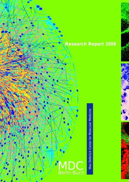
Legend to Cover Figure:<br />
A human protein-protein interaction map<br />
(this report page 170)<br />
Graph representation of 3,186 putative interactions<br />
between 1,705 different human proteins (drawn<br />
using the Pajek program package). Reprinted from<br />
Cell 122 Vol. 6, Stelzl et al. A human proteinprotein<br />
interaction network: a novel resource for<br />
annotating the proteome. 957-968, Copyright<br />
(2005), with permission from Elsevier.
<strong>Research</strong> <strong>Report</strong><br />
<strong>2006</strong><br />
Covers the period 2004/2005
2<br />
Content<br />
Inhalt<br />
Foreword<br />
Vorwort ................................................................................................................................................................ 7<br />
Cardiovascular and Metabolic Diseases<br />
Herz-Kreislauf- und Stoffwechselerkrankungen ............................................................................................ 12<br />
Hypertension, Vascular Disease, and Kidney Disease<br />
Molecular Cardiovascular <strong>Research</strong><br />
Thomas E. Willnow ................................................................................................... 16<br />
Molecular Biology of Peptide Hormones<br />
Michael Bader .......................................................................................................... 20<br />
Hypertension, Vascular Disease, Genetics, and Nephrology<br />
Friedrich C. Luft ....................................................................................................... 22<br />
Cytochrome P450-dependent Eicosanoids<br />
in the Regulation of Cellular and Organ Function<br />
Wolf-Hagen Schunck ............................................................................................... 25<br />
Smooth Muscle Cell Electrophysiology, Ion Channel,<br />
and Transporter Function<br />
Maik Gollasch (Helmholtz Fellow) .............................................................................. 27<br />
Cardiovascular and Metabolic Regulation<br />
Jens Jordan ............................................................................................................. 31<br />
Functional Characterization of Newly Identified Human Importin α Proteins<br />
Matthias Köhler (Helmholtz Fellow) ........................................................................... 34<br />
Mechanisms of Hypertension-induced Target Organ Damage<br />
Dominik N. Müller (Helmholtz Fellow) ........................................................................ 36<br />
Heart Disease<br />
Cardiovascular Molecular Genetics<br />
Ludwig Thierfelder .................................................................................................... 39<br />
Genetic Disorders of the Cardiovascular System<br />
Brenda Gerull (Helmholtz Fellow) .............................................................................. 41<br />
Myocardial Regeneration and Heart Failure<br />
Rainer Dietz ............................................................................................................. 43<br />
Cardiovascular Magnetic Resonance<br />
Jeanette Schulz-Menger ........................................................................................... 46<br />
Myocardial Signal Transduction in Heart Failure<br />
Martin W. Bergmann ................................................................................................ 48<br />
Molecular Muscle Physiology<br />
Ingo L. Morano ........................................................................................................ 50
3<br />
Cell Polarity and Epithelial Formation<br />
Salim Abdelilah-Seyfried ........................................................................................... 53<br />
Molecular and Cell Biology of the (Epi)genome<br />
M. Cristina Cardoso ................................................................................................. 56<br />
Immunology of Cardiovascular Diseases<br />
Gerd Wallukat .......................................................................................................... 59<br />
Neuromuscular and Cardiovascular Cell Biology<br />
Michael Gotthardt .................................................................................................... 61<br />
Metabolic Diseases, Genetics, Genomics, and Bioinformatics<br />
Bioinformatics<br />
Jens Reich ............................................................................................................... 63<br />
Medical Genomics and Gene Mapping Center<br />
Norbert Hübner ........................................................................................................ 66<br />
Genetics of Atopic Disease<br />
Young-Ae Lee .......................................................................................................... 68<br />
Cancer <strong>Research</strong><br />
Krebsforschungsprogramm ............................................................................................................................. 72<br />
Signalling Pathways, Cell Biology, and Cancer<br />
Epithelial Signal Transduction, Invasion, and Metastasis<br />
Walter Birchmeier ..................................................................................................... 80<br />
Genetics of Tumor Progression and Metastasis<br />
Ulrike S. Ziebold (funded by a Marie-Curie Excellence Grant) .................................... 84<br />
Signaling Mechanisms in Embryonic Stem Cells<br />
Daniel Besser (Helmholtz Fellow) .............................................................................. 86<br />
Surgical Oncology<br />
Peter M. Schlag ....................................................................................................... 88<br />
Molecular Genetics of Cell Differentiation & Tumorigenesis<br />
Achim Leutz ............................................................................................................. 91<br />
Cancer Stem Cells and Transcription Factors<br />
Frank Rosenbauer (Helmholtz Fellow) ....................................................................... 95<br />
Signal Transduction in Tumor Cells<br />
Claus Scheidereit ..................................................................................................... 97<br />
Intracellular proteolysis<br />
Thomas Sommer ................................................................................................... 100<br />
Regulation of Nuclear Transport Processes<br />
Katrin Stade (Helmholtz Fellow) .............................................................................. 103<br />
Post-Translational Modifications<br />
Gunnar Dittmar ...................................................................................................... 104<br />
Control of DNA Replication<br />
Manfred Gossen ..................................................................................................... 105
4<br />
Nuclear Signalling and Chromosome Structure<br />
Harald Saumweber ................................................................................................ 107<br />
Evolution, Regulation and Genetic Applications<br />
of Transposable Elements in Vertebrates<br />
Zoltán Ivics ............................................................................................................. 109<br />
Mobile DNA Elements in Vertebrates<br />
Zsuzsanna Izsvák ................................................................................................... 111<br />
Structural and Functional Genomics<br />
Macromolecular Structure and Interaction<br />
Udo Heinemann ..................................................................................................... 113<br />
Computer Simulation of Biomolecular Structures, Dynamics,<br />
and Interactions<br />
Heinz Sklenar ......................................................................................................... 116<br />
Nucleoside Analogs as Inhibitors of HBV and HCV Replication<br />
Eckart Matthes ....................................................................................................... 118<br />
Tumor Genetics<br />
Siegfried Scherneck ............................................................................................... 120<br />
Tumor Immunology<br />
Differentiation and Growth Control in Lymphocyte Development<br />
and Immunopathogenesis<br />
Martin Lipp ............................................................................................................. 123<br />
Biology and Targeted Therapy of Lymphoma<br />
Bernd Dörken ........................................................................................................ 127<br />
Molecular Mechanisms of Immune Evasion in Tumor Biology<br />
and Herpesvirus Infection<br />
Armin Rehm (Helmholtz Fellow) .............................................................................. 130<br />
Cell-Biological Determinants of Treatment Response and Prognosis<br />
in Acute Leukemias<br />
Wolf-Dieter Ludwig ................................................................................................. 133<br />
Cancer Genetics and Cellular Stress Responses<br />
Clemens A. Schmitt ............................................................................................... 135<br />
Clinical and Molecular Oncology<br />
Peter Daniel ........................................................................................................... 137<br />
Molecular Immunotherapy<br />
Antonio Pezzutto .................................................................................................... 140<br />
Molecular Immunology and Gene Therapy<br />
Thomas Blankenstein ............................................................................................. 142<br />
Molecular Cell Biology and Gene Therapy<br />
Wolfgang Uckert .................................................................................................... 144<br />
Cellular Immunology of Autoimmune Reactions<br />
Kirsten Falk<br />
Olaf Rötzschke ....................................................................................................... 146
5<br />
Experimental Pharmacology<br />
Iduna Fichtner ........................................................................................................ 148<br />
Bioethics and Science Communication<br />
Christof Tannert ...................................................................................................... 150<br />
Function and Dysfunction of the Nervous System<br />
Funktion und Dysfunktion des Nervensystems ............................................................................................ 154<br />
Pathophysiological Mechanisms of Neurological and Psychiatric Disorders<br />
Imaging of the Living Brain<br />
Signalling Pathways in the Nervous System<br />
Mouse Genetics – Tools for the Functional Analysis of Genes<br />
that are Important for Development and Disease<br />
Carmen Birchmeier-Kohler ...................................................................................... 157<br />
Molecular Control of Spinal Cord and Peripheral Nervous<br />
System Development<br />
Stefan Britsch (Helmholtz Fellow) ............................................................................ 160<br />
Defining Novel Molecular Components of the Pain Pathway<br />
Alistair N. Garratt (Helmholtz Fellow) ....................................................................... 162<br />
Cellular Neurosciences<br />
Helmut Kettenmann ............................................................................................... 164<br />
Brain Energy Metabolism<br />
Susanne Arnold (Emmy Noether <strong>Research</strong> Group) .................................................. 167<br />
Proteomics and Molecular Mechanisms of Neurodegenerative Disorders<br />
Erich E. Wanker ..................................................................................................... 169<br />
Developmental Neurobiology<br />
Fritz G. Rathjen ...................................................................................................... 173<br />
Growth Factors and Regeneration<br />
Gary Lewin ............................................................................................................. 175<br />
Neurodegeneration<br />
Christiane Alexander .............................................................................................. 177<br />
Neuronal Stem Cells<br />
Gerd Kempermann ................................................................................................. 179<br />
Molecular Neurobiology<br />
Inés Ibañez-Tallon ................................................................................................... 181
6<br />
Academics<br />
Akademische Aktivitäten<br />
Academic Appointments<br />
Berufungen .......................................................................................................... 184<br />
Awards<br />
Preise ................................................................................................................... 188<br />
Helmholtz Fellows<br />
Helmholtz-Stipendiaten ...................................................................................... 189<br />
International PhD Program<br />
Internationales PhD-Programm ......................................................................... 190<br />
Congresses and Scientific Meetings<br />
Kongresse und Wissenschaftliche Tagungen ................................................... 191<br />
Seminars<br />
Seminare .............................................................................................................. 193<br />
Overview<br />
Überblick<br />
Creation of a Translational <strong>Research</strong> Center: The ECRC<br />
Errichtung eines Zentrums für Translationsforschung: Das ECRC ................. 206<br />
The <strong>MDC</strong> Berlin-Buch and the Helmholtz Association<br />
Das <strong>MDC</strong> Berlin-Buch und die Helmholtz-Gemeinschaft ................................. 209<br />
The <strong>MDC</strong> Berlin-Buch and the Campus Berlin-Buch<br />
Das <strong>MDC</strong> Berlin-Buch und der Campus Berlin-Buch ....................................... 210<br />
Organizational Structure<br />
Organisationsstruktur ......................................................................................... 213<br />
Facts and Figures<br />
Fakten und Kennzahlen ...................................................................................... 217<br />
Financing<br />
Finanzierung .......................................................................................................... 217<br />
Personnel<br />
Personal ................................................................................................................ 218<br />
Technology Transfer<br />
Technologietransfer ................................................................................................ 221<br />
Index ..................................................................................................................... 222<br />
Organigram<br />
Organigramm ............................................................................................... 230/231<br />
Campus Map .................................................................................. Inside Back Cover<br />
Campusplan ............................................................................. Innenumschlag hinten<br />
How to find your way to the <strong>MDC</strong> ................................................ Inside Back Cover<br />
Wie gelangen Sie zum <strong>MDC</strong> .................................................... Innenumschlag hinten
7<br />
Foreword<br />
Vorwort<br />
It is my pleasure to present you with the <strong>2006</strong> <strong>Research</strong><br />
<strong>Report</strong> of the Max Delbrück Center for Molecular Medicine<br />
(<strong>MDC</strong>) in Berlin-Buch, which covers the research periods<br />
2004 and 2005. Founded in 1992, the <strong>MDC</strong> is a young research<br />
institute that is sponsored by the German Federal<br />
government (90%) and the State of Berlin (10%) and is a<br />
member of the Helmholtz Association of National <strong>Research</strong><br />
Centers (Helmholtz-Gemeinschaft Deutscher Forschungszentren).<br />
In addition to federal and state funds, the <strong>MDC</strong><br />
augments its total research budget via third-party financial<br />
resources.<br />
<strong>Research</strong> at the <strong>MDC</strong> focuses on the molecular analysis and<br />
treatment of the most prevalent diseases in the population,<br />
namely cardiovascular diseases, cancer, and neurological diseases.<br />
Two research clinics of the Charité University Medical<br />
School in Berlin-Buch, the Franz Volhard Clinic for Cardiovascular<br />
Diseases as well as the Robert Rössle Cancer Clinic,<br />
are connected to the <strong>MDC</strong>. Regarding patient care, these two<br />
clinics are part of the HELIOS Clinics GmbH. The relationships<br />
of the <strong>MDC</strong> to the Charité’s neurobiological clinics sector<br />
are being developed at present. Together with the <strong>MDC</strong>’s<br />
partner institute, the <strong>Research</strong> Institute for Molecular Pharmacology<br />
(FMP), and the around 30 companies on the campus in<br />
Berlin-Buch, an entire repertoire of technologies is available<br />
on the campus for characterizing diseases molecularly, identifying<br />
new starting points in diagnostics and treatment, and<br />
realizing them in clinical application. Along with genetic and<br />
cell biological characterizations of diseases, scientists in<br />
Berlin-Buch can analyze the structure of essential macromolecules<br />
and develop substances to interact with them.<br />
Based on this model, the <strong>MDC</strong> and Charité plan to establish a<br />
jointly run Experimental and Clinical <strong>Research</strong> Center<br />
(ECRC) on the Berlin-Buch campus. The ECRC will intensify<br />
the exchange of scientific ideas between the laboratory and<br />
the clinic in both directions and, hence, accelerate the transmission<br />
of scientific findings directly into clinical applications.<br />
An optimum scientific and technical environment will<br />
be created within the ECRC in which the most promising joint<br />
projects between the <strong>MDC</strong> and Charité can be conducted in<br />
Es ist mir eine große Freude, Ihnen hiermit den wissenschaftlichen<br />
Bericht <strong>2006</strong> des Max-Delbrück-Centrums für Molekulare<br />
Medizin (<strong>MDC</strong>) Berlin-Buch vorzulegen, der die Forschungsperiode<br />
2004 und 2005 umfasst. Das <strong>MDC</strong> ist ein<br />
junges Forschungsinstitut, begründet 1992, das vom Bund<br />
und dem Land Berlin im Verhältnis 90 zu 10 getragen wird<br />
und zur Helmholtz-Gemeinschaft Deutscher Forschungszentren<br />
gehört. Das <strong>MDC</strong> ist für seine Forschung stark auf die<br />
Einwerbung von Drittmitteln angewiesen, die einen großen<br />
Teil seiner effektiven Forschungsmittel ausmachen.<br />
Das <strong>MDC</strong> beschäftigt sich wissenschaftlich mit der molekularen<br />
Analyse und Therapie der wichtigsten Volkskrankheiten,<br />
Herz-Kreislauf-, Krebs- und neurologischen Erkrankungen.<br />
Angeschlossen sind dem <strong>MDC</strong> zwei Forschungskliniken<br />
der Charité – Universitätsmedizin Berlin in Berlin-Buch, die<br />
Franz-Volhard-Klinik für Herz-Kreislauf-Erkrankungen sowie<br />
die Robert-Rössle-Krebsklinik. In der Krankenversorgung<br />
sind diese beiden Kliniken in die HELIOS Kliniken GmbH<br />
integriert. Die Beziehungen des <strong>MDC</strong> zu den Kliniken der<br />
Charité im neurobiologischen Bereich werden zur Zeit ausgebaut.<br />
Zusammen mit dem Partner-Institut des <strong>MDC</strong>, dem Forschungsinstitut<br />
für Molekulare Pharmakologie (FMP), und<br />
den rund 30 Firmen auf dem Bucher Campus steht in Berlin-<br />
Buch somit nahezu das gesamte Repertoire an Techniken zur<br />
Verfügung, um Krankheiten molekular zu charakterisieren,<br />
neue Ansatzpunkte in Diagnostik und Therapie zu identifizieren<br />
und in der klinischen Anwendung umzusetzen. Neben<br />
der genetisch/zellbiologischen Beschreibung der Krankheiten<br />
können die Bucher Wissenschaftlerinnen und Wissenschaftler<br />
die Struktur von essentiellen Makromolekülen analysieren<br />
und Substanzen entwickeln, die mit ihnen in Wechselwirkung<br />
treten. Auf dieser Grundlage planen <strong>MDC</strong> und Charité, ein gemeinsam<br />
getragenes Experimental and Clinical <strong>Research</strong><br />
Center (ECRC) auf dem Campus Berlin-Buch zu errichten.<br />
Das ECRC wird den Austausch wissenschaftlicher Ideen zwischen<br />
Labor und Klinik in beide Richtungen verstärken und<br />
auf diese Weise die Übertragung, Translation, wissenschaftlicher<br />
Ergebnisse in die klinische Anwendung beschleunigen.<br />
Mit dem ECRC wird ein optimales wissenschaftliches und
8<br />
Der damalige Bundespräsident Johannes Rau und seine Frau im Januar 2004 im Gläsernen Labor auf dem<br />
Campus Berlin-Buch. Photo: Thomas Oberländer/Copyright: Helios Klinikum Berlin-Buch<br />
The former German President, Johannes Rau, and his wife in January 2004 in the Life Science Learning<br />
Laboratory at the Campus Berlin-Buch. Photo: Thomas Oberländer/Copyright: Helios Klinikum Berlin-Buch<br />
the three areas of cardiovascular, cancer, and neurobiological<br />
research. In this way, the ECRC will reinforce the existing<br />
synergies on the campus and raise them to a new level.<br />
I would like to mention a few of the many high-impact scientific<br />
publications of scientists from the <strong>MDC</strong> and its partner<br />
clinics that arose during the report period. Ludwig Thierfelder's<br />
group discovered mutations in the plakophilin 2 gene in<br />
more than 25% of patients with arrhythmic right ventricular<br />
cardiomyopathies (ARVC) (Gerull et al., 2004, Nature Genetics).<br />
This means that high mutation rates, which usually only<br />
occur with frequent cancer types, are now also known with<br />
cardiomyopathies. Regarding the sequence analysis of the rat<br />
genome, the group led by Norbert Hübner generated a SNP<br />
map (Single Nucleotide Polymorphism Point Mutations) of<br />
this genome (Zimdahl et al., 2004, Science). The comparison<br />
of the cDNAs (complementary DNA) of three different laboratory<br />
rat strains with the standard rat genome showed over<br />
12,000 variations here. This “card” is a valuable instrument<br />
for comparative gene analysis with other mammals and can<br />
help identify medically important genes. Clemens Schmitt’s<br />
group was able to identify senescence as a new type of tumor<br />
suppressor mechanism which limits the transformation capacity<br />
of oncogenes (Braig et al., 2005, Nature). An inactivation<br />
of this mechanism leads to aggressive lymphomas that are<br />
apoptosis-competent. When mitogenic oncogenes are activated,<br />
a cell security program is activated that leads to either<br />
apoptosis (cell suicide) or senescence (the suspension of the<br />
cell cycle). Thomas Willnow’s group was able to explain a<br />
fundamentally new mechanism as to how sex hormones are<br />
transported to those sites where they are functionally necessary<br />
(Hammes et al., 2005, Cell). The steroid hormones are<br />
bound to the cell surface in the complex with their plasma<br />
transport proteins by a receptor, megalin, and transported to<br />
the nucleus. Lack of this receptor in knockout mice leads to<br />
technisches Umfeld geschaffen werden, in dem die aussichtsreichsten<br />
Kooperationsprojekte zwischen <strong>MDC</strong> und Charité<br />
in den Schwerpunkten Herz-Kreislauf-Forschung, Krebsforschung,<br />
Neurobiologische Forschung bearbeitet werden<br />
können. Auf diese Weise wird das ECRC die bestehenden<br />
Synergien auf dem Campus verstärken und auf ein neues<br />
Niveau heben.<br />
Ich möchte für die Berichtsperiode unter vielen wissenschaftlichen<br />
Arbeiten des <strong>MDC</strong> und seiner Partner-Kliniken in sogenannten<br />
High-Impact-Journalen einige wenige besonders<br />
erwähnen. Die Gruppe von Ludwig Thierfelder hat Mutationen<br />
im Gen Plakophilin-2 bei über 25 % der Patienten<br />
mit Arrythmischer Rechtsventrikulärer Kardiomyopathie<br />
(ARVC) gefunden (Gerull et al., 2004, Nature Genetics). Damit<br />
sind nun auch bei Kardiomyopathien hohe Mutationsraten<br />
bekannt, die sonst nur bei häufigen Krebsarten auftreten. Im<br />
Rahmen der Sequenzanalyse des Rattengenoms hat die<br />
Gruppe von Norbert Hübner eine SNP-Karte (Single Nucleotide<br />
Polymorphisms – Punktmutationen) dieses Genoms vorgelegt<br />
(Zimdahl et al., 2004, Science). Der Vergleich der<br />
cDNAs (complementary DNA) von drei verschiedenen Laborrattenstämmen<br />
mit dem „Standardgenom“ der Ratte zeigte dabei<br />
über 12.000 Variationen. Diese „Karte“ ist ein wertvolles<br />
Instrument für die vergleichende Genomanalyse mit anderen<br />
Säugern und kann helfen, medizinisch wichtige Gene zu identifizieren.<br />
Die Gruppe von Clemens Schmitt konnte Seneszenz<br />
als einen neuartigen Tumorsuppressor-Mechanismus<br />
identifizieren, der die Transformationskapazität von Onkogenen<br />
limitiert (Braig et al., 2005, Nature). Eine Inaktivierung<br />
dieses Mechanismus führt zu aggressiven, aber dennoch<br />
Apoptose-kompetenten Lymphomen. Bei der Aktivierung<br />
mitogener Onkogene wird ein Sicherheitsprogramm der Zelle<br />
aktiviert, das entweder zur Apoptose, Selbstmord der Zelle,<br />
oder zur Seneszenz, Stopp des Zellzyklus, führt. Die Gruppe
9<br />
the inability to react to sexual hormones (steroid insensitivity)<br />
and, as a consequence, to incompletely formed sexual organs.<br />
The group led by Thomas Blankenstein was able to show that,<br />
contrary to previous knowledge, immunogenic tumors do not<br />
have to escape detection through T-cells (Willimsky und<br />
Blankenstein, 2005, Nature). This mainly affects so-called<br />
spontaneous tumors that develop in the absence of external<br />
influences. Erich Wanker’s group generated a genome-wide<br />
interaction map of human proteins that shows 3,186 protein<br />
interactions among 1,705 proteins (Stelzl et al., 2005, Cell).<br />
These include previously unknown interaction partners of 195<br />
proteins associated with diseases.<br />
The <strong>Research</strong> <strong>Report</strong> contains three main sections reflecting<br />
the three foci of research at the <strong>MDC</strong>: cardiovascular research,<br />
cancer research, and neurological sciences. These sections<br />
are presented in English and intended for scientists and<br />
students as well as for new co-workers. Essential parts of the<br />
report are presented in German to make this report accessible<br />
to a broader public. Finally, the report provides a general<br />
overview of the <strong>MDC</strong> including a description of the newly<br />
planned ECRC, administrative structure, financial statistics,<br />
as well as currently funded scientific projects.<br />
The <strong>MDC</strong> has been able to recruit new group leaders for the<br />
<strong>MDC</strong> (see “Academic Appointments”). The German Cancer<br />
Prize for 2005 was awarded for a second time to <strong>MDC</strong> scientists:<br />
Claus Scheidereit and Bernd Dörken. This prize had previously<br />
been awarded to Walter Birchmeier and Peter M.<br />
Schlag in 1999. A further highlight was the completion and<br />
operational start of the new animal facility and the new<br />
research building for medical genome research. Both buildings<br />
are joint projects of the <strong>MDC</strong> and the Institute for Molecular<br />
Pharmacology (FMP). The medical genome research<br />
building has an area of about 3200m 2 of new research space<br />
available. In this context, three W3/C4 professorships in medical<br />
genome research, bioinformatics/system biology, and<br />
cardiovascular and metabolic diseases were filled. Norbert<br />
Hübner was appointed to the new W2/C3 professorship for<br />
genetics/genomics.<br />
We hope you enjoy reading the <strong>MDC</strong> <strong>Research</strong> <strong>Report</strong> <strong>2006</strong><br />
Walter Birchmeier<br />
Scientific Director<br />
von Thomas Willnow konnte einen grundlegend neuen<br />
Mechanismus aufklären, wie Sexualhormone an ihre Wirkorte<br />
gelangen (Hammes et al., 2005, Cell). Die Steroidhormone<br />
werden im Komplex mit ihren Plasmatransportproteinen von<br />
einem Rezeptor, Megalin, auf der Zelloberfläche erkannt und<br />
in das Zellinnere transportiert. Ein Fehlen des Rezeptors in<br />
Knockout-Mäusen führt zum Unvermögen, auf Sexualhormone<br />
anzusprechen (Steroidinsensitivität) und, als Konsequenz,<br />
zu unvollständig ausgebildeten Geschlechtsorganen. Die<br />
Gruppe von Thomas Blankenstein konnte zeigen, dass entgegen<br />
bisheriger Erkenntnisse immunogene Tumore der Erkennung<br />
durch T-Zellen nicht entkommen müssen (Willimsky<br />
und Blankenstein, 2005, Nature). Dies trifft vor allem auf<br />
sogenannte spontane, ohne äußere Einflüsse entstandene<br />
Tumore zu. Die Gruppe von Erich Wanker hat eine genomweite<br />
Interaktionskarte von menschlichen Proteinen vorgelegt,<br />
die 3.186 Protein-Wechselwirkungen zwischen 1.705<br />
Proteinen darstellt (Stelzl et al., 2005, Cell). Darunter befinden<br />
sich auch bislang unbekannte Interaktionspartner von 195<br />
krankheits-assoziierten Proteinen.<br />
Sie sehen drei Teile im Wissenschaftlichen Bericht, die sich<br />
mit der Forschung in diesen drei Bereichen – Herz-Kreislaufforschung,<br />
Krebsforschung und Neurowissenschaften – beschäftigt.<br />
Sie sind in Englisch geschrieben und für Wissenschaftler<br />
und Studenten, auch potentielle neue Mitarbeiter,<br />
gedacht. Essentielle Teile des Berichtes sind in Deutsch verfasst,<br />
um diesen Bericht auch einer breiteren Öffentlichkeit<br />
zugänglich zu machen. Zusätzliche Teile des Berichtes beschäftigen<br />
sich mit dem geplanten ECRC sowie neuen Entwicklungen<br />
in der Verwaltung, einer Übersicht zur Finanzierung<br />
und derzeit geförderten wissenschaftlichen Projekten.<br />
Im Berichtszeitraum konnten neue Nachwuchswissenschaftler<br />
für das <strong>MDC</strong> gewonnen werden. Der Deutsche Krebspreis<br />
ging im Jahr 2005 zum zweiten Mal an Wissenschaftler des<br />
<strong>MDC</strong>: Claus Scheidereit und Bernd Dörken wurden 2005 mit<br />
dem Deutschen Krebspreis ausgezeichnet. Schon 1999 hatten<br />
Walter Birchmeier und Peter M. Schlag diese Auszeichnung<br />
erhalten. Ein weiterer Höhepunkt war die Fertigstellung und<br />
Inbetriebnahme des neuen Tierhauses 2004 sowie des neuen<br />
Forschungsgebäudes für Medizinische Genomforschung Anfang<br />
<strong>2006</strong>. Beide Gebäude sind Gemeinschaftsprojekte von<br />
<strong>MDC</strong> und dem Forschungsinstitut für Molekulare Pharmakologie<br />
(FMP). In dem Gebäude für Medizinische Genomforschung<br />
stehen ca. 3200 m 2 an neuen Forschungsflächen zur<br />
Verfügung. In diesem Zusammenhang werden eine W3/C4-<br />
Professur für Medizinische Genomforschung (gemeinsam mit<br />
dem FMP), eine W3/C4-Professur für Bioinformatik/Systembiologie<br />
sowie eine W3/C4-Professur für Herz-Kreislauf- und<br />
Stoffwechselerkrankungen neu besetzt. Auf die neu zu besetzende<br />
W2/C3-Professur für Genetik/Genomik wurde Nobert<br />
Hübner berufen.<br />
Beim Studium dieses Forschungsberichtes wünsche ich Ihnen<br />
viel Vergnügen.<br />
Walter Birchmeier<br />
Wissenschaftlicher Stiftungsvorstand
Cardiovascular and<br />
Metabolic Diseases<br />
Hypertension, Vascular Disease,<br />
and Kidney Disease<br />
Coordinator: Thomas Willnow<br />
Heart Disease<br />
Coordinator: Ludwig Thierfelder<br />
Metabolic Diseases, Genetics,<br />
Genomics, and Bioinformatics<br />
Coordinator: Jens Reich
12<br />
Cardiovascular and Metabolic<br />
Diseases <strong>Research</strong><br />
Herz-Kreislauf- und<br />
Stoffwechselerkrankungen<br />
Michael Bader<br />
Norbert Hübner<br />
Friedrich Luft<br />
Jens Reich<br />
Ludwig Thierfelder<br />
Thomas E. Willnow<br />
Michael Bader<br />
Norbert Hübner<br />
Friedrich Luft<br />
Jens Reich<br />
Ludwig Thierfelder<br />
Thomas E. Willnow<br />
Introduction<br />
Diseases of the cardiovasculature and the metabolism are the<br />
major cause of morbidity and mortality in our society. Because<br />
such disorders particularly affect the elderly, the socioeconomic<br />
impact of these disease entities is expected to rise<br />
even further in aging populations of the Western world. <strong>Research</strong><br />
in this program aims at elucidating the genes and genetic<br />
pathways that regulate the normal function of the cardiovascular<br />
system and the metabolism and that cause human<br />
disease in these areas. Ultimately, identification of disease<br />
genes will lead to a better understanding of cardiovascular<br />
disease processes, to improved diagnosis, and to new concepts<br />
in therapy.<br />
Towards these goals, we use functional genomics approaches<br />
to study disease processes in many systems that provide utilitarian<br />
models including fruit fly, frog, mouse, and rat, and we<br />
compare our findings to studies conducted in human subjects<br />
(and vice versa). Our studies are performed by scientists that<br />
lead research groups at the <strong>MDC</strong> in close collaboration with<br />
clinicians at the Franz-Volhard-Clinic for Cardiovascular<br />
Diseases (FVK). <strong>Research</strong> activities are coordinated in three<br />
topics that are of particular relevance to this program, namely<br />
(1) Hypertension, Vascular Disease, and Kidney Disease<br />
(2) Heart Disease<br />
(3) Metabolic Diseases, Genetics, Genomics, and Bioinformatics.<br />
Einführung<br />
Erkrankungen des kardiovaskulären Systems und des Stoffwechsels<br />
sind die Hauptursache für Morbidität und Mortalität<br />
in unserer Gesellschaft. Auf Grund eines deutlichen Anstiegs<br />
der durchschnittlichen Lebenserwartung in unserer Bevölkerung<br />
und des erhöhten Risikos älterer Menschen, an kardiovaskulären<br />
Komplikationen zu erkranken, ist davon auszugehen,<br />
dass die Belastungen unserer Gesundheitssysteme durch<br />
die Folgekosten kardiovaskulärer Krankheiten zukünftig dramatisch<br />
ansteigen werden. Ziel unserer Forschungsanstrengungen<br />
vor diesem Hintergrund ist es, die genetischen Mechanismen<br />
aufzuklären, welche die normalen Funktionen von<br />
Herz-Kreislauf und Stoffwechsel regeln und welche für krankhafte<br />
Veränderungen dieser Systeme beim Patienten verantwortlich<br />
sind. Letztlich wird die Identifizierung grundlegender<br />
genetischer Mechanismen zu einem besseren Verständnis kardiovaskulärer<br />
Krankheitsprozesse, zu verbesserter Diagnostik<br />
und zu neuen therapeutischen Ansätzen führen.<br />
Um dieses Ziel zu erreichen, verfolgen wir ein Konzept der<br />
vergleichenden Genomforschung, bei dem wir normale physiologische<br />
Prozesse und krankhafte Veränderungen des<br />
kardiovaskulären Systems parallel in Patienten sowie in<br />
experimentellen Tiermodellen untersuchen und miteinander<br />
vergleichen. Aus den Informationen, welche wir in Modellsystemen<br />
wie der Fruchtfliege, dem Krallenfrosch oder<br />
Nagern gewinnen, lassen sich wichtige Rückschlüsse auf relevante<br />
Krankheitsprozesse beim Menschen ziehen und neue<br />
Strategien zu deren Therapie entwickeln. Unsere Arbeiten<br />
sind das Ergebnis einer erfolgreichen Zusammenarbeit von<br />
Grundlagenwissenschaft am <strong>MDC</strong> und klinischer Forschung<br />
an der Franz-Volhard-Klinik für Herz-Kreislauferkrankungen<br />
(FVK) der Charité – Universitätsmedizin Berlin auf dem<br />
Campus Berlin-Buch. Unsere Forschungsaktivitäten konzentrieren<br />
sich auf drei Themenfelder mit besonders hoher Relevanz<br />
für kardiovaskuläre Erkrankungen:<br />
(1) Hypertonie, Gefäß- und Nierenerkrankungen<br />
(2) Herzerkrankungen<br />
(3) Genetik, Genomik, Bioinformatik und Metabolismus
13<br />
Hypertension, Vascular Disease, and Kidney Disease<br />
Hypertension is a complex regulatory disorder that results in<br />
increased blood pressure. The heart, the blood vessels, and the<br />
kidney are involved either as a primary cause or as a secondary<br />
target of this disease. With the elucidation of hitherto unknown<br />
genetic mechanisms contributing to hypertension, vascular<br />
disease, and kidney disease, new therapies may become<br />
possible. In the past two years, scientists in this topic have<br />
made important contributions towards this goal.<br />
Antibody-mediated rejection is thought to account for around<br />
one-third of all rejections following kidney transplantation.<br />
The association between HLA antibodies present at the time<br />
of transplant and graft loss has been well established. Now,<br />
Duska Dragun, Dominik Müller, Gerd Wallukat and their colleagues<br />
uncovered that a non-HLA, angiotensin II type 1<br />
(AT1)-receptor meditated pathway may be involved in kidney<br />
transplant rejection. The scientists studied 33 kidney transplant<br />
recipients who presented with antibody-mediated rejection<br />
following kidney allograft. Of these, 13 had donor-specific<br />
anti-HLA antibodies. Sixteen of the remaining 20 patients<br />
without anti-HLA antibodies were shown to have both agonistic<br />
antibodies targeting the AT1-receptor and malignant<br />
hypertension. Furthermore, when agonistic antibodies were<br />
transferred to rats that had received kidney transplants, hypertension<br />
and vasculopathy were induced. Of the 16 patients<br />
with agonistic antibodies, seven underwent plasmapheresis<br />
for the removal of the antibodies and received the AT1-receptor<br />
blocker losartan. As compared to those control patients<br />
who received standard treatments, these seven fared better in<br />
terms of renal function and graft survival. Thus, novel therapies<br />
for the treatment of antibody-mediated rejection may be<br />
developed that involve the removal of AT1-receptor antibodies<br />
or the blockage of AT1 receptors.<br />
Ralph Kettritz and his colleagues have elucidated central signaling<br />
pathways leading to systemic vasculitis, an inflammatory<br />
process in medium to small blood vessels caused by antibodies<br />
directed against neutrophil components. They found<br />
that integrins and cytokines activate nuclear transcription factor-kappaB<br />
in human neutrophils. They showed that beta (2)<br />
integrins provide co-stimulatory signals allowing soluble mediators<br />
to activate the NF-kappaB pathway when the cells are<br />
fixed to matrix, even when they are not capable of doing so<br />
when the cells are in suspension. This effect may become important<br />
when human neutrophils leave the circulating blood<br />
and migrate through extracellular matrix during inflammation.<br />
Michael Bader and colleagues have identified the role of<br />
the mas protooncogene as a receptor for angiotensin (1-7) and<br />
the cardioprotective actions of this angiotensin-II metabolite.<br />
The group of Thomas Willnow, in collaboration with colleagues<br />
at the University of Aarhus, has uncovered the existence of<br />
endocytic pathways that govern the tissue-specific uptake of<br />
steroid hormones such as androgens and estrogens. Previously,<br />
the delivery of steroids to their respective target<br />
tissues was believed solely to depend on non-specific diffusion<br />
of the hormones through the plasma membrane. The<br />
identification of active uptake pathways for androgens and<br />
estrogens challenges a central dogma in steroid hormone biology<br />
and holds tremendous potential for therapeutic strategies<br />
Hypertonie, Gefäß- und Nierenerkrankungen<br />
Hypertonie, die krankhafte Erhöhung des Blutdrucks, ist eine<br />
komplexe Regulationsstörung des Kreislaufs, welche in unserer<br />
Bevölkerung weit verbreitet ist. Fehlfunktionen des Herzens,<br />
der Gefäße oder der Nieren sind primäre Ursache dieser<br />
Störung oder treten sekundär als Folge pathologischer Veränderungen<br />
beim Hypertoniker auf. Durch die Entschlüsselung<br />
bislang unbekannter genetischer Mechanismen, die zu Bluthochdruck,<br />
zu Gefäßerkrankungen oder zu Nierendefekten<br />
führen, hoffen wir die Ursachen der Hypertonie aufzuklären<br />
und neue Strategien zu deren Prävention entwickeln zu können.<br />
In den vergangenen zwei Jahren gelang es uns, wichtige<br />
neue Erkenntnisse in dieser Hinsicht zu gewinnen.<br />
Autoantikörper-induzierte Abstoßungsreaktionen sind die Ursache<br />
für etwa 30% aller Fälle von Organverlust bei Nierentransplantation.<br />
In neueren Arbeiten konnten Duska Dragun,<br />
Dominik Müller, Gerd Wallukat und ihre Kollegen zeigen,<br />
dass aktivierende Autoantikörper gegen den Angiotensin II<br />
type 1 (AT1)-Rezeptor ursächlich für die Entstehung von Hypertonie<br />
und Nierentransplantat-Abstoßung im Patienten sind.<br />
Dies ließ sich durch den Transfer von anti-AT1-Antikörpern<br />
in Ratten dokumentieren, die dadurch eine fulminante Hypertonie<br />
und Gefäßschädigungen entwickelten. In einem Pilotexperiment<br />
konnten mittels Plasmapherese anti-AT1-Autoantikörper<br />
aus Serum von Patienten mit einer Spenderniere<br />
entfernt und damit eine signifikante Verbesserung renaler<br />
Funktionen und der Transplantat-Lebensdauer erzielt werden.<br />
Der Arbeitsgruppe von Ralph Kettritz gelang es, zentrale Signaltransduktionsmechanismen<br />
in der Entstehung systemischer<br />
Vaskulitis, einer entzündlichen Erkrankung kleiner und<br />
mittlerer Gefäße, aufzuklären. Dieser Krankheitsprozess beruht<br />
auf einer Immunreaktion des Körpers gegen zelluläre Bestandteile<br />
von Neutrophilen. Die Wissenschaftler konnten zeigen,<br />
dass die Aktivierung des NF-kappaB-Signalweges über<br />
Integrine und Cytokine eine wichtige stimulatorische Wirkung<br />
auf die Entstehung und das Fortschreiten entzündlicher<br />
Prozesse in den Gefäßen hat. Michael Bader und Mitarbeiter<br />
konnten erstmals die Rolle des mas Proto-Oncogens als<br />
Rezeptor für Angiotensin 1–7 dokumentieren sowie die<br />
kardioprotektive Funktion dieses Angiotensin-II-Metaboliten<br />
aufzeigen.<br />
Der Arbeitsgruppe von Thomas Willnow gelang in Zusammenarbeit<br />
mit Kollegen der Universität Aarhus der Nachweis,<br />
dass Steroidhormone wie Androgene und Östrogene mittels<br />
rezeptor-vermittelter Endozytose in Zellen aufgenommen<br />
werden können. Diese Befunde widerlegen die gängige Lehrmeinung,<br />
dass Steroidhormone ausschließlich über unspezifische,<br />
freie Diffusion in Zielgewebe des Körpers gelangen.<br />
Die Blockade solcher spezifischer Aufnahmemechanismen<br />
für Steroidhormone sind möglicherweise neue therapeutische<br />
Ansätze zur Behandlung steroidabhängiger Tumore der Brust<br />
und Prostata.
14<br />
aimed at modulating delivery of steroid hormones to target<br />
tissues in patients.<br />
Heart Disease<br />
Cardiac development, myocardial injury and repair, hereditary<br />
forms of heart failure and sudden death, and molecular<br />
and clinical imaging are the main areas of research interests.<br />
Several milestones have been achieved recently. Rüdiger von<br />
Harsdorf and Rainer Dietz characterized the role of pro- and<br />
anti-apoptotic molecules in the heart, of which ARC seems to<br />
have a protective role against circulatory stress. Inhibition of<br />
apoptosis and induction of cardiac cell division (not just<br />
mitosis!) may, some day, provide therapeutic options to heal<br />
infarcted myocardium. The elucidation of new factors essential<br />
for cardiac morphogenesis is one focus of Salim Abdelilah-<br />
Seyfried’s group. The recent characterization of the molecular<br />
defects in the heart and soul (has) and nagie oko (nok) zebrafish<br />
mutants provide insights into early phases of cardiac morphogenesis<br />
(cardiac tube formation).<br />
Another lesson from embryonic cardiac pathology lead to the<br />
discovery of a new cardiomyopathy disease gene in humans.<br />
When plakophilin-2 is ablated in the mouse, no regular cellcell<br />
contacts can form and mouse embryos die early as shown<br />
by Walter Birchmeier’s group. Ludwig Thierfelder and colleagues<br />
hypothesized that plakophilin-2 might play a role in<br />
patients suffering from a hereditary cardiomyopathy associated<br />
with arrhythmias and sudden cardiac death. In a large<br />
cohort of these cardiomyopathy patients, a high number of<br />
disease causing mutations were discovered and such patients<br />
can now be readily identified and receive effective and even<br />
prophylactic treatment.<br />
Another translational approach leading to several clinical<br />
hypotheses was generated from a basic science lab (Gerd<br />
Wallukat’s group) when a number of autoantibodies directed<br />
against various cell surface receptors were shown to adversely<br />
modify the course of patients with heart failure, preeclampsia,<br />
or renal graft rejection. Therapeutic removal of such autoantibodies<br />
will soon be implemented in clinical practice.<br />
Metabolic Diseases, Genetics, Genomics, and<br />
Bioinformatics<br />
Elucidation of the human and other mammalian genomes heralds<br />
a new area in biomedical research. Major challenges in<br />
the future will be to assign functions to the wealth of sequence<br />
information generated in the various genome programs. Thus,<br />
high throughput sequence analysis and bioinformatics technologies<br />
have to be developed and applied to the positional cloning<br />
of disease genes in monogenic and complex traits of<br />
metabolic disturbances.<br />
Towards these goals, recent major achievements in this program<br />
include the study by Hans Knoblauch and Friedrich C.<br />
Luft who, in a collaboration with Peter Nürnberg and Jens<br />
Reich, performed a detailed investigation of common lipidgene<br />
variants and lipid levels in 250 German families. The<br />
subjects numbered over 1000 and spanned 3 generations. The<br />
Herzerkrankungen<br />
Die Entschlüsselung genetischer Grundlagen der Herzentwicklung,<br />
der Schädigung und Regeneration des Myokards,<br />
familärer Erkrankungen des Herzversagens sowie die Entwicklung<br />
molekularer und klinischer bildgebender Verfahren<br />
sind zentrale Forschungsfelder dieses Programmthemas. Eine<br />
Vielzahl wichtiger Meilensteine konnten in diesen Forschungsfeldern<br />
in den vergangenen 2 Jahren erreicht werden. Rüdiger<br />
von Harsdorf, Rainer Dietz und Mitarbeiter haben die Rolle<br />
pro- und anti-apoptotischer Faktoren im Myokard untersucht<br />
und dabei einen wichtigen Beitrag des Proteins ARC als protektivem<br />
Faktor vaskulärer Stressbedingungen identifiziert.<br />
Zukünftig könnte die Intervention bei pro-apoptotischen zellulären<br />
Vorgängen ein wichtiges Instrument zur Therapie des<br />
geschädigten Herzmuskelgewebes darstellen.<br />
Die Aufklärung neuer genetischer Signalwege in der Herzentwicklung<br />
ist Schwerpunkt der Arbeiten von Salim Abdelilah-<br />
Seyfried. Seiner Gruppe gelang in neueren Studien die<br />
Beschreibung der molekularen Defekte in den Zebrafischmutanten<br />
heart and soul (has) und nagie oko (nok), welche wichtige<br />
neue Einblicke in die frühe Phase der Herzmorphogenese<br />
geliefert haben.<br />
Neue Erkenntnisse aus dem Studium kardialer Entwicklungsdefekte<br />
in Tiermodellen führten ebenfalls zur Aufklärung der<br />
molekularen Ursachen häufiger Kardiomyopathien beim Patienten.<br />
Wie die Gruppe von Walter Birchmeier zeigen konnte,<br />
bedingt die Inaktivierung des Plakophilin-2-Gens in Mausmodellen<br />
den Verlust der Zell-Zell-Interaktionen im embryonalen<br />
Herzen und frühe Lethalität. Ausgehend von diesen<br />
Befunden gelang es Ludwig Thierfelder und Mitarbeitern,<br />
vergleichbare genetische Defekte im Plakophilin-2-Gen beim<br />
Patienten als Ursache einer häufigen Form familiärer Kardiomyopathie<br />
(mit Arrhythmie und plötzlichem Herztod) nachzuweisen.<br />
Basierend auf diesen Befunden lassen sich betroffene<br />
Patienten jetzt frühzeitig diagnostisch erfassen und mit<br />
geeigneten interventionellen und prophylaktischen Maßnahmen<br />
behandeln.<br />
Ein anderer translationaler Forschungsansatz der Gruppe um<br />
Gerd Wallukat führte zur Aufklärung der zentralen Rolle von<br />
Autoantikörpern gegen verschiedene Oberflächenrezeptoren<br />
als Auslöser pathologischer Prozesse bei Patienten mit Herzversagen,<br />
Präeklampsie und Nierentransplantatabstoßung.<br />
Therapeutische Maßnahmen zur Entfernung solcher Autoantikörper<br />
aus dem Serum Betroffener mittels Plasmapherese<br />
werden zukünftig Eingang in die klinische Praxis finden.<br />
Metabolische Erkrankungen, Genetik, Genomik und<br />
Bioinformatik<br />
Die Entschlüsselung des Erbguts des Menschen und das anderer<br />
Säuger ist ein Meilenstein der modernen biomedizinischen<br />
Forschung. Noch größer jedoch als die Herausforderung der<br />
Entschlüsselung des menschlichen Erbguts ist die Aufgabe,<br />
die gewonnene genetische Information auszuwerten und<br />
funktionell zu charakterisieren. Ein wesentliches Ziel des Forschungsschwerpunktes<br />
Genetik, Genomik und Bioinformatik<br />
besteht darin, neue molekulargenetische, bioinformatische
15<br />
group performed a comprehensive SNP analysis in 13 genes.<br />
The relative effects of the individual genes on the lipid phenotypes<br />
were elucidated in their study. Their work ushers in<br />
the 500K SNP analyses that are on the horizon.<br />
The group of Norbert Hübner has focused on a novel approach<br />
of integrating genome-wide expression profiling with linkage<br />
to identify genes underlying complex traits. The researchers<br />
applied this approach to the regulation of gene expression in<br />
rat recombinant inbred strains, a leading resource for genetic<br />
analysis of the metabolic syndrome. In two tissues important<br />
to the pathogenesis of this syndrome, they mapped cis- and<br />
trans-regulatory control elements for expression of many<br />
genes across the genome. This data set lead to new insights<br />
into genes and regulatory pathways underlying the extensive<br />
range of metabolic and cardiovascular disease phenotypes<br />
that segregate in these recombinant inbred strains.<br />
Previous studies in animal models suggested an important<br />
contribution of infiltrating monocytes in adipose tissues to the<br />
pathological alteration in metabolism during adipositas.<br />
Using microdialysis experiments, Jens Jordan and co-workers<br />
now have confirmed this central mechanism in human subjects.<br />
und biostatistische Verfahren zu entwickeln, um die Flut genetischer<br />
Informationen analysieren zu können und für funktionelle<br />
Untersuchungen bei der Kartierung monogener und<br />
komplexer genetischer Ursachen von Stoffwechselstörungen<br />
einzusetzen.<br />
Im Rahmen ihrer Studien haben Hans Knoblauch und Friedrich<br />
C. Luft in Zusammenarbeit mit Peter Nürnberg und<br />
Jens Reich eine detaillierte Kartierung häufiger genetischer<br />
Varianten in Genen des Lipidstoffwechsels durchgeführt und<br />
deren Einfluss auf die Höhe individueller Plasmalipidspiegel<br />
in über 250 Familien bestimmt.<br />
Die Arbeitsgruppe von Norbert Hübner hat sich auf die Aufklärung<br />
komplexer genetischer kardiovaskulärer Erkrankungen<br />
mittels Genom-weiter Expressionsanalyse konzentriert.<br />
Die Wissenschaftler haben diese Methodik eingesetzt, um<br />
Genexpressionsprofile in Ratten-Inzuchtstämmen metabolischer<br />
Störungen zu erstellen und cis- und trans-regulatorische<br />
Elemente zu identifizieren, welche eine Vielzahl metabolischer<br />
und kardiovaskulärer Parameter im Säugerorganismus<br />
bestimmen.<br />
Ausgehend von Untersuchungen in Tiermodellen, die nahelegen,<br />
dass in Fettgewebe eingewanderte Monozyten wesentlich<br />
zu pathologischen Veränderungen bei Adipositas beitragen,<br />
haben Jens Jordan und Mitarbeiter diese Hypothese<br />
mittels Microdialyse-Untersuchungen im Patienten geprüft<br />
und validiert.
Hypertension, Vascular Disease,<br />
and Kidney Disease<br />
Molecular Cardiovascular <strong>Research</strong><br />
Thomas E. Willnow<br />
Introduction<br />
The low-density lipoprotein (LDL) receptor is a 150 kDa<br />
endocytic receptor that mediates the cellular uptake of lipoprotein<br />
particles and plays a central role in the removal of<br />
lipids from the systemic circulation. In patients with a genetic<br />
defect of the LDL receptor (Familial Hypercholesterolemia),<br />
a massive increase in the concentration of circulating plasma<br />
lipoproteins results in hyperlipidemia and, consequently, in<br />
atherosclerosis and coronary artery disease. In recent years, a<br />
number of novel receptors have been identified that are structurally<br />
related to the LDL receptor and that are designated<br />
members of the LDL receptor gene family. Given the central<br />
role of the LDL receptor in the cardiovascular system, equally<br />
important roles for other receptors in this gene family are<br />
anticipated. The focus of our studies is the elucidation of the<br />
functions that LDL receptor-related receptors (LRPs) play in<br />
the (patho)physiology of the cardiovascular system, particularly<br />
in the systemic and cellular lipid metabolism. Towards<br />
this goal, we are using gene-targeting approaches to generate<br />
mouse models with ubiquitous or conditional LRP deficiencies<br />
and to analyze the consequences of the receptor gene<br />
defects in vivo. In the past two years, we have identified important<br />
new functions of lipoprotein receptors in the development<br />
of the central nervous system and the reproductive<br />
organs, and the molecular mechanisms underlying human<br />
diseases in these areas.<br />
Role of lipoprotein receptors in forebrain<br />
development<br />
Megalin is an LRP expressed in the neuroepithelium and the<br />
yolk sac of the early embryo. The receptor acts as multi-ligand<br />
scavenging receptor mediating the cellular uptake of lipoproteins<br />
and lipid/carrier complexes. Loss of megalin expression<br />
in knockout mice results in forebrain defects and in holoprosencephaly<br />
(HPE), indicating an essential yet unidentified<br />
function in brain development. HPE is defined as a failure of<br />
the prosencephalon to separate along the mid-sagital axis into<br />
discrete hemispheres. This defect is likely to be the result of<br />
defective patterning during development involving improper<br />
specification of the rostral portion of the neural tube. HPE is<br />
the most common brain anomaly of the human embryo and affects<br />
1 in 250 pregnancies. Disturbances in cholesterol metabolism<br />
seem to play a causal role in this disease as indicated<br />
by the holoprosencephalic defects seen in patients suffering<br />
from Smith-Lemli-Opitz Syndrome, a heritable defect of cholesterol<br />
biosynthesis. We have used mice with conditional megalin<br />
gene inactivation in the yolk sac and/or the embryo proper<br />
to uncover the role of the receptor in forebrain formation.<br />
We demonstrated that expression of megalin in the neuroepithelium<br />
but not in the yolk sac is required for brain development.<br />
In the neuroepithelium, megalin deficiency causes<br />
defects in dorso-ventral patterning of the telencephalon including<br />
loss of expression of sonic hedgehog (Shh) in the ventral<br />
(figure 1) and enhanced expression of bone morphogenic protein<br />
4 (Bmp4) in the dorsal neural tube. These findings indicate<br />
a crucial role for the lipoprotein receptor megalin in the<br />
regulation of the activity of SHH and BMP4, two morphogens<br />
that act as central regulators of neural tube patterning.<br />
Endocytic receptors for steroid hormones<br />
According to current concepts, steroid hormones enter target<br />
cell by free diffusion through the plasma membrane. However,<br />
we have shown previously that some tissues use endocytosis<br />
to actively internalize the steroid hormone 25-OH<br />
vitamin D3 bound to carrier proteins, thus acquiring large<br />
amounts of this important regulator independent of diffusion<br />
processes. Initially considered a unique feature of vitamin D<br />
metabolites, we now have proof that endocytosis is a general<br />
concept that also applies to other steroid hormones such as<br />
androgens and estrogens. We demonstrated that megalin, an<br />
endocytic receptor for lipid/carrier complexes, is expressed in<br />
the principal cells of the epididymis and in the uterus, tissues<br />
that require large amounts of sex steroids for normal function.<br />
We further showed that megalin mediates the cellular uptake<br />
of androgens and estrogens bound to the carrier sex hormone-
17<br />
A<br />
B<br />
bpd<br />
Zli<br />
bpd<br />
Zli<br />
+/+ -/-<br />
Figure 1 Shh expression in wild type (A) and megalin -/- (B) embryos.<br />
In situ hybridization of frontal head aspects of wild type (A) and megalin -/- E10.5 embryos (B) depicting Shh expression<br />
in the rostral ventral neural tube (arrowheads) of wild type but not knockout animals. Expression of Shh in other<br />
areas of the forebrain such as in the basal plate of the diencephalon (bpd) or the zona limitans intrathalamica (Zli) is<br />
identical in both genotypes.<br />
binding globulin (SHBG) (figure 2). Finally, we uncovered<br />
that megalin deficiency in mice results in impaired descent of<br />
the testes into the scrotum in males and in blockade of vagina<br />
opening in females, two processes that are critically dependent<br />
on sex steroid signaling. Similar defects are seen in rodents<br />
treated with androgen or estrogen receptors antagonists,<br />
indicating insensitivity to steroid hormones as the underlying<br />
molecular defect in megalin deficient mice. Our findings<br />
demonstrate a direct link between endocytosis of steroid<br />
hormones and sex steroid action in vivo, and they provide<br />
further evidence for a paradigm shift in our concept of the<br />
metabolism and action of steroid hormones.<br />
SorLA, a neuronal lipoprotein receptor that regulates<br />
processing of the amyloid precursor protein<br />
Recently, a novel receptor, SorLA-1, was uncovered that<br />
combines motifs of the LDL receptor gene family with structural<br />
elements found in the yeast vacuolar sorting receptor<br />
Vps10p. Although, SorLA has been shown to bind ligands relevant<br />
for lipoprotein metabolism, such as apolipoprotein E,<br />
the physiological function of this receptor was unclear. However,<br />
its homology to sorting receptors that shuttle between<br />
plasma membrane, endosomes, and Golgi suggested a related<br />
function in neuronal trafficking processes. Because expression<br />
of SorLA is reduced in the brain of patients with Alzheimer’s<br />
disease (AD), we tested the involvement of this receptor<br />
in intracellular transport and processing of the amyloid<br />
precursor protein (APP) to the amyloid β-peptide (Aβ), the<br />
principal component of senile plaques. APP follows a complex<br />
trafficking pathway that determines alternative processing<br />
into the soluble sAPP· fragment (non-amyloidogenic<br />
pathway) or into the Aβ peptide (amyloidogenic pathway).<br />
The cellular mechanisms that direct APP into either of the two<br />
processing pathways remain elusive. Here, we demonstrated<br />
that SorLA interacts with APP in vitro and in living cells<br />
(figure 3), and that both proteins co-localize in endosomal<br />
and Golgi compartments. Overexpression of SorLA in neurons<br />
causes redistribution of APP to the Golgi and decreased<br />
processing to Aβ whereas ablation of SorLA expression in<br />
knockout mice results in increased levels of Aβ in the brain<br />
similar to the situation in patients with AD. Thus, SorLA acts<br />
as a sorting receptor that protects APP from processing into<br />
Aβ and thereby reduces the burden of amyloidogenic peptide<br />
formation. Consequently, reduced receptor expression in<br />
human brain may increase Aβ production and senile plaque<br />
formation, and promote spontaneous AD.<br />
Selected Publications<br />
1. Andersen, OM, Reiche, R, Schmidt, V, Gotthardt, M,<br />
Spoelgen, R, Behlke, J, von Arnim, CAF, Breiderhoff, T,<br />
Jansen, P, Wu, X, Bales, KR, Cappai, R, Masters, CL,<br />
Gliemann, J, Mufson, EJ, Hyman, BT, Paul, SM, Nykjær,<br />
N, Willnow, TE. (2005). SorLA/LR11, a neuronal sorting<br />
receptor that regulates processing of the amyloid precursor<br />
protein. Proc. Natl. Acad. Sci. USA (in press)<br />
2. Hammes, A, Andreassen, TK, Spoelgen, R, Raila, J,<br />
Huebner, N, Schulz, H, Metzger, J, Schweigert, FJ, Luppa,<br />
PB, Nykjaer, A, Willnow, TE. (2005). Role of Endocytosis<br />
in Cellular Uptake of Sex Steroids. Cell 122, 751-762.<br />
3. Spoelgen, R, Hammes, A, Anzenberger, U, Zechner, D,<br />
Jerchow, B, Andersen, OM, Willnow, TE. (2005).<br />
LRP2/megalin is required for patterning of the ventral<br />
telencephalon. Development 132, 405-414.<br />
4. Nykjaer, A, Lee, R, Teng, TT, Nielsen, MS, Jansen, P,<br />
Madsen, P, Jacobsen, C, Kliemannel, M, Willnow, TE,<br />
Hempstead, B, Petersen, CM. (2004). A novel neurotrophin<br />
receptor essential for proNGF induced neuronal cell<br />
death. Nature 427, 843-848.<br />
5. Petersen, HH, Hilpert, H, Militz, D, Zander, V, Jacobsen,<br />
C, Roebroek, AJM, Willnow, TE. (2003). Functional interaction<br />
of megalin with the megalin-binding protein<br />
(megbp), a novel tetratrico peptide repeat-containing<br />
adaptor molecule J. Cell Sci. 116, 453-461.
18<br />
FITC-DHT/SHBG<br />
- RAP + RAP<br />
Merge<br />
FITC-DHT<br />
anti-SHBG<br />
Figure 2<br />
Endocytic uptake of dihydrotestosterone/SHBG complexes in cells expressing megalin.<br />
Cells were incubated with preformed complexes of FITC-labeled dihydrotestosterone(DHT)/SHBG in the presence or<br />
absence of RAP, an inhibitor of ligand binding to megalin. Subsequently, the cellular uptake of FITC-DHT and SHBG<br />
(anti-SHBG antibody, followed by secondary alexa 660-labeled IgG) were detected by confocal immunofluorescence<br />
microscopy. Endocytic uptake of androgen/carrier complexes can be seen in cells with megalin activity (-RAP) but not<br />
in cells treated with receptor antagonists (+RAP).<br />
Patent Applications<br />
1. Patent application US pat. Appl. No. 10/131,597:<br />
Agents for prevention of organ damage induced by therapeutic<br />
agents<br />
2. Patent application PCT/IB02/01393:<br />
Agents for prevention of organ damage induced by therapeutic<br />
agents<br />
3. Patent application PA 2003 00459:<br />
Use of compounds for the prevention of drug-induced cell<br />
toxicity.<br />
4. Patent application PCT/DK03/00919:<br />
Modulation of activity of neurotrophins
19<br />
Donor Acceptor Lifetime<br />
A<br />
Alexa488-α-sorLA<br />
B<br />
Alexa488-α-sorLA<br />
Cy3-α-APP<br />
300 ps 2100 ps<br />
Figure 3<br />
Fluorescence lifetime imaging microscopy (FLIM) of APP and SorLA interaction in N2A cells.<br />
The figure displays intensity images of SorLA immunocytochemistry using donor fluorophore Alexa488-conjugated<br />
antibodies (left), and pseudo coloured FLIM images (middle) and lifetime histograms (right), indicating shortening of<br />
lifetime from blue to orange/red color in the absence (A) or presence (B) of acceptor fluorophore Cy3. (A) Cells<br />
stained for the amino terminal domain of SorLA with donor fluorophore Alexa488 in the absence of acceptor<br />
(lifetime 2007 ± 12 psec). (B) Cells stained for SorLA with donor fluorophore Alexa488 in the presence of acceptor<br />
Cy3 on the ectodomain of APP. The shortening of the fluorescence lifetime on SorLA to 1365 ± 139 psec indicates<br />
a close physical association with the acceptor on APP.<br />
Structure of the Group<br />
Group Leader<br />
Prof. Dr. Thomas E. Willnow<br />
Scientists<br />
Dr. Olav M. Andersen<br />
Dr. Tilmann Breiderhoff<br />
Dr. Annette Hammes-Lewin<br />
Graduate students<br />
Uwe Anzenberger<br />
Chandresh Ganjera<br />
Daniel Militz<br />
Juliane Reiche<br />
Vanessa Schmidt<br />
Technical Assistants<br />
Susanne Schütz<br />
Donate Vetter<br />
Jana Zenker*<br />
Secretariat<br />
Verona Kuhle<br />
* part of the period reported
20<br />
Molecular Biology of Peptide<br />
Hormones<br />
Michael Bader<br />
The kallikrein-kinin system (KKS) is an important hormone<br />
system for cardiovascular regulation mostly counteracting the<br />
effects of the RAS. As models for the functional analysis of<br />
the KKS in intact animals, transgenic rats were generated expressing<br />
different components of the system, such as tissue<br />
kallikrein, the kinin B1 or the B2 receptor, either ubiquitously<br />
or specifically in cardiovascular organs. These animals supported<br />
the protective role of the KKS in kidney and heart<br />
against ischemic, diabetic, and hypertrophic injury. Furthermore,<br />
the analysis of transgenic rats overexpressing B1 receptors<br />
in vessels and of knockout mice lacking one or both kinin<br />
receptors showed that the KKS is essential for the hypotension<br />
occurring after septic shock.<br />
Natriuretic peptide system<br />
There are 3 natriuretic peptides, ANP, BNP, and CNP, which<br />
interact with two receptors, NPR-A and NPR-B, to induce a<br />
multitude of actions in heart, kidney, and vessels. The receptors<br />
are dimeric molecules which, after activation, synthesize<br />
cyclic GMP. We have shown that dimerization is essential for<br />
the activation of the receptors and have designed dominant<br />
negative mutants to downregulate the activity of the receptors<br />
in cells and transgenic animals.<br />
Serotonin system<br />
The group is interested in the molecular biology and function<br />
of hormone systems involved in cardiovascular regulation.<br />
Besides the cloning and characterization of genes for the components,<br />
the physiological functions of the systems are analyzed<br />
by the production and analysis of transgenic and genetargeted<br />
animal models.<br />
Renin-angiotensin system<br />
The renin-angiotensin system (RAS) is of central importance<br />
in blood pressure regulation and in the initiation of target<br />
organ damage. In particular, local angiotensin-II generating<br />
systems in tissues, such as brain, heart, and kidney, are involved<br />
in these processes. Therefore, transgenic rats with<br />
local up- or downregulation of RAS components in brain,<br />
heart, and vessels (e.g. by the organ-specific expression of<br />
antisense-RNA or of a peptide-liberating protein) were produced.<br />
Using these rats, we could show that central angiotensin<br />
modulates circadian blood pressure rhythms, the baroreceptor<br />
reflex and, as a non-cardiovascular parameter, alcohol consumption.<br />
Furthermore, it is involved in the hypertensive and<br />
hypertrophic effects of circulating angiotensin. Other genetically<br />
altered mouse and rat models for non-classical RAS<br />
components, such as ACE2, the renin receptor, the mas protooncogene,<br />
and angiotensin(1-7), have elucidated the physiological<br />
function of these factors.<br />
Kallikrein-kinin system<br />
Serotonin is at the same time a very important neurotransmitter<br />
in the brain and a major factor released by platelets in the<br />
circulation. After our discovery of the second serotonin<br />
synthesizing enzyme, tryptophan hydroxylase 2 (TPH2),<br />
which is responsible for serotonin synthesis in the central nervous<br />
system, we could show that this gene shows linkage to<br />
psychiatric diseases. Mice deficient in the peripheral enzyme,<br />
TPH1, exhibited defects in platelet function due to a blunted<br />
release of α-granules containing von Willebrand factor at sites<br />
of vessel injury. Serotonin stimulates the release of α-granules<br />
by a novel signalling pathway, the serotonylation of small<br />
GTPases. Unexpectedly, TPH1-deficient animals have a completely<br />
normal gut function despite a very high activity of<br />
TPH1 in this organ of normal mice. However, homeostasis of<br />
the mammary gland is severely impaired by the lack of TPH1.<br />
Transgenic and stem cell technology<br />
The group is also interested in developing transgenic and stem<br />
cell technology in the mouse and even more importantly in the<br />
rat. In order to detect crucial molecules for the development<br />
of the differentiation of serotonergic neurons, mouse embryonic<br />
stem (ES) cells are genetically modified and selected during<br />
in vitro differentiation to enrich for serotonergic precursors.<br />
In order to allow targeted genetic alterations in the rat,<br />
several techniques are being employed. Rat ES cells were isolated<br />
but did not allow germline transmission. Therefore,<br />
nuclear transfer technologies have been developed and optimized<br />
for the rat. Furthermore, transgenic rats are produced<br />
carrying constructs which express small interference RNAs<br />
suited to downregulate specific genes.<br />
Selected Publications<br />
Walther, D.J., Peter, J.U., Bashammakh, S., Hörtnagl, H.,<br />
Voits, M., Fink, H., and Bader, M. (2003) Synthesis of serotonin<br />
by a second tryptophan hydroxylase isoform. Science<br />
299, 76
21<br />
Nuclei (DAPI) Neurons (TuJ) Serotoninergic neurons (5-HT)<br />
Figure 1<br />
Serotonergic neurons developed in-vitro from mouse embryonic stem cells.<br />
The same in-vitro differentiated embryonic stem cells were stained for nuclei (DAPI) to show all cells, for tubulin β-III (TuJ) to mark<br />
neurons and for serotonin (5-HT) to highlight serotonergic neurons.<br />
Walther, D.J., Peter, J.U., Winter, S., Höltje, M., Paulmann,<br />
N., Grohmann, M., Vowinckel, J., Alamo-Bethencourt, V.,<br />
Wilhelm, C.S., Ahnert-Hilger, G., and Bader, M. (2003) Serotonylation<br />
of small GTPases is a signal transduction pathway<br />
that triggers platelet α-granule release. Cell 115: 851-862<br />
Matsuda, M., Imaoka, T., Vomachka, A.J., Gudelsky, G.A.,<br />
Hou, Z., Mistry, M., Bailey, J.P., Nieport, K.M., Walther, D.J.,<br />
Bader, M., Horseman, N.D. (2004) Serotonin regulates mammary<br />
gland development via a novel autocrine-paracrine loop.<br />
Dev. Cell 6:193-203.<br />
Langenickel, T., Buttgereit, J., Pagel, I., Dietz, R., Willenbrock,<br />
R., Bader, M. (2004) Forced homodimerization by sitedirected<br />
mutagenesis alters guanylyl cyclase activity of natriuretic<br />
peptide receptor B. Hypertension 43: 460-465.<br />
Maul, B., Siems, W.E., Krause, W., Pankow, A., Becker, M.,<br />
Gembardt, F., Alenina, N., Walther, T., Bader, M. (2005) Central<br />
angiotensin II controls alcohol consumption via its AT1<br />
receptor. FASEB J. 19: 1474-1481<br />
Structure of the Group<br />
Group Leader<br />
Prof. Dr. Michael Bader<br />
Scientists<br />
Dr. Natalia Alenina<br />
Dr. Claudia Wilhelm*<br />
Dr. Ovidiu Baltatu*<br />
Dr. Cibele Campos Cardoso<br />
Dr. Luciana Aparecida Campos*<br />
Dr. Tanja Shmidt<br />
Dr. Alexander Krivokharchenko<br />
Dr. Elena Popova<br />
Dr. Mihail Todiras*<br />
Dr. Martin Würtele<br />
Dr. Fatimunissa Qadri*<br />
Dr. Franziska Hampich<br />
Dr. Robert Fischer<br />
Graduate Students<br />
Marcos Eduardo Barbosa<br />
Saleh Bashammakh<br />
Celine Burckle<br />
Jens Buttgereit<br />
Vanessa Ferreira Merino<br />
Anna Panek<br />
Brit Rentzsch<br />
Larissa Vilianovich<br />
Katja Tenner<br />
Alberto Navarro*<br />
Dana Kikic*<br />
Ping Xu<br />
Technical Assistants<br />
Adelheid Böttger<br />
Monika Nitz<br />
Lieselotte Winkler<br />
Sabine Grüger*<br />
Irene Strauss<br />
Vera Saul*<br />
Marleen Klann*<br />
Secretariat<br />
Dana Lafuente<br />
Manuela Friede-Strauch*<br />
* part of the period reported
22<br />
Hypertension, Vascular Disease,<br />
Genetics, and Nephrology<br />
Friedrich C. Luft<br />
extracellular matrix during inflammation. In other studies, the<br />
group showed that human hematopoietic stem cells could be<br />
cultivated into human neutrophils that express PR3 on their<br />
surface (figure). This membrane PR3 expression is strongly<br />
influenced by genetic variance and is of great clinical importance<br />
to the disease process. Finally, Ralph Kettritz is busy investigating<br />
the effect of fever-like temperatures on neutrophil<br />
signaling. Fever-like temperatures protect circulating neutrophils<br />
from TNF-alpha-mediated apoptosis, but not the adherent<br />
cells. The findings support the notion that fever is a parsimonious<br />
protective response.<br />
Systems biology in mice<br />
Summary<br />
The group is interested in molecular and genetic mechanisms<br />
contributing to blood pressure regulation, cardiovascular, and<br />
renal diseases. Ralph Kettritz is pursuing mechanisms responsible<br />
for proteinase-3 (PR3) and myeloperoxidase (MPO)<br />
antibody-induced vasculitis. The neutrophil is the key cell in<br />
these diseases. He elucidated novel signaling pathways, genetic<br />
control of PR3 cell surface expression, and successfully<br />
studied neutrophil differentiation and PR3 expression from<br />
stem cells. Volkmar Gross pursued long-term telemetric physiological<br />
studies of gene-disrupted mice. Hans Knoblauch<br />
performed a detailed SNP analysis of 13 lipid-relevant genes<br />
in 250 families. Atakan Aydin elucidated the SNPs in long-QT<br />
syndrome genes. Sylvia Bähring and Martin Kann pursued the<br />
role of inversions, deletions, and reinsertion rearrangements<br />
in families with autosomal-dominant hypertension and<br />
brachydactyly.<br />
Vasculitis<br />
Systemic vasculitis (inflammation of medium to small blood<br />
vessels) is caused to a large extent by antibodies directed<br />
against neutrophil components, notably against proteinase-3<br />
(PR3) and myeloperoxidase (MPO). A characteristic immunofluorescent<br />
staining pattern termed anticytoplasmic neutrophil<br />
antibodies (C-ANCA) is seen with the former (Wegener’s<br />
granulomatosis), while a perinuclear staining pattern termed<br />
perinuclear anticytoplasmic neutrophil antibodies (P-ANCA)<br />
is observed with the latter. ANCA-induced vasculitis is a common<br />
cause of end-stage renal failure, upper airway disease,<br />
and pulmonary disease. Ralph Kettritz and his group focused<br />
on neutrophil signaling. They found that integrins and cytokines<br />
activate nuclear transcription factor-kappaB in human<br />
neutrophils. They showed that beta(2) integrins provide costimulatory<br />
signals allowing soluble mediators to activate the<br />
NF-kappaB pathway when the cells are fixed to matrix even<br />
when they are not capable of doing so when the cells are in<br />
suspension. This effect may become important when human<br />
neutrophils leave the circulating blood and migrate through<br />
Volkmar Goss and Michael Obst have continued to perfect sophisticated,<br />
long-term, telemetric cardiovascular studies in<br />
gene-modified mice. The group showed increased systemic<br />
blood pressure and enhanced vascular smooth muscle contractility<br />
in TRPC6-/- mice. They found that mice deficient in the<br />
regulator of G protein signaling (RGS2) have hypertension,<br />
increased sympathetic tone, and baroreflex resetting. They<br />
investigated renal function and concentrating ability of mice<br />
deficient in uromodulin. The group tracked cardiac output,<br />
peripheral vascular resistance, and systemic blood pressure in<br />
two models of evolving hypertension, L-NAME and DOCA-<br />
Salt. They found that contrary to what was previously believed,<br />
both models generate increased peripheral vascular<br />
resistance rather than increased cardiac output during the<br />
development of hypertension. Finally, in a closely coordinated<br />
“Of Mice and Men” study, the group investigated spontaneous<br />
baroreflex sensitivity in conscious mice through parasympathetic<br />
activation. Parallel studies were performed in<br />
human subjects. Both studies were published back-to-back<br />
and gave similar insight into central regulation of parasympathetic<br />
tome via adrenergic alpha2 receptor activation. The<br />
conscious murine 600-bpm heart rate and 100-breaths/min<br />
respiratory rate were challenges that had to be surmounted.<br />
Molecular genetics<br />
Hans Knoblauch and Friedrich C. Luft (Franz Volhard Clinic)<br />
collaborated with Peter Nürnberg and Jens Reich (<strong>MDC</strong>) and<br />
performed a detailed investigation of common lipid-gene variants<br />
and lipid levels in 250 German families. The subjects<br />
numbered over 1000 and their ages spanned 3 generations.<br />
The group performed a comprehensive SNP analysis in 13<br />
genes. The relative effects of the individual genes on the lipid<br />
phenotypes were elucidated in their study. Their work ushers<br />
in the 500K SNP analyses that are on the horizon.<br />
Atakan Aydin identified SNPs in the genes responsible for the<br />
long-QT syndrome, a condition causing sudden cardiac death.<br />
He found 35 SNPs, 10 of which were not previously known.<br />
Ten SNPs were in KCNE1, six in HERG, eight in KCNQ1,<br />
four in KCNE2, and seven in SCN5A. Four SNPs were associated<br />
with QTc interval in his subjects, one in KCNE1, one in<br />
KCNE2, and two in SCN5A. Two of these SNPs had also not<br />
previously been described. Atakan Aydin patented two new<br />
methods for rapid SNP identification and genotyping. His<br />
PhD thesis was judged summa cum laude.
23<br />
Differentiation of CD34+ hematopoietic stem cells with G-CSF. Expanded CD34+ cells at day 0 and after 4 and 14 d of treatment with<br />
G-CSF were cytospun and stained with Wright-Giemsa. Microscopy indicates the progressive appearance of cells with morphologic<br />
signs of neutrophilic maturation. A typical of two independent experiments is depicted. Magnification, x40 in left images; x100 in right<br />
images.<br />
Sylvia Bähring, Martin Kann, Atakan Aydin, and Friedrich C.<br />
Luft have directed their energies on elucidating autosomaldominant<br />
hypertension and brachydactyly, a condition that<br />
they mapped earlier to the short arm of chromosome 12. The<br />
group showed recently that the problem resides in a complex<br />
chromosomal rearrangement on chromosome 12p instead of a<br />
mutated gene within the linkage interval. Additional complex<br />
phenotyping in the Clinical <strong>Research</strong> Center of the Franz<br />
Volhard Clinic showed that three candidate genes in the interval,<br />
PDE3A, Kir6.1, and SUR2, all functioned normally and<br />
were therefore declared innocent. Very recently, Martin Kann<br />
performed a detailed 24-clone BAC contig interphase FISH<br />
investigation of four families and one isolated patient with the<br />
syndrome. He found that inversions of the centromeric part of<br />
the locus were present in all affected persons; they were unique<br />
for any given family. However, the inversions lacked a<br />
common breakpoint and differed greatly in size. In one instance,<br />
the inversion even extended beyond the boundaries of<br />
the previously mapped linkage interval. None of the inversions<br />
led to disruption of any known coding sequence. Since<br />
no known genes are affected by the mutations, possible causes<br />
for the syndrome include as yet unknown genes or non-coding<br />
RNA. A position effect must also be considered that could<br />
alter the interplay between a regulatory element and its<br />
respective gene. We are currently investigating these possibilities.<br />
Hypertension-induced vasculopathy<br />
This topic has become the responsibility of Dominik N.<br />
Müller (see Helmholtz Fellows). Anette Fiebeler has pursued<br />
the roll of mineralocorticoid receptor signaling in vascular injury.<br />
Several important papers have emanated from her work.<br />
Anette Fiebeler recently achieved faculty rank through her<br />
research on the role of the mineralocorticoid receptor in the<br />
heart and elsewhere as a signaling operator for remodeling<br />
events.
24<br />
Milestones<br />
This group, the group led by Jens Jordan, and the Helmholtz<br />
Fellows Dominik N. Müller, Maik Gollasch, and Matthias<br />
Köhler, constitute the research section of the Department of<br />
Nephrology and Hypertension, Medical Faculty of the<br />
Charité, Campus-Buch, chaired by Friedrich C. Luft. Jens<br />
Jordan and Ralph Kettritz were appointed Professors of<br />
Medicine, Campus-Buch in 2004. Maik Gollasch, who had<br />
served as Associate Professor of Physiology, Louisiana State<br />
University, New Orleans, Louisiana, USA, was appointed as<br />
Professor of Medicine, Campus-Virchow Klinikum in 2005.<br />
Adrian Schreiber, Kai Schmidt-Ott, and Roland Schmitt are<br />
currently in the USA pursuing DFG-sponsored research<br />
fellowships. The latter two are recipients of the prestigious<br />
Emmy Noether Award.<br />
Selected Publications<br />
Kettritz, R., Choi, M., Rolle, S., Wellner, M., Luft, F.C. (2004)<br />
Integrins and cytokines activate nuclear transcription factorkappaB<br />
in human neutrophils. J Biol Chem 23, 2657-65.<br />
Schreiber, A., Otto, B., Ju, X., Zenke, M., Goebel, U., Luft,<br />
F.C., Kettritz, R. (2005) Membrane proteinase 3 expression in<br />
patients with Wegener's granulomatosis and in human hematopoietic<br />
stem cell-derived neutrophils. J Am Soc Nephrol 16,<br />
2216-24.<br />
Salanova, B., Choi, M., Rolle, S., Wellner, M., Scheidereit,<br />
C., Luft, F.C., Kettritz, R. (2005) The effect of fever-like temperatures<br />
on neutrophil signaling. FASEB J 19, 816-8.<br />
Gross, V., Tank, J., Obst, M., Plehm, R., Blumer, K.J.,<br />
Diedrich, A., Jordan, J., Luft, F.C. (2005) Autonomic nervous<br />
system and blood pressure regulation in RGS2-deficient mice.<br />
Am J Physiol 288:R1134-42.<br />
Structure of the Group<br />
Group Leader<br />
Prof. Dr. Friedrich C. Luft<br />
Scientists<br />
Dr. Atakan Aydin<br />
Dr. Sylvia Bähring<br />
Dr. Mira Choi<br />
PD Dr. Anette Fiebeler<br />
Prof. Dr. Ursula Göbel<br />
Dr. Maolian Gong<br />
Dr. Volkmar Gross<br />
PD Dr. Volker Homuth<br />
Dr. Martin Kann<br />
Prof. Dr. Ralph Kettritz<br />
Dr. Hans Knoblauch<br />
Dr. Birgit Salanova<br />
Graduate Students<br />
Philipp Maass<br />
Björn Otto<br />
Michael Obst<br />
Nico Ruf<br />
Technical Assistants<br />
Ilona Kamer<br />
Eireen Klein<br />
Astrid Mühl<br />
Yvette Neuenfeld<br />
Susanne Rolle<br />
Regina Uhlmann<br />
Manager of Sponsored Programs<br />
Susanne Wissler<br />
Fiebeler, A., Nussberger, J., Shagdarsuren, E., Rong, S.,<br />
Hilfenhaus, G., Al-Saadi, N., Dechend, R., Wellner, M.,<br />
Meiners, S., Maser-Gluth, C., Jeng, A.Y., Webb, R.L., Luft,<br />
F.C., Muller, D.N. (2005) Aldosterone synthase inhibitor<br />
ameliorates angiotensin II-induced organ damage. Circulation<br />
111:3087-94.<br />
Knoblauch, H., Bauerfeind, A., Toliat, M.R., Becker, C.,<br />
Luganskaja, T., Gunther, U.P., Rohde, K., Schuster, H., Junghans,<br />
C., Luft, F.C., Nurnberg, P., Reich, J.G. (2004). Haplotypes<br />
and SNPs in 13 lipid-relevant genes explain most of the<br />
genetic variance in high-density lipoprotein and low-density<br />
lipoprotein cholesterol. Hum Mol Genet 13, 993-1004.<br />
Bahring, S., Rauch, A., Toka, O., Schroeder, C., Hesse, C.,<br />
Siedler, H., Fesus, G., Haefeli, W.E., Busjahn, A., Aydin, A.,<br />
Neuenfeld, Y., Muhl, A., Toka, H.R., Gollasch, M., Jordan, J.,<br />
Luft, F.C. (2004). Autosomal-dominant hypertension with<br />
type E brachydactyly is caused by rearrangement on the short<br />
arm of chromosome 12. Hypertension 43, 471-6.
25<br />
Cytochrome P450-dependent<br />
Eicosanoids in the Regulation of<br />
Cellular and Organ Function<br />
Wolf-Hagen Schunck<br />
Summary<br />
The laboratory is interested in the role of arachidonic acid<br />
(AA) metabolizing cytochrome P450 (CYP) enzymes in the<br />
regulation of vascular tone, renal function, and the control of<br />
inflammation. In collaboration with other groups at the <strong>MDC</strong><br />
and the Franz Volhard Clinic, we have examined pathophysiological<br />
changes in renal CYP-dependent AA metabolism<br />
using mice and rat models of hypertension and end-organ<br />
damage. Our previous studies revealed that angiotensin II<br />
(Ang II)-induced hypertension is associated with a downregulation<br />
of CYP enzymes required for the production of<br />
epoxyeicosatrienoic acids (EETs) in the kidney. Since EETs<br />
are mediators in the action of vasodilatory hormones, have<br />
anti-inflammatory properties, and promote salt excretion,<br />
these findings led to the hypothesis that a shortage of these<br />
eicosanoids may contribute to the development of high blood<br />
pressure and renal damage.<br />
Cardiovascular disease<br />
We have focused on PPAR-alpha activation, CYP-dependent<br />
AA metabolism, and renal damage. Recently, we addressed<br />
the question whether hypertension and target organ damage<br />
can be ameliorated by treatments that improve the expression<br />
and activity of EET-producing CYP-enzymes. We found that<br />
an activator of the peroxisome proliferator-activated receptor<br />
alpha (PPAR-alpha) strongly induced renal CYP-dependent<br />
EET generation and protected from Ang II-induced hypertension<br />
and renal injury. These experiments were performed<br />
using double transgenic rats (dTGR) that overexpress both the<br />
human renin and angiotensinogen genes. Untreated dTGR developed<br />
severe hypertension and died of cardiac and renal damage.<br />
Treatment with the PPAR-alpha activator fenofibrate<br />
reduced mortality to zero and normalized blood pressure and<br />
albuminuria. Fenofibrate strongly induced renal microsomal<br />
AA-epoxygenase activities and the expression of the major<br />
CYP-isoform responsible in the rat kidney for EET production<br />
(CYP2C23). In the course of these studies, we found a<br />
novel catalytic capacity of the CYP2C23 enzyme. CYP2C23<br />
is highly efficient in epoxidizing 20-hydroxyeicosatetraenoic<br />
acid (20-HETE). The main product was identified as 20-hydroxy,<br />
8,9-epoxyeicosatrienoic acid (HEET). HEETs are<br />
endogenous high-affinity PPAR-alpha ligands. Therefore, we<br />
speculate that fenofibrate triggered a positive feed back<br />
mechanism that involved PPAR-alpha activation, CYP2C23<br />
induction, production of EETs and HEETs which then enhanced<br />
PPAR-alpha activation. We have also focused on<br />
omega-3 polyunsaturated fatty acids (n-3 PUFAs), CYP-dependent<br />
metabolism, and beneficial cardiovascular effects.<br />
Numerous studies have demonstrated that diets rich in n-3<br />
PUFAs, such as fish oil, have beneficial cardiovascular<br />
effects. We demonstrated that treatment of dTGR with n-3<br />
PUFAs protected against Ang II-induced end-organ damage.<br />
This protection may have involved an anti-inflammatory<br />
action of the n-3 PUFAs since we found a down-regulation of<br />
the proinflammatory transcription factors AP-1 and NFkappaB.<br />
However, the molecular mechanisms of n-3 PUFA<br />
action are largely unknown and probably complex. We speculated<br />
that alterations in CYP-dependent eicosanoid production<br />
could contribute to the vasodilatory and anti-inflammatory<br />
effects of dietary n-3 PUFAs. As a first step to test this hypothesis,<br />
we studied the ability of AA-metabolizing CYP-isoforms<br />
to accept eicosapentaenoic acid (EPA) and docosahexaenoic<br />
acid (DHA), the main n-3 PUFAs contained in fish oil,<br />
as alternative substrates. Our results demonstrate that both<br />
EET- and 20-HETE-producing isoforms of human, rat, and<br />
mouse convert EPA and DHA with high catalytic efficiencies.<br />
CYP4A and CYP4F enzymes generated predominantly<br />
19-/ 20-OH-EPA and 21-/22-OH-DHA whereas CYP2C and<br />
CYP2J enzymes produced different regio- and stereoisomeric<br />
epoxides of EPA and DHA. Interestingly, the n-3 double bond<br />
was a preferred site of epoxidation. The resulting metabolites<br />
(17,18-epoxy-EPA and 19,20-epoxy-DHA) are unique in having<br />
no homologs in the series of AA metabolites. 17,18-<br />
epoxy-EPA showed the capacity to dilate rat renal microvessels<br />
substantiating our previous finding that this metabolite is<br />
a strong activator of calcium-dependent potassium channels<br />
in vascular smooth muscle cells. Taken together, these studies<br />
show that n-3 PUFAs have the capacity to compete with AA<br />
for the conversion by CYP enzymes and the generation of biologically<br />
active eicosanoids. Based on these results, we are<br />
now interested in the signal transduction pathways influenced<br />
by CYP-dependent n-3 PUFA metabolites in endothelial cells,<br />
vascular smooth muscle cells, and cardiac myocytes.<br />
Genetics of CYP polymorphisms and CYP enzyme<br />
metabolic function<br />
In collaboration with the Department of Clinical Pharmacology<br />
at Charité Midtown Campus, we have studied the<br />
hydroxylation activity of common allelic human CYP1A1<br />
variants. We found that the CYP1A1.2 (Ile(462)Val) variant<br />
exhibits superior activity because of a higher V(max). This<br />
observation is highly relevant to the risk of estrogen-induced<br />
cancers and also to cardiovascular disease. We have also<br />
expressed CYP1A1 and human NADPH-cytochrome P450<br />
reductase in Spodoptera frugiperda insect cells. With the help<br />
of this system, we found that the capacity of human CYP1A1<br />
to metabolize AA and EPA greatly affects the production of
26<br />
A<br />
CYP2C23<br />
metabolism by cytochrome p450 enzymes of the CYP2C subfamily.<br />
Biochem Biophys Res Commun 329, 1275-81.<br />
Theuer, J., Shagdarsuen, E., Muller, D.N., Kaergel, E.,<br />
Honeck, H., Park, J.K., Fiebeler, A., Dechend, R., Haller, H.,<br />
Luft, F.C., Schunck, W.H. (2005) Inducible NOS inhibition,<br />
eicosapentaenoic acid supplementation, and angiotensin II-induced<br />
renal damage. Kidney Int 67, 248-58.<br />
B<br />
CYP4A<br />
Schwarz, D., Kisselev, P., Ericksen, S.S., Szklarz, G.D,<br />
Chernogolov, A., Honeck, H., Schunck, W.H., Roots, I. (2004)<br />
Arachidonic and eicosapentaenoic acid metabolism by human<br />
CYP1A1: highly stereoselective formation of 17 ® , 18(S)-<br />
epoxyeicosatetraenoic acid. Biochem Pharmacol 67,1445-57.<br />
Muller, D.N., Theuer, J., Shagdarsuren, E., Kaergel, E.,<br />
Honeck, H., Park, J.K., Markovic, M., Barbosa-Sicard, E.,<br />
Dechend, R., Wellner, M., Kirsch, T., Fiebeler, A., Rothe, M.,<br />
Haller, H, Luft, F.C., Schunck, W.H. (2004) A peroxisome<br />
proliferatior-activated receptor-alpha activator induces renanl<br />
CYP2C23 activity and protects from angiotensin II-induced<br />
renal injury. Am J Pathol 164, 521-32.<br />
C<br />
Structure of the Group<br />
Group Leader<br />
Dr. Wolf-Hagen Schunck<br />
Graduate Students<br />
Marija Markovic<br />
Cosima Schmidt<br />
Michael Oechsner<br />
Technical Assistants<br />
Christel Andree<br />
Ramona Zummach<br />
CYP2C23 (A) and CYP4A immunoreactivity (B) in renal cortical tubules of untreated<br />
double transgenic rats (dTGR), fenofibrate treated dTGR (dTGR+Feno) and Sprague-<br />
Dawley control rats (SD). Fenofibrate, a PPAR-alpha agonist, restores CYP2C23 activity<br />
and induces CYP4A protein in this model of angiotensin II-induced target organ damage.<br />
(C): CYP4A (red) and CYP2C23 (green) are co-localized in several cortical tubules<br />
(orange in the merged picture). The enzymes cooperate to produce 20-hydroxy,8,9-<br />
epoxyeicosatrienoic acid (HEET). HEETs are endogenous high-affinity PPAR-alpha<br />
ligands.<br />
active metabolites. The relevance of these observations has to<br />
do with inducing the enzyme by polycyclic aromatic hydrocarbons.<br />
Selected Publications<br />
Kisselev, P., Schunck, W.H., Roots, I., Schwarz, D. (2005)<br />
Association of CYP1A1 polymorphisms with differential<br />
metabolic activation of 17 beta-estradiol and estrone. Cancer<br />
Res 65, 2972-8<br />
Barbosa-Sicard, E., Markovic, M., Honeck, H., Christ, B.,<br />
Muller, D.N., Schunck, W.H. (2005) Eicosapentaenoic acid
27<br />
Smooth Muscle Cell Electrophysiology,<br />
Ion Channel, and<br />
Transporter Function<br />
Maik Gollasch<br />
(Helmholtz Fellow)<br />
Control of arterial tone by perivascular fat<br />
Virtually all blood vessels are surrounded by variable<br />
amounts of adipose tissue. We established the concept of the<br />
paracrine role for periadventitial adipose tissue in the regulation<br />
of arterial tone, an advential-derived relaxing factor<br />
(ADRF), in earlier reports. We showed that perivascular<br />
adipose tissue induces vasorelaxation by activating smooth<br />
muscle voltage-dependent K v channels. In collaboration with<br />
Dr. W.-H. Schunck’s group, we are investigating the effects of<br />
P450-dependent epoxygenation of eicosapentaenoic acid on<br />
potassium channels. We have initiated a major effort to isolate<br />
this material using various purification methods. We believe<br />
that perturbations in ADRF release may provide the connection<br />
between obesity and arterial hypertension.<br />
Transport properties of INDY<br />
Summary<br />
The work in the laboratory focuses on the ionic mechanisms<br />
responsible for the onset and maintenance of myogenic vascular<br />
tone in small arteries. A second area of research is directed<br />
at identifying a factor produced by perivascular fat as a<br />
modulator of arterial tone. We termed this factor the advential-derived<br />
relaxing factor. A third area of area of research<br />
focuses on the dicarboxylate transporter encoded by the lifeextending<br />
gene family Indy (I’m not dead yet).<br />
Calcium sparks and control of myogenic tone<br />
We used gene knockout animals and were successful in showing<br />
that the large conductance calcium-activated potassium<br />
(BK) channel represents the molecular target of calcium<br />
sparks to reduce myogenic tone. Deletion of the pore-forming<br />
BK alpha subunit disrupted coupling between calcium sparks<br />
and BK channels, leading to increased arterial tone, hyperaldosteronism,<br />
and an increase in systemic blood pressure in<br />
mice. We also found that the canonical transient receptor potential<br />
channel 6 (TRPC6) is important to the control of vascular<br />
smooth muscle tone. We showed that constitutively<br />
active TRPC3 type channels are upregulated in TRPC6-gene<br />
deficient smooth muscle cells, leading to an increase in contractile<br />
response. The TRPC6 gene-deficient mice also have<br />
systemic hypertension. We are currently examining the role of<br />
Cav1.2 L-type Ca 2+ channels in triggering the release of calcium<br />
sparks in arterial smooth muscle cells using smooth<br />
muscle cell-specific inactivation of the Ca v 1.2 L-type Ca 2+<br />
channel gene in mice. These mice are termed SMAKO mice.<br />
They appear to have a malfunction of the calcium spark/BK<br />
channel pathway which, together with TRPC6 type channels,<br />
could contribute to hypertension. We are hopeful that these<br />
basic research directions will lead to similar hypotheses being<br />
tested in humans.<br />
Felix Knauf joined the group as a medical student. He had<br />
completed a two-year research fellowship at Yale University<br />
with Dr. Peter Aronson, a noted nephrologist and transporter<br />
physiologist. Felix needed a few crucial transport experiments<br />
to complete his paper. We established the Xenopus oocyte<br />
system in the laboratory and relied on electrophysiology techniques<br />
that were available in the laboratory. Felix was assisted<br />
by Nilufar Mohebbi, a nephrologist from the clinic. The study<br />
Felix was able to complete here in our <strong>MDC</strong> laboratory is<br />
cited in the report and includes his U.S. collaborators. However,<br />
the studies elucidating Indy were expanded and continued.<br />
Using two electrode voltage clamp and flux measurements<br />
in Xenopus oocytes, we were able to show that the<br />
life-extending gene Indy encodes an exchanger for Krebscycle<br />
intermediates. We propose that the effect of decreasing<br />
INDY activity, as in long-lived Indy Drosophila mutants, may<br />
be to alter energy metabolism in a manner that favors life span<br />
extension. This work is currently under review.<br />
Selected Publications<br />
Knauf, F., Rogina, B., Jiang, Z., Aronson, P.S., Helfand, S.L.<br />
(2002) Functional characterization and immunolocalization of<br />
the transporter encoded by the life-extending gene Indy. Proc.<br />
Natl. Acad. Sci. U S A. 29;99,14315-9.<br />
Verlohren, S., Dubrovska, G., Tsang, S.Y., Essin, K., Luft,<br />
F.C., Huang, Y., Gollasch, M. (2004) Visceral periadventitial<br />
adipose tissue regulates arterial tone of small mesenteric arteries.<br />
Hypertension 44, 271-6.<br />
Gollasch, M., Dubrovska, G. (2004) Paracrine role for periadventitial<br />
adipose tissue in the regulation of arterial tone.<br />
Trends Pharmacol Sci. 25, 647-53.<br />
Sausbier, M., Arntz, C., Bucurenciu, I., Zhao, H., Zhou, X.B.,<br />
Sausbier, U., Feil, S., Kamm, S., Essin, K., Sailer, C.A., Abdullah,<br />
U., Krippeit-Drews, P., Feil, R., Hofmann, F., Knaus, H.G.,<br />
Kenyon, C., Shipston, M.J., Storm, J.F., Neuhuber, W., Korth,<br />
M., Schubert, R., Gollasch, M., Ruth, P. (2005) Elevated blood<br />
pressure linked to primary hyperaldosteronism and impaired<br />
vasodilation in BK channel deficient mice. Circulation 112, 60-8.
28<br />
A<br />
B<br />
Figure 1<br />
Panel A: Calcium spark in a rat tibial artery smooth muscle cell.<br />
Series of 8 consecutive laser scanning confocal images (17 ms<br />
apart) illustrating a representative Ca2+ spark. The Ca 2+ spark<br />
area is marked by a square and the peak of the Ca 2+ spark is<br />
indicated by arrows (panel B). The myocyte was loaded with the<br />
Ca 2+ indicator dye fluo-3. Two-dimensional (2D) images were<br />
obtained using a Nipkow spinning disk confocal microscope.<br />
Panel B: Three-dimensional plot of fluorescence intensity of the<br />
cell shown in panel A at 68 ms. The Ca 2+ spark occurred in<br />
close proximity to the plasma membrane. Panel C: Time course<br />
of the Ca 2+ spark in the marked area. The amplitude is expressed<br />
as F/Fo. F is the fluorescence intensity in the marked area<br />
where the spark appeared. Fo is fluorescence intensity of the<br />
same cell area in the absence of Ca 2+ spark. Panel D: Confocal<br />
line-scan image of a fluo-3-loaded control (WT) cell with the<br />
time course of Ca 2+ sparks indicated below (upper panel). The<br />
fluorescence time course of the Ca 2+ sparks was determined<br />
over the line indicated by the two arrows. Each line-scan image<br />
is a plot of fluorescence along a scanned line (ordinate) vs. time<br />
(abscissa). The line scan image duration was 5 s, and each line<br />
was 4 ms. Confocal line-scan image of a fluo-3-loaded SMAKO<br />
cell, with the time course of a Ca 2+ spark indicated below (lower<br />
panel). Amplitudes of Ca 2+ sparks are expressed as absolute<br />
values (delta[Ca 2+ ] i ) relative to the global resting cytosolic<br />
[Ca 2+ ] r at F0 using equation 1. Panel E: Comparison of spatialtemporal<br />
characteristics of Ca2+ sparks in WT and SMAKO<br />
cells.<br />
C<br />
D<br />
E
29<br />
A<br />
Skeletal Muscle<br />
(mechanical coupling)<br />
L-type Ca 2+ channel (Ca v 1.1)<br />
Plasma membrane<br />
Ca 2+ spark<br />
RyR1 channels<br />
Sarcoplasmic<br />
Reticulum<br />
B<br />
Cardiac Muscle<br />
(Ca 2+ sparklet)<br />
L-type Ca 2+ channel (Ca v 1.2)<br />
Plasma membrane<br />
Ca 2+ spark<br />
RyR2 channels<br />
Sarcoplasmic<br />
Reticulum<br />
C<br />
Arterial Smooth Muscle<br />
L-type Ca 2+ channel (Ca v 1.2)<br />
Plasma membrane<br />
Ca 2+ spark<br />
SMAKO mice<br />
Sarcoplasmic<br />
Reticulum<br />
RyR channels (RyR2 )<br />
Figure 2<br />
Role of Ca v 1.x L-type channels in Ca 2+ spark formation in muscle cells. Panel A: In skeletal muscle, the Cav1.1 L-type channel is physically coupled to one or multiple RyR1 channels<br />
in the SR (red wave-like line). Ca 2+ efflux through RyR activates neighboring RyRs (red arrows). Panel B: In cardiac muscle, RyR2 channels are activated by local Ca 2+ influx<br />
through an individual, co-localized (
30<br />
Dietrich, A., Mederos, Y., Schnitzler, M., Gollasch, M., Gross,<br />
V., Storch, U., Dubrovska, G., Obst, M., Yildirim, E., Salanova,<br />
B., Kalwa, H., Essin, K., Pinkenburg, O., Luft, F.C.,<br />
Gudermann, T., Birnbaumer, L. (2005) Increased vascular<br />
smooth muscle contractility in TRPC6 -/- mice. Mol. Cell.<br />
Biol. 25, 6980-9.<br />
Structure of the Group<br />
Group Leader<br />
Prof. Dr. Dr. Maik Gollasch<br />
Postdoctoral Fellows<br />
Dr. Galina Dubrovska<br />
Dr. Nilufar Mohebbi<br />
Graduate Students<br />
Kirill Essin<br />
Stefan Verlohren<br />
Felix Knauf<br />
Carsten Teichert<br />
Gabor Fesüs<br />
Technical Assistant<br />
Diana Herold<br />
Manager of Sponsored Programs<br />
Susanne Wissler
31<br />
Cardiovascular and Metabolic<br />
Regulation<br />
Jens Jordan<br />
Over the last years, we established an internationally leading<br />
center on disorders of the autonomic nervous system. Autonomic<br />
failure is one of the conditions we are interested in.<br />
Autonomic failure patients suffer from severe orthostatic<br />
hypotension, anhydrosis, decreased saliva and tear production,<br />
impaired bladder emptying, and erectile dysfunction<br />
among other symptoms. In many patients, an underlying etiology<br />
cannot be found. A subgroup of patients with subacute<br />
autonomic neuropathy has serum autoantibodies against<br />
ganglionic acetylcholine receptors (AChR). However, a causal<br />
relationship between ganglionic AChR antibodies and autonomic<br />
failure has not been convincingly demonstrated in<br />
humans. We encountered a patient with long-standing severe<br />
autonomic neuropathy. Responses to autonomic function<br />
tests, baroreflex function, and plasma catecholamine concentrations<br />
were highly pathologic. He tested positive for ganglionic<br />
AChR antibodies. The antibodies blocked the intracellular<br />
signaling to acetylcholine in a dose dependent fashion<br />
(figure 1). Removal of antibodies by plasma exchange resulted<br />
in a dramatic clinical improvement. Furthermore, responses<br />
to autonomic function tests, baroreflex function, and catecholamine<br />
concentrations improved after treatment. Our<br />
study is the first to show that ganglionic AChR antibodies<br />
cause an endogenous ganglionic blockade. The important<br />
clinical implication is that patients with autonomic failure not<br />
otherwise explained should be tested for presence of AChR<br />
antibodies as a potentially treatable cause for autonomic<br />
failure. We plan additional experiments. We will try to transfer<br />
autoimmunity passively to a suitable animal model.<br />
Furthermore, we would like to develop more specific treatment<br />
strategies.<br />
Adipose tissue as an endocrine and inflammatory<br />
organ<br />
The main interest of the group is basic (mechanism-oriented)<br />
patient-oriented research in the field of clinical autonomic<br />
disorders, arterial hypertension, obesity, and the metabolic<br />
syndrome. We run a clinical research center (CRC) that provides<br />
the infrastructure for these studies. One intention of our<br />
group is to combine patient-oriented research with basic<br />
science and genetics in the field of cardiovascular and metabolic<br />
diseases. The purpose of our research is to develop new<br />
treatment strategies for patients with obesity, the metabolic<br />
syndrome, arterial hypertension, orthostatic intolerance, and<br />
autonomic failure based on a better understanding of the pathophysiology<br />
of these conditions. We believe that studies on<br />
rare human diseases that are associated with low blood pressure<br />
may also yield important insight into the mechanisms of<br />
essential and obesity-associated hypertension. We are involved<br />
ongoing projects on the International Space Station<br />
(ISS). To elucidate the potential influence of candidate genes,<br />
we participate in twin studies together with HealthTwist. We<br />
have a close collaboration with other groups at the <strong>MDC</strong> to<br />
confirm hypotheses that are generated in humans in existing<br />
or in newly created animal models.<br />
Autoimmune mediated autonomic failure<br />
Adipose tissue secretes a large number of products that have<br />
been implicated in the pathogenesis of cardiovascular disease.<br />
We are particularly interested in adipose tissue-derived angiotensin<br />
II, leptin, and endocannabinoids. The renin-angiotensin-aldosterone<br />
system has been causally implicated in obesity-associated<br />
hypertension. We studied the influence of<br />
obesity and weight reduction on the circulating and adipose<br />
tissue renin-angiotensin-aldosterone system in menopausal<br />
women. We showed that obese women had higher circulating<br />
angiotensinogen, renin, aldosterone, and angiotensin-converting<br />
enzyme than lean women, and lower angiotensinogen<br />
gene expression in adipose tissue. A 5% weight loss in the<br />
obese group decreased circulating angiotensinogen, renin,<br />
aldosterone, and angiotensin-converting enzyme activity.<br />
Angiotensinogen expression in adipose tissue decreased<br />
markedly. The mechanisms may have a beneficial effect on<br />
blood pressure.<br />
Leptin circulates in a receptor-bound and in a free form. We<br />
previously showed that bound and free leptin appears to have<br />
different biological functions. To further address the issue, we<br />
determined free leptin, bound leptin, and leptin receptor concentrations<br />
in monozygotic (MZ) and in dizygotic (DZ) twins.<br />
Our data are consistent with a strong genetic influence on both<br />
leptin receptor and bound leptin concentrations and a weaker<br />
genetic influence on free leptin concentrations.<br />
Activation of the central endocannabinoid system increases<br />
food intake and promotes weight gain. We measured circulating<br />
endocannabinoid concentrations and studied the expression<br />
of CB1 and the main degrading enzyme, fatty acid amide<br />
hydrolase (FAAH), in adipose tissue of lean and obese<br />
women. Circulating levels of anandamide and 1/2-arachidonoylglycerol<br />
were increased in obese women. Adipose tissue<br />
mRNA levels of CB1 and FAAH were reduced in the obese<br />
group. FAAH expression in adipose tissue and circulating endocannabinoids<br />
were negatively correlated (figure 2). Expression<br />
of CB1 and FAAH was increased in mature human adipocytes<br />
compared to preadipocytes. Our findings support the<br />
presence of a peripheral endocannabinoid system that is upregulated<br />
in human obesity.
32<br />
Antibody<br />
[nM]<br />
0<br />
0.001<br />
Acetylcholine<br />
[10 -7 M]<br />
Novel pathways for lipid mobilization in humans<br />
The goal of obesity treatment is to mobilize excess fat from<br />
adipose tissue. Traditional approaches to achieve lipid mobilization<br />
were focused on the adrenergic system. However, the<br />
utility is limited given the potential for cardiovascular side<br />
effects. In previous studies, atrial natriuretic peptide (ANP) in<br />
pharmacological concentrations was shown to stimulate lipid<br />
mobilization in humans. We tested ANP in physiologically<br />
relevant concentrations. Serum non-esterified fatty acids<br />
(NEFA) and glycerol concentrations increased dose dependently<br />
with ANP infusion. Using the microdialysis technique,<br />
we showed that ANP mobilized lipids in adipose tissue but not<br />
in skeletal muscle (figure 3). Thus, the natriuretic-peptide<br />
system may be a novel target for obesity treatment. Excess<br />
ANP release may contribute to cardiac cachexia.<br />
Fluorescence<br />
0.1<br />
300<br />
200<br />
100<br />
0<br />
0 nM<br />
0.001 nM<br />
0.1 nM<br />
0 50 100 sec<br />
Hypoxia training to “reprogram” oxidative<br />
metabolism and mitochondriogenesis in the metabolic<br />
sysndrome<br />
Reductions in the number, aberrant location, and morphological<br />
changes of mitochondria have been described in insulinresistant<br />
individuals. Microarray studies showed decreased<br />
expression of genes involved in oxidative phosphorylation in<br />
type 2 diabetic patients. These changes may be mediated<br />
through the PCG-1 pathway. The cellular energy sensor AMP<br />
kinase may also be involved. We hypothesize that stimulation<br />
of the system through hypoxia in combination with endurance<br />
exercise may activate mitochondrial biogenesis in adipose<br />
tissue and in skeletal muscle and reverse the metabolic abnormalities.<br />
We have recently established and opened a normobaric<br />
hypoxia program at our Clinical <strong>Research</strong> Center. This<br />
facility, coupled with our expertise in metabolic research,<br />
cardiovascular physiology, and in sports medicine, enables us<br />
to perform novel studies that cannot be performed elsewhere.<br />
Furthermore, our established program in obesity metabolic<br />
research at the molecular level gives us access to the<br />
necessary populations.<br />
Figure 1<br />
Effects of the patient’s autoantibodies on intracellular calcium responses are displayed.<br />
Upper panel: IMR-32 cells before (left) and after (right) addition of 10-7 M acetylcholine<br />
in the presence or absence of serum with known antibody concentration. Lower panel:<br />
Typical time course of intracellular calcium concentration after addition of acetylcholine<br />
in a single cell. Addition of the antibodies attenuated the intracellular calcium response<br />
to acetylcholine in a concentration dependent manner.<br />
Animal studies suggest that differentially expressed systemic<br />
activation of monocytes contributes to obesity-associated<br />
metabolic and cardiovascular complications. We tested the<br />
hypothesis that systemic monocyte activation is associated<br />
with changes in adipose tissue and skeletal muscle metabolism.<br />
Our data demonstrated that human monocyte activation<br />
is associated with tissue-specific changes in glucose and lipid<br />
metabolism. These findings may be explained in part by<br />
monocyte/macrophage infiltration of adipose tissue which<br />
appears to interfere with insulin responsiveness.<br />
Selected Publications<br />
Birkenfeld, A.L., Boschmann, M., Moro, C., Heusser, K.,<br />
Schroeder, C., Berlan, M., Luft, F.C., Lafontan, M., Jordan, J.<br />
(2005). Metabolic responses to atrial natriuretic peptide in<br />
humans. J Clin Endocrinol Metabol 90,3622-8.<br />
Boschmann, M., Engeli, S., Adams, F., Gorzelniak, K.,<br />
Franke, G., Klaua, S., Kreuzberg, U., Luedtke, S., Kettritz, R.,<br />
Sharma, A.M., Luft, F.C., Jordan, J. (2005) Adipose tissue<br />
metabolism and CD11b expression on monocytes in obese<br />
hypertensives. Hypertension 46,130-6.<br />
Engeli, S., Bohnke, J., Gorzelniak, K., Janke, J., Schling, P.,<br />
Bader, M., Luft, F.C., Sharma, A.M. (2005). Weight loss and<br />
the renin-angiotensin-aldosterone system. Hypertension 45,<br />
356-62.<br />
Engeli S, Böhnke J, Feldpausch M, Gorzelniak K, Janke J,<br />
Batkai S, Pacher P, Harvey-White J, Luft FC, Sharma AM,
33<br />
Figure 2 Circulating anandamide (AEA) and arachidonoylglycerols (1/2-AG) are negatively correlated with the expression of the FAAH gene in 40 human adipose tissue samples<br />
Figure 3 Correlation between serum ANP concentrations and venous NEFA concentrations and microdialysate glycerol concentrations in adipose tissue and skeletal muscle<br />
Jordan J. Activation of the peripheral endocannabinoid system<br />
in human obesity. Diabetes 2005;54:2838-43.<br />
Schroeder C, Vernino SA, Birkenfeld AL, Tank J, Heusser K,<br />
Lipp A, Benter T, Kettritz R, Luft FC, Jordan J. Plasma<br />
Exchange for primary autoimmune autonomic failure (2005).<br />
New England Journal of Medicine 353:1585-90.<br />
Structure of the Group<br />
Group Leader<br />
Prof. Dr. Jens Jordan<br />
Scientists<br />
Frauke Adams<br />
Dr. Michael Boschmann<br />
Dr. Andreas Birkenfeld<br />
Petra Budziarek<br />
Dr. Dipl.-Biol. Stefan Engeli<br />
Dr. Kerstin Gorzelniak<br />
PD Dr. Karsten Heußer<br />
Dr. Jürgen Janke<br />
Susanne Klaua<br />
Dr. Heidrun Mehling<br />
Dr. Christoph Schröder<br />
Dr. Jens Tank<br />
Graduate Students<br />
Sven Haufe<br />
Antje Friederike Mayer<br />
Iryna Moldovanova<br />
Technical Assistants<br />
Henning Damm<br />
Gabi Franke<br />
Christine Junghans<br />
Nadine Krüger<br />
Elke Nickel-Szczech<br />
<strong>Research</strong> Nurses<br />
Iris Gottschalk<br />
Claudia Preißner<br />
Anke Strauss<br />
Grit Stoffels<br />
Dietician<br />
Jana Böhnke<br />
Secretariat<br />
Karin Chiluvane<br />
Student Assistant<br />
Melanie Wiesner<br />
Manager of Sponsored Programs<br />
Susanne Wissler
34<br />
Functional Characterization of<br />
Newly Identified α Importins<br />
Matthias Köhler<br />
(Helmholtz Fellow)<br />
binding site of importin α3 for NF-κappaB proteins was identified.<br />
The study showed that nuclear import of NF-κappaB is<br />
a highly regulated process mediated by a subset of importin<br />
α molecules.<br />
Currently, Beate Friedrich, Franziska Hampich, and Tanja<br />
Schmidt are working in close collaboration with the groups of<br />
Michael Bader, Enno Hartmann, and Thomas Sommer on<br />
further identification of various in vitro and in vivo importin α<br />
a functions by the construction of several knock out mice.<br />
Selected Publications<br />
Wang, W, Yang, X, Kawai, T, López de Silanes, I, Mazan-<br />
Mamczarz, K, Chen, P, Chook, Y, Quensel, C, Köhler, M, and<br />
Gorospe, M. (2004). AMP-activated protein kinase-regulated<br />
phosphorylation and acetylation of importin α1. J. Biol.<br />
Chem. 279: 48376-48388.<br />
Summary<br />
The group is interested in nucleocytoplasmic protein transport.<br />
Various soluble transport factors, so called importins or<br />
karyopherins, mediate translocation of macromolecules into<br />
the cell nucleus. Importin α acts as an adapter by binding both<br />
the import substrate and importin β. The trimeric import complex<br />
docks to the NPC via importin β and translocates into the<br />
nucleus. After the cloning of several novel human β importins,<br />
our group continued to investigate the functional relevance<br />
of the various isoforms.<br />
Alpha Importins<br />
In collaboration with the group of Myriam Gorospe, Christina<br />
Quensel investigated the import pathway of HuR, a shuttling<br />
RNA-binding protein associated with reduced stability of its<br />
target mRNAs. She and her co-investigators showed that<br />
AMP-activated kinase (AMPK) plays a key role in the acetylation<br />
and phosphorylation of importin α1. Further results indicated<br />
that importin α1 is a key mediator of the AMPK-triggered<br />
HuR nuclear import.<br />
Although previous in vitro studies suggested that the various<br />
α importins differ in their substrate specificity, their in vivo<br />
relevance was still unknown. Christina Quensel and Beate<br />
Friedrich therefore analyzed substrate specificity of human<br />
α importins in living cells via RNA interference. They were<br />
able to show that importin α3 is the only α importin responsible<br />
for nuclear import of RCC1 in living cells. Furthermore,<br />
down-regulation of importins α3, α5, and α7 strongly inhibited<br />
cellular proliferation whereas down-regulation of importins<br />
α1 and α4 had no or only minor effects.<br />
Quensel, C, Friedrich, B, Sommer, T, Hartmann, E, and<br />
Köhler, M. (2004). In vivo analysis of importin α proteins reveals<br />
cellular proliferation inhibition and substrate specificity.<br />
Mol. Cell. Biol. 24, 10246-10255.<br />
Fagerlund, R, Kinnunen L, Köhler, M, and Julkunnen, I.<br />
(2005). NF-κB is transported into the nucleus by importin α3<br />
and importin α4. J. Biol. Chem. 280, 15942-15951.<br />
Structure of the Group<br />
Group Leader<br />
PD Dr. Matthias Köhler*<br />
Scientists<br />
Dr. Christina Quensel*<br />
Dr. Franziska Hampich<br />
Dr. Tanja Schmidt<br />
Graduate Student<br />
Beate Friedrich<br />
Technical Assistant<br />
Brigitte Nentwig*<br />
Manager of Sponsored Programs<br />
Susanne Wissler<br />
* part of the time reported<br />
Together with the group of Krister Melen, we further investigated<br />
the role of α importins in nuclear import of the transcription<br />
factor NF-κappaB. We showed that importin α3 directly<br />
binds to previously characterized nuclear localization<br />
signals of NF-κappaB p50 and p65 proteins. In this study, the
35<br />
A<br />
recombinant<br />
protein added<br />
siRNA no α 3 α 5<br />
α 3<br />
α 5<br />
1 min<br />
3 min 1 min 1 min 3 min<br />
B<br />
100<br />
90<br />
1 min<br />
3 min<br />
% cells with RCC1 in the nucleus<br />
80<br />
70<br />
60<br />
50<br />
40<br />
30<br />
20<br />
10<br />
0<br />
α 3<br />
α 3<br />
α 5<br />
α 3<br />
α 3<br />
α 5<br />
α 5<br />
α 5<br />
siRNA<br />
recombinant<br />
protein added<br />
Coinjection of recombinant importin α3 but not of importin α5 restores nuclear RCC1 accumulation in HeLa cells with decreased expression of importin α3 or α5. (A) RCC1 was<br />
microinjected either alone or in combination with recombinant importin α3 or α5, and subcellular distribution was analyzed by confocal microscopy at the time points indicated.<br />
Representative images of two independent experiments analyzing at least 10 cells per experiment are shown. Stars indicate no nuclear accumulation, but artifact caused by injection.<br />
(B) Summary of the experiments described in the legend to panel A. Subcellular distribution of microinjected RCC1-fl was analyzed after 1 and 3 min by using confocal microscopy,<br />
and the percentage of cells with stronger nuclear than cytoplasmic staining was determined. Coinjection of importin α-3 caused fast RCC1 import, in less than 1 min, in 100%<br />
of importin α3 and α5 knockdown cells. Coinjection of importin α5 resulted in nuclear accumulation after 1 or 3 min in 12 or 28% and 35 or 47% of cells depleted of importin α3 or<br />
α5, respectively.
36<br />
Mechanisms of Hypertensioninduced<br />
Target Organ Damage<br />
Dominik N. Müller<br />
(Helmholtz Fellow)<br />
Summary<br />
The group has focused on hypertension/angiotensin (Ang)<br />
II-induced target organ damage. Dominik Müller was awarded<br />
a Helmholtz Fellowship in 2004. He collaborates with<br />
<strong>MDC</strong> scientists, as well as clinician-scientists from the Franz<br />
Volhard Clinic and elsewhere. The idea is to combine in vitro<br />
mechanistic studies, in vivo experimental animal studies, and<br />
patient-oriented research. Together with Ralf Dechend, Gerd<br />
Wallukat, and Duska Dragun, the group studied unique agonisitic<br />
antibodies capable of stimulating the Ang II AT1 receptor.<br />
They produced a novel model of preclampsia that develops<br />
these antibodies. Furthermore, they found that kidneytransplanted<br />
patients who develop C4D-negative humoral<br />
rejection also harbor these antibodies. Dominik Müller and<br />
colleagues produced findings similar to the human findings in<br />
transplanted rats infused with these antibodies. Other recent<br />
highlights include elucidation of PPAR-alpha and CYP2C23-<br />
dependent mechanisms in target organ damage, production of<br />
a cardiac cachexia animal model complete with gene expression,<br />
a study focused on the role of aldosterone in Ang II-induced<br />
target-organ damage, evaluation of a novel human<br />
renin inhibitor, and discovery that vascular smooth muscle<br />
cells express increased complement C3 levels when they<br />
exhibit a synthetic phenotype.<br />
Cytochrome P450 (CYP) enyzmes and target organ<br />
damage<br />
Dominik Müller has collaborated actively with Wolf-Hagen<br />
Schunck (<strong>MDC</strong>). In a DFG funded project, they studied the<br />
role of CYP enzymes and arachidonic acid (AA) metabolites<br />
in target-organ damage. They identified CYP2C23 as the major<br />
AA epoxygenase in the rat kidney. They also found that the<br />
PPAR-alpha agonist fenofibrate induced CYP2C23 activity.<br />
Chronic fenofibrate treatment cured double transgenic rats<br />
(dTGR) of Ang II-induced vasculopathy. As a result, fenofibrate<br />
induced renal microsomal epoxygenase activity which<br />
led to an increased 11,12-epoxyeicosatrienoic acid (EET) and<br />
hydroxy-epoxyeicosatrienoic acid (HEET) generation. The<br />
role of EETs and HEETs on cell proliferation and migration is<br />
the focus of present studies.<br />
Agonistic AT1 receptor antibodies (AT1-AA)<br />
Agonistic AT1 receptor antibodies (AT1-AA) were discovered<br />
in patients with malignant hypertension and preeclampsia by<br />
Gerd Wallukat (<strong>MDC</strong>) and collaborating clinicians from the<br />
Franz Volhard Clinic. Ralf Dechend and Dominik Müller observed<br />
that dTGR develop these same antibodies under certain<br />
circumstances. One example is when females harboring the<br />
human angiotensinogen gene are crossed with males carrying<br />
the human renin gene. While pregnant, the dams develop a<br />
preeclampsia-like syndrome featuring the antibodies. The rats<br />
exhibit proteinuria and renal histology very similar to human<br />
preeclampsia. Duska Dragun focused on kidney transplant patients<br />
who develop a humoral rejection that does not involve<br />
the complement protein C4d. She found that these patients<br />
also develop AT1-AA. An intense collaboration between<br />
Dominik Müller and the clincian-scientists culminated in a rat<br />
transplant model that provided strong evidence implicating<br />
AT1-AA in this form of rejection.<br />
Cardiac cachexia, terminal heart failure, and gene expression<br />
Half of untreated dTGR die at age week 7. The rats lose about<br />
a quarter of their body weight and exhibit severe cachexia<br />
with signs of terminal heart failure. Maren Wellner, Dominik<br />
Müller, and others performed a cardiac gene expression study<br />
and found that mitochondrial respiratory chain genes and lipid<br />
catabolism genes were reduced in expression while genes encoding<br />
transcription factors (CEBP-beta, c-fos, Fra-1), coagulation,<br />
remodeling/repair components (HSP70, HSP27, heme<br />
oxygenase), immune system (complement components, IL-6),<br />
and metabolic pathway were differentially expressed. Treating<br />
dTGR with losartan improved cardiac function and altered<br />
the gene expression patterns to those observed in non-transgenic<br />
healthy Sprague-Dawley control rats.<br />
Role of aldosterone in Ang II-induced target-organ<br />
damage<br />
Anette Fiebeler and Dominik Müller tested the hypothesis that<br />
reducing aldosterone by inhibiting CYP11B2 or by adrenalectomy<br />
(ADX) ameliorates target-organ damage in dTGR. In<br />
recent studies, they found that aldosterone plays a major role<br />
in the pathogenesis of end-organ damage. Inhibition of the<br />
mineralocorticoid receptor as well as blockade of the aldosterone<br />
synthase with the novel CYP11B2 inhibitor FAD286<br />
ameliorates Ang II-induced renal and cardiac damage.<br />
Interestingly, removal of the adrenals results in an even more<br />
potent end-organ protection. The group also elucidated the<br />
source of cardiac aldosterone. They found that local cardiac<br />
aldosterone is produced in the adrenals. Another interesting<br />
finding is that aldosterone potentiates Ang II-induced signal<br />
transduction. The exact molecular mechanism is the focus of a<br />
current project. Altogether, the data indicate that crosstalk of<br />
Ang II and aldosterone play a key role in the pathogenesis of<br />
Ang II-induced organ damage.
37<br />
C3c<br />
C5b-9<br />
dTGR<br />
dTGR + ALISKIREN<br />
The human renin inhibitor, aliskiren, reduces complement C3c and C5b-9 in angiotensin II-induced renal damage.<br />
A human renin inhibitor<br />
The renin-angiotensin system cascade would be best blocked<br />
at the renin step rather than at subsequent sites. However, the<br />
compounds had poor bioavailability which limited their use.<br />
Furthermore, the compounds could only be tested in primates,<br />
since the renins are species-specific. Aliskiren circumvents<br />
the bioavailability problem. The dTGR carry the human genes<br />
and, thus, provide an ideal animal model. The group studied<br />
aliskiren in the dTGR model. Aliskiren, in essence, eliminated<br />
hypertension and target organ damage in the dTGR model<br />
completely. Vascular smooth muscle cells isolated from dTGR<br />
exhibited a synthetic phenotype in that they grew faster and<br />
react differently in response to stimuli compared to wild type<br />
smooth muscle cells. One interesting finding involved the<br />
production of complement by the cells in response to TNF-α<br />
and C-reactive protein. The dTGR model showed strong<br />
evidence for complement activation and participation of the<br />
membrane attack complex in the production of vasculopathy<br />
(figure). These observations demonstrate the role of innate<br />
immune mechanisms that were hitherto fore not appreciated.<br />
Selected Publications<br />
Dechend, R., Gratze, P., Shagdarsuren, E., Wallukat, G.,<br />
Bräsen, J-H., Schneider, W., Plehm, R., Luyten, C., Pijnenborg,<br />
R., Luft, F.C., Müller, D.N. (2005) Agonistic antibodies<br />
to the AT1 receptor in a transgenic rat model of preclampsia.<br />
Hypertension 45,742-6.<br />
Dragun, D., Müller, D.N., Bräsen, J-H., Fritsche, L., Nieminen,<br />
M., Dechend, R., Kintscher, U., Rudolph, B., Hoebeke,<br />
J., Eckert, D., Mazak, I., Plehm, R., Schönemann, C., Unger,<br />
T., Budde, K., Neumayer, H-H., Luft, F.C., Wallukat, G.<br />
(2005). Angiotensin II type 1 receptor-activating antihodies in<br />
renal allograft rejection. N Engl J Med 352,558-69.<br />
Wellner, M., Park, J-K., Shagdarsuren, E., Dechend, R.,<br />
Al-Saach, N., Kirsch, T., Fiebeler, A., Haller, H., Luft, F.C.,<br />
Müller, D.N. (2005) Altered gene expression in rats with<br />
terminal heart failure and cachexia. Physiol Genomics<br />
20,256-67.<br />
Fiebeler, A., Nussberger, J., Shagdarsuren, E., Rong, S,<br />
Hilfenhaus, G., Al-Saadi, N., Dechend, R., Wellner, M.,
38<br />
Meiners, S., Maser-Gluth, C., Jeng, A.Y., Webb, R.L., Luft,<br />
F.C., Müller, D.N. (2005) An aldosterone synthase inhibitor<br />
ameliorates angiotensin II-induced end-organ damage. Circulation<br />
111,3087-94.<br />
Shagdarsuren, E, Wellner, M, Braesen, J-H, Park, J-K, Fiebeler,<br />
A, Henke, N, Dechend, R, Gratze, P, Luft, FC, Müller,<br />
DN. Complement activation in angiotensin II-induced organ<br />
damage. Circ Res 2005;97:716-24.<br />
Structure of the Group<br />
Group Leader<br />
Dr. Dominik N. Müller<br />
Scientists<br />
Dr. Erdenechimeg Shagdarsuren<br />
Dr. Petra Gratze<br />
Dr. Maren Wellner<br />
Doctoral Students<br />
Florian Herse<br />
Karolina Swiderska<br />
Sandra Feldt<br />
Technical Assistants<br />
Gabi N’diaye<br />
Mathilde Schmidt<br />
May-Britt Köhler<br />
Study Nurse<br />
Susann Tzaniilidis<br />
Manager of Sponsored Programs<br />
Susanne Wissler
Heart Disease<br />
Cardiovascular Molecular Genetics<br />
Ludwig Thierfelder<br />
Arrhythmogenic Right Ventricular Cardiomyopathy<br />
and the Prevention of Sudden Cardiac Death<br />
Sudden cardiac death (SCD) is a devastating complication in<br />
heart failure, a complex clinical syndrome with a myriad of<br />
etiologies. We study and search for genetic components of<br />
familial forms of heart failure in order to better understand<br />
structural and functional pathways of ventricular remodeling.<br />
Arrhythmogenic right ventricular cardiomyopathy (ARVC) is<br />
a difficult-to-diagnose structural cardiac disorder associated<br />
with SCD and heart failure. ARVC is often familial in origin.<br />
In a large cohort of 120 ARVC patients, we identified mutations<br />
in plakophilin-2 (PKP2), a component of the cardiac<br />
desmosome, in more than 25% of cases. The majority of<br />
PKP2 mutations cause truncations of palkophilin-2 in these<br />
patients. In fact, western blot analysis of a cardiac biopsy<br />
from an ARVC patient with a PKP2 deletion mutation revealed<br />
no abnormally sized plakophilin-2, suggesting<br />
haploinsufficiency as one operant mechanism. Although SCD<br />
in ARVC can occur in early adulthood, family members of<br />
ARVC index cases with PKP2 mutations show a wide range<br />
of desease onset. Furthermore, disease penetrance in ARVC<br />
caused by PKP2 mutations is less than 0.5.<br />
SCD due to ventricular tachyarrhythmias can be successfully<br />
treated by the implantation of internal cardiac defibrillators<br />
(ICD). In a large founder population of ARVC patients from<br />
Canada, we have genetically mapped the defect to a 2 Mbp interval<br />
on chromosome 3p25 and haplotyped several hundred<br />
of these patients. As ARVC in this population is associated<br />
with a life expectancy of 39 years in males, we have started a<br />
program to identify mutation carriers by clinical and genetic<br />
analysis and provide those who carry the affected chromosome<br />
3p25 ARVC haplotype with ICDs. We showed that this<br />
strategy can efficiently prevent SCD in these patients when<br />
compared to historical well matched but untreated controls.<br />
Molecular Genetic Studies in Other Forms<br />
of Familial Heart Failure<br />
Isolated non-compaction of the left ventricle (INVC) is a rare<br />
disorder characterized by wide intertrabecular spaces due to<br />
an arrest of endomyocardial morphogenesis. It has been<br />
known for some time that infantile INVC is an X-chromosomal<br />
disease and is caused by mutations in G4.5, a gene with a<br />
yet unknown function. We studied a large population of adult<br />
INVC patients to assess whether genetic defects can also be<br />
accounted for in this population. In one large pedigree, INVC<br />
segregated as an autosomal dominant trait and a new locus on<br />
chromosome 11p15 was identified in a genomewide linkage<br />
analysis. Analyses of the putative disease gene are under way.<br />
Dilated cardiomyopathy (DCM) in two large kindreds is associated<br />
with early onset of disease, sudden cardiac death, and<br />
diffuse myocardial fibrosis. DCM in these families is unlinked<br />
to the known DCM loci. In a genome wide screen, we<br />
identified a new tentative locus and are currently searching for<br />
the disease causing mutation.<br />
Molecular Genetics of Pseudoxanthoma lasticum<br />
(PXE)<br />
Pseudoxanthoma elasticum (PXE) is a heritable systemic disorder<br />
of the elastic tissue characterized by degenerative calcification<br />
with subsequent disintegration and destruction of the<br />
elastic tissue of several organs. Cardiovascular disease<br />
encompasses a wide clinical spectrum from mental fatigue<br />
syndrome to early cardiovascular death due to myocardial<br />
infarction or, very rarely, gastrointestinal hemorrhage. We had<br />
previously mapped the PXE locus to a 500 kb interval on<br />
chromosome 16p13.1 and have since shown that mutations in<br />
a transmembrane transporter protein, ABC-C6 (also known as<br />
MRP-6), cause PXE. Recently, an extensive mutation screen<br />
of 81 PXE families revealed 59 distinct ABC-C6 mutations.<br />
The types of mutations indicate loss-of-function as the genetic<br />
mechanism for the PXE phenotype. In 76 of the 81 families,<br />
the affected individuals were either homozygous for the<br />
same mutation or compound heterozygous for two PXE mutations.<br />
In the remaining five families with one uncovered<br />
mutation, affected individuals showed allelic compound<br />
heterozygosity for the cosegregating PXE haplotype. This<br />
demonstrates pseudo-dominance as the relevant inheritance<br />
mechanism, since disease transmission to the next generation<br />
always requires one mutant allelic variant from each parent.<br />
In contrast to other studies, our results show evidence only for<br />
recessive forms of PXE.
40<br />
Partial schematic of the plakophilin-2 gene (PKP2) with ARVC causing mutations. In a mutation screen of 120 ARVC patients, 25 different PKP2 mutations were identified in 32 index<br />
cases. The majority of the PKP2 mutations introduce premature stop signals causing trunctions of plakophilin 2. Three PKP2 mutations have been identified in several independent<br />
cases.<br />
Selected Publications<br />
Gerull B, Heuser A, Wichter T, Paul M, Basson CT, McDermott<br />
DA, Lerman BB, Markowitz SM, Ellinor PT, MacRae<br />
CA, Peters S, Grossmann KS, Drenckhahn J, Michely B,<br />
Sasse-Klaassen S, Birchmeier W, Dietz R, Breithardt G,<br />
Schulze-Bahr E, Thierfelder L. (2004). Mutations in the desmosomal<br />
protein plakophilin-2 are common in arrhythmogenic<br />
right ventricular cardiomyopathy. Nat Genet. 36, 1162-4<br />
Hodgkinson KA, Parfrey PS, Bassett AS, Kupprion C,<br />
Drenckhahn J, Norman MW, Thierfelder L, Stuckless SN,<br />
Dicks EL, McKenna WJ, Connors SP. (2005). The impact of<br />
implantable cardioverter-defibrillator therapy on survival in<br />
autosomal-dominant arrhythmogenic right ventricular cardiomyopathy<br />
(ARVD5). J Am Coll Cardiol. 45, 400-8.<br />
Structure of the Group<br />
Group Leader<br />
Prof. Dr. Ludwig Thierfelder<br />
Scientists<br />
Arnd Heuser<br />
Sabine Sasse-Klaassen<br />
Bertold Struk<br />
Manuela Reisbeck<br />
Technical Assistants<br />
Sigrid Milan<br />
Constanze Nandy*<br />
* part of time reported<br />
Sasse-Klaassen S, Probst S, Gerull B, Oechslin E, Nurnberg P,<br />
Heuser A, Jenni R, Hennies HC, Thierfelder L. (2004). Novel<br />
gene locus for autosomal dominant left ventricular noncompaction<br />
maps to chromosome 11p15. Circulation. 109, 2720-<br />
2723<br />
Miksch S, Lumsden A, Guenther UP, Foernzler D, Christen-<br />
Zach S, Daugherty C, Ramesar RK, Lebwohl M, Hohl D,<br />
Neldner KH, Lindpaintner K, Richards RI, Struk B. (2005).<br />
Molecular genetics of pseudoxanthoma elasticum: type and<br />
frequency of mutations in ABCC6.Hum Mutat. 26, 235-248.<br />
Gerull B, Gramlich M, Atherton J, McNabb M, Trombitás K,<br />
Sasse-Klaassen S, Seidman JG, Seidman CE, Granzier H,<br />
Labeit S, Frenneaux M, Thierfelder L. (2002). Mutations of<br />
TTN, encoding the giant muscle filament titin, cause familial<br />
dilated cardiomyopathy. Nat Genet. 30, 201-204.
41<br />
Genetic Disorders of the<br />
Cardiovascular System<br />
Brenda Gerull<br />
(Helmholtz Fellow)<br />
Genetic causes of heart failure and sudden cardiac<br />
death<br />
Congestive heart failure is a complex syndrome resulting<br />
from various disease states with inadequate cardiac output.<br />
Familial forms of congestive heart failure can be studied by<br />
genetic analyses. Cardiomyopathies (CMPs) are heart muscle<br />
disorders with a strong genetic component. Dilated cardiomyopathy<br />
(DCM) is an etiologically heterogeneous cardiac<br />
disease characterized by left ventricular dilatation and systolic<br />
dysfunction. Approximately 25 to 30% of DCM patients<br />
show a family history of autosomal dominant inheritance. We<br />
have demonstrated that mutations in the giant muscle filament<br />
titin (TTN) can cause DCM. The prevalence of TTN mutations<br />
in familial DCM is unknown.<br />
TTN encodes for the largest known protein, titin. Titin serves<br />
as a scaffold in the sarcomere, plays a role in myofilament<br />
turnover and, probably, in myocyte signal transduction. More<br />
than 360 exons code for multiple alternatively spliced isoforms<br />
of approximately 1-3MDa in size which are expressed<br />
in cardiac and skeletal muscle. A role for titin in non-muscle<br />
tissue (where it may be expressed at low levels) is less clear.<br />
Because of the extensive genomic sequence, studies to screen<br />
the entire coding sequence for mutations in larger populations<br />
are not feasible. However, due to its enormous size and multiple<br />
functions, titin is a prominent target for mutations and may<br />
account for a significant proportion of the genetic etiology of<br />
familial DCM.<br />
In a large DCM kindred, a segregating 2bp insertion mutation<br />
(c.43628insAT) in titin exon 326 causes a frame shift, thereby<br />
truncating A-band titin. The truncated 2MDa protein is expressed<br />
in skeletal muscle but Western blot studies with epitope-specific<br />
anti-titin antibodies suggest its proteolytic processing<br />
into a 1.14MDa subfragment by site-specific cleavage<br />
within the PEVK region. Interestingly, in a cardiac biopsy<br />
sample taken from an affected patient, the truncation mutation<br />
seems not to be expressed (or actively degraded) at the cDNA<br />
or protein level. A mouse model expressing the truncation mutation<br />
should provide further insight.<br />
In two other large DCM families, linked to chromosome 2q31,<br />
we have found mutations in TTN: a missense mutation,<br />
W930R, is predicted to disrupt a highly conserved hydrophobic<br />
core sequence of an immunoglobulin fold located in the<br />
Z-disc/I-band transition zone, and a frameshift mutation,<br />
K20963fsX20972, which results in a premature stop codon<br />
and truncates the A-band titin. Further linkage studies of<br />
families and more efficient screening strategies in larger populations<br />
will be required to survey the frequency of titin<br />
mutations in inherited forms of DCM. In addition, further investigations<br />
of molecular pathways will provide new insights<br />
into pathogenetic mechanisms of DCM caused by TTN mutations.<br />
Arrhythmogenic right ventricular cardiomyopathy (ARVC) is<br />
a genetically heterogeneous heart muscle disorder clinically<br />
characterized by ventricular arrhythmias and heart failure. We<br />
have identified a common disease gene for ARVC. Heterozygous<br />
mutations in PKP2, which encodes plakophilin-2, an<br />
arm-repeat protein of the desmosome, cause more than 25%<br />
of ARVC cases. Genes encoding other junctional components<br />
than PKP2 will be tested for the presence of mutations in a<br />
large cohort of ARVC patients.<br />
Genetics of antiphospholipid antibody syndrome<br />
and thrombozytopenia linked to chromosome<br />
10p12<br />
The antiphospholipid antibody syndrome (APS) is a complex,<br />
usually aquired hypercoagulation disorder, clinically characterized<br />
by thromboembolism, stillbirth, and thrombozytopenia<br />
in the presence of antiphospholipid antibodies. It is<br />
largely unknown whether genetic factors play a role in this<br />
complex syndrome. We have identified a large kindred (A)<br />
with multiple members clinically affected by one or more<br />
features of APS. Segregation, linkage, and molecular analyses<br />
have identified at least two genetic defects in Kindred A, one<br />
being a factor V Leiden mutation in a nuclear pedigree of<br />
kindred A and a yet unknown defect on chromosome 10p12<br />
causing thrombozytopenia. Other APS features in kindred A<br />
(e.g., presence of antiphopholipd antibodies, stillbirth) have<br />
not yet been mapped. We are currently screening for the<br />
chromosome 10p12 gene carrying the mutation responsible<br />
for thrombozytopenia in kindred A.<br />
Selected Publications<br />
Gerull B, Heuser A, Wichter T, Paul M, Basson CT, McDermott<br />
DA, Lerman BB, Markowitz SM, Ellinor PT, Macrae<br />
CA, Peters S, Grossmann KS, Michely B, Sasse-Klaassen S,<br />
Birchmeier W, Dietz R, Breithardt G, Schulze-Bahr E, Thierfelder<br />
L. Mutations in the desmosomal protein plakophilin-2<br />
are common in arrhythmogenic right ventricular cardiomyopathy.<br />
(2004). Nat Genet. Nov; 36(11):1162-64.
42<br />
Fujita H, Labeit D, Gerull B, Labeit S, Granzier H. Titin<br />
isoform-dependent effect of calcium on passive myocardial<br />
tension. (2004). Am J Physiol Heart Circ Physiol. Dec; 287:<br />
H2528-34.<br />
Gerull B, Gramlich M, Atherton J, McNabb M, Trombitás K,<br />
Sasse-Klaassen S, Seidman JG, Seidman CE, Granzier H,<br />
Labeit S, Frenneaux M, Thierfelder L. (2002). Mutations of<br />
TTN, encoding the giant muscle filament titin, cause familial<br />
dilated cardiomyopathy. Nat Genet 30(2):201-4.<br />
Sasse-Klaassen S, Probst S, Gerull B, Oechslin E, Nürnberg P,<br />
Heuser A, Jenni R, Hennies HC, Thierfelder L., A novel gene<br />
locus for autosomal dominant left ventricular noncompaction<br />
maps to chromosome 11p15. (2004). Circulation. Jun 8;<br />
109(22):2720-3.<br />
Structure of the Group<br />
Group Leader<br />
Dr. Brenda Gerull<br />
Scientists<br />
Michael Gramlich<br />
Graduate Students<br />
Susanne Probst<br />
Technical Assistants<br />
Ilona Trippmacher
43<br />
Myocardial Regeneration and Heart<br />
Failure<br />
Rainer Dietz<br />
The cardiovascular research group of Rainer Dietz consists of<br />
three distinct subgroups focussing on myocardial regeneration<br />
and cardioprotection, on heart failure and congenital heart<br />
disease, and on molecular mechanisms of preeclampsia. For a<br />
better presentation of the different research activities, the<br />
groups will be described separately as follows:<br />
1. Myocardial Regeneration and Cardioprotection<br />
When fully differentiated, the mammalian heart is composed<br />
of cardiomyocytes which have withdrawn from the cell cycle.<br />
Thus, the heart is not able to compensate for cell loss rendering<br />
it biologically inert in regard to regeneration.<br />
Inducing mitosis and cytokinesis in postmitotic cardiomyocytes<br />
Cardiomyocytes lose their ability to divide during early neonatal<br />
period attaining a terminally differentiated state. We<br />
hypothesized that ectopic expression of cell cycle factors promotes<br />
cardiomyocyte division. Isolated postnatal and adult rat<br />
ventricular cardiomyocytes were adenoviral transfected with<br />
E2F2, Cyclin D1 and cdk4 either alone or in combination. 72<br />
hours after transfection, cells were prepared for indirect immunocytochemistry<br />
using antibodies against phosphorylated<br />
histone H3 (PiH3) for monitoring mitotic cells. Additionally<br />
antibodies to alpha tubulin in conjunction with PiH3 were employed<br />
for detecting cardiomyocytes undergoing cytokinesis.<br />
Each of the cell cycle factors employed is capable of inducing<br />
mitosis in neonatal cardiomyocytes on its own. However adult<br />
cardiomyocytes require their combined activity.<br />
In conclusion, our study provides sustained evidence that<br />
terminally differentiated cardiomyocytes can divide when<br />
challenged with cell cycle activating factors.<br />
2. ARC is required for cardioprotection in response<br />
to biomechanical and ischemic stress<br />
ARC is a master regulator of cardiac death signaling, as it is<br />
the only known factor that specifically inhibits both apoptotic<br />
death pathways. By generating ARC deficient mice, we<br />
attempted to elucidate the physiological role of ARC. ARC<br />
null mice developed normally to adulthood and had no abnormality<br />
in cardiac morphology and function under resting<br />
conditions. Upon biomechanical stress induced by aortic<br />
banding, ARC null mice developed accelerated cardiomyopathy<br />
compared with littermate controls that was characterized<br />
by reduced contractile function, cardiac enlargement, and<br />
myocardial fibrosis. Likewise, ischemia-reperfusion injury of<br />
ARC null mice resulted in markedly increased myocardial<br />
infarct sizes. The pathophysiological relevance was underscored<br />
by specimens from failing human hearts showing<br />
markedly reduced ARC protein levels. Thus, we identified a<br />
tissue-specific anti-apoptotic factor which is downregulated<br />
in human failing myocardium and which is required for<br />
cardioprotection in pressure overload and ischemia.<br />
3. Clinical and Molecular <strong>Research</strong> in Heart Failure<br />
and Congenital Heart Disease<br />
CardioGenetic Laboratory at the Clinical <strong>Research</strong> Center<br />
The group is focussed on “disease oriented research” in the<br />
field of heart muscle diseases. We are mainly interested in the<br />
pathogenesis and molecular mechanisms of cardiomyopathies<br />
and heart failure, especially inherited forms of hypertrophic and<br />
dilated cardiomyopathy are examined in different projects. A<br />
further area of interest is genetic aspects of congenital heart diseases<br />
where we cooperate with the pediatric cardiologists from<br />
Charité and the German Heart Center Berlin. Genetic analysis<br />
of patients is done using candidate gene approach and linkage<br />
analysis (in close cooperation with the <strong>MDC</strong> Gene Mapping<br />
Center). A number of mutations in different genes were detected.<br />
Recently, we were able to identify a new disease gene for<br />
cardiomyopathy, the muscle LIM protein (MLP) gene.<br />
4. Molecular mechanisms of preeclampsia<br />
Preeclamspia afflicts 5% of women in Europe. The condition<br />
is the commonest cause of morbidity and mortality for both<br />
mother and child in the perinatal period.<br />
Preeclampsia at the Franz Volhard Clinic<br />
Ralf Dechend, Volker Homuth, Dominik N. Müller, Gerd Wallukat,<br />
and Friedrich C. Luft showed that AT1-AA and Ang II both<br />
stimulate the AT1 receptor and initiate a signaling cascade resulting<br />
in tissue factor expression. Their results showed an action of<br />
AT1-AA on human cells that could contribute to the pathogenesis<br />
of preeclampsia. The group next showed that the NADPH<br />
oxidase, the major producer of reactive oxygen species (ROS) in<br />
blood vessels, is potentially an important source of ROS that may<br />
upregulate NF-kappaB in preeclampsia. They suggested that<br />
AT1-AA through activation of NADPH oxidase could contribute<br />
to ROS production and inflammation. These provocative findings<br />
have since been confirmed by the group of Rodney Kellems<br />
from the University of Texas, Houston, TX, USA.
44<br />
Renal Pathology<br />
Fibrin<br />
IgG<br />
PAS<br />
female TGR (hAogen)<br />
x<br />
male TGR (hRen)<br />
female TGR (hRen)<br />
x<br />
male TGR (hAogen)<br />
female SD<br />
x<br />
male TGR (hRen)<br />
Immunofluorescence for fibrin, IgG, and periodic acid Schiff (PAS). The female hAogen x male hRen cross showed glomerular staining, whereas the other crosses did not. Similarly,<br />
a PAS stain showed evidence of endothelial cell swelling (endotheliosis) in the affected cross.<br />
Ralf Dechend works closely with Volker Homuth, and Friedrich<br />
C. Luft at the Franz Volhard Clinic and with Dominik N.<br />
Müller and Gerd Wallukat at the <strong>MDC</strong>. This project is an<br />
example of interdisciplinary research that the Franz Volhard<br />
Clinic (Departments of Cardiology and Nephrology and<br />
<strong>MDC</strong>) are pledged to uphold.<br />
Selected Publications<br />
Hauck, L, von Harsdorf, R. (2005). E2F transcription factors<br />
and pRb pocket proteins in cell cycle progression. Methods<br />
Mol Biol. 296, 239-245.<br />
von Harsdorf, R. (2004). “Fas-ten” your seat belt: anti-apoptotic<br />
treatment in heart failure takes off. Circ Res. 95, 554-<br />
556.
45<br />
von Harsdorf, R, Poole-Wilson, PA, Dietz, R. (2004). Regenerative<br />
capacity of the myocardium: implications for treatment<br />
of heart failure. Lancet. 363, 1306-1313.<br />
Perrot A, Schmidt-Traub H, Hoffmann B, Prager M, Bit-Avragim<br />
N, Rudenko RI, Usupbaeva DA, Kabaeva Z, Aldashev A,<br />
Mirrakhimov MM, Dietz R, Wycisk A, Tendera M, Geßner R,<br />
Osterziel KJ (2005). Prevalence of cardiac beta-myosin heavy<br />
chain gene mutations in patients with hypertrophic cardiomyopathy.<br />
J Mol Med 83, 468-477.<br />
Geier C, Perrot A, Özcelik C, Binner P, Counsell D, Hoffmann<br />
K, Pilz B, Martiniak Y, Gehmlich K, van der Ven PFM, Fürst<br />
DO, Vornwald A, von Hodenberg E, Nürnberg P, Scheffold T,<br />
Dietz R, Osterziel KJ (2003). Mutations in the human muscle<br />
LIM protein gene in families with hypertrophic cardiomyopathy.<br />
Circulation 107, 1390-1395.<br />
Dechend, R., Viedt, C., Muller, D.N., Ugele, B., Brandes,<br />
R.P., Wallukat, G., Park, J.K., Janke, J., Barta, P., Theuer, J.,<br />
Fiebeler, A., Homuth, V., Dietz, R., Haller, H., Kreuzer, J.,<br />
Luft, F.C. (2003) AT1 receptor agonistic antibodies from preeclamptic<br />
patients stimulate NADPH oxidase. Circulation<br />
107:1632-9.<br />
Dechend, R., Gratze, P., Wallukat, G., Shagdarsuren, E.,<br />
Plehm, R., Brasen, J.H., Fiebeler, A., Schneider, W.,<br />
Caluwaerts, S., Vercruysse, L., Pijnenborg, R., Luft, F.C.,<br />
Muller, D.N. (2005) Agonistic autoantibodies to the AT1<br />
receptor in a transgenic rat model of preeclampsia. Hypertension<br />
45:742-6.<br />
Structure of the Group<br />
Group Leader<br />
Prof. Dr. Rainer Dietz<br />
Leading Scientist (Projects 1 + 2)<br />
Prof. Dr. Rüdiger v. Harsdorf<br />
Scientists<br />
Dr. Ludger Hauck<br />
Dr. Peifeng Li<br />
Dr. Felix Mehrhof<br />
Dr. Stefan Donath<br />
Thomas Knaus<br />
Junfeng An<br />
Carolin Stocker<br />
Graduate and Undergraduate Students<br />
Alan M. Punnoose<br />
Jana Bröcker<br />
Scientists<br />
Dr. Christian Geier<br />
Dr. Sabine Haßfeld<br />
Dr. Katrin Wenzel<br />
Dr. Maximilian Posch*<br />
Andreas Perrot, Dipl.-Ing. Biotech.<br />
Graduate and Undergraduate Students<br />
Veronica Leon Perez*<br />
Claudia Noack*<br />
Aline Megne Taba*<br />
Peter Simeonov*<br />
David Lindner<br />
Technical Assistants<br />
Ursula Weiher<br />
Andrea Köstner<br />
Secretariat<br />
Kerstin Seidler<br />
* part of the time reported<br />
Leading Scientist (Project 4)<br />
PD Dr. Ralf Dechend<br />
Associated Scientists<br />
PD Dr. Dominik N. Müller<br />
PD Dr. Volker Homuth<br />
PD Dr. Anette Fiebeler<br />
PD Dr. Gerd Wallukat<br />
Fellows<br />
Dr. Norbert Henke<br />
Dr. Petra Gratze<br />
Graduate Students<br />
Florian Herse<br />
Claudia Meister<br />
Technical Assistants<br />
Astrid Schiche<br />
Jutta Meisel<br />
May-Britt Köhler<br />
Study Nurse<br />
Susann Tzaniilidis<br />
Manager of Sponsored Programs<br />
Susanne Wissler<br />
Technical Assistants<br />
Marlies Grieben<br />
Daniela Grothe<br />
Janet Lips<br />
Leading Scientist (Project 3)<br />
Prof. Dr. Karl-Josef Osterziel
46<br />
Cardiovascular Magnetic<br />
Resonance<br />
Jeanette Schulz-Menger<br />
the direct quantification of magnetic relaxation properties of<br />
the myocardium was generated and is now implemented. The<br />
diagnostic value of this new method will be studied in various<br />
clinical settings, with a focus on patients with inflammatory<br />
diseases.<br />
As known, inflammation also can impair endothelial function.<br />
We showed with standard tests for endothelial dysfunction<br />
that blood-oxygen-level-dependent (BOLD) MRI can detect<br />
dissociation of tissue hemoglobin oxygenation from blood<br />
flow. BOLD MRI may be a useful adjunct in assessing endothelial<br />
dysfunction.<br />
Coronary Artery Disease and Arteriosclerosis<br />
Summary<br />
The Cardiovascular Magnetic Resonance (CMR) group at the<br />
Franz Volhard Clinic has focused its research on the in vivo<br />
assessment of functional and structural myocardial abnormalities<br />
related to inflammatory diseases and coronary heart<br />
disease. Using two 1.5 clinical MRI-scanners with dedicated<br />
cardiac software, we developed new approaches for the differentiation<br />
of tissue changes in myocardial diseases. The application<br />
of this equipment as a research tool and the translation<br />
into a clinical setting are the main interests of the group.<br />
Inflammation<br />
The diagnosis of myocarditis and the assessment of myocardial<br />
tissue changes during follow-up are challenging tasks in<br />
cardiovascular research and clinical cardiology. Clinical presentation<br />
of patients with myocarditis often mimics other<br />
disorders and may vary from flu-like symptoms or subclinical<br />
disease to acute heart failure and sudden cardiac death.<br />
Cardiovascular magnetic resonance (CMR) has the capability<br />
to differentiate between the various forms of myocardial injuries<br />
(e.g. edema, hyperemia, and fibrosis). In the late 1990s,<br />
we developed an approach for the non-invasive detection of<br />
acute myocarditis by CMR. In 2005, we published a new<br />
multi-sequential approach with a significantly increased diagnostic<br />
accuracy. Myocardial inflammation also has a high<br />
prognostic impact in different systemic diseases. However,<br />
early assessment is difficult. We used CMR technology in<br />
patients with sarcoidosis and were able to detect myocardial<br />
involvement in those patients with preserved left-ventricular<br />
function. <strong>Research</strong> in Churg Strauss syndrome and amyloidosis<br />
is ongoing. The current multi-sequential approach<br />
allows the quantitative assessment of edema, as well as the detection<br />
of reversible and irreversible contrast-enhancement<br />
(corresponding to hyperemia, capillary leakage, and fibrosis,<br />
respectively). Further technical developments will improve<br />
the diagnostic accuracy of diffuse homogeneous abnormalities<br />
of the myocardium. A new pulse sequence which enables<br />
The detection of coronary artery stenosis is a growing field in<br />
CMR. We compared the performance of a contrast-enhanced<br />
to a non-contrast breath-hold 3D-SSFP-pulse sequence and<br />
showed that extensive parts of the coronary arteries are depictable<br />
without application of contrast media. The need to<br />
differentiate acute from chronic and irreversible myocardial<br />
injury, as well as to evaluate the impact of myocardial injury,<br />
is a common challenge in clinical decision-making and often<br />
represents a limit for currently available imaging modalities.<br />
We have shown that combined sequences offer the capability<br />
to differentiate acute from chronic myocardial infarction and<br />
the impact of reperfusion injury after acute myocardial infarction.<br />
Elective percutaneous interventions are associated with<br />
intermittent impairment of myocardial perfusion and its<br />
impact on prognosis and therapeutic consequences are under<br />
investigation. It is known that the degree of a vessel stenosis<br />
is not a strong marker for vulnerability. A new field in CMR is<br />
the work on the atherosclerotic plaque differentiation. We<br />
started to assess plaque morphology in the carotid artery. In a<br />
clinical setting, we identified MR signals suggestive of<br />
vulnerable plaques, a finding which was independent of the<br />
degree of stenosis.<br />
Selected Publications<br />
Abdel-Aty H, Boye P, Zagrosek A, Wassmuth R, Kumar A,<br />
Messroghli D, Bock P, Dietz R, Friedrich MG and Schulz-<br />
Menger J. (2005). Diagnostic performance of cardiovascular<br />
magnetic resonance in patients with suspected acute myocarditis:<br />
comparison of different approaches. J Am Coll Cardiol<br />
45(11):1815-22.<br />
Messroghli DR, Radjenovic A, Kozerke S, Higgins DM,<br />
Sivananthan MU and Ridgway JP. (2004). Modified Look-<br />
Locker inversion recovery (MOLLI) for high-resolution T1<br />
mapping of the heart. Magn Reson Med 52(1):141-6.<br />
Schulz-Menger J, Wassmuth R, Abdel-Aty H, Franke A, Dietz<br />
R and Friedrich M. (2005). Patterns of Myocardial Inflammation<br />
and Scarring in Sarcoidosis as Assessed by Cardiovascular<br />
Magnetic Resonance, HEART (in press).<br />
Utz W, Jordan J, Niendorf T, Stoffels M, Luft FC, Dietz R and<br />
Friedrich MG. (2005). Blood Oxygen Level-Dependent MRI<br />
of Tissue Oxygenation. Relation to Endothelium-Dependent
47<br />
and Endothelium-Independent Blood Flow Changes. Arterioscler<br />
Thromb Vasc Biol..<br />
Zagrosek A, Noeske R, Abdel-Aty H, Friedrich M, Dietz R<br />
and Schulz-Menger J. (2005). MR Coronary Angiography<br />
Using 3D-SSFP With and Without Contrast Application.<br />
JCMR (in press).<br />
Structure of the Group<br />
Group Leader<br />
Dr. Jeanette Schulz-Menger<br />
Scientists<br />
Dr. Ralf Wassmuth<br />
Dr. Petra Bock<br />
Steffen Bohl<br />
Philipp Boye<br />
Dr. Mirko Fröhlich<br />
Dr. Daniel Messroghli<br />
Dr. med. Dipl.-Phys. Wolfgang Utz<br />
Dr. Anja Zagrosek<br />
Graduate Students<br />
Franziska Herrmann<br />
Ilka Kriedemann<br />
Natalie Siegle<br />
Nico van der Meer<br />
André Rudolph<br />
Study Nurse<br />
Melanie Bochmann
48<br />
Myocardial Signal Transduction in<br />
Heart Failure<br />
Martin W. Bergmann<br />
(Helmholtz Fellow)<br />
Heart failure is the leading cause of hospitalization in western<br />
civilized countries. In recent years, the balance between the<br />
three cellular phenomenons of programmed cell death<br />
(apoptosis), cardiomyocyte hypertrophy, and possibly regeneration<br />
has been determined as the cellular basis of heart failure<br />
development. The current pharmacological treatments rely on<br />
the unspecific blockade of cell-surface receptors previously<br />
determined to negatively influence the heart’s adaptation to<br />
chronic stress after myocardial infarct or pressure overload in<br />
arterial hypertension. Recent genetic approaches have centered<br />
around specific intra-cellular signaling cascades mediating<br />
particularly cardiomyocyte hypertrophy, which precedes<br />
the development of open heart failure. Most prominently, the<br />
calcium-controlled transcription factors NF-AT and MEF2<br />
have been identified as mediators of cardiac mal-adaptation.<br />
The Bergmann group located at the Max Delbrück Center of<br />
Molecular Medicine and closely linked to the Franz Volhard<br />
Clinic, Charité Campus Berlin-Buch, has focused on the role<br />
of other specific transcription factors involved in cardiac<br />
hypertrophy. Techniques used include both isolated neonatal<br />
and adult primary cardiomyocytes as well as transgenic mice<br />
generated by Cre/lox techniques. Similar to NF-AT, the transcription<br />
factor NF-κB was first identified for its prominent<br />
role in regulating inflammatory cytokines. However, NF-κB<br />
was recently found to be important for B-cell proliferation in<br />
Hodgkins’s disease and shown to be necessary for G-protein<br />
coupled receptor – induced cardiomyocyte hypertrophy in<br />
vitro as well as survival of cardiomyocytes. Therefore the<br />
group tested the hypothesis, that NF-κB is also involved in<br />
cardiomyocyte growth. Similarly, the transcription factor<br />
cAMP response element binding protein (CREB) was studied<br />
in cardiomyocyte hypertrophy induced by transient hypoxia.<br />
In order to translate these findings to patient treatment, the<br />
effect of currently used drugs on the activity of these transcription<br />
factors was investigated. The studies have established<br />
a role of these two factors in maladaptive cardiac remodelling,<br />
which is now further investigated.<br />
NF-κB is a suitable target to improve left ventricular<br />
remodeling in vivo<br />
Mice with heart-specific NF-κB inhibition were generated by<br />
mating α-myosin heavy chain Cre-recombinase mice (Michael<br />
D. Schneider, Houston, Texas) to loxP-IκBΔN mice in<br />
collaboration with R. Schmidt-Ullrich, group Scheidereit<br />
(<strong>MDC</strong>). The mice have been characterized at baseline as well<br />
as after 14 days of AngII infusion by osmotic minipumps.<br />
Histologic, echocardiography, and gene expression analysis<br />
revealed diminished hypertrophy in mice with heart-specific<br />
NF-κB inhibition compared to control animals. Gene chip<br />
analysis comparing adult cardiomyocytes with adenoviral<br />
overexpression of NF-κB inhibitor IκBΔN to control virus<br />
transfected cells revealed a set of NF-κB targets, which seem<br />
to be heart specific as a control of these genes by NF-κB has<br />
not been described before. One of these target genes, the IL-6<br />
cytokine receptor gp130, has been validated in the above mentioned<br />
mouse model and could explain the beneficial effects<br />
of NF-κB inhibition on heart remodelling. After finishing this<br />
part of the study the group now aims to study the effect of<br />
NF-κB inhibition in heart failure development after chronic<br />
pressure overload by transverse aortic banding. The hypothesis<br />
that NF-κB inhibition might serve as a new strategy in<br />
preventing heart failure development and/or worsening will<br />
be tested.<br />
CREB is essential for hypoxia/reoxygenation<br />
induced cardiomyocyte hypertrophy<br />
Another set of experiments has focused on cardiomyocyte<br />
hypertrophy induced by hypoxia followed by reoxygenation<br />
similar to the ventricular remodeling observed in vivo after<br />
myocardial infarct. While hypertrophy was not altered by<br />
inhibiting NF-κB activation, a role for CREB downstream of<br />
the PI3-kinase/AKT/GSK3β signaling pathway was identified<br />
in these studies. Interestingly, GSK3‚ did not alter CREB<br />
serine133 phosphorylation, the common endpoint of CREB<br />
stimulation regulating transactivation of CREB-responsive<br />
genes. Instead, GSK3‚ regulated CREB DNA binding, possibly<br />
by a second phosphorylation at CREB serine 129.<br />
Statins protect cardiomyocytes from apoptosis by<br />
inactivating GSK3‚<br />
Our data imply differential sets of transcription factors involved<br />
in cardiac remodeling preceding heart failure. These<br />
studies prompted us to investigate the effect of currently used<br />
drugs on cardiomyocyte nuclear signaling. Statins are used for<br />
their effect on cholesterol levels in blood. However, recent<br />
evidence suggests a direct effect on cardiac remodeling independent<br />
of vascular protection. Experiments with isolated rat<br />
cardiomyocytes revealed activation of the well-known<br />
PI3-kinase/AKT/GSK3‚ pathway resulting in reduced apoptosis.<br />
Downstream of GSK3‚, the transcription factor β-catenin<br />
was stabilized as an effect of statin treatment
49<br />
Selected Publications<br />
Freund, C., Schmidt-Ullrich, R., Baurand, A., Dunger, S.,<br />
Schneider, W., Loser, P., El-Jamali, A., Dietz, R., Scheidereit,<br />
C. & Bergmann, M.W. Requirement of NF-kappaB in angiotensin<br />
II- and isoproterenol- induced cardiac hypertrophy in<br />
vivo. (2005). Circulation 111, 2319-25.<br />
Bergmann, M.W., Rechner, C., Freund, C., Baurand, A.,<br />
El Jamali, A. & Dietz, R. Statins inhibit reoxygenation-induced<br />
cardiomyocyte apoptosis: role for glycogen synthase<br />
kinase 3[beta] and transcription factor [beta]-catenin. (2004).<br />
Journal of Molecular and Cellular Cardiology 37, 681-690.<br />
El Jamali, A., Freund, C., Rechner, C., Scheidereit, C., Dietz,<br />
R. & Bergmann, M.W. Reoxygenation after severe hypoxia<br />
induces cardiomyocyte hypertrophy in vitro: activation of<br />
CREB downstream of GSK3beta. (2004). Faseb J 18, 1096-8.<br />
Structure of the Group<br />
Group Leader<br />
Martin W. Bergmann, MD<br />
Scientists<br />
Christian Freund, PhD<br />
Anthony Baurand, PhD<br />
Sandra Dunger, PhD<br />
Russell Betney, PhD<br />
Technical Assistants<br />
Bärbel Pohl
50<br />
Molecular Muscle Physiology<br />
Ingo L. Morano<br />
force generation, shortening velocity, rate of force development<br />
as well as Ca 2+ sensitivity than normal ventricular preparations<br />
without ALC-1. Ca 2+ -calmodulin dependent second<br />
messenger pathways, in particular calcineurin-NFAT and<br />
CaMK IV were involved in the activation of the human<br />
ALC-1 promoter. Transgenic overexpression of the human<br />
ALC-1 in the rat heart significantly improved the contractility<br />
of the whole isolated perfused, electrically driven heart.<br />
Elucidation of the latch-state: Non-muscle myosins<br />
are the latch-bridges<br />
Sustained activation of smooth muscle elicits an initial phasic<br />
contraction and a subsequent tonic contraction. Tonic contraction<br />
of smooth muscle is unique since it is generated at almost<br />
basal free Ca 2+ , reduced oxygen consumption, ATPase<br />
Contractility of cardiac and smooth muscle is regulated by<br />
calcium ions (Ca 2+ ) which enter the cells through voltagegated<br />
L-type Ca 2+ channels and, subsequently, induce the<br />
release of high amounts of Ca 2+ from the sarcoplasmic reticulum<br />
into the myoplasm through calcium release channels.<br />
Ca 2+ activates both intracellular signalling pathways and contraction<br />
of the myofibrils. In cardiomyocytes, Ca 2+ activates<br />
the myofibrils by binding to troponin C, which turns the thin<br />
filament from an “off” into an “on” state, allowing the molecular<br />
motor myosin to interact with the thin filament to produce<br />
force and shortening. In smooth muscle cells, Ca 2+ forms<br />
a complex with calmodulin which activates the myosin light<br />
chain kinase, an enzyme which phosphorylates a 20kDa regulatory<br />
light chain of myosin, thus allowing the smooth muscle<br />
myosins to generate contraction upon interaction with the thin<br />
filaments. Because of their key-roles in muscle, we are studying<br />
the expression regulation, post-translational modifications,<br />
and functional roles of the subunits of L-type Ca 2+ channel,<br />
proteins of the Ca 2+ signalling pathways, and Type II<br />
myosin in cardiac and smooth muscle. Any change in these<br />
key proteins, by mutation, differential gene expression, alternative<br />
splicing of the transcripts, or post-translational modification,<br />
modulates cardiac and smooth muscle function.<br />
Understanding muscle contraction regulation at the molecular<br />
and functional levels provides an opportunity to develop new<br />
therapies for the treatment of cardiac and smooth muscle<br />
dysfunction.<br />
Atrial essential myosin light chain invigorates<br />
cardiac contractility<br />
Type II myosin isoenzymes are hexamers of about 500 kDa<br />
composed of two heavy chains (MyHC) and 4 light chains<br />
(MLC). Atrium- and ventricle-specific essential (ALC-1 and<br />
VLC-1, respectively) and regulatory (ALC-2 and VLC-2, respectively)<br />
MLC exist in the human heart. Cardiomyocytes of<br />
hypertrophied human ventricles re-expressed ALC-1, while<br />
MyHC isoenzymes did not change. Ventricular in vitro preparations<br />
with myosin associated with ALC-1 revealed a higher<br />
activity, and shortening velocity (“latch state”). The latch<br />
mechanism remained obscure for decades. Smooth muscle<br />
cells express three MyHC genes, namely one smooth-musclespecific<br />
(SM-MyHC) as well as two non-muscle-MyHC<br />
(NM-MyHCA and NM-MyHCB). We eliminated expression<br />
of the SM-MyHC by gene targeting technology. Smooth muscle<br />
from knock-out neonatal mice did not exhibit initial phasic<br />
contraction while tonic contraction remained normal.<br />
Intracellular Ca 2+ transients of smooth muscle cells from<br />
wild-type and knock-out animals were similar. Thus, initial<br />
phasic contraction is generated by SM-MyHC recruitment<br />
while the sustained tonic contraction state can be produced by<br />
NM-MyHC activation, which represents the latch crossbridges<br />
in smooth muscle.<br />
Elucidation of the missing link: Ahnak confers the<br />
sympathetic stimulation of cardiac L-type calcium<br />
channel activity<br />
Sympathetic tone is a major determinant of the L-type calcium<br />
channel activity, thus regulating influx of calcium ions<br />
(Ca) from exterior into the cytosol of cardiomyocytes (ICaL).<br />
Sympathetic stimulation of ICaL is known to be mediated by<br />
a cascade of reactions involving beta-adrenergic receptors,<br />
G-protein coupled adenylyl cyclase, and protein kinase A<br />
(PKA). The molecular basis of this regulation, in particular<br />
the site(s) targeted by PKA, as well as the mechanism by<br />
which phosphorylation increased IcaL, remained obscure. In<br />
fact, the postulated link between Cav1.2 phosphorylation and<br />
enhanced ICaL could not be demonstrated in the Xenopus<br />
oocyte expression system. Thus, PKA-dependent regulation<br />
of IcaL may require additional unidentified components.<br />
Searching for those “missing links“ led us to the identification<br />
of the 700kDa phosphoprotein ahnak in mammalian cardiomyocytes<br />
which could initially be characterized by co-precipitation<br />
with the β2 subunit and in vivo phosphorylation in<br />
response to sympathetic stimulation of the heart.<br />
Ahnak is encoded by an intronless gene on human chromosome<br />
11q12-13. In the myocardium, we localized ahnak to<br />
cardiomyocytes, endothelial cells and smooth muscle cells of<br />
blood vessels. At the subcellular level, ahnak locates to the<br />
cytoplasmic aspect of the sarcolemma in cardiomyocytes. It
51<br />
Figure 1<br />
Confocal images depicting the localization of ahnak-C2 in the human heart. Longitudinal (A) and cross (B) sections of human myocardium were stained for ahnak-C2 (green) and<br />
nuclei (red). Ahnak-C2 labels the T-tubular system (small arrows), the surface sarcolemma (star), and the intercalated discs (big arrow) C) High magnification of a transversal section<br />
showing one myocyte. The T-tubular system (arrows) is oriented radially inside the myocyte (from: Hohaus et al. 2002, FASEB 16: 1205-1216).<br />
Figure 2<br />
Proposed model for sympathetic control of ICaL by ahnak/Ca 2+ channel binding. Under basal conditions, ICaL carried by the ß1C-subunit is reprimed by strong ahnak-C1/ß2-subunit<br />
binding (left panel). Upon sympathetic stimulation, PKA sites in ahnak and/or in ß2 become phosphorylated. This attenuates ahnak-C1/ß2-subunit binding resulting in increased<br />
ICaL since ß2-subunit is more available for ß1C (right panel). Hence, we propose ahnak-C1/ß2-subunit binding serves as physiological inhibitor of 1C conductance. Relief from this<br />
inhibition is proposed as pathway used by the sympathetic signal cascade. Likewise the missense mutation Ile5236Thr attenuated ahnak/ß2 interaction thus increasing ICaL.<br />
interacts with the channel β2-subunit via multipoint attachment<br />
mediated by ahnak’s carboxy-terminal domains, ahnak-<br />
C1 and ahnak-C2. The most C-terminal ahnak-C2 domain has<br />
actin-binding and actin-bundling capacity. As such it provides<br />
a link to the subsarcolemma cytoskeleton and stabilizes<br />
muscle contractility. Recent patch-clamp experiments on rat<br />
ventricular cardiomyocytes showed that targeting the high<br />
affinity ahnak-C2/β2-subunit interaction by a peptide competition<br />
approach leads to an increase in the Ca 2+ current amplitude<br />
and a slowing of channel inactivation. These results<br />
suggested that endogenous ahnak exerts a sustained inhibitory<br />
effect on ICaL by strong β2-subunit binding via the ahnak-C2<br />
domain. Furthermore, the interaction between ahnak-C1 and<br />
β2-subunit plays a critical role for the sympathetic regulation<br />
of L-type Ca 2+ channel activity: PKA phosphorylation significantly<br />
reduced interaction between ahnak-C1 and the<br />
β2-subunit, thus relieving its inhibitory effect on IcaL.<br />
We screened a patient cohort with hypertrophic cardiomyopathy<br />
in order to identify naturally occurring, genetic ahnak<br />
variants. The identification of the coding genetic variant<br />
Ile5236Thr-ahnak prompted us to study functional consequences<br />
of this mutation on β2-subunit binding and Ca 2+<br />
channel function. We found that Ile5236Thr ahnak interfered<br />
with the classic beta-adrenergic regulation of IcaL.
52<br />
Selected Publications<br />
Haase, H., Alvarez, J., Petzhold, D., Doller, A., Behlke, J.,<br />
Erdmann, J., Hetzer, R., Regitz-Zagrosek, V., Vassort, G.,<br />
Morano, I. (2005) “Ahnak is critical for cardiac Ca (v) 1.2<br />
calcium channel function and its beta-adrenergic regulation”<br />
FASEB J; in press<br />
Woischwil,l C., Karczewski, P., Bartsch, H., Kott, M., Haase,<br />
H., Morano, I. (2005) „Regulation of the human atrial myosin<br />
light chain 1 promoter by Ca 2+ -calmodulin-dependent signaling<br />
pathways“ FASEB J; 19 (6): 503-511<br />
Abdelaziz, A.I., Segaric, J., Bartsch, H., Petzhold, D.,<br />
Schlegel, W.-P., Kott, M., Seefeldt, I., Klose, J., Bader, M.,<br />
Haase, H., Morano, I. (2004) “Functional characterization of<br />
the human atrial essential myosin light chain (hALC-1) in a<br />
transgenic rat model” J Mol Med; 82: 265-274<br />
Alvarez, J., Hamplova, J., Hohaus, A., Morano, I., Haase, H.,<br />
Vassort, G.; “L-type Ca (2+) current of rat cardiomyocytes is<br />
modulated by carboxy-terminal ahnak peptides” J Biol.<br />
Chem., 2004, 279 (13); 12456-12461<br />
Haase, H., Pagel, I., Khalina, Y., Zacharzowsky, U. Person, V.,<br />
Lutsch, G., Petzhold, D. Kott, M., Schaper, J., Morano, I.<br />
(2004) “The carboxy-terminal ahnak domain induces actinbundling<br />
and stabilizes muscle contraction.” FASEB J; 18 (7):<br />
839-841<br />
Structure of the Group<br />
Group Leader<br />
Prof. Dr. Ingo Morano<br />
Scientists<br />
Dr. Hannelore Haase<br />
Dr. Peter Karczewski<br />
Graduate and Undergraduate Students<br />
Christiane Woischwill<br />
Daria Petzhold<br />
Lars Schultz<br />
Technical Assistants<br />
Petra Pierschalek<br />
Steffen Lutter<br />
Wolfgang-Peter Schlegel<br />
Secretariat<br />
Manuela Kaada
53<br />
Cell Polarity and Epithelial<br />
Formation<br />
Salim<br />
Abdelilah-Seyfried<br />
Summary<br />
Epithelial cells polarize along their apico-basal axis and separate<br />
apical from basolateral membrane compartments during<br />
development. Mature epithelial cells are highly polarized with<br />
separate apical and baso-lateral membrane compartments,<br />
each with a unique composition of lipids and proteins. Within<br />
mature epithelial tissues, cell polarity regulates cellular morphology,<br />
intracellular signaling, asymmetric cell division, cell<br />
migration, and cellular and tissue physiology as well as complex<br />
organ morphogenesis. We are interested in the molecular<br />
mechanisms that regulate the polarization of epithelial cells<br />
and are using zebrafish and fruitfly Drosophila as our experimental<br />
systems. We would like to understand: How do the<br />
different protein complexes that establish cell polarity interact<br />
with each other What are the signals by which cell polarity is<br />
mediated within cells How is cell polarity regulated within<br />
epithelial sheets during morphogenesis of tissues and organs<br />
How is cell polarity linked to the morphogenesis of the early<br />
zebrafish heart Several zebrafish mutants with defects of<br />
epithelial cell layers will help us to address this issue. Our<br />
long-term interest is to understand how the cellular mechanisms<br />
controlling cell polarity shape our own bodies.<br />
Recent Projects<br />
Cell polarity and zebrafish cardiac morphogenesis<br />
Vertebrate heart morphogenesis involves the generation of a<br />
simple tube that is shaped into a complex functional organ.<br />
The types of cellular and tissue rearrangements driving this<br />
process are largely unknown. In zebrafish, heart morphogenesis<br />
involves fusion of two bilateral populations of cardiac progenitor<br />
cells at the midline of the embryo followed by the formation<br />
of the three-dimensional heart cone, a transitional<br />
structure that consists of a dorsal ventricular and a ventral<br />
atrial part. Subsequently, the heart cone moves from the dorsal-ventral<br />
into the anterior-posterior plane, a process referred<br />
to as tilting. As a consequence, the ventricular portion of the<br />
heart cone comes to lie more posteriorly. Finally, the heart<br />
cone elongates along the anterior-posterior axis into the heart<br />
tube that is converted into a looped, two-chambered organ.<br />
The zebrafish mutation heart and soul (has) causes severe defects<br />
in cardiac morphogenesis in which tilting of the heart<br />
cone is blocked and heart tube elongation is impaired. The has<br />
gene encodes Protein Kinase C iota (PRKCi) which is required<br />
for the establishment of apical-basal polarity of epithelial<br />
cells. PRKCs are components of the apical Par3 protein<br />
complex which has been linked to the apical Crumbs-Protein<br />
associated with Lin-seven 1 (Pals1)/MAGUK p55 subfamily<br />
member 5 (Mpp5) protein complex. Consistent with a function<br />
in apical-basal cell polarity, has mutants show defective<br />
formation and maintenance of several embryonic epithelia.<br />
During heart fusion and cone formation, myocardial cells are<br />
organized into two bilateral cell populations that have some<br />
characteristics of polarized epithelia. At these early stages,<br />
PRKCs are localized to the apical junctions of myocardial<br />
cells.<br />
We have now shown that zebrafish Has/PRKCi is required tissue-autonomously<br />
within the myocardium for normal heart<br />
morphogenesis. We have also performed a functional analysis<br />
of Has/PRKCi by using a combination of antisense oligonucleotide<br />
morpholino mediated gene “knock down” and coexpression<br />
of mutant has/prkci mRNAs. This approach provides<br />
conclusive evidence that the catalytic activity of Has/<br />
PRKCi is essential within the context of cell polarity and heart<br />
morphogenesis. We conclude that phosphorylation targets of<br />
Has/PRKCi that are relevant for heart morphogenesis are<br />
present within the myocardium.<br />
The zebrafish mutation nagie oko (nok) also causes severe<br />
epithelial defects similar to has. nok encodes a membrane<br />
associated guanylate kinase (MAGUK) family protein with<br />
functional homology to mammalian Pals1/Mpp5. Using nok/<br />
mpp5 antisense oligonucleotide morpholino mediated gene<br />
“knock down”, we have identified an early function of this<br />
gene in the polarized epithelial organization of myocardial<br />
cells prior to heart cone formation, whereby heart cone fusion<br />
is severely impaired. Moreover, zygotic nok/mpp5 mutants<br />
have later myocardial defects including an incomplete heart<br />
tube elongation corresponding with a failure of myocardial<br />
cells to correctly expand in size. Together, these results demonstrate<br />
that cardiac morphogenesis depends on the polarized<br />
organization and coherence of the myocardium and that the<br />
expansion of myocardial cell size contributes to the transformation<br />
of the heart cone into an elongated tube.<br />
<strong>Research</strong> in our laboratory is currently directed towards identifying<br />
and characterizing the direct downstream phosphorylation<br />
targets of Has/PRKCi in the context of cell polarity<br />
and organ morphogenesis. Furthermore, we are involved<br />
in the cloning and characterization of other zebrafish mutations<br />
that affect cellular polarity, epithelial integrity, and<br />
organ morphogenesis. The identification of the molecular<br />
pathways involved in vertebrate epithelial morphogenesis<br />
may lead to relevant animal models for human epithelial<br />
pathologies and to the development of novel therapeutic<br />
approaches.
54<br />
Figure 1<br />
Zebrafish heart tube assembly is disrupted in nok/mpp5 morphants. All images represent reconstructions of confocal Z-stack<br />
sections imaged on whole-mounts. cardiac myosin light chain2: GFP, green (nuclear GFP within myocardial cells); PRKC/aPKC,<br />
red; ZO-1, blue. PRKC and ZO-1 were used as a counterstain to visualize the embryonic midline (see also dotted line in D). (A) At<br />
the 16-somite stage, wildtype myocardial cells are organized as two bilateral sheets of cells. (B) Both sheets converge onto the<br />
midline where they fuse to form the heart cone around the 20-somite stage. (C) Heart cone tilting places the heart into the anteriorposterior<br />
orientation by the 28-somite stage. The atrium (arrowhead) is located to the left and anterior whereas the ventricle (arrow)<br />
is oriented towards the midline and posterior. (D-I) All images are details imaged from 16-somite stage whole-mounts. (D-F) At the<br />
16-somite stage, wildtype myocardial cells have an epithelial organization with junctional distribution of PRKC and ZO-1. (G-I) At<br />
the 16-somite stage, nok/mpp5 morphants, display a diffuse distribution of PRKC along membranes and ZO-1 junctional belts are<br />
disrupted and appear as spots. Orientation: Dorsal view and anterior to the top.<br />
Figure 2<br />
The zygotic function of nok/mpp5 is required<br />
for myocardial cell remodeling. (A,C)<br />
36 hpf wt and (B,D) nok s305 in the Tg(cardiac<br />
myosin light chain2: GFP) transgenic background.<br />
Images are reconstructions of confocal<br />
Z-stack sections. (A,B) Arrows indicate<br />
atrial myocardial and arrowheads ventricular<br />
myocardial cells. Shape and density of<br />
nok s305 mutant atrial and ventricular cells are<br />
similar. (C,D) Comparison of atrial myocardial<br />
cells in wt and nok s305 mutant hearts, indicating<br />
that nok s305 mutant atrial myocardial<br />
cells fail to expand in size and resemble<br />
ventricular myocardial cells. A, atrium; V,<br />
ventricle. Orientation: Dorsal view and anterior<br />
to the top.
55<br />
Selected Publications<br />
Rohr, S, Bit-Avragim, N, and Abdelilah-Seyfried, S. (2005).<br />
Heart and soul/PRKCi and Nagie oko/Mpp5 regulate myocardial<br />
coherence and remodeling during cardiac morphogenesis.<br />
Development 133, 107-115.<br />
Abdelilah-Seyfried, S, Cox, DN, and Jan, Y.N. (2003).<br />
Bazooka is a permissive factor for the invasive behavior of<br />
discs large tumor cells in Drosophila ovarian follicular<br />
epithelia. Development 130, 1927-1935.<br />
Hauptmann, G, Belting HG, Wolke U, Lunde K, Soll I,<br />
Abdelilah-Seyfried S, Prince V, and Driever W. (2002). spiel<br />
ohne grenzen/pou2 is required for zebrafish hindbrain<br />
segmentation. Development 129, 1645-55.<br />
Structure of the Group<br />
Group Leader<br />
Dr. Salim Abdelilah-Seyfried<br />
Scientists<br />
Dr. Nana Bit-Avragim<br />
Dr. Eva Hartfuss*<br />
Graduate Students<br />
Elena Cibrian-Uhalte<br />
David Hava<br />
Cecile Otten*<br />
Sabine Seipold<br />
Stefan Rohr<br />
Technical Assistants<br />
Petra Heere*<br />
Jana Richter<br />
* part of the period reported
56<br />
Molecular and Cell Biology of the<br />
(Epi)genome<br />
M. Cristina Cardoso<br />
methods, including fluorescence photobleaching, fluorescence<br />
correlation microscopy and single molecule tracking. In<br />
addition to established molecular techniques to introduce such<br />
molecular markers via DNA vectors, we are currently exploiting<br />
methods to introduce molecules directly via peptide transducing<br />
domains that can cross cellular membranes. We are<br />
now exploring their application in new molecular therapy<br />
strategies to improve heart function (in collaboration with I.<br />
Morano).<br />
Dynamic organization of chromatin replication<br />
DNA replication is the central event of the cell division cycle<br />
and is linked to cell cycle regulation and the cellular response<br />
to DNA damage in many ways. The precise and coordinated<br />
duplication of genetic information is critical for genome stability.<br />
Errors in DNA replication may trigger or promote cancer<br />
progression.<br />
Although the nucleus (from the Latin word ‘nux’) is the hallmark<br />
of eukaryotic cells, we still know remarkably little about<br />
its structure and function. For a long time, the nucleus has<br />
been underestimated as a mere repository of the genetic information<br />
packed into chromatin, freely floating like noodles in<br />
a soup of amorphous nucleoplasm. However, in the last decades,<br />
the development of antibodies to nuclear components<br />
combined with the ability to fluorescently tag proteins uncovered<br />
a different picture of the nucleus with many discrete and<br />
distinguishable subnuclear compartments involved in DNA or<br />
RNA metabolism. Unlike cytoplasmic organelles, subnuclear<br />
compartments are not limited by membranes raising the question<br />
as to how they arise and are maintained. Our goal is to understand<br />
how factors are assembled and recruited or excluded<br />
from their sites of action to control their biological function<br />
and, thereby, elucidate the principles and consequences of the<br />
dynamic nuclear organization. In particular, we focus on how<br />
the genetic and epigenetic information is replicated at every<br />
cell division and how it is “translated” during development<br />
into different gene expression programs defining the specific<br />
cell types and functions. Elucidation of mechanisms maintaining<br />
and changing the epigenome will lead to new approaches<br />
in disease prevention as well as therapy. In particular, epigenetic<br />
reprogramming may lend an entirely new perspective to<br />
regenerative medicine.<br />
Dynamics of chromatin accessibility and proteome<br />
distribution in the nucleus<br />
To elucidate the basic mechanisms responsible for this nuclear<br />
organization and its consequences, we are systematically<br />
probing the macromolecular features (size, charge, biochemical<br />
affinity) which determine the access to and interactions<br />
with different subnuclear compartments, such as transcriptionally<br />
active or inactive chromatin, the interchromatin space,<br />
and the nucleolus. To this end, we have developed and evaluated<br />
live-cell fluorescent markers for various subnuclear<br />
compartments and apply molecular and cell biology techniques<br />
in combination with quantitative live-cell microscopy<br />
Replication of the mammalian genome starts at tens of thousands<br />
of origins that are activated at specific times during S<br />
phase raising the question as to how this replication program<br />
is coordinated. Importantly, the spatio-temporal progression<br />
of DNA replication is inherited through consecutive cell division<br />
cycles. We are studying the coordination of the multiple<br />
enzymatic activities involved in the replication of the genome<br />
preceding every mitotic division. With fluorescent fusion proteins<br />
and high resolution multidimensional time lapse microscopy,<br />
we could show that replication patterns within the<br />
nucleus change in a characteristic manner throughout S phase.<br />
Using biochemical in situ extractions and fluorescence photobleaching/activation<br />
techniques, we could show that the<br />
PCNA (proliferating cell nuclear antigen) clamp was tightly<br />
bound at replication sites, showing only little exchange over<br />
minutes, while other replication factors exchanged within seconds.<br />
These results suggest that the PCNA clamp forms a stable<br />
core bound to replicating DNA throughout the synthesis of<br />
several Okazaki fragments while other factors transiently<br />
associate with different kinetics. We are currently testing this<br />
hypothesis by simultaneously measuring the on/off rates of<br />
PCNA and PCNA-binding replication factors in living cells as<br />
well as by single molecule tracking.<br />
A careful examination of the temporal and spatial assembly of<br />
new PCNA molecules by overlaying images collected at consecutive<br />
times indicated that PCNA assembles at adjacent<br />
sites, suggesting that activation of neighboring replication<br />
origins may occur by a “domino effect” possibly involving<br />
local changes in chromatin structure and accessibility. We are<br />
now trying to dissect the mechanisms that control the ordered<br />
activation of later replication origins and thus determine the<br />
replication program. Furthermore, we are analyzing links between<br />
DNA replication and repair, in particular the recruitment<br />
of DNA synthesis factors to DNA damage sites and the consequences<br />
for cell cycle progression and genome stability.<br />
In addition, these studies have yielded a precise and direct<br />
way to identify in situ all cell cycle stages, which opens up<br />
new experimental approaches to study cell cycle-dependent<br />
protein dynamics in living cells.
57<br />
Figure 1<br />
Law and order in the nucleus.<br />
The graphic illustrates several of the different compartments identified in the nucleus with some of their functions highlighted and their relation to interphase chromatin organization.<br />
Figure 2<br />
Cell cycle progression markers.<br />
The graphic in A shows cells in different cell cycle stages where only the mitotic phase can be identified by its characteristic morphology. Visualization of fluorescent replication<br />
factors in B (e.g., DNA ligase I or PCNA) allows identification of the S phase and its progression. The combination with DNA methyltransferase I in C, further distinguishes the G1 and<br />
G2 phases.<br />
Replication and translation of epigenetic<br />
information<br />
Most cells of multicellular organisms contain identical genetic<br />
information but differ in their epigenetic information. The<br />
latter is encoded at the molecular level by post-replicative<br />
methylation of certain DNA bases (in mammals, 5-methyl<br />
cytosine at CpG sites) and multiple histone modifications. In<br />
addition, higher-order chromatin structures are established<br />
during cellular differentiation which impact genome expression<br />
and stability. As with the genetic information, the epigenetic<br />
information also needs to be duplicated and maintained<br />
over many cell generations. Our focus is on the role and regulation<br />
of DNA methylation. This epigenetic modification is<br />
essential for proper mammalian development and has been<br />
linked to human diseases (ie.g., cancer, Rett syndrome, and
58<br />
Chromocenters<br />
(major satellite)<br />
nucleus<br />
MeCP2<br />
MBD1<br />
MBD2<br />
MBD4<br />
Figure 3<br />
Methyl CpG binding proteins induce clustering of heterochromatin during differentiation.<br />
A 3D view through the nucleus (contour in red) of a living mouse myoblast cell (left) showing multiple heterochromatin centers (chromocenters, in green). Increasing the level of MeCP<br />
family members during terminal differentiation (myotubes, right) results in a large-scale reorganization and clustering of the highly methylated satellite DNA at chromocenters.<br />
ICF syndrome). We are analyzing different proteins involved<br />
in the maintenance and change of this epigenetic modification<br />
and their dynamic interaction with the replication machinery<br />
and chromatin. Recently, we found that the main DNA<br />
methyltransferase Dnmt1 binds to the DNA replication machinery<br />
during S phase via its interaction with PCNA and<br />
binds to pericentric heterochromatin during G2 and M phase<br />
via a separate targeting sequence. This dual regulation might<br />
be required to reestablish the epigenetic information after<br />
DNA replication and to maintain stable gene expression<br />
patterns.<br />
The epigenetic information needs to be “translated” to define<br />
specific cell types with specific sets of active and inactive<br />
genes, collectively called the epigenome. The methyl-cytosine<br />
binding proteins (MeCP2, MBD1-4) are involved in the<br />
translation of epigenetic information. These proteins are<br />
known to bind methylated cytosines and recruit other chromatin<br />
modifiers such as histone deacetylases. We have recently<br />
found that MBDs induce large-scale heterochromatin reorganization<br />
during terminal muscle differentiation. Based on this<br />
finding, we want to dissect the mechanisms responsible for<br />
this chromatin reorganization by a combination of in vitro and<br />
in vivo approaches including biochemical and photodynamic<br />
assays. This should help to elucidate the role of nuclear topology<br />
in cellular differentiation and provide new ways to manipulate<br />
the phenotypic plasticity of cells for application in cell<br />
replacement therapies in regenerative medicine.<br />
Selected Publications<br />
Solomon, D. A., Cardoso, M. C. and Knudsen, E. S. (2004).<br />
Dynamic targeting of the replication machinery to sites of<br />
DNA damage. J. Cell Biol., 166: 455-463.<br />
Easwaran, H. P., Schermelleh, L., Leonhardt, H. and Cardoso,<br />
M. C. (2004). Replication-independent chromatinloading of<br />
Dnmt1 during G2 and M phases. EMBO Rep., 5: 1181-1186.<br />
Brero, A., Easwaran, H. P., Nowak, D., Grunewald, I.,<br />
Cremer, T., Leonhardt, H. and Cardoso, M. C. (2005).<br />
MethylCpG binding proteins induce large-scale chromatin<br />
reorganization during terminal differentiation. J. Cell Biol.,<br />
169: 733-743.<br />
Sporbert, A., Domaing, P., Leonhardt, H. and Cardoso, M. C.<br />
(2005). PCNA acts as a stationary loading platform fortransiently<br />
interacting Okazaki fragment proteins. NAR, 33: 3521-<br />
3528.<br />
Schermelleh, L., Spada, F., Easwaran, H. P., Zolghadr, K.,<br />
Margot, J. B., Cardoso, M. C. and Leonhardt, H. (2005).<br />
Trapped in action – direct visualization of Dnmt1 activity in<br />
living cells. Nature Methods, In Press.<br />
Structure of the Group<br />
Group Leader<br />
Dr. M. Cristina Cardoso<br />
Scientists<br />
Dr. Alessandro Brero*<br />
Dr. Volker Buschmann*<br />
Dr. Vadim Chagin* (guest)<br />
Dr. Hariharan P. Easwaran*<br />
Dr. Sabine M. Görisch*<br />
Dr. Tanja Hardt*<br />
Dr. Anje Sporbert*<br />
Graduate and Undergraduate Students<br />
Robert M. Martin<br />
Noopur Agarwal*<br />
Danny Nowak (part time)<br />
Gisela Tünnemann (part time)<br />
Technical Assistants<br />
Marion Fillies* (part time)<br />
Petra Domaing<br />
Anja Gahl<br />
Ingrid Grunewald<br />
* part of the period reported
59<br />
Immunology of Cardiovascular<br />
Diseases<br />
Gerd Wallukat<br />
Based on our autoimmune hypothesis, we proposed new therapeutic<br />
possibilities to treat patients with endstage dilated<br />
cardiomyopathy. One is unspecific immunoadsorption using<br />
columns that remove all IgG immunoglobulins from the patient’s<br />
plasma. After this treatment, a marked improvement in<br />
cardiac function and normalization of the cardiac size were<br />
observed. The strong correlation observed between the reduction<br />
in the number of circulating autoantibodies to the β 1 -adrenoceptor<br />
and the improvement of heart function support the<br />
hypothesis that the anti-β 1 -adrenoceptor antibodies may play<br />
a role in the pathophysiology of myocarditis and DCM.<br />
To confirm this hypothesis, we developed a specific immunoadsorption<br />
column. Based on our epitope analysis, a peptide<br />
column was generated that selectively removes the anti-β 1 -<br />
adrenoceptor autoantibodies. In a pilot study, it was shown<br />
that the treatment of DCM patients with this specific adsorption<br />
improves cardiac function.<br />
The interest of our group is focussed on immunological processes<br />
in cardiovascular diseases. In several cardiovascular<br />
diseases, we discovered functional autoantibodies against<br />
extracellular structures of G-protein coupled receptors. We<br />
observed autoantibodies against adrenergic receptors and<br />
AT1-receptors in the sera of patients with myocarditis, dilated<br />
cardiomyopathy (DCM), peripartum cardiomyopathy, preeclampsia,<br />
vascular mediated kidney rejection, and essential<br />
and pulmonary hypertension. These autoantibodies recognize<br />
epitopes on the first or second extracellular loop of the receptors<br />
and act like the corresponding pharmacological agonists.<br />
In patients with myocarditis and dilated cardiomyopathy, but<br />
also in Chagas’ disease, the autoantibodies recognize the<br />
β 1 -adrenoceptor and, in some patients, the muscarinic M2<br />
receptor as well. In recent years, we have investigated in more<br />
detail the effects of these autoantibodies. We believe that the<br />
antibodies stabilize the agonistic dimeric confirmation of the<br />
receptors resulting in the agonist-like effect.<br />
Autoantibodies in myocarditis, dilated cardiomyopathy<br />
(DCM), and peripartum cardiomyopathy<br />
(PPCM)<br />
The suggestion that the anti-β1-adrenoceptor autoantibody<br />
may play a role in the pathogenesis of DCM is supported by<br />
similar findings in patients with myocarditis, a disease widely<br />
held to be a precursor of DCM. We observed in patients with<br />
myocarditis that these autoantibodies disappeared spontaneously<br />
during the process of healing. In parallel, the left ventricular<br />
ejection fraction and heart rate were normalized.<br />
Moreover, it was shown that immunization of animals with<br />
the β 1 -adrenoceptor or with peptides corresponding to this<br />
receptor caused a disease that corresponded to dilated cardiomyopathy.<br />
Similar antibodies against the β1-adrenoceptor were also observed<br />
in patients with peripartum cardiomyophathy (PPCM).<br />
This disease occurs 1 to 5 months after delivery and is very<br />
rare in Europe. However, the prevalence is high in the black<br />
population of South Africa (1:1000). In South Africa, 25% of<br />
the women with PPCM died. In this disease, the AAB recognize<br />
exclusively an epitope on the second extracellular loop of<br />
the β 1 -adrenoceptor. The epitope is localized near the<br />
N-terminal part of this loop. Recently, we treated a patient<br />
with PPCM with immunoadsorption and observed a rapid improvement<br />
of the cardiac function after the removal of the β 1 -<br />
adrenoceptor autoantibodies.<br />
Autoantibodies in essential, refractory, and<br />
pulmonary hypertension<br />
Furthermore, we have investigated the role of autoantibodies<br />
in essential, in therapy refractory, and in pulmonary hypertension.<br />
In the sera of patients with this disease, we detected<br />
autoantibodies directed against the α1-adrenoceptor. These<br />
autoantibodies recognize epitopes on the first or second extracellular<br />
loop of the α1-adrenergic receptor and act like α1-adrenergic<br />
agonists. In patients with refractory hypertension,<br />
more than 80% were antibody positive. In patients with<br />
malignant hypertension, we observed autoantibodies against<br />
the angiotensin II AT1-receptor.<br />
Autoantibodies in preeclampsia and acute vascular<br />
kidney rejection<br />
In both diseases, we detected autoantibodies against the angiotensin<br />
II AT1 receptor. In patients with preeclampsia, this<br />
IgG3 subclass antibody recognizes one epitope on the second<br />
extracellular loop. These agonist-like anti-AT1-receptor antibodies<br />
induce the formation of the transcriptions factors AP-1<br />
and NFκB and activate NADPH oxidase. It appears that the<br />
antibodies were induced in the early phase of pregnancy by<br />
disturbances of the placental perfusion. The antibodies may<br />
play a role in elevating vascular resistance and promoting<br />
hypertension in these patients.<br />
The AT1-receptor antibodies found in patients with acute vascular<br />
kidney rejection after transplantation bind to two different<br />
epitopes on the second extracellular loop. These antibodies<br />
are antibodies of the IgG1 and IgG3 subclass. The
60<br />
agonistic effect of these antibodies were blocked by AT1-receptor<br />
antagonists and neutralized by peptides corresponding<br />
to the second extracellular loop of the AT1-receptor. As these<br />
antibodies activate –(beside their functional effect) transcription<br />
factors and the tissue factor, we assumed that they may be<br />
involved in the vascular mediated rejection of the kidney after<br />
transplantation. Based on our observation, we developed a<br />
new therapy which successfully prevents the allograft rejection.<br />
Selected Publications<br />
Dragun, D., Müller, D.N., Bräsen, J.H., Fritsche, L.,<br />
Nieminen-Kelhä, M., Dechend, R., Kutscher, U., Rudolph, B.,<br />
Hoebeke, J., Eckert, D., Mezah, I., Plehm, R., Schönemann,<br />
L., Unger, T., Budde, K., Neumayer, H.H., Luft, F.C., and<br />
Wallukat, G. (2005). Angiotensin II Type 1-Receptor Activating<br />
Antibodies in Renal-Allograft Rejection. New Engl. J.<br />
Med. 352, 558-69.<br />
Peter, J.-C., Wallukat, G., Tugler, J., Maurice, D., Roegel,<br />
J.C., Briand, J.-P., Hoebeke, J. (2004). Modulation of the M2<br />
muscarinic acetylcholine receptor activity with monoclonal<br />
anti-M2 receptor antibody fragments. J. Biol. Chem. 279,<br />
55697-706.<br />
Wallukat, G., Neichel, D., Nissen, E., Homuth, V., and Luft,<br />
F.C. (2003). Agonistic autoantibodies directed against the<br />
angiotensin II AT1 receptor in patients with preeclampsia.<br />
Can J Physiol Pharmacol 81, 79-83.<br />
Wallukat, G., Müller, J., and Hetzer, R. (2002). Specific<br />
removal of beta1-adrenergic autoantibodies from patients<br />
with idiopathic dilated cardiomyopathy. NEJM 347, 1806.<br />
Structure of the Group<br />
Group Leader<br />
PD Dr. Gerd Wallukat<br />
Scientists<br />
Dr. Sabine Bartel<br />
Dr. Wolfgang Schulze<br />
Dr. Ludmila Okruhlicova*<br />
Graduate and Undergraduate Students<br />
Jeanette Freier<br />
Fu Qin<br />
Christin Wegner*<br />
Technical Assistants<br />
Karin Karczewski<br />
Secretariat<br />
Dana Lafuente<br />
* part of the period reported
61<br />
Neuromuscular and Cardiovascular<br />
Cell Biology<br />
Michael Gotthardt<br />
To investigate the stretch signaling pathway, we apply<br />
mechanical strain in vivo (plaster cast for skeletal muscle;<br />
aortic banding for the heart) and in tissue culture (cultivation<br />
on elastic membranes). The resulting changes in protein<br />
expression and localization in our titin kinase and spring<br />
element deficient animals are used to map the mechanotransduction<br />
pathway.<br />
Smooth muscle and non-muscle titins<br />
Only recently, the muscle protein titin has been proposed to<br />
perform non-muscle functions, following its localization to<br />
various cell compartments such as the chromosomes of drosophila<br />
neuroblasts and the brush border of intestinal epithelial<br />
cells. Titin has been implicated in cytokinesis through localization<br />
to stress fibers/cleavage furrows and in chromosome<br />
condensation through localization to mitotic chromosomes.<br />
Drosophila melanogaster deficient in the titin homologue<br />
D-titin show chromosome undercondensation, premature<br />
sister chromatid separation, and aneuploidy.<br />
Introduction<br />
Titin is a protein with multiple elastic and signaling functions<br />
derived from a complex subdomain structure. In striated<br />
muscle, it forms a continuous filament system and serves as a<br />
template to assemble the sarcomere providing multiple binding<br />
sites (see figure).<br />
Titin is relevant in human disease, not only for its role in late<br />
stage cardiomyopathies, where changes in titin-isoform<br />
expression impair tissue elasticity, but also as the primary defect<br />
in various cardiac as well as skeletal muscle diseases.<br />
Currently, it is not known why the titin mutations identified so<br />
far cause either cardiac diseases or skeletal muscular dystrophy,<br />
although the mutated titin segments are expressed in all<br />
striated muscles.<br />
Our long-term goal is to establish the role of titin in muscle<br />
and non-muscle function and disease, with the main emphasis<br />
on signal transduction and biomechanics. We have established<br />
the structure of the mouse titin gene and used gene targeting to<br />
create a set of knockout mice, which can be induced to express<br />
various mutant titin molecules lacking critical subdomains.<br />
With these mice, we have demonstrated that titin is<br />
crucial for embryonic development, assembly of the sarcomere,<br />
and muscle function.<br />
Our preliminary data indicate that titin is present in virtually<br />
every cell-type tested, albeit at lower levels than in striated<br />
muscle. Nevertheless, our knockout of titin’s M-line exon 1<br />
and 2 does not show an obvious non-muscle phenotype, such<br />
as a defect in implantation or in cell-migration. Accordingly,<br />
we have extended the analysis of our titin knockout animals to<br />
actin-filament dependent functions (assembly of the brush<br />
border) and to vascular smooth muscle (elastic recoil of the<br />
aorta or “Windkessel Effect”).<br />
Functional analysis of individual titin domains<br />
To lay the groundwork for the in vivo analysis of titin’s multiple<br />
signaling, elastic, and adaptor domains, we have generated<br />
various titin mutant mice (knock-in and conditional knockout<br />
animals) and established a tissue culture system to study<br />
titin’s muscle and non-muscle functions. We utilize a combination<br />
of cell-biological, biochemical, and genetic tools to<br />
establish titin as a stretch sensor converting mechanical into<br />
biochemical signals.<br />
Understanding structural and biomechanical, as well as signaling<br />
and metabolic functions of titin, will help elucidate the<br />
pathomechanism of various cardiovascular diseases and, ultimately,<br />
aid the development of suitable therapeutic strategies.<br />
Mechanotransduction<br />
Titin is a unique molecule that contains elastic spring elements<br />
and a kinase domain, as well as multiple phosphorylation sites.<br />
Therefore, it has been frequently speculated that titin and invertebrate<br />
giant titin-like molecules could act as a stretch sensor<br />
in muscle. More recently, this concept has been supported<br />
by studies on human dilative cardiomyopathies which suggest<br />
an impaired interaction of titin with its regulatory ligands<br />
Tcap/telethonin and MLP protein. However, so far it is unclear<br />
as to how the stretch signal is processed (i.e. how the mechanical<br />
stimulus stretch is converted into a biochemical signal).<br />
Selected Publications<br />
Peng, J., Raddatz, K., Labeit, S., Granzier, H., Gotthardt, M.,<br />
(2005). Muscle atrophy in Titin M-line deficient mice. J<br />
Muscle Res and Cell Moltility. In press.<br />
Witt, C.C., Ono. Y., Puschmann, E., McNabb, M., Wu, Y.,<br />
Gotthardt, M., Witt, S.H., Haak, M., Labeit, D., Gregorio,<br />
C.C., Sorimachi, H., Granzier, H., Labeit, S. (2004) Induction<br />
and myofibrillar targeting of CARP, and suppression of the<br />
Nkx2.5 pathway in the MDM mouse with impaired titin-based<br />
signaling. J Mol Biol. 336(1), 145-54.
62<br />
Schematic diagram of the sarcomere (modified from Gregorio et al., Curr. Opin. Cell. Biol. 11: 18-25, 1999). Titin forms a continuous filament system along the muscle fiber in<br />
vertebrate striated muscle overlapping in the M-line (titin C-terminus) and in the Z-disc (N-terminus). The titin kinase is found near the edge of the M-line region, while the elastic<br />
PEVK resides in the I-band. Titin interacts with a plethora of sarcomeric proteins, such as T-cap and C-protein.<br />
Boucher, P.#, Gotthardt, M. #, Li, W.P., Anderson, R.G., Herz,<br />
J. (2003). LRP: Role in Vascular Wall Integrity and Protection<br />
from Atherosclerosis. Science 300, 329-32 (# = equal contribution).<br />
Gotthardt, M., Hammer, H.E., Hübner, N., Monti, J., Witt,<br />
C.C., McNabb, M.M., Richardson, J., Granzier, H., Labeit, S.,<br />
Herz, J. (2003). Conditional expression of mutant M-line<br />
titins results in cardiomyopathy with altered sarcomere structure.<br />
J. Biol. Chem. 278, 6059-65<br />
Holtwick, R., Gotthardt, M., Skryabin, B., Steinmetz, M.,<br />
Potthast, R., Zetsche, B., Hammer, R.E., Herz, J., and Kuhn,<br />
M. (2002). Smooth muscle-selective deletion of guanylyl<br />
cyclase-A prevents the acute but not chronic effects of ANP<br />
on blood pressure. PNAS 99, 7142-7147.<br />
Structure of the Group<br />
Group Leader<br />
Prof. Dr. Michael Gotthardt<br />
Scientists<br />
Agniszka Pietas*<br />
Yu Shi*<br />
Graduate and Undergraduate Students<br />
Michael Radke<br />
Katy Raddatz<br />
Steffi Weinert<br />
Nora Bergmann*<br />
Jessica Oder*<br />
Technical Assistants<br />
Beate Goldbrich<br />
Katrin Räbel**<br />
Cathrin Rudolph**<br />
* part of the period reported<br />
** guest, part of the period reported
Metabolic Diseases, Genetics,<br />
Genomics and Bioinformatics<br />
Bioinformatics<br />
Jens Reich<br />
The lipid network group continued its long-term project,<br />
which is a study, in collaboration with the Franz-Volhard-<br />
Clinic (Prof. F. Luft) and the <strong>MDC</strong> Genotyping Center<br />
(P. Nürnberg, now headed by N. Hübner), aiming at the dissection<br />
of the genetic factors that contribute to the cholesterolcontaining<br />
blood lipoproteins LDL and HDL and to the concentration<br />
of triglycerides. All these metabolites are indicators<br />
of metabolic risk, in particular of developing arteriosclerosis<br />
and hyperlipidemia, which can be followed by myocardial infarction<br />
and cerebral stroke. It has been shown by previous<br />
studies that the relative contribution of genetic factors (so-called<br />
“heritability”) to the risk or protection status of an individual<br />
is higher than 30% in most populations and conditions, in<br />
some circumstances (when the complementary non-genetic<br />
factors, i.e. lifestyle and eating habits) even 50% and higher.<br />
Such a value shows that a trait is to a similar degree caused by<br />
genetic and non-genetic factors. Since the latter are difficult to<br />
standardize and vary even between individuals and even<br />
within families, the dissection of the genetic factor is subject<br />
to a large statistical scatter due to the non-genetic confounding<br />
factors.<br />
A risk status is clinically defined by a high LDL and a low<br />
HDL value, whereas a relatively protected metabolic status is<br />
characterized inversely by high HDL and low LDL. A genetic<br />
component of a trait in the population is usually revealed by<br />
statistical evaluation of family studies. Such high genetic influence<br />
is caused by variants in the DNA of lipid genes, which<br />
are transmitted from parents to children according to the rules<br />
of Mendelian genetics. Variation in the lipid status, insofar as<br />
being caused by inheritance, can be located in the relevant<br />
gene sequences, since the most widespread genetic variants<br />
on the population levels are single-nucleotide variants<br />
(acronym SNPs). Therefore, the specific task of the project is<br />
to find SNPs that segregate in families, to study their statistical<br />
properties, and to associate their occurrence in an individual<br />
with its lipid status. From general biochemical and physiological<br />
knowledge, one could predict that about 10 to 20<br />
genes contribute collectively to the individual lipid status.<br />
Approximately every 10,000 base pairs occurs such a SNP<br />
(frequency > 3%) in most genes, so the average gene locus<br />
contains about 5-10 loci. It is necessary to study not only isolated<br />
SNPs but also their allelic association on individual<br />
chromosomes (so-called “SNP haplotypes”). The team of<br />
Klaus Rohde (also in the bioinformatics group) has longstanding<br />
theoretical and practical experience for this analytic<br />
task.<br />
Preliminary theoretical estimates predicted that a populationrepresentative<br />
sample of about 250 extended families<br />
(parents, several children, and perhaps relatives) with a total<br />
of about 1,200 subjects would be required in order to find the<br />
gene loci responsible for the lipid status. Such a “sample” was<br />
patiently recruited over many years by the Franz-Volhard<br />
group. They also measure the clinical status of the lipids. The<br />
genotyping center established the SNP status at 100 single<br />
nucleotide loci in 13 lipid-relevant genes. K. Rohde and his<br />
team then derived, by computer analysis, 230 haplotypes from<br />
the family distribution of SNPs.<br />
We then performed variance component analysis, assuming a<br />
standard genetic model, of the influence of the 13 genes on the<br />
lipid status. The genetic variance on LDL explained 26%, on<br />
HDL explained 38%, and on LDL/HDL explained 28% of the<br />
total variance, respectively. The association of haplotypes in<br />
all the genes tested explained a major fraction of the genetic<br />
phenotypic variance component. For LDL, the association<br />
with haplotypes explained 67% and for HDL 58% of the genetic<br />
variance. We concluded that these haplotypes explain<br />
most of the genetic variance in LDL, HDL, and LDL/HDL in<br />
these representative German families. An analysis of the contribution<br />
to the genetic variance at each locus showed that<br />
APOE (50%), CETP (28%), LIPC (9%), APOB (8%), and<br />
LDLR (5%) influenced variation in LDL. LIPC (53%), CETP<br />
(25%), ABCA1 (10%), LPL (6%), and LDLR (6%) influenced<br />
the HDL variance. This study was the first extensive analysis<br />
of multiple haplotypes in multiple genes to predict the genetic<br />
variance on a complex trait.<br />
Comparative study in independent population samples is a<br />
highly recommended, but rarely performed, methodological<br />
approach in genetic studies of complex traits. Its rationale is<br />
based on the assumption that a true association between varia-
64<br />
tion at a candidate locus and variation of a phenotypic trait is<br />
likely to be confirmed across different population samples,<br />
while a spurious association will lead to discordant results. An<br />
opportunity to perform such a comparative study was provided<br />
by an epidemiology group at the University of Geneva<br />
(Switzerland). Nevertheless, unaware of the parallel Geneva<br />
study, our group had independently pursued a similar hypothesis,<br />
namely that common variants in multiple lipid-metabolism<br />
genes could predict lipid phenotypes. In Geneva,<br />
1,708 French-speaking middle-aged adults were recruited and<br />
tested, while our group comprised 218 families with 1,054 individuals.<br />
These data were combined for replicate analysis.<br />
The results are at present being prepared for publication. We<br />
found that the genetic factors and the attribution to individual<br />
genes could be reproduced in both European populations. This<br />
confirmation is of high importance for the predictive value of<br />
SNPs for the lipid risk status of a person.<br />
The team of K. Rohde is part of the Genetic-Epidemiological<br />
Methodological Centers (GEM) in the framework of the<br />
NGFN, and works in two NGFN-funded projects.<br />
One project deals with the analysis of gene chip data for the<br />
10K to 500K chips of Affymetrix, one of the most advanced<br />
and promising techniques for generating genome-wide SNP<br />
genotypes for population- and family-based studies, which<br />
allows coverage of the whole genome with a set of 10K,<br />
100K, and, in the future, even more preselected SNP. This<br />
technology and its further development will form the basis of<br />
future genome-wide genotyping at the GMC. We plan in<br />
cooperation with the Cologne Center for Genomics (P. Nuernberg)<br />
to implement and establish a software basis for handling<br />
and analysing this large amount of genotypic data, to find<br />
linkage and association to genetic traits, and to use the technology<br />
for studying the genetic structure of the under-lying<br />
population. Already established in a program ALOHOMORA<br />
is the quality control and data conversion for linkage analysis.<br />
The program checks the gender of individuals and the correct<br />
family structure, identifies mendelian and non-mendelian<br />
errors (unlikely genotypes), and converts the data into the<br />
linkage format for Allegro, Genehunter, Merlin, and Simwalk2.<br />
Due to the constraints of linkage programs, the calculation<br />
can be performed optionally with sets of markers in the way of<br />
a moving window. Three genetic maps, DeCode, Marshfield,<br />
and SLM1 as well as allele frequencies of three ethnic populations,<br />
African-Americans, Asians, and Caucasians (provided<br />
by Affymetrix), can be selected. Further implementations will<br />
be interfaces to Mendel5.5 and association analysis programs.<br />
All data genotyped at the GMC shall be tested and compared<br />
for population stratification to characterize the underlying<br />
population.<br />
The second project deals with the problem of linkage disequilibrium<br />
for multiple SNP markers along the genome and their<br />
association to complex genetic traits. Large-scale association<br />
studies have been proposed to be more promising for the identification<br />
of predisposing genes for complex diseases. Genome-wide<br />
linkage disequilibrium (LD) mapping of common<br />
disease could be more powerful than linkage analysis if the<br />
appropriate density of polymorphic markers were known and<br />
if the genotyping effort and costs were reduced. There is considerable<br />
evidence that discrete blocks of low diversity and<br />
high LD exist within the human genome, however their number<br />
is unknown. Within such blocks, information from some<br />
multiple single nucleotide polymorphisms (SNPs) may be redundant;<br />
the non-redundant subset of haplotype tagging SNPs<br />
(htSNPs) can distinguish the majority of haplotypes. Haplotypes<br />
capture the local LD information and are, therefore, more<br />
informative than single markers, because haplotypes contain<br />
more information on historical recombination and mutation<br />
events. The genotyping effort can potentially be reduced by<br />
estimating the optimal htSNPs using a small subsample of the<br />
study population that have been genotyped for a dense SNP<br />
map, so that genotyping of the whole sample is restricted to<br />
these htSNPs. With the decreased costs for individual genotyping,<br />
potentially larger study samples may be included,<br />
which should increase study power. Therefore, association<br />
studies using case-control designs with unrelated individuals<br />
or family controls may be more informative and cost-effective<br />
when based on haplotypes, whether to test a direct effect of a<br />
candidate gene or to fine-map an unknown gene by exploiting<br />
the pattern of LD. At present, work is in progress to estimate<br />
the number of stable LD blocks in the genome in their dependence<br />
on SNP marker choice and density using data of the<br />
HapMap project. On the basis of our research in the field of<br />
linkage disequilibrium and the introduction of an novel multilocus<br />
LD measure and the experience of assessing association<br />
of genetic traits to haplotype to multiple SNP genotypes of unknown<br />
phase by EM-algorithm and Markov Chain Monte<br />
Carlo technique, the aim of this project is to clarify some basic<br />
questions regarding association methods for mapping genes in<br />
complex diseases using SNPs, to incorporate results of these<br />
investigations into new approaches, and to give recommendations<br />
for the application of haplotype-based methods for<br />
genetic mapping of complex diseases.<br />
Selected Publications<br />
Knoblauch, H., Bauerfeind, A., Toliat, M.R., Becker, C.,<br />
Luganskaja, T., Günther, U.P., Rohde, K., Schuster, H., Junghans,C.,<br />
Luft, F.C., Nürnberg, P., and Reich, J.G. (2004).<br />
Haplotypes and SNPs in 13 lipid-relevant genes explain most<br />
of the genetic variance in high-density lipoprotein and lowdensity<br />
lipoprotein cholesterol. Hum. Mol. Genet. 13, 993-<br />
1004.<br />
Pospisil, H., Herrmann, A., Bortfeldt, R.H., and Reich, J.<br />
(2004). EASED: Extended alternatively spliced EST database.<br />
Nucleic Acids Res. 32, D70-D74.<br />
Rüschendorf, F, Nürnberg, P, (2005). ALOHOMORA: a tool<br />
for linkage analysis<br />
using 10K array data. Bioinformatics 21, 2123-5.<br />
Rohde, K, Fürst, R, (2003). Association of genetic traits to<br />
estimated haplotypes from SNP genotypes using EM<br />
algorithm and Markov Chain Monte Carle techniques. Hum.<br />
Hered. 56, 41-7.
65<br />
Patent Applications<br />
Use of haplotypes and SNPs in lipid-relevant genes fort the<br />
analysis and diagnosis of cardiovascular diseases.<br />
Inventors:<br />
Nürnberg, Reich, Luft, Knoblauch,<br />
Bauerfeind, Luganskaja, Rohde<br />
<strong>MDC</strong>-Number: <strong>MDC</strong> 0401 / EP<br />
European Patent<br />
Registration: 04 003 654.3<br />
Structure of the Group<br />
Group Leader<br />
Univ. Prof. em. Dr. Jens G. Reich<br />
Scientists<br />
Dr. Peer Bork<br />
Dr. Francesca Ciccarelli*<br />
Dr. Stefan Dahm*<br />
Robert Fürst*<br />
Dr. Wilfried Gunia*<br />
Andreas Kuntzagk<br />
Dr. Hans Lucius*<br />
Dr. Michael Nothnagel<br />
Dr. Heike Pospisil*<br />
Dr. Klaus Rohde<br />
Dr. Franz Rüschendorf*<br />
Dr. Willy Schmidt*<br />
Dominik Seelow*<br />
Dr. Holger Thiele*<br />
Alexej Tschapek*<br />
Graduate Students<br />
Anja Bauerfeind<br />
Katrin Hafez*<br />
Alexander Herrmann<br />
Jan Korbel*<br />
Parantu Shah<br />
Technical Assistants<br />
Tatjana Luganskaja<br />
Gudrun Nürnberg*<br />
Brunhilde Poppe<br />
Verena Thiele*<br />
Edelgard Wolf<br />
* part of the period reported
66<br />
Medical Genomics and Gene<br />
Mapping Center<br />
Norbert Hübner<br />
Determining the molecular basis of natural phenotypic variation,<br />
including inter-individual susceptibility to common<br />
diseases, is a central challenge of post-genome genetics. Our<br />
group is investigating the molecular genetic basis of common<br />
cardiovascular risk factors and disorders in experimental<br />
rodent and human populations. Within the Gene Mapping<br />
Center (GMC) we are providing state-of-the art platforms for<br />
high throughput genotyping of single nucleotide polymorphisms<br />
(SNPs) and microsatellite markers for linkage and<br />
association mapping as well as genome wide expression profiling.<br />
Genome approaches to dissecting cardiovascular<br />
and metabolic disease<br />
Heritable differences in gene expression have been proposed<br />
to play critical roles in biomedical phenotype and evolution<br />
including inter-individual susceptibility to common diseases.<br />
The spontaneously hypertensive rat (SHR) is a widely studied<br />
model of human hypertension and also has many features of<br />
the metabolic syndrome. SHR and Brown Norway (BN) are<br />
founder strains for the BXH/HXB recombinant inbred (RI)<br />
strains, one of the largest rodent RI panels for analysis of cardiovascular<br />
and metabolic phenotypes. The reference BN genome<br />
sequence, together with availability of tens of thousands<br />
of rat SNPs, a dense genetic map and the previous mapping of<br />
over 70 physiological and pathophysiological phenotypes in<br />
this RI panel, make SHR an excellent model for dissection of<br />
complex cardiovascular and metabolic traits.<br />
We applied integrated gene expression profiling and linkage<br />
analysis to the regulation of gene expression in fat, kidney,<br />
and adrenal tissue in the BXH/HXB panel of rat RI strains.<br />
About 15% of the eQTLs detected independently in the three<br />
tissues investigated were commonly regulated, with the<br />
majority acting in cis. This suggests that the preponderance of<br />
trans-acting eQTLs observed in the three separate tissues belong<br />
to tissue-specific networks for control of gene regulation.<br />
To investigate the overall effect of polymorphisms on gene<br />
expression levels, we compared the SNP frequency across the<br />
genome with the SNP frequency in eQTL genes. We found a<br />
highly significant enrichment of SNPs in the cis-regulated<br />
eQTL genes compared with either the trans-regulated eQTL<br />
genes or the observed rate across the genome. Cis-acting<br />
eQTLs are of particular interest as positional candidate genes<br />
for QTLs. We applied a comparative mapping strategy to<br />
explore the applicability of the detected cis-acting eQTLs to<br />
human hypertension. By forming a robust dataset of cis-acting<br />
eQTL genes with a false discovery rate
67<br />
Locations of cis-acting eQTLs and previously mapped SHR pQTLs. Chromosomes are shown in blue. The arrowheads on the left of each chromosome (yellow, fat; red, kidney;<br />
green, shared) represent the location of the probe set for each cis-acting eQTL with a false discovery rate < 5%. Previously identified pQTLs in SHR and the RI strains are shown on<br />
the right of each chromosome. Gray boxes represent pQTLs for which both flanking markers are mapped. White boxes represent pQTLs for which incomplete flanking marker information<br />
was available, in which case the flanking marker(s) are estimated to be 10Mbp from the linkage peak<br />
Zimdahl, H, Nyakatura, G, Brandt, P, Schulz, H, Hummel, O,<br />
Fartmann, B, Brett, D, Droege, M, Monti, J, Lee, YA, Sun, Y,<br />
Zhao, S, Winter, E, Ponting, C, Chen, Y, Kasprzyk, A,<br />
Birney, E, Ganten, D, Hubner, N. (2004). A SNP map of the<br />
rat genome generated from transcribed sequences. Science.<br />
303, 807.<br />
Gong, M, Zhang, H, Schulz, H, Lee, YA, Sun, K, Bähring, S,<br />
Luft, FC, Nürnberg, P, Reis, A, Rohde, K, Ganten, D, Hui, R,<br />
Hubner, N. (2003). Genome-wide linkage reveals a locus for<br />
human essential (primary) hypertension on chromosome 12p.<br />
Human Molecular Genetics. 12, 1273-1277.<br />
Structure of the Group<br />
Group Leader<br />
Prof. Dr. Norbert Hübner<br />
Graduate Students<br />
Lalitha Kota<br />
Henrike Maatz<br />
Judith Fischer<br />
Technical Assistants<br />
Heide Kistel<br />
Anita Müller<br />
Sabine Schmidt<br />
Susanne Blachut<br />
Giannino Patone<br />
Matthias Gerhardt<br />
Gabi Born<br />
Heike Fischer<br />
Secretariat<br />
Kornelia Dokup<br />
Scientists<br />
Dr. Claudia Gösele<br />
Dr. Heike Zimdahl<br />
Dr. Kathrin Saar<br />
Dr. Franz Rüschendorf<br />
Dr. Herbert Schulz<br />
Oliver Hummel<br />
Dr. Maolian Gong
68<br />
Genetics of Atopic Disease<br />
Young-Ae Lee<br />
We have previously mapped a major susceptibility locus for<br />
atopic dermatitis to chromosome 3q21. Strong functional candidate<br />
genes within the 90% support interval of the AD locus<br />
were investigated for association with the disease phenotypes.<br />
CD80 and CD86 are type 1 membrane proteins of the immunoglobin<br />
superfamily that mediate important costimulatory<br />
signals for T cell activation and have been implicated in the<br />
activation of the Th2 subset of CD4+ T helper lymphocytes<br />
that play a pivotal role in mediating allergic inflammation.<br />
Genetic polymorphisms were evaluated for association with<br />
atopic dermatitis and atopy and no association with AD or<br />
atopy was detected.<br />
Subregions on chromosome 3q21 were prioritized according<br />
to gene content for SNP identification and genotyping. In<br />
view of the overlapping linkage findings for the chronic<br />
inflammatory skin diseases atopic dermatitis and psoriasis, a<br />
1,2 Mb subregion surrounding the SLC12A8 gene was targeted<br />
that had been shown to be linked and associated with<br />
psoriasis in a Swedish cohort. Microsatellite markers and<br />
SNPs in the region were examined for association with atopic<br />
dermatitis and atopy. No significant association was detected.<br />
We conclude that on chromosome 3q21 distinct genetic determinants<br />
for atopic dermatitis and psoriasis are operative and<br />
that the overlap of linkage findings may reflect close linkage<br />
of functionally related genes.<br />
The allergic diseases, particularly atopic dermatitis, food<br />
allergy, asthma, and hay fever, are among the most common<br />
chronic diseases in humans. The prevalence of atopic diseases<br />
has increased to epidemic dimensions over the past decades.<br />
In the industrialized countries, 25-30% of the population are<br />
affected. A strong genetic component in atopy and has been<br />
recognized. Our group is using genetic and genomic<br />
approaches to identify genes and genetic variants that predispose<br />
to atopic dermatitis and atopy. The identification of the<br />
molecular pathways underlying allergic disease will provide<br />
novel targets for preclinical diagnosis, disease prevention, and<br />
therapeutic intervention.<br />
Clinical phenotyping<br />
Genetic studies in complex human traits requires meticulous<br />
phenotyping of numerous patients and their families. Patient<br />
recruitment is performed at the Charité Children’s Hospital.<br />
Atopic dermatitis in early childhood is an important risk<br />
factor for the development of asthma and hayfever. Most of<br />
the index children were below school age at the time of initial<br />
enrollment and may not yet have manifested allergic airways<br />
disease. The goal of the clinical phenotyping group is therefore<br />
to perform a prospective reevaluation of the families for<br />
atopic dermatitis and allergic airways disease by questionnaires,<br />
physical examination, and lung function testing. To<br />
obtain additional sub-phenotypes, a subset of families is being<br />
investigated for genome-wide gene expression levels from<br />
peripheral blood leukocytes.<br />
Dissection of a locus for atopic dermatitis on<br />
chromosome 3q21<br />
We have used the positional cloning approach to identify the<br />
susceptibility gene for atopic dermatitis on chromosome<br />
3q21. To obtain higher resolution in the candidate region,<br />
linkage disequilibrium mapping has been performed. Genetic<br />
markers have been identified by sequence analysis and have<br />
been selected for genotyping. The family cohorts recruited in<br />
the NGFN during the first funding period have been used to<br />
replicate positive association findings.<br />
Evaluation of GPRA in atopic dermatitis<br />
The common genetic background of the allergic diseases is reflected<br />
by the atopic march in which a susceptible child passes<br />
a characteristic sequence of transient or persistent disease<br />
stages that begin with atopic dermatitis in the young infant<br />
and continue with the development of asthma and allergic<br />
rhinitis later in life. The close familial and intra-individual<br />
association of these disease entities strongly suggests shared<br />
genetic determinants. Recently, the gene encoding G proteincoupled<br />
receptor for asthma susceptibility (GPRA) was<br />
shown to be involved in the pathogenesis of atopy and asthma.<br />
The expression pattern of the disease associated B isoform,<br />
including tissues commonly associated with allergic reactions,<br />
such as skin keratinocytes, bronchial smooth muscle cells,<br />
bronchial epithelium, and intestinal epithelium, render the<br />
GPRA gene a prime candidate for the joint genetic origin of<br />
allergic disease. We have therefore evaluated the GPRA gene<br />
as a genetic risk factor for atopic dermatitis.<br />
Using two large European family cohorts, including 826<br />
children with atopic dermatitis, we conducted a family-based<br />
association test to avoid potential sources of error due to<br />
population admixture or stratification. No association with<br />
atopic dermatitis was detected. We conclude that the GPRA<br />
risk-haplotypes for asthma do not play a major role in the<br />
development of atopic dermatitis and that the GPRA gene<br />
does not contribute to the shared genetic predisposition of<br />
atopic dermatitis and allergic airways disease in our study<br />
population.
69<br />
Selected Publications<br />
Söderhäll C, Marenholz I, Nickel R, Grüber C, Kehrt R,<br />
Griffioen RW, Meglio P, Tarani L, Gustafsson D, Hoffmann<br />
U, Gerstner B, Müller S, Wahn U, Lee YA. Lack of association<br />
of the GPRA (G protein-coupled receptor for asthma<br />
susceptibility) gene with atopic dermatitis. J All Clin Immunol<br />
2005;116(1):220-1.<br />
Altmuller J, Seidel C, Lee YA, Loesgen S, Bulle D, Friedrichs<br />
F, Jellouschek H, Kelber J, Keller A, Schuster A, Silbermann<br />
M, Wahlen W, Wolff P, Ruschendorf F, Schlenvoigt G,<br />
Nurnberg P, Wjst M. Phenotypic and genetic heterogeneity in<br />
a genome-wide linkage study of asthma families. BMC Pulm<br />
Med. 2005;5(1):1<br />
Lee, Y.A., Wahn, U., Kehrt, R., Tarani, L., Businco, L.,<br />
Gustafsson, D., Andersson, F., Oranje, A.P., Wolkertstorfer,<br />
A., Berg, A., Hoffmann, U., Kuster, W., Wienker, T.,<br />
Ruschendorf, F., Reis, A. (2000) A major susceptibility locus<br />
for atopic dermatitis maps to chromosome 3q21. Nature<br />
Genet 26: 470-473.<br />
Structure of the Group<br />
Group Leader<br />
Prof. Young-Ae Lee<br />
Scientists<br />
Dr. Cilla Söderhäll<br />
Dr. Ingo Marenholz<br />
Dr. med. Christoph Grüber<br />
Technical Assistants<br />
Sabine Enigk<br />
Christina Flachmeier<br />
Jenny Pech<br />
Monika Schwarz<br />
Secretariat<br />
Ulrike Ohnesorge
Cancer <strong>Research</strong><br />
Cancer <strong>Research</strong> Program<br />
Coordinator: Walter Birchmeier<br />
Signalling Pathways, Cell Biology,<br />
and Cancer<br />
Coordinator: Achim Leutz<br />
Structural and Functional Genomics<br />
Coordinator: Udo Heinemann<br />
Tumor Immunology<br />
Coordinator: Martin Lipp
72<br />
Cancer <strong>Research</strong> Program<br />
Walter Birchmeier<br />
Achim Leutz<br />
Martin Lipp<br />
Udo Heinemann<br />
Krebsforschungsprogramm<br />
Walter Birchmeier<br />
Achim Leutz<br />
Martin Lipp<br />
Udo Heinemann<br />
Cancer is a collective term of heterogeneous diseases that<br />
arise in different organs by mutations of the genome. The<br />
unifying premise of cancer cells is that they escape the natural<br />
“neighborhood watch” of growth control: cancer cells divide<br />
in an uncontrolled fashion, they escape natural cell death and<br />
the control of the immune system, and settle and grow elsewhere<br />
in the body by migrating through the blood- and lymph<br />
system (metastasis). The objective of the <strong>MDC</strong> Cancer<br />
<strong>Research</strong> Program is to understand how cancer develops and<br />
progresses and to use that knowledge to improve the diagnosis<br />
and, ultimately, the treatment of cancer.<br />
How does cancer arise The human genome consists of between<br />
30,000 to 40,000 genes. Some of these genes are of particular<br />
importance in the regulation of cellular behavior. Many of<br />
these crucial genes are recurring targets of genetic alterations<br />
that provoke the emergence of cancer. These genes are categorized<br />
as either “oncogenes” or “tumor suppressor genes” and<br />
code for regulatory and structural proteins that control cell proliferation,<br />
differentiation, apoptosis, and cell migration. Many<br />
of them are also active during embryogenesis or during the development<br />
of distinct cell types. This is why tumor cells appear<br />
to share characteristics of embryonic cells.<br />
The <strong>MDC</strong> Cancer <strong>Research</strong> Program consists of several scientific<br />
research groups that work in the fields of signal transduction<br />
and growth control, structural genome research, and<br />
tumor immunology. Knowledge in various basic biomedical<br />
and clinical disciplines is pooled to investigate the causes and<br />
the emergence of cancer and to find rational treatments. Cancer<br />
studies are conducted in close collaboration with clinically<br />
orientated groups at the Robert-Roessle Cancer Clinic of the<br />
Charité – Medical Faculty Berlin and at the HELIOS Clinic.<br />
The aim of the Cancer <strong>Research</strong> Program is to discover and to<br />
characterize genes that are responsible for the emergence of<br />
cancer and to determine how these gene products function in<br />
the above-mentioned crucial cellular processes and during<br />
progression of the disease. The resulting knowledge lays the<br />
essential groundwork for the development of future cancer<br />
treatments.<br />
Krebs ist ein Sammelbegriff für heterogene Erkrankungen<br />
malignen Zellwachstums, die durch Mutationen des Genoms<br />
entstehen können. Die Vorgeschichte von Krebszellen ist ähnlich:<br />
Krebszellen entziehen sich der natürlichen „Überwachung“<br />
durch Nachbarzellen. Sie teilen sich unkontrolliert,<br />
werden unsterblich, schirmen sich von der Kontrolle des Immunsystems<br />
ab, wandern über das Blut- und Lymphsystem<br />
und siedeln an verschiedenen Körperstellen, um Tochtergeschwulste<br />
(Metastasen) zu bilden. Das Ziel des <strong>MDC</strong>-Krebsforschungsprogramms<br />
besteht darin zu verstehen, wie Krebs<br />
sich entwickelt und fortschreitet, um diese Kenntnisse zur<br />
Verbesserung der Diagnose und auch zur Behandlung von<br />
Krebs anwenden zu können.<br />
Wie entsteht Krebs Das menschliche Genom besteht aus<br />
30.000 bis 40.000 Genen. Einige dieser Gene sind für die<br />
Regulation des Zellverhaltens von besonderer Bedeutung.<br />
Viele dieser Schlüsselgene sind wiederkehrende Ziele genetischer<br />
Veränderungen, die die Entstehung von Krebs hervorrufen.<br />
Diese Gene werden als „Onkogene“ oder „Tumorsuppressor“-Gene<br />
kategorisiert und kodieren Proteine, die die<br />
Zellteilung, -differenzierung, -apoptose und -wanderung kontrollieren.<br />
Viele dieser Gene sind auch während der Embryogenese<br />
oder der Entwicklung bestimmter Zelltypen aktiv.<br />
Das Krebsforschungsprogramm des <strong>MDC</strong> setzt sich aus mehreren<br />
wissenschaftlichen Gruppen zusammen, die auf den Gebieten<br />
der Signalumwandlung und Wachstumskontrolle, der<br />
Strukturgenom-Forschung und der Tumorimmunologie tätig<br />
sind. Diese Untersuchungen werden in enger Zusammenarbeit<br />
mit den klinisch orientierten Gruppen der Robert-Rössle-<br />
Krebsklinik der Charité-Universitätsmedizin Berlin und der<br />
HELIOS-Klinik durchgeführt. Das Ziel des Krebsforschungsprogramms<br />
des <strong>MDC</strong> besteht darin, Gene zu entdecken und<br />
zu charakterisieren, die für das Entstehen von Krebs verantwortlich<br />
sind und zu ermitteln, welche Rolle diese Genprodukte<br />
in den o. g. entscheidenden Zellprozessen sowie im<br />
weiteren Verlauf der Krebserkrankung spielen. Das hieraus<br />
resultierende Wissen bildet den Grundstein für die Entwicklung<br />
zukünftiger Krebsbehandlungen.
73<br />
Signalling Pathways, Cell Biology, and Cancer<br />
NFκB is a pleiotropic regulator of gene expression involved<br />
in immune response, cell proliferation, and stress response.<br />
Normally, NFκB functions are held in check by inhibitory<br />
molecules, however, in Hodgkin disease NFκB is constitutively<br />
activated, as found earlier by Claus Scheidereit and<br />
Bernd Dörken. Both researchers have continued to explore the<br />
impact of dysregulated NFκB in Hodgkin Lymphoma and<br />
have now discovered a gene expression signature that reveals<br />
a plethora of NFκB target genes, all involved in advancing the<br />
growth of Reed-Sternberg tumor cells in Hodgkin disease.<br />
Identification of NFκB as one of the major gene regulators in<br />
Hodgkin disease immediately suggests inhibitors of the NFκB<br />
activation pathways as potential therapeutic reagents. Claus<br />
Scheidereit and Bernd Dörken were awarded the prestigious<br />
German Cancer Price in 2005 for their seminal work on Hodgkin<br />
disease achieved by a close collaboration between basic<br />
and clinical science and thus enabling a more rapid development<br />
of novel therapeutic approaches.<br />
Two remarkable interdisciplinary scientific collaborations<br />
between Cancer <strong>Research</strong> and Cardiovascular <strong>Research</strong> at the<br />
<strong>MDC</strong> have yielded unexpected and far reaching results with<br />
respect to the understanding of and the therapeutic intervention<br />
in pathological processes. Walter Birchmeier’s group has<br />
experimentally eliminated Plakophilin-2 in the mouse, a relative<br />
of β-catenin that plays a major causative role in human<br />
carcinoma. Plakophilin-2 is the binding partner of desmosomal<br />
cadherins and intermediate filaments at the cell surface in<br />
cell junctions (desmosomes). Drastic effects of Plakophilin-2<br />
deletion on heart development in mice were observed. Now,<br />
Ludwig Thierfelder (research cardiologist at the <strong>MDC</strong>-associated<br />
Franz-Volhard-Clinic/Helios/Charité University Medical<br />
Center) and his group have examined patients with a specific<br />
heart disease called Arrhythmogenic Right Ventricular<br />
Cardiomyopathy (ARVC) for mutations in the Plakophilin-2<br />
gene. The researchers were able to show that 25 – 30% of the<br />
ARVC patients have mutations in Plakophilin-2, linking for<br />
the first time a highly mutational-hot spot in a structural gene<br />
to cardiovascular disease. Asymptomatic mutation carriers<br />
can now be identified through genetic screening and treated<br />
prophylactically by implantation of a defibrillator to prevent<br />
heart attacks. In 2005, Walter Birchmeier was elected member<br />
of the renowned European Molecular Biology Organization<br />
(EMBO) for his work.<br />
In addition to lymphomagenesis, NFκB has now also been implicated<br />
in hypertension. A problematic complication in<br />
hypertension is the thickening of the heart due to an increased<br />
work load by pumping blood through the body. Claus<br />
Scheidereit and Ruth Schmidt-Ullrich together with Martin<br />
Bergmann from the Franz-Volhard-Clinic were able to show<br />
that NFκB signaling is an essential trigger of heart hypertrophy,<br />
opening the possibility of preventing heart hypertrophy<br />
by blocking NFκB activity. Both examples (i.e. Plakophilin<br />
mutations and NFκB action in heart disease) represent major<br />
advances in cardiovascular research as a direct result of interdisciplinary<br />
collaboration between cell biologists and clinicians<br />
in cancer and cardiovascular research.<br />
Signalwege, Zellbiologie, und Krebs<br />
NFκB ist ein pleiotrophischer Regulator der Gen-Expression,<br />
der eine entscheidende Rolle in der Zelldifferenzierung, der<br />
Immunantwort und der Reaktion auf Stressbedingungen<br />
spielt. Unter normalen Umständen wird die NFκB-Aktivität/<br />
Funktion durch die Wirkung inhibitorischer Moleküle kontrolliert.<br />
Von Claus Scheidereit und Bernd Dörken wurde jedoch<br />
bereits vor einiger Zeit eine kontinuierliche Aktivierung<br />
von NFκB im Hodgkin-Lymphom festgestellt. Die beiden<br />
Forscher haben nun eine Gen-Expressions Signatur entdeckt,<br />
die eine Fülle von NFκB-regulierten Zielgenen einschließt,<br />
welche am Wachstum von Reed-Sternberg-Tumorzellen im<br />
Hodgkin-Lymphom beteiligt sind. Die Identifikation NFκBs<br />
als zentraler Genregulator im Hodgkin-Lymphom eröffnet<br />
damit neue Möglichkeiten in der Chemotherapie dieser<br />
Krankheiten. In der Zukunft könnten Inhibitoren der NFκB<br />
Aktivierung als Therapeutika eingesetzt werden. Für ihre herausragende<br />
Arbeit am Hodgkin-Lymphom und die enge Zusammenarbeit<br />
zwischen Grundlagenforschung und klinischer<br />
Forschung, sowie für die Entwicklung der Grundlage neuer<br />
therapeutischer Ansätze, wurden Claus Scheidereit und Bernd<br />
Dörken mit dem Deutschen Krebspreis 2005 ausgezeichnet.<br />
Mit zwei bemerkenswerten Kooperationen zwischen der<br />
Krebsforschung und Herz-Kreislauf Forschung am <strong>MDC</strong><br />
wurden zukunftweisende Ergebnisse in Bezug auf therapeutische<br />
Ansätze erzielt. In der Arbeitsgruppe Walter Birchmeiers<br />
wurde das Plakophilin-2 Gen in der Maus eliminiert. Plakophilin-2<br />
ist ein mit β-Catenin verwandtes Molekül, welches<br />
eine Hauptrolle in der Karzinogenese spielt. Plakophilin-2 ist<br />
der Bindungspartner von Desmosomalen Cadherinen und Intermediärer<br />
Filamente auf der Zelloberfläche an Zellverbindungen<br />
(Desmosomen). Es zeigte sich, dass die Deletion von<br />
Plakophilin-2 in Mäusen einen drastischen Effekt auf die<br />
Herzentwicklung hat. Aufgrund des Zusammenhangs zwischen<br />
Herzentwicklung und Plakophilin-2 Funktion untersuchten<br />
nun Ludwig Thierfelder (Kardiologe an der <strong>MDC</strong>-assoziierten<br />
Franz-Volhard-Klinik/HELIOS/Charité Medizinzentrum<br />
der Universität) und seine Gruppe daraufhin Patienten mit<br />
einem spezifischen Herzfehler, der Arrhythmogenen Rechts-<br />
Ventrikulären Kardiomyopathie (ARVC), auf Mutationen im<br />
Plakophilin-2-Gen. Der Forschungsgruppe gelang der Nachweis,<br />
dass zwischen 25-30% der ARVC-Patienten eine Mutation<br />
im Plakophilin-2 Gen tragen. Mit diesem Forschungsergebnis<br />
konnte zum ersten Mal ein Mutations „Hot-Spot“,<br />
wie man solche oft in Tumoren findet, mit einer verbreiteten<br />
Herzkrankheit in Verbindung gebracht werden. Träger der<br />
Mutation, bei denen die Krankheit bislang symptomfrei verlaufen<br />
ist, können nunmehr durch genetisches Screening identifiziert<br />
werden und durch prophylaktische Implantation eines<br />
Defibrillators vor dem Herztod gerettet werden. 2005 wurde<br />
Walter Birchmeier für seine Arbeiten zum Mitglied der angesehenen<br />
EMBO-Organisation (EMBO-Member) gewählt.<br />
Neben der Lymphomagenese spielt NFκB auch im Bluthochdruck<br />
eine Rolle. Eine der Hauptkomplikationen bei Bluthochdruck<br />
ist die Vergrößerung des Herzmuskels (Hypertrophie)<br />
als Folge seiner erhöhten Arbeitslast. Claus Scheidereit<br />
und Ruth Schmidt-Ullrich haben zusammen mit Martin<br />
Bergmann von der Franz-Volhard-Klinik herausgefunden,<br />
dass NFκB ein entscheidender Auslöser der Herz-Hyper-
74<br />
Metastasis is the major pathological complication in solid<br />
tumors. Metastasis is often associated with an epithelialmesenchymal<br />
transition that permits cancer cells to migrate<br />
elsewhere in the body to initiate secondary tumors. The laboratory<br />
of Walter Birchmeier has now discovered that the<br />
product of the BCL9 gene induces epithelial-mesenchymal<br />
transition by interfering with β-catenin binding to the cell<br />
membrane. In addition, BCL9 helps keeping β-catenin in the<br />
nucleus and increases the transcriptional activity of β-catenin.<br />
Peter Schlag’s research group has identified two proteins predictive<br />
for cancer and metastasis development which might<br />
function as potential future therapeutic targets. One protein,<br />
termed prognostin, appears to be predictive for colon cancer<br />
formation and is functionally involved in receptor tyrosine<br />
kinase signaling and enhanced tumor growth. A second protein,<br />
metastasin, was identified as a direct target of Wnt/β-catenin<br />
signalling. Metastasin is involved in cell migration and<br />
invasion, and might become an important marker of metastasis<br />
formation.<br />
Cell division of normal and leukemic blood cells depends on<br />
the product of the Myb proto-oncogene. A mutant version of<br />
Myb was already isolated 70 years ago in a retrovirus that induces<br />
myeloblastosis in birds. It was later discovered that viral<br />
Myb is a recombined descendant of the normal cellular<br />
Myb counterpart. Cellular Myb acts as gene regulator balancing<br />
proliferation and differentiation in blood cells, a process<br />
disrupted in leukaemia. Now, researchers in the laboratory of<br />
Achim Leutz have shown that Myb assists in the modification<br />
of histone proteins by acetylation of lysine residues. This<br />
mechanism is crucial during chromatin remodelling and<br />
determines whether genes are expressed or remain silent. The<br />
normal Myb protein helps catalyze the acetylation of histones<br />
at genes involved in myeloid differentiation, whereas the<br />
leukemic Myb version has lost this ability and, thus, prevents<br />
cell differentiation. Achim Leutz was elected member of the<br />
prestigious European Molecular Biology Organization<br />
(EMBO) in 2005 for his work.<br />
It has been previously demonstrated that cells have a cancer<br />
protection program termed cellular senescence which irrevocably<br />
arrests cells in G1 phase and stops them from further<br />
cell division. Now Clemens Schmitt and co-workers have<br />
shown in a landmark discovery that the anti-cancer senescence<br />
program prevents the ras-oncoprotein from induction<br />
of lymphoma. The mechanism they were able to discover involves<br />
an enzyme already known to shut down genes by altering<br />
the methylation status of their respective chromatin targets<br />
at lysine residues on histone tails. This senescence<br />
pathway works in concert with the apoptosis pathway and is<br />
also downstream of p53. In contrast to p53 mutations frequent<br />
in cancer development, mutations of the senescence<br />
pathway leave the cells responsive to therapeutic treatment<br />
capable of inducing apoptosis, however.<br />
An intriguing feature of cancer cells is their dysregulated cell<br />
cycle which induces untimely cell division. Accurate replication<br />
of cellular DNA is a prerequisite for successful cell division.<br />
We know surprisingly little about how replication of<br />
nuclear DNA originates in higher eukaryotes although it is<br />
trophie ist. Diese Erkenntnis eröffnet die Möglichkeit der<br />
Behandlung von Hypertrophie durch Blockieren NFκB-assoziierter<br />
Signaltransduktionswege. Beide Beispiele, Plakophilin<br />
und NFκB bei der Entstehung von Herz-Kreislauf-Krankheiten<br />
und von Krebs, sind von weitreichender Bedeutung<br />
und dokumentieren ein erfolgreiches, zukunftsweisendes<br />
Ergebnis der interdisziplinären Forschung zwischen Zellbiologen<br />
und Klinikern am <strong>MDC</strong>.<br />
Die Metastasierung von malignen Tumoren ist eines der<br />
Hauptprobleme bei der Behandlung von Krebs. Der Prozess<br />
der Metastasierung geht häufig mit einer epithelial zu mesenchymalen<br />
Umwandlung der Krebszellen einher, in deren<br />
Folge sich Krebszellen im Körper verbreiten und die Bildung<br />
neuer Tochtertumore initiieren können. In der Gruppe Walter<br />
Birchmeiers wurde nun herausgefunden, dass das Produkt des<br />
BCL9-Gens den mesenchymalen Übergang fördert, indem es<br />
die Bindung β-Catenins an die Zellmembran verhindert. Zudem<br />
hält das BCL9-Genprodukt β-Catenin im Zellkern fest<br />
und führt so zu einer Verstärkung der β-Catenin-vermittelten<br />
Genaktivierung.<br />
Auf der Suche nach neuartigen zellulären Zielstrukturen zur<br />
Diagnose und Behandlung metastasierender Tumore konnten<br />
in der Arbeitsgruppe Peter Schlags zwei Proteine charakterisiert<br />
werden: Prognostin und Metastasin. Prognostin ist möglicherweise<br />
ein Marker zur Erkennung einer Kolonkarzinom-<br />
Entstehung, spielt eine Rolle in der Signalübertragung durch<br />
Rezeptor-Tyrosin-Kinasen und fördert das Tumorwachstum.<br />
Das weitere Protein, Metastasin, wurde als direktes Zielgen<br />
des Wnt/β-Catenin-Signal-Transduktionweges identifiziert.<br />
Das Metastasin-Protein ist an der Zellmigration und -invasion<br />
beteiligt und könnte sich als wichtiger Marker für die Entstehung<br />
von Metastasen erweisen.<br />
Die Zellvermehrung von normalen als auch von leukämischen<br />
Blutzellen wird durch das Produkt des c-Myb-Gens gesteuert.<br />
Eine mutierte Version von Myb wurde bereits vor 70 Jahren in<br />
einem Retrovirus entdeckt, das in Vögeln Myeloblastosen<br />
hervorruft. Später wurde auch das zelluläre Gegenstück von<br />
Myb entdeckt, welches durch einen Rekombinationsvorgang<br />
in mutierter Form in das Virus integrierte und nun Leukämie<br />
verursacht. Zelluläres Myb agiert als Transkriptionsfaktor und<br />
stellt die Balance zwischen Zellproliferation und Differenzierung<br />
ein. Dieser Prozess ist in Leukämien gestört. In der<br />
Arbeitsgruppe Achim Leutz’ konnte nun gezeigt werden, dass<br />
Myb an der Modifikation eines Histon-Proteins durch Azetylierung<br />
beteiligt ist und somit die Struktur des Chromatins ändert.<br />
Dieser Mechanismus ist ein essenzieller Schritt bei der<br />
Genregulation, die von der Struktur des Chromatins abhängt.<br />
Das normale Myb-Protein unterstützt die Azetylierung von<br />
Histonen an Genen, die an der myeloiden Zelldifferenzierung<br />
mitwirken. Die leukämogene Myb-Version kann diesen Prozess<br />
nicht durchführen und verhindert dadurch die Zelldifferenzierung.<br />
Achim Leutz wurde 2005 für seine Arbeiten zum<br />
Mitglied der angesehenen EMBO-Organisation (EMBO-<br />
Member) gewählt.<br />
Vor einigen Jahren wurde gezeigt, dass Zellen vor unkontrollierter<br />
Zellteilung durch Zellalterung (Seneszenz) geschützt<br />
sind. Dieses Programm der zellulären Alterung arretiert Zellen<br />
in der G1-Phase des Zellzyklus und verhindert dadurch
75<br />
evident that many potential targets for future cancer therapies<br />
might be found in the machinery that regulates DNA replication.<br />
The group of Manfred Gossen has now made considerable<br />
progress in analyzing the protein complex required for initiating<br />
replication in vertebrate nuclear DNA.<br />
The secretory pathway of eukaryotic cells harbors an elaborate<br />
protein quality control system, which prevents the accumulation<br />
of misfolded or unassembled proteins in the secretory<br />
pathway. This system is localized in the Endoplasmic<br />
Reticulum (ER). ER associated protein degradation (ERAD)<br />
is an important component of this quality assurance system<br />
and directs misfolded proteins for destruction by the cytoplasmic<br />
ubiquitin-proteasome pathway. The group of Thomas<br />
Sommer has now identified a new component, Ubx2/Sel1p,<br />
necessary for ERAD. In addition, the scientists have shown<br />
that Ubx2/Sel1p function is not limited to ERAD substrates<br />
but is also involved in breakdown of cytosolic substrates that<br />
are processed by membrane-bound ubiquitin ligases.<br />
Evolution of the genomes is strongly affected by molecular<br />
parasites termed transposable elements. Transposable elements<br />
are mobile segments of DNA that are ubiquitous in<br />
most living organisms. In humans, approximately half of the<br />
genome consists of such or related elements. Transposable<br />
elements may excise and integrate at different locations, thus<br />
inducing DNA damage and dysregulating gene expression.<br />
DNA damage repair and gene silencing mechanisms appear<br />
to have evolved closely together in order to restrict transposon<br />
functions. The laboratories of Zoltan Ivics and Zsuzsanna<br />
Izsvak are working on unravelling the molecular mechanisms<br />
involved in transposition. Their research is aimed at elucidating<br />
how transposition is regulated in vertebrates, and by<br />
which molecular interactions host DNA repair mechanisms<br />
function to minimize DNA damage inflicted by transposons,<br />
a process which has enabled a peaceful coexistence during<br />
evolution. Their findings will contribute not only to the understanding<br />
of the molecular events leading to cancer by<br />
failure of DNA damage repair mechanisms, but also to the<br />
development of safer gene therapeutic vectors. Zsuzsanna<br />
Izsvak was awarded the “European Young Investigator<br />
Award”, which enabled her to establish her own independent<br />
research group with a prestigious and generous grant, thus<br />
intensifying transposon research at the <strong>MDC</strong>.<br />
Further newcomers to the <strong>MDC</strong> are Daniel Besser, working<br />
on signalling in human embryonic stem cells, Ulrike Ziebold,<br />
working on mechanisms of tumor progression and metastasis,<br />
and Frank Rosenbauer, working on the concept of cancer<br />
stem cells and transcription factor function in hematopoietic<br />
and leukemic stem cells. Both Ulrike Ziebold (Marie Curie<br />
Excellence Grant, FP6) and Frank Rosenbauer (Helmholtz-<br />
University Young Investigators Group) have been able to<br />
raise extramural funding to establish independent research<br />
groups at the <strong>MDC</strong>. Moreover, Ulrike Ziebold has been<br />
awarded with the Monika Kutzner Prize by the Berlin-<br />
Brandenburg Academy of Science.<br />
weitere Zellteilung. Clemens Schmitt hat zusammen mit seinen<br />
Mitarbeitern herausgefunden, dass das Ras-Onkoprotein<br />
durch das Anti-Tumor-Zellalterungprogramm an der Induktion<br />
von Lymphomen gehindert wird. Der von der Forschungsgruppe<br />
beschriebene molekulare Mechanismus basiert<br />
auf einem Enzym, das bereits durch seine Fähigkeit<br />
bekannt war, die Gen-Expression durch die Änderung des<br />
Methylierungszustandes von Histon-Proteinen zu ändern. Der<br />
Signalweg zur Zellalterung arbeitet in engem Zusammenspiel<br />
mit Apoptose-Signalwegen und wird zudem von dem Tumorsuppressor-Protein<br />
p53 reguliert. Im Gegensatz zu Tumoren,<br />
die Mutationen in p53 tragen, bleibt bei Tumoren mit Mutationen<br />
im Zellalterungssignalweg die Empfindlichkeit gegenüber<br />
Krebstherapien erhalten.<br />
Eine bemerkenswerte Eigenschaft von Krebszellen ist ihr deregulierter<br />
Zellzyklus und damit die Eigenschaft der unkontrollierten<br />
Zellteilung. Voraussetzung für eine erfolgreiche<br />
Zellteilung ist die genaue Replikation der DNA. Obgleich die<br />
DNA-Replikationsmaschinerie wahrscheinlich eine Fülle geeigneter<br />
Zielstrukturen für neuartige Krebstherapien bereithält,<br />
ist über die molekularen Mechanismen eukaryontischer<br />
DNA-Replikation überraschend wenig bekannt. Manfred<br />
Gossen hat mit seiner Arbeitgruppe beachtliche Fortschritte in<br />
der Analyse von eukaryontischen Proteinkomplexen erzielt,<br />
die an der DNA-Replikation beteiligt sind.<br />
Der Sekretionsweg eukaryontischer Zellen enthält ein effizientes<br />
Qualitätssicherungssystem, das die Akkumulation von<br />
falsch gefalteten oder nicht assemblierten Sekretproteinen<br />
verhindert. Dieses System ist im Endoplasmatischen Retikulum<br />
(ER) lokalisiert und funktionell an die ER-assoziierte<br />
Proteolyse (ERAD) gekoppelt. ERAD sorgt dafür, dass die als<br />
falsch erkannten Proteine zurück in das Zytosol transportiert<br />
werden und dort einem ubiquitin- und proteasomabhängigen<br />
Abbau unterliegen. Die Gruppe von Thomas Sommer konnte<br />
eine neue zentrale Komponente dieses ERAD Systems,<br />
Ubx2/Sel1p, identifizieren und charakterisieren. Die Funktion<br />
von Ubx2/Sel1p ist nicht nur auf ERAD beschränkt, sondern<br />
erstreckt sich auch auf den Abbau zytosolischer oder nukleärer<br />
Proteine die durch Ubiquitinligasen der ER-Membran prozessiert<br />
werden.<br />
Die Evolution des Genoms wurde in großem Maße von so<br />
genannten Transposablen Elementen mitgestaltet. Beispielsweise<br />
besteht das menschliche Genom etwa zur Hälfte aus<br />
solchen oder ähnlichen Elementen. Transposons konnten bislang<br />
im Erbgut von Bakterien, Pflanzen und Vertebraten nachgewiesen<br />
werden. Sie haben die Eigenschaft, sich aus der<br />
DNA herauszuschneiden und sich später an anderer Stelle<br />
wieder in die DNA zu integrieren. Diese Vorgänge können zu<br />
DNA-Schäden und zur Fehlregulation der Gen-Expression<br />
führen. Um dies zu verhindern, haben sich zelluläre DNA-Reparaturmechanismen<br />
herausgebildet, die das eigenständige<br />
Agieren von Transposons unterdrücken. Die molekularen<br />
Mechanismen dieses Vorganges werden am <strong>MDC</strong> von Zoltán<br />
Ivics und Zsuzsanna Izsvák untersucht. Besonderes Augenmerk<br />
gilt ihnen dabei dem Wechselspiel der Erbgutschädigung<br />
durch Transposons und der DNA-Reparaturmaschinerie,<br />
die Schäden durch Transposons begrenzt. Neben einem besseren<br />
Verständnis der Transposonbiologie und ihrem Beitrag zur<br />
Krebsentwicklung, bietet die Forschung beider Wissenschaft-
76<br />
Human breast cancer is a very heterogeneous disease. The<br />
majority of sporadic breast cancers result from an accumulation<br />
of gene mutations in somatic cells. In addition to highly<br />
penetrant inherently predisposing mutations in at least two<br />
genes, BRCA1 and BRCA2, more genetic factors with variable<br />
penetrance in causing breast cancer are awaiting identificaler<br />
eine Grundlage für die Entwicklung sicherer Vektoren für<br />
die Gentherapie. Als Anerkennung ihrer wichtigen wissenschaftlichen<br />
Arbeit wurde Zsuzsanna Izsvák der „European<br />
Young Investigator Award“ zuerkannt, der ihr die Einrichtung<br />
einer eigenen unabhängigen Forschungsgruppe zur Intensivierung<br />
der Transposonforschung am <strong>MDC</strong> ermöglichte.<br />
Zu den Neuzugängen am <strong>MDC</strong> zählen auch Daniel Besser mit<br />
seiner Forschung an Signaltransduktionswegen in humanen<br />
embryonalen Stammzellen, Ulrike Ziebold mit ihrer Forschung<br />
an der Tumorprogression und Metastasierung, sowie<br />
Frank Rosenbauer, der das Konzept von Krebs-Stammzellen<br />
bei der Leukämieentstehung untersucht. Ulrike Ziebold<br />
(Marie Curie Excellence Grant der EU) und Frank Rosenbauer<br />
(Junior-Gruppe aus dem Fonds des Helmholtz-Präsidenten)<br />
haben sich zudem erfolgreich für Drittmittel zum Aufbau<br />
einer eigenen Arbeitsgruppe am <strong>MDC</strong> bewerben können.<br />
Außerdem ist Ulrike Ziebold mit dem Monika-Kutzner-Preis<br />
der Berlin-Brandenburgischen Akademie der Wissenschaften<br />
ausgezeichnet worden.<br />
Structural and Functional Genomics<br />
Structural studies of proteins and their interactions with other<br />
molecules benefit greatly from technical developments made<br />
recently within the context of structural genomics (structural<br />
proteomics) initiatives. In the laboratory of U. Heinemann,<br />
these techniques are used to characterize the structural basis<br />
of protein-DNA interaction, vesicular transport, and cellular<br />
processes related to human diseases. The plasmid RP4-encoded<br />
KorB protein, involved in gene regulation and plasmid<br />
partitioning, was shown to recognize its DNA target sequence<br />
in an unusual way where protein-DNA contacts made by a<br />
helix-turn-helix motif play a secondary role and sequence<br />
read-out is achieved by contacts outside this motif. In the area<br />
of vesicle-based intracellular transport, studies of the TRAPP<br />
tethering complex, localized to the Golgi membrane, have<br />
yielded the crystal structures of the palmitoylated subunit<br />
BET3 and a further subunit, TPC6. These two proteins were<br />
shown to form heterodimers, representing the first sub-complex<br />
of TRAPP identified so far.<br />
Current treatment protocols for hepatitis B virus (HBV)<br />
infection are effective but hampered by the selection of drug<br />
resistant HBV strains. Within a research program aimed<br />
at developing new antiviral compounds against HBV, new<br />
L-nucleosides and their corresponding triphosphates, targeting<br />
the catalytic site of HBV DNA polymerase, were synthesized<br />
(E. Matthes). Between the newly synthesized compounds,<br />
β-L-2’,3’-didehydro-2’,3’-dideoxy-N4-hydroxycytid<br />
in (hydroxy-ddeC) and N4-hydroxy-thiacytidine (hydroxy-<br />
3TC) proved to be the most active in suppressing HBV replication.<br />
Surprisingly, hydroxy-3TC was completely inactive<br />
against HIV infections.<br />
Strukturelle und funktionelle Genomik<br />
Strukturuntersuchungen an Proteinen und ihren Wechselwirkungen<br />
mit anderen Molekülen bedienen sich zunehmend<br />
methodischer Entwicklungen, die kürzlich im Rahmen von<br />
Initiativen der Strukturellen Genomik (Strukturellen Proteomik)<br />
stattfanden. Im Labor von Udo Heinemann werden diese<br />
Techniken eingesetzt, um die strukturelle Basis von Protein-<br />
DNA-Interaktionen, des vesikulären Transports und krankheitsbezogener<br />
zellulärer Prozesse aufzuklären. Für das vom<br />
Plasmid RP4 kodierte und in Genregulation und Plasmidpartitionierung<br />
involvierte Protein KorB konnte ein ungewöhnlicher<br />
Modus der DNA-Bindung aufgezeigt werden, in dem das<br />
„Helix-Turn-Helix“-Motiv des Proteins eine sekundäre Rolle<br />
spielt, wohingegen die Sequenzerkennung über Kontakte<br />
außerhalb dieses Motivs realisiert wird. Auf dem Gebiet des<br />
vesikelgestützten intrazellulären Transports haben Arbeiten<br />
am Golgi-lokalisierten Anheftungskomplex TRAPP zu Kristallstrukturen<br />
der palmitoylierten Untereinheit BET3 sowie<br />
einer weiteren Untereinheit, TPC6, geführt. Die Bildung von<br />
Heterodimeren aus diesen beiden Proteinen, die damit einen<br />
ersten identifizierten Sub-Komplex von TRAPP repräsentieren,<br />
konnte nachgewiesen werden.<br />
Aktuelle medikamentöse Protokolle zur Behandlung von<br />
Infektionen mit dem Hepatitis B-Virus (HBV) sind wirksam,<br />
führen jedoch oft zur Selektion resistenter HBV-Stämme. Als<br />
Teil eines Forschungsprogramms zur Entwicklung gegen<br />
HBV gerichteter antiviraler Verbindungen wurden neue<br />
L-Nukleoside und die entsprechenden Triphosphate, die gegen<br />
das katalytische Zentrum der DNA-Polymerase von HBV<br />
gerichtet sind, synthetisiert (E. Matthes). Von den neu synthetisierten<br />
Substanzen bewirkten β-L-2’,3’-Didehydro-2’,3’-dideoxy-N4-hydroxycytidin<br />
(Hydroxy-ddeC) und N4-Hydroxythiacytidin<br />
(Hydroxy-3TC) die ausgeprägteste Suppression<br />
der Replikation von HBV. Überraschenderweise war Hydroxy-3TC<br />
gegen HIV-Infektionen völlig inaktiv.
77<br />
tion. A search for genes and gene networks involved in breast<br />
cancer uses a combined functional approach of microcell<br />
mediated chromosome transfer and expression difference<br />
analysis (S. Scherneck). With this approach, γ-aminobutyric<br />
acid receptor-associated protein, a novel tumor suppressor<br />
showing reduced expression in breast cancer was characterized<br />
and ST18, located at human chromosome 8q11.2, was<br />
identified as a breast cancer tumor suppressor gene.<br />
A comprehensive analysis of the sequence-dependent<br />
structure and dynamics of double-stranded DNA can only be<br />
achieved using computer simulation. As part of a large international<br />
research consortium, Heinz Sklenar has obtained<br />
conformational parameters for the 136 unique tetranucleotides<br />
occurring in DNA using Molecular Dynamics and Monte<br />
Carlo approaches. A computer modeling study of Methylene<br />
Blue binding to DNA has revealed both minor-groove binding<br />
and base-pair intercalation as possible modes of association.<br />
Modeling techniques developed in the laboratory have been<br />
used to reveal the structural and energetic origins of sequencespecific<br />
DNA binding by the papilloma virus E2 protein.<br />
Brustkrebs ist eine sehr heterogene Erkrankung. Die Mehrheit<br />
der sporadischen Fälle von Brustkrebs ist das Ergebnis einer<br />
Akkumulation von Genmutationen in somatischen Zellen. Zusätzlich<br />
zu den bekannten Genen BRCA1 und BRCA2, deren<br />
Mutationen mit hoher Penetranz Brustkrebs auslösen, gilt es<br />
weitere, mit variabler Penetranz Brustkrebs auslösende Gene<br />
zu identifizieren. In einer Suche nach Genen und Gen-Netzwerken<br />
mit Bezug zur Entstehung von Brustkrebs wird ein<br />
kombinierter funktioneller Ansatz eingesetzt, der Mikrozellen-vermittelten<br />
Chromosomentransfer mit differenzieller Expressionsanalyse<br />
verbindet (S. Scherneck). Mit diesem Ansatz<br />
wurde das γ-Aminobuttersäurerezeptor-assoziierte Protein,<br />
ein neuer Tumorsuppressor mit reduzierter Expression in<br />
Brustkrebsgewebe, charakterisiert. Daneben wurde das auf<br />
dem menschlichen Chromosom 8q11.2 lokalisierte Gen ST18<br />
als Brustkrebs-Tumorsuppressor-Gen identifiziert .<br />
Eine umfassende Analyse der sequenzabhängigen Struktur<br />
und Dynamik doppelsträngiger DNA ist nur durch Computeranalyse<br />
möglich. Als Mitglied eines großen internationalen<br />
Forschungs-Konsortiums hat Heinz Sklenar Konformationsparameter<br />
für die 136 einzigartigen Tetranukleotide der DNA<br />
mittels Molecular-Dynamics- und Monte-Carlo-Ansätzen bestimmt.<br />
Die Computersimulation der Bindung von Methylenblau<br />
an DNA identifizierte sowohl Einlagerung in die kleine<br />
Furche, als auch Interkalation zwischen die Basenpaare als<br />
mögliche Modi der Assoziation. In der Arbeitsgruppe entwickelte<br />
Modellierungstechniken erlaubten schließlich die<br />
Aufklärung der strukturellen und energetischen Grundlagen<br />
der sequenzspezifischen DNA-Bindung des E2-Proteins des<br />
Papillomavirus.<br />
Tumor Immunology<br />
In Hodgkin’s lymphoma, several molecular defects have been<br />
associated with the deregulation of cell proliferation, differentiation,<br />
and apoptosis (B. Dörken). Hodgkin-Reed Sternberg<br />
(HRS) cells show constitutive activity of transcription factors<br />
such as AP-1 and NFκB. The NFκB signaling pathway appears<br />
to be partly responsible for the apoptosis resistance of<br />
HRS cells. This involves the up-regulation of c-FLIP proteins<br />
and the inhibition of the classical cell death pathways involving<br />
CD95 and TRAIL. In addition, cell proliferation and<br />
apoptosis of HRS cells seem to be influenced by Notch signaling<br />
as the interaction between Notch1 on tumor cells and its<br />
ligand Jagged1 induces proliferation and inhibition of apoptosis<br />
in vitro. Remarkably, ligand-induced Notch signaling was<br />
also identified as a critical growth factor for cultured and primary<br />
multiple myeloma cells suggesting that these interactions<br />
contribute to myelomagenesis in the bone marrow<br />
microenvironment.<br />
Little is known about how spontaneous, non-viral tumors efficiently<br />
manage to escape recognition and elimination by the<br />
immune system, a process termed immunosurveillance. The<br />
so-called immunoediting hypothesis postulates that tumors<br />
escape T cell recognition by losing or down-modulating molecules,<br />
which can trigger strong immune responses. In addition,<br />
there is evidence that tumors themselves form microenvironments,<br />
which paralyze immune cells, like dendritic<br />
cells and T cells, necessary for tumor-specific immunosur-<br />
Tumor-Immunologie<br />
In Hodgkin Lymphomen wird die deregulierte Proliferation,<br />
Differenzierung und Apoptose von Tumorzellen mit einer<br />
Anzahl molekularer Defekte in diesen Zellen assoziiert<br />
(B. Dörken). Hodgkin-Reed-Sternberg (HRS) Zellen zeigen<br />
beispielsweise eine konstitutive Aktivität der Transkriptionsfaktoren<br />
NFκB und AP-1. Insbesondere NFκB kommt dabei<br />
eine zentrale Funktion bei der Apoptose-Resistenz der HRS-<br />
Zellen zu, die mit einer verstärkten Expression von FLIP Proteinen<br />
und der Inhibition klassischer Signalwege der<br />
Apoptose über CD95 und TRAIL einhergeht. Zusätzlich<br />
scheint die Signalvermittlung über Notch-Rezeptoren das<br />
Wachstum von HRS-Zellen zu beeinflussen, da die Interaktion<br />
zwischen Notch1 auf der Oberfläche von Tumorzellen<br />
und seinem Liganden Jagged1 in vitro die Proliferation der<br />
Hodgkin-Zellen stimuliert und gleichzeitig die Apoptose inhibiert.<br />
Die über Notch vermittelte Signaltransduktion ist darüber<br />
hinaus ein wichtiger Faktor für das Wachstum primärer<br />
und kultivierter Plasmozytom-Zellen und ist daher vermutlich<br />
auch im Mikromilieu des Knochenmarks an der Entstehung<br />
von Plasmozytomen beteiligt.<br />
Bisher ist wenig darüber bekannt, wie spontan entstehende,<br />
nicht durch Viren verursachte Tumoren der Immunüberwachung<br />
entkommen können. Eine Theorie besagt, dass Tumoren<br />
wachsen können, indem sie durch „Immunoediting“ der<br />
Erkennung durch T-Zellen dadurch entgehen, dass sie die Produktion<br />
derjenigen Faktoren herunterregulieren oder einstel-
78<br />
veillance. The development of a novel transgenic animal<br />
model, in which the viral cancer-promoting SV40 large<br />
T protein is activated randomly in various tissues, allows the<br />
analysis of the immune response against sporadic tumors<br />
arising from single cells (T. Blankenstein). Interestingly,<br />
although these spontaneous tumors retained their immunogenicity<br />
after transplantation, they were unable to induce or<br />
sustain a protective immune response. Instead, they induced<br />
tolerance associated with the expansion of non-functional<br />
T cells. Although the underlying mechanisms are yet unknown,<br />
these results argue against immunosurveillance of<br />
spontaneous cancer.<br />
A critical aspect in cancer immunotherapy is the generation<br />
of a strong, tumor-specific immune response. Genetically<br />
modified T cells expressing T cell receptors (TCR) specific<br />
for tumor-associated antigens may facilitate cancer therapy<br />
(W. Uckert). The retroviral vector MP71, based on the mouse<br />
myeloproliferative sarcoma virus, yielded up to 75-fold<br />
higher transgene expression than a vector based on the<br />
Moloney murine leukaemia virus (MLV). This suggests that<br />
MP71 is generally applicable for high-expression gene transfer<br />
into T cells. This reprogramming of TCR specificity could<br />
be used to generate T cells directed against the tumor and thus<br />
support cancer therapy.<br />
Autoreactive T cells which have escaped the regulatory<br />
mechanisms of the immune system may cause severe autoimmune<br />
diseases. Several mechanisms have been described<br />
which might control these cells (O. Rötzschke/K. Falk). One<br />
of these are regulatory CD25+CD4+ T cells (Treg cells),<br />
which constitute a central element of peripheral tolerance.<br />
The phenotypic and functional characteristics of these cells<br />
are poorly defined with regard to memory. Interestingly, the<br />
chemokine receptor CCR6 is expressed on a distinct memory-like<br />
subset of mouse and human Treg cells, which are<br />
generated in vivo from CCR6-CD25+ T cells after antigen<br />
encounter. These CCR6+ Treg cells accumulate in the central<br />
nervous system after induction of experimental autoimmune<br />
encephalomyelitis (EAE). The positioning and phenotype of<br />
these Treg cells suggests that they are meant to control potentially<br />
destructive autoimmune responses directly in inflamed<br />
tissues.<br />
Homeostatic trafficking of lymphocytes through secondary<br />
lymphoid organs as well as extralymphoid tissues is required<br />
for immune surveillance and the establishment of self-tolerance<br />
(M. Lipp). The homeostatic chemokine receptors CCR7<br />
and CXCR5 control lymphocyte trafficking to and within<br />
secondary lymphoid organs. However, their role in regulating<br />
lymphocyte recirculation through peripheral tissues under<br />
non-inflammatory conditions is not well understood.<br />
Remarkably, the chemokine receptor CCR7 also controls<br />
homeostatic recirculation of lymphocytes through body<br />
cavities and non-lymphoid tissues. Therefore, naïve and<br />
memory lymphocytes accumulate in body cavities and mucosal<br />
tissues of CCR7-/- mice due to impaired lymphocyte<br />
egress via afferent lymphatics (U. E. Höpken). Thus, CCR7<br />
appears to be a key regulator of homeostatic recirculation of<br />
lymphocytes through peripheral tissues.<br />
len, die die Immunreaktion anfangs ausgelöst haben. Es gibt<br />
aber auch experimentelle Hinweise darauf, dass Tumorzellen<br />
selbst ein Mikromilieu schaffen, das Immunzellen, wie dendritsche<br />
Zellen oder T-Zellen, in Anergie versetzt und damit<br />
lahmlegt. Ein neuartiges transgenes Tiermodell, indem ein virales<br />
Onkogen, das sogenannte SV40 T-Antigen, zufällig in<br />
unterschiedlichen Geweben aktiviert wird, ermöglicht es, die<br />
Immunantwort gegen sporadische, aus einer einzelnen Zelle<br />
entstandene Tumore zu untersuchen (T. Blankenstein). Obwohl<br />
diese Tumore nach Transplantation in gesunde Tiere<br />
noch eine starke Immunantwort hervorrufen können, hatten<br />
sie diese Fähigkeit in den ursprünglich erkrankten, transgenen<br />
Tieren verloren. Stattdessen entwickelten die betroffenen<br />
Tiere eine nicht protektive Immunantwort verbunden mit der<br />
Bildung funktionsloser T-Zellen. Diese Ergebnisse zeigen<br />
erstmals, dass diese spontanen Tumore nicht durch Immunoediting<br />
der Immunüberwachung entkommen, sondern Toleranz<br />
hervorrufen.<br />
Ein wichtiger Aspekt der Immuntherapie bei Krebs ist die<br />
Generierung einer effizienten Tumor-spezifischen Immunantwort.<br />
T-Zellen, die mit Hilfe retroviraler Vektoren genetisch<br />
so modifiziert wurden, dass ihre T-Zell-Rezeptoren<br />
(TCR) spezifisch ein Tumor-assoziiertes Antigen erkennen,<br />
sind für eine adoptive T-Zell Therapie besonders geeignet (W.<br />
Uckert). Der von dem myoproliferativen Sarkomvirus der<br />
Maus abgeleitete retrovirale Vektor MP71 zeigte eine bis zu<br />
75-fache höhere Expression des Transgens in T-Zellen im<br />
Vergleich zu konventionellen Vektoren und konnte damit sehr<br />
effizient zur Tumorantigen-spezifischen Umprogrammierung<br />
von T-Zellen mit Hilfe rekombinanter TCR eingesetzt werden.<br />
Autoreaktive T-Zellen, die den Kontrollmechanismen des Immunsystems<br />
entkommen, können schwere Autoimmunerkrankungen<br />
hervorrufen (K. Falk/O. Rötzschke). Ihre Kontrolle<br />
wird im Wesentlichen durch sogenannte regulatorische<br />
CD25+CD4+ T-Zellen (Treg) sichergestellt, die zwar eine<br />
zentrale Bedeutung bei der Aufrechterhaltung der peripheren<br />
Toleranz spielen, deren funktionelle Eigenschaften vor allem<br />
bezogen auf die Entwicklung eines regulatorischen immunologischen<br />
Gedächtnisses aber nur unvollkommen verstanden<br />
sind. Eine neue, beim Menschen und der Maus konservierte<br />
Subpopulation von Treg-Zellen mit typischen Eigenschaften<br />
von Gedächtniszellen ist durch die Expression des Chemokinrezeptors<br />
CCR6 charakterisiert und kann aus CCR6-<br />
CD25+ T-Zellen nach Antigen-Kontakt generiert werden. Interessanterweise<br />
reicherten sich diese CCR6+ Treg-Zellen<br />
nach Induktion einer Experimentellen Autoimmun-Enzephalitis<br />
(EAE) im Gehirn von Mäusen an und scheinen direkt an<br />
der Kontrolle von Autoimmun-Prozessen in entzündeten Geweben<br />
beteiligt zu sein.<br />
Die homeostatische Rezirkulation von Lymphozyten durch<br />
sekundäre lymphatische Organe aber auch durch nicht-lymphatische<br />
Gewebe ist von essentieller Bedeutung für die Immunüberwachung<br />
und die Etablierung der sogenannten<br />
Selbsttoleranz (M. Lipp). Während die Einwanderung von<br />
Lymphozyten und dendritischen Zellen in die T- und B-Zell<br />
Zonen sekundärer lymphatischer Organe maßgeblich durch<br />
die homeostatischen Chemokinrezeptoren CXCR5 und<br />
CCR7 gesteuert wird, weiß man wenig über die Mechanis-
79<br />
men, die die Migration von Lymphozyten durch periphere<br />
Gewebe unter nicht-inflammatorischen Bedingungen steuern.<br />
Offensichtlich reguliert CCR7 auch die Migration von<br />
Lymphozyten durch nicht-lymphatische Organe. In CCR7-<br />
defizienten Mäusen reichern sich deshalb naive und Gedächtnis<br />
T-Zellen in der Peritonealhöhle und Mukosa-assoziierten<br />
Geweben, wie Magen und Dickdarm, an, da die Lymphozyten<br />
nicht über die afferenten lymphatischen Gefäße in die<br />
Lymphknoten auswandern können (U. E. Höpken). CCR7<br />
stellt damit ein Schlüsselmolekül für die Regulation der<br />
homeostatischen Rezirkulation von Lymphozyten durch peripheres<br />
Gewebe dar.
Signalling Pathways, Cell Biology,<br />
and Cancer<br />
Epithelial Signal Transduction,<br />
Invasion, and Metastasis<br />
Walter Birchmeier<br />
The molecular and functional analysis of cell adhesion and<br />
signaling pathways in development and tumor progression<br />
has been the major focus of our laboratory for many years. We<br />
defined vital functions of the E-cadherin/β-catenin system,<br />
which is important for cell-cell adhesion of epithelial cells<br />
and for preventing invasion and metastasis. Moreover, we discovered<br />
that β-catenin, which is also a central component of<br />
the canonical Wnt signaling pathway, binds to the transcription<br />
factors LEF/TCF. This interaction enables β-catenin to<br />
translocate to the nuclei of cells, where it regulates gene expression.<br />
We also demonstrated that in the absence of Wnt signals,<br />
β-catenin is degraded by a protein complex containing<br />
Conductin/Axin, APC, Diversin, GSK3β, and CK1ε. Nuclear<br />
signalling of β-catenin is essential in many developmental<br />
processes as well as in tumor progression. Mice deficient in<br />
β-catenin die early in embryogenesis (at E 8.0) due to defective<br />
anterior-posterior axis formation and absence of mesoderm.<br />
We have also used conditional gene ablation to study the role<br />
of β-catenin in the skin, the limbs, and the brain, and essential<br />
functions of β-catenin in these organs have been established.<br />
In the skin, β-catenin is essential for specification of stem<br />
cells to the hair, but not epidermal lineage. Moreover, our analyses<br />
of plakoglobin (γ-catenin) knock-out mice have established<br />
a role of this junctional molecule in heart stability and<br />
junction formation.<br />
In addition, we have a long-standing interest in the role of<br />
scatter factor/hepatocyte growth factor (SF/HGF) and its receptor,<br />
the c-Met tyrosine kinase, both of which play a role in<br />
tumor progression and metastasis. Signaling elicited by these<br />
molecules is vital for the migration and morphogenesis of<br />
epithelial cells. An example is the downstream effector Gab1,<br />
which mediates the signal responsible for branching morphogenesis<br />
of epithelial cells. Gab1-deficient mice die during<br />
mid-gestation due to placenta and liver defects as well as<br />
show an absence of migration of muscle precursors into<br />
limbs.<br />
Requirements of plakophilin 2 for heart morphogenesis<br />
and cardiac junction formation<br />
Katja S. Grossmann, Joerg Huelsken, Martin Behrend,<br />
Bettina Erdmann, in collaboration with Christine Grund and<br />
Werner W. Franke (DKFZ, Heidelberg)<br />
Plakophilins are proteins of the armadillo family (as are β-catenin<br />
and plakoglobin) that function in embryonic development<br />
and in the adult and, when mutated, can cause disease.<br />
We have ablated the plakophilin 2 gene in mice. The resulting<br />
mutant mice exhibit lethal alterations in heart morphogenesis<br />
and stability at mid-gestation (E10.5–E11), characterized by<br />
reduced trabeculation, disarrayed cytoskeleton, ruptures of<br />
cardiac walls, and blood leakage into the pericardiac cavity. In<br />
the absence of plakophilin 2, the cytoskeletal linker protein<br />
desmoplakin dissociates from the plaques of the adhering<br />
junctions that connect the cardiomyocytes and forms granular<br />
aggregates in the cytoplasm. By contrast, embryonic epithelia<br />
show normal junctions. Thus, we conclude that plakophilin 2<br />
is important for the assembly of junctional proteins and represents<br />
an essential morphogenic factor and architectural component<br />
of the heart.<br />
Hereditary human cardiomyopathies are characterized by impaired<br />
myocardial contractility and ventricular dilatation. Mutations<br />
in genes coding for components of the contractile apparatus<br />
have been identified in cardiomyopathy. After our<br />
report that ablation of the plakoglobin gene in mice leads to<br />
heart rupture, it was found that Naxos disease is caused by<br />
mutation of plakoglobin that results in cardiomyopathy and<br />
skin defects. Mutations of desmoplakin and other junctional<br />
proteins in humans were recently also found to be associated<br />
with complex heart and skin disorders. Based on our studies<br />
with mice, it was therefore tempting to speculate that alterations<br />
of the plakophilin 2 gene might also impair heart function<br />
and play a role in human heart disease. This has now been<br />
found by Brenda Gerull, Arnd Heuser, Ludwig Thierfelder,<br />
<strong>MDC</strong>, and other researchers, in collaboration with our laboratory.<br />
Arrhythmogenic right ventricular cardiomyopathy<br />
(ARVC) is associated with fibrofatty replacement of cardiac<br />
myocytes, ventricular tachyarrhythmias, and sudden cardiac<br />
death. In 32 of 120 unrelated individuals with ARVC, heterozygous<br />
mutations in the plakophilin 2 gene were discovered.<br />
In two kindreds with ARVC, disease was incompletely penetrant<br />
in most carriers of plakophilin 2 mutations.
81<br />
Wildtype Plakophilin 2–/–<br />
Major changes of the adhering junctions in the intercalated<br />
disks of hearts in plakophilin 2-deficient E10.75<br />
mouse embryos.<br />
In immunofluorescence microscopy of cardiac tissue, specific<br />
combinations of antibodies were used: desmoplakin<br />
(marked by green fluorescence) and antibodies to diverse<br />
other cardiac adhering junction components (red fluorescence):<br />
(a, b) plakoglobin; (c, d) N-cadherin; (e, f) β-catenin;<br />
(g, h) plakophilin 2. The merged fluorescence images<br />
are shown. In the wildtype embryos, the adhering junctions<br />
of the intercalated disks show colocalization (yellow) of<br />
desmoplakin with other junctional proteins (a, c, e, g). By<br />
contrast, in the plakophilin 2-/- embryos, none of the other<br />
plaque proteins colocalizes with desmoplakin (b, d, f).<br />
Instead desmoplakin is dispersed over the cytoplasm, often<br />
appearing in granular aggregates (green dots). (h) Shows<br />
the complete absence of plakophilin 2 immunostaining in<br />
the mutant embryos. Scale bars, 50 µm.<br />
Wildtype<br />
Desmosome<br />
Plakophilin 2–/–<br />
Intermediate Juncttion<br />
Schematic structure of cardiac adhering junctions and<br />
changes in junctional composition in hearts of plakophilin<br />
2-deficient E10.75 mouse embryos.<br />
In wildtype hearts, typical desmosomes, adherens junctions<br />
and intermediate junctions are present. Loss of plakophilin<br />
2 leads to the dissociation of desmoplakin from<br />
the junctional plaques. Desmoplakin forms aggregates in<br />
the cytoplasm and is often still found to be associated with<br />
the intermediatefilament cytoskeleton, that forms unordered<br />
swirls around the desmoplakin aggregate. In addition,<br />
the expression levels of plakoglobin and desmosomal cadherins<br />
are reduced in mutant hearts, so that a stable desmosomal<br />
junction is not formed. In contrast typical adherens<br />
junction proteins such as the classical cadherin<br />
N-cadherin or β-catenin and α-catenin are normally localized<br />
to mutant intercalated disks.<br />
plakoglobin<br />
plakophilin 2<br />
α-catenin<br />
desmoplakin<br />
Desmosome<br />
Intermediate Juncttion<br />
β-catenin<br />
cadherin
82<br />
BCL9-2 in normal epithelial cells induces epithelial-mesenchymal transition and nuclear translocation of b-catenin. (A) Domain structure of vertebrate BCL9-2 proteins.<br />
BCL9-2 contains seven short, highly conserved domains: N-HD, novel N-terminal homology domain; PyBD, pygopus binding domain; β-catBD, β-catenin binding domain; NLS,<br />
domain with a classical nuclear localization signal; C-HD1, C-HD2, C-HD3, C-terminal homology domain-1, -2, -3. (B) Morphology of normal epithelial <strong>MDC</strong>K cells that were stably<br />
transfected with BCL9-2 or control vector. Cells were also treated with hepatocyte growth factor (0.5 and 1 U ml-1 HGF) for 18 hrs. (C) Immunofluorescence microscopy of<br />
β-catenin and BCL9-2 in BCL9-2 transfected <strong>MDC</strong>K cells, and overlapping fluorescence in the nuclei: endogenous β-catenin (red), tagged BCL9-2 (green), and merged fluorescence<br />
(yellow).
83<br />
Essential role of BCL9-2 in the switch between<br />
β-catenin’s adhesive and transcriptional functions<br />
Felix H. Brembeck, Thomas Schwarz-Romond, Sabine<br />
Wilhelm, in collaboration with Jeroen Bakkers and Matthias<br />
Hammerschmidt (Max Planck Institute for Immunobiology,<br />
Freiburg)<br />
β-Catenin controls both cadherin-mediated cell adhesion and<br />
activation of Wnt target genes. We have demonstrated that the<br />
β-catenin-binding protein BCL9-2, a homolog of the human<br />
proto-oncogene product BCL9, induced epithelial-mesenchymal<br />
transitions of nontransformed cells and increases β-catenin-dependent<br />
transcription. RNA interference of BCL9-2<br />
in carcinoma cells induces an epithelial phenotype and translocated<br />
β-catenin from the nucleus to the cell membrane. The<br />
switch between β-catenin’s adhesive and transcriptional functions<br />
is modulated by phosphorylation of Tyr 142 of β-catenin,<br />
which can be induced by SF/HGF and c-Met, favors<br />
BCL9-2 binding, and precludes interaction with α-catenin.<br />
We have also shown that during zebrafish embryogenesis,<br />
BCL9-2 acts in the Wnt8 signaling pathway and regulates<br />
mesoderm patterning.<br />
Epithelial-mesenchymal transitions occur during critical<br />
phases of embryonic development. Such transitions are also<br />
observed late in the progression of carcinomas and provide a<br />
possible metastatic mechanism. It was known that epithelialmesenchymal<br />
transitions are initiated by a breakdown of the<br />
E-cadherin/β-catenin/α-catenin complex at the plasma membrane<br />
and a dissociation of this adhesive complex from the<br />
cytoskeleton, which can be induced by tyrosine phosphorylation.<br />
We have demonstrated that BCL9-2 contributes to<br />
epithelial-mesenchymal transitions and oncogenicity by two<br />
mechanisms: (1) interfering with cadherins, which act as<br />
tumor suppressor genes, and (2) by increased signaling of<br />
family members of the Wnt pathway, which contains many<br />
oncogenes and tumor suppressor genes. Furthermore, our data<br />
suggest that BCL9-2 acts as a specific modulator of canonical<br />
Wnt signaling at particular developmental stages, rather than<br />
as a general component of Wnt signaling.<br />
Selected Publications<br />
Schuetz, G., M. Rosário, J. Grimm, T.M. Boeckers, E.D. Gundelfinger,<br />
and W. Birchmeier (2004): The neuronal scaffold<br />
protein Shank3 mediates signaling and biological function of<br />
the receptor tyrosine kinase Ret in epithelial cells. J. Cell Biol.<br />
167, 945-952<br />
Brembeck, F.H., Schwarz-Romond, T., Bakkers, J., Wilhelm,<br />
S., Hammerschmidt, M., and Birchmeier, W. (2004): Essential<br />
role of BCL9-2 in the switch between beta-catenin’s adhesive<br />
and transcriptional functions. Genes & Development 18,<br />
2225-2230<br />
Grossmann, K., Grund, C., Huelsken, J., Behrend, M., Erdmann,<br />
B., Franke, W.W., and Birchmeier, W. (2004): Requirement<br />
of plakophilin 2 for heart morphogenesis and cardiac<br />
junction formation. J. Cell Biol. 167, 149-160<br />
Gerull, B., Heuser, A., Wichter, T., Paul, M., Basson, C.T.,<br />
McDermott, D.A., Lerman, B.B., Markowitz, S.M., Ellinor,<br />
P.T., MacRae, C.A., Peters, S., Grossmann, K., Michely, B.,<br />
Sasse-Klaassen, S., Birchmeier, W., Dietz, R., Breithardt, G.,<br />
Schulze-Bahr, E., and Thierfelder, L. (2004): Mutations in the<br />
desmosomal protein plakophilin-2 are common in arrhythmogenic<br />
right ventricular cardiomyopathy. Nature Genetics 36,<br />
1162-1164<br />
Stelzl, U., Worm, U., Lalowski, M., Hanig, C., Brembeck, F.,<br />
Goehler, H., Stroedicke, M., Zenkner, M., Schoenherr, A.,<br />
Koeppen, S., Timm, J., Mintzlaff, S., Abraham, C., Bock, N.,<br />
Kietzmann, S., Goedde, A., Toksöz, E., Droege, A., Krobitsch,<br />
S., Kor, B., Birchmeier, W., Lehrach, and Wanker, E.E.<br />
(2005): A human protein-protein interaction network: A<br />
resource for annotating the proteome. Cell 122, 1-12<br />
Hill, T.P., Später, D., Taketo, M.M., Birchmeier, W., and Hartmann,<br />
C. (2005): Canonical Wnt/β-catenin signaling prevents<br />
osteoblasts from differentiating into chondrocytes. Developmental<br />
Cell 8, 727-738<br />
Structure of the Group<br />
Group Leader<br />
Prof. Dr. Walter Birchmeier<br />
Scientists<br />
Dr. Felix Brembeck<br />
Dr. Johannes Fritzmann<br />
Dr. Klaus Hellmuth*<br />
Dr. Li Li*<br />
Dr. Markus Morkel*<br />
Dr. Marta Rosário<br />
Dr. Hans-Jörg Schaeffer*<br />
Dr. Ute Schaeper*<br />
Dr. Gunnar Schütz<br />
Dr. Dietmar Zechner<br />
Graduate Students<br />
Jolanta Chmielowiec<br />
Katja Großmann<br />
Tamara Grigoryan*<br />
Heinz Möller<br />
Alexandra Klaus<br />
Peter Wend*<br />
Technical Assistants<br />
Renate Franke<br />
Lysann Hinz*<br />
Frauke Kosel*<br />
Marion Müller*<br />
Regina Vogel<br />
Ingrid Walther<br />
Sabine Wilhelm*<br />
Secretariat<br />
Irmgard Wiznerowicz<br />
Gerhild Richter*<br />
* part of the time reported
84<br />
Genetics of Tumor Progression and<br />
Metastasis<br />
Ulrike S. Ziebold<br />
(funded by a<br />
Marie-Curie Excellence Grant)<br />
The differentiation capacity of tumor suppressors<br />
in murine embryonic stem cells<br />
The differentiation capacity of mES-cells is immense. We exploit<br />
this capacity for the characterization of G1/S-phase<br />
checkpoint molecules using the “hanging drop-method”<br />
(pioneered by A.Wobus) to form embryoid bodies. These intricate<br />
cell-aggregates obtained by differentiation from undifferentiated<br />
mES-cells recapitulate many steps of the early mammalian<br />
development. With the aid of inducible GFP-tagged<br />
transgenes and known differentiation, markers we are characterizing<br />
the developmental inducing capacity of tumor suppressors<br />
and oncogenes on an individual cell-by-cell base in<br />
embryoid bodies.<br />
Finding molecules that regulate progression of<br />
tumors and metastasis<br />
In collaboration with P. Schlag RRK/Charité and<br />
W. Birchmeier, <strong>MDC</strong><br />
The central hypothesis our laboratory questions is whether<br />
cell-cycle, differentiation and tumorsuppression are innately<br />
connected processes. This hypothesis is based on observations<br />
that in many tumors uncontrolled proliferation, inappropriate<br />
developmental programs, or de-differentiation are observed<br />
simultaneously. Since in most, if not all, human tumors the<br />
signaling pathway of the retinoblastoma tumor suppressor<br />
protein (pRB) is lost or mutated, we believe that this pathway<br />
is the critical hinge to explore this tight connection of cellcycle,<br />
differentiation, and tumor suppression. Thus, we are<br />
dissecting pRB/E2F3-dependent processes using genetic and<br />
molecular tools with an emphasis on the mouse as our model<br />
system. First, we rely on genetically modified murine embryonic<br />
stem cells (mES-cells) and second, we apply established as<br />
well as newly created mouse cancer models.<br />
Using murine embryonic stem cells for a gene-trap<br />
screen<br />
Undifferentiated mES-cells have the potential to grow tumors<br />
if transplanted into hosts. Once these mES-cells are differentiated,<br />
they loose this ability. Concomitant with this change in<br />
tumorigenic capacity, mES-cells impose or significantly<br />
lengthen their G1/S-phase. This transition of the cell-cycle,<br />
central to our hypothesis, is yet poorly defined. Therefore, we<br />
have used genechip-micro-array as a first step to monitor all<br />
transcriptional changes within the first six days of differentiation,<br />
providing companion markers to oct 3/4 or nanog. Currently,<br />
we are characterizing this transition phase by devising<br />
a gene-trap screen. Next, we want to molecularly dissect the<br />
phenotypic changes of this transition using mutant mES-lines<br />
of our screen. Lastly, we wish to directly assess in vivo functions<br />
of promising candidates in the mouse in order to find<br />
new molecules and mechanisms that connect the control of<br />
embryonic stem cell proliferation, differentiation, and the<br />
capacity to form tumors.<br />
We have previously shown that Rb/E2f3 mutant mice develop<br />
aggressive mouse medullary thyroid carcinomas which<br />
metastasize to numerous organs. Using these tumors, we hope<br />
to gain insight into the nature of common and distinct regulators<br />
of metastasis. With micro-array gene-chips, we have<br />
compared metastatic and non-metastatic mouse tumors. The<br />
differentially expressed transcripts represent novel ‘metastatic<br />
markers’. Our candidates are now being tested in functional<br />
migration and invasion assays. In the future, we will challenge<br />
if these markers are direct pRB/E2F3 target genes and how<br />
they impinge upon tumor progression and metastasis.<br />
The tumorigenic capacity of E2F3 in mice and man<br />
In collaboration with R. Bernards, NKI Amsterdam, The<br />
Netherlands<br />
We have shown that E2F3, one member of the E2F-family, is<br />
a key downstream target of pRB. In addition, we showed that<br />
E2F3-loss was able to promote or suppress the development<br />
of specific tumors in the mouse. Importantly, over-expression<br />
of E2F3 was now identified in a subset of human epithelial tumors<br />
(as ovarian cancer). These findings underscore the need<br />
to understand the consequences of E2F3 de-regulation. Therefore,<br />
we use a knock-in strategy to regulate E2F3 levels in the<br />
mouse. This mouse will than be crossed to an ovarian-specific<br />
Tamoxifen inducible Cre-line currently being constructed.<br />
Ultimately, we wish to realize the full biological tumor<br />
functions of E2F3 and pRB.<br />
Selected Publications<br />
Ziebold, U, Caron, A, Bronson, R, Lees, JA. (2003). E2F3-<br />
loss has opposing effects on different pRb-deficient tumors,<br />
resulting in suppression of pituitary tumors but metastasis of<br />
medullary thyroid carcinomas. Mol. Cell. Biol. 18, 6542-<br />
6552.<br />
Lee, EY, Cam, H, Ziebold, U, Rayman, J.B, Lees, JA, Dynlacht,<br />
BD. (2002). E2F4 Loss suppresses Tumorigenesis in
85<br />
RB Mutant Mice Through a Novel Mechanism. Cancer Cell 2,<br />
463-472.<br />
Cloud, JE, Rogers, C, Reza, TL, Ziebold, U, Stone, JR,<br />
Picard, MH, Caron, AM, Bronson, RT, Lees, JA. (2002).<br />
Mutant mouse models reveal the relative roles of E2F1 and<br />
E2F3 in vivo. Mol. Cell. Biol. 22, 2663-2672.<br />
Ziebold, U, Reza, T, Caron, A, Lees, JA. (2001). E2F3 contributes<br />
both to the proliferation and to the apoptosis arising in<br />
Rb mutant embryos. Genes Dev. 15, 386-391.<br />
Structure of the Group<br />
Group Leader<br />
Dr. Ulrike S. Ziebold<br />
Scientist<br />
Dr. Maria Törnkvist<br />
Graduate and Undergraduate Students<br />
Caroline Boden<br />
Christina Chap<br />
Kirsten Vormbrock<br />
Technical Assistants<br />
Gitta Blendinger<br />
Susanne Lützkendorf<br />
Secreteriat<br />
Sonja Giering<br />
Petra Haink
86<br />
Signaling Mechanisms in<br />
Embryonic Stem Cells<br />
Daniel Besser<br />
(Helmholtz Fellow)<br />
TGFβ signaling and inhibition of BMP signaling. Although it<br />
has been suggested that BIO by the activation of Wnt signaling<br />
pathway maintains the pluripotent state, recent data in our<br />
laboratory indicate that the activation of TGFβ signaling and<br />
the maintenance of pluripotency appears to be the consequence<br />
of a yet unknown target of BIO. The characterization<br />
of this new target and its interaction with TGFβ signaling is<br />
one major focus of our research and will be important for the<br />
understanding of the signaling events in undifferentiated<br />
hESCs.<br />
Secreted factors from mouse embryo fibroblasts<br />
(MEF)<br />
Embryonic stem cells (ESC) are pluripotent cells which can<br />
proliferate indefinitively and participate in the formation of<br />
basically all cell types. Recently, researchers have started to<br />
analyze human ESCs (hESC) allowing a glimpse into early<br />
human embryogenesis and the development of tools for pharmacology<br />
and regenerative medicine. While our understanding<br />
of the basic biology of mouse ESCs (mESC) is quite advanced,<br />
we know very little about the biological properties of<br />
hESCs. Studying human cells is of special importance since<br />
their properties differ from mouse cells. This finding is not<br />
unexpected given the differences in the early embryonic<br />
development between mice and humans. One of the foremost<br />
questions in the study of hESC is the identification of factors<br />
required for maintenance of the pluripotent state. Our laboratory<br />
focuses particularly on the maintenance of the undifferentiated,<br />
pluripotent state in human embryonic stem cells and<br />
the underlying molecular events in the transduction of signals<br />
and the regulation of genes. Currently, three major projects<br />
are being pursued in the laboratory.<br />
TGFβ and BMP signaling in hESCs<br />
We and others have shown that activation of TGFβ signaling<br />
and inhibition of BMP signaling play critical roles in the<br />
maintenance of the undifferentiated state in hESCs. We have<br />
identified downstream target genes in hESCs for both pathways.<br />
TGFβ signaling by the activation of transcription factors<br />
Smad2/3 specifically regulates the expression of at least<br />
three known target genes (i.e. lefty-A, lefty-B and nodal)<br />
while target genes for the BMP pathway via Smad1/5/8 (i.e.<br />
id-1 and msx-2) are inhibited in the undifferentiated state and<br />
activated immediately upon differentiation. These pathways<br />
establish extensive crosstalk between each other and this<br />
crosstalk appears to be essential to determine the maintenance<br />
of pluripotency and the induction of differentiation.<br />
Moreover, we have shown that a chemical inhibitor of GSK3,<br />
6-bromoindirubin-3’oxime (BIO), which is able to keep<br />
hESCs in the undifferentiated state, leads to the activation of<br />
We are also focusing on the identification of unknown secreted<br />
factors that are produced by mouse embryonic fibroblasts<br />
(MEFs). Conditioned medium from MEFs is sufficient to<br />
maintain hESCs undifferentiated suggesting that secreted factors<br />
produced by these cells regulate the molecular program in<br />
the undifferentiated state of human embryonic stem cells. We<br />
found that treatment with the extracellular factor FGF-2 is required<br />
to induce the expression of these unknown secreted<br />
factors. Using a global expression analysis, we have identified<br />
15 secreted molecules produced by MEFs in the presence but<br />
not in the absence of FGF-2 that are candidates for the regulation<br />
of the undifferentiated state in hESCs. Expression of<br />
these factors will allow us to identify further molecular processes<br />
required for pluripotency. We have collected expression<br />
plasmids for these factors and are currently expressing<br />
them in cell lines which do not support the undifferentiated<br />
growth of hESCs. In this project, we also want to establish<br />
how these secreted factors influence the regulation of transcription<br />
factors which are known to be important for the undifferentiated<br />
state of embryonic stem cells (i.e. Pou5F1<br />
(Oct3/4), Sox2, and Nanog).<br />
We are in the process of establishing small hairpin (sh) RNA<br />
constructs to allow the knock-down of these factors via expression<br />
of small interfering (si) RNAs. This technique will<br />
be used to establish a global knock-down approach using<br />
shRNA constructs in hESCs and identify further genes that influence<br />
the undifferentiated state and the differentiation of the<br />
cell into specific cell types. The identification of these factors,<br />
signaling pathways, and downstream target genes as well as<br />
their crosstalk is of major importance for the future research<br />
on hESCs and, ultimately, for their use in pharmacological<br />
and medical applications.<br />
Signaling in human mesenchymal stem cells<br />
(hMSCs) and their differentiation to cardiomyocytes<br />
and hepatocytes<br />
(START-MSC Consortium, BMBF joint project grant)<br />
Recently, we have initiated a project in which signaling mechanisms<br />
in hMSCs in their undifferentiated state and during the<br />
differentiated into two clinically relevant cell types, cardiomyocytes<br />
and hepatocytes, will be studied. The signaling<br />
mechanisms in hMSCs, which are potentially of high relevance<br />
for cell-based therapies in the near future, will be analyzed<br />
as part of a consortium with in vivo approaches, analy-
87<br />
Besser, D. (2004) Expression of Nodal, Lefty-A, and Lefty-B<br />
in undifferentiated human embryonic stem cells requires<br />
activation of Smad2/3. J. Biol. Chem. 279, 45076-45084<br />
James, D, Levine, AJ, Besser, D, Brivanlou, AH. (2005)<br />
TGFβ/activin/nodal signaling is necessary for the maintenance<br />
of pluripotency in human embryonic stem cells. Development<br />
132, 1273-1282<br />
Structure of the Group<br />
Group Leader<br />
Dr. Daniel Besser<br />
Graduate Students<br />
Angel Quiroga-Negreira<br />
Torben Redmer<br />
Technical Assistant<br />
Françoise André<br />
Differentiation of H1 hESCs on Matrigel. Undifferentiated H1 cells in conditioned medium<br />
(CM) and differentiating H1 cells without conditioned medium for 24 and 72 hours.<br />
The differentiation can be documented by the flattened morphology of the cells.<br />
ses of structural cell components, genomics and proteomics,<br />
and clinical efficacy of the derived cell types. Our findings<br />
will also be compared to the molecular events in the undifferentiated<br />
state of hESCs, the main focus of the laboratory.<br />
Selected Publications<br />
Yang, E, Lerner, L, Besser, D, Darnell, JE. (2003) Independent<br />
and cooperative activation of chromosomal c-fos promoter<br />
by STAT3. J. Biol. Chem. 278, 15794-15799<br />
Wolfrum, C, Besser, D, Luca, E, Stoffel, M. (2003) Insulin<br />
regulates the activity of forkhead transcription factor HNF3β/<br />
Foxa-2 by Akt-mediated phosphorylation and nuclear/cytosolic<br />
localization. Proc. Natl. Acad. Sci. USA 100, 11624-11629
88<br />
Surgical Oncology<br />
Peter M. Schlag<br />
involved in - for taylor-made intervention strategies with respect<br />
to cancer therapy.<br />
On the way to a prognostic chip for colorectal<br />
carcinomas<br />
W. Kemmner, U. Stein, J. Fritzmann, P.M. Schlag. In cooperation<br />
with M. Morkel, W. Birchmeier (<strong>MDC</strong>) and Invitek<br />
GmbH, Berlin<br />
Identification of genes predictive for human colon<br />
cancer metastasis and their involvement in cellular<br />
signalling pathways<br />
U. Stein, W. Walther, P.M. Schlag. In cooperation with<br />
W. Birchmeier (<strong>MDC</strong>), E.D. Harris, S.D. Mertins, and R.H.<br />
Shoemaker (National Cancer Institute, Frederick, MD),<br />
T. Waldman (Georgetown University, Washington, DC),<br />
C. Heizmann (University Zürich), D. Allard (University Liverpool,<br />
UK)<br />
We focussed on a better understanding of the molecular mechanisms<br />
associated with tumor progression and metastasis of<br />
colorectal cancer and searched for genes which are involved<br />
or causal for these biological processes. We identified a novel<br />
gene without significant homology to known sequences in the<br />
data bank, referred to as prognostin. It is predictive for colon<br />
cancer metastasis formation, with higher expression levels in<br />
surgical cancer specimens, which metachronically developed<br />
metastases compared to those which did not develop any<br />
distant metastasis. The putative protein harbors domains for<br />
protein interaction and is involved in the HGF/Met/MAPKsignalling<br />
pathway. Prognostin induced cell migration and<br />
proliferation in vitro which are reverted by gene-specific<br />
siRNA. Enhanced tumor growth and liver metastasis were<br />
observed in several in vivo models following transplantation<br />
of prognostin-expressing tumor cells.<br />
Other genes such as S100A4/metastasin are known to be highly<br />
associated with cancer metastasis. We identified, that<br />
S100A4/metastasin is a direct target of the Wnt/β-catenin<br />
signalling pathway in colon cancer cells. It induces cell migration<br />
and invasion in cell culture, which is knocked down<br />
by gene-specific siRNA. S100A4/metastasin has potential<br />
value as a predictor of colon cancer metastasis formation in<br />
patients. Connection of oncogenic β-catenin signalling with<br />
this metastasis-associated gene adds to the existing data on<br />
dependent genes related to proliferation or metastasis. Taken<br />
together, our findings represent both, identification of genes<br />
predictive for metastasis of human colorectal cancer, and<br />
identification of targets – genes/signalling cascades they are<br />
Colorectal cancer remains a significant health care problem<br />
worldwide. To date, the tumor–node–metastasis (TNM)<br />
system represents the main tool for identifying prognostic differences.<br />
Thus, identifying genes whose differential expression<br />
affects the survival of patients after primary tumor<br />
surgery is a major aim of clinical cancer research. On the one<br />
hand, factors relevant for tumor angiogenesis and glycosylation<br />
are examined. To this end, we have developed a oligonucleotide<br />
microarray containing probes for 150 of the most<br />
relevant glycosyltransferases and carbohydrate metabolizing<br />
enzymes. On the other hand, expression profiles of colonic<br />
carcinomas have been studied by whole genome microarrays.<br />
This analysis led to identification of a number of candidate<br />
genes. Evaluation of candidate gene expression by quantitative<br />
Taqman RT-PCR supports the results found by GeneChip<br />
analysis. Tissue-specific expression of the putative marker<br />
genes was also evaluated by in-situ hybridization and immunohistochemistry.<br />
Based on data sets from all cooperation<br />
projects with the group of Walter Birchmeier, about 300 candidate<br />
genes have been selected and spotted for generation of<br />
a customized chip. Currently, colorectal carcinomas, available<br />
from the comprehensive tumor bank of our hospital, are<br />
examined using these low-density chips.<br />
Gene expression profiling of metastasis in colorectal<br />
cancer<br />
J. Fritzmann, M. Krause, P.M. Schlag. In cooperation with M.<br />
Morkel, W. Birchmeier (<strong>MDC</strong>) and J. Budczies (GSF Munich)<br />
Much has been learned in recent years about mutations that<br />
control the initiation and progression of colorectal cancer but<br />
less is known about molecular events that are crucial in metastasis<br />
formation. As metastasis is the main cause of mortality<br />
in individuals with cancer, the identification of genes that are<br />
important for this process is essential. Gene expression profiling<br />
of 91 human, locally advanced colorectal adenocarcinomas<br />
has been used to identify genes whose expression characterizes<br />
metastatic tumors. Therefore RNA was isolated from<br />
laser captured microdissected tissue and examined using<br />
Affymetrix expression profiling. We found consistent differences<br />
in the expression profiles of metastatic primary tumors<br />
and lymphatic, hepatic, and pulmonary metastases when compared<br />
to non-metastatic tumors. Our findings suggest the existence<br />
of a common gene expression signature that indicates<br />
the metastatic potential of a large subset of colorectal tumors<br />
and metastases. Using a classifier based on this signature, we<br />
were able to distinguish metastatic and non-metastatic cancer<br />
with high accuracy. Using in situ hybridisation, we confirmed<br />
the differential expression of several metastasis-associated<br />
genes. This metastasis classifier may therefore provide a strategy<br />
to select patients with a high risk of metastasis and thus
89<br />
appropriate candidates for chemotherapy after surgical resection<br />
of the primary tumor.<br />
fold up- or downregulated in metastatic gastric cancer. The<br />
detailed biostatistical analysis is currently ongoing.<br />
In vivo application of heat-induced nonviral gene<br />
therapy for tumor treatment<br />
W. Walther, U. Stein, P. M. Schlag. In cooperation with EMS<br />
Medical Systems SA, Nyon, Switzerland and EPO GmbH,<br />
Berlin<br />
We have characterized in detail the proximal human multidrug<br />
resistance gene 1 (MDR1) promoter and identified heatshock<br />
(HSE) elements. In vitro studies showed, that these elements<br />
permit heat-inducibility by specific binding of heat<br />
shock factor-1. This particular feature can be exploited for the<br />
construction of heat-regulable expression vectors. We therefore<br />
established vector constructs, in which the HSE-harboring<br />
portion of the mdr1-promoter is driving the expression of<br />
the human tumor necrosis factor alpha (TNF) gene. For the intratumoral<br />
in vivo gene transfer, the nonviral jet-injection<br />
technology was employed to efficiently transduce naked vector-DNA.<br />
In vivo experiments, in which colon carcinoma<br />
bearing mice were jet-injected with the heat-inducible<br />
vectors, revealed the significant heat-induced, temperature-,<br />
and time-dependent increase of TNF-expression at mRNAand<br />
protein level. More importantly, the therapeutic approach<br />
of combined hyperthermia-induced TNF-expression and adriamycin<br />
treatment demonstrated significantly reduced tumor<br />
growth associated with tumor necrosis of large tumor portions.<br />
In contrast, animals which were treated with either<br />
hyperthermia or adriamycin, showed only mild reduction in<br />
tumor growth. Therefore, gene therapy of heat-induced TNFexpression<br />
sensitizes colon carcinomas towards drug treatment<br />
resulting in an improved therapeutic efficacy. This supports<br />
the concept that heat-induced MDR1-promoter-driven<br />
expression of therapeutic genes is feasible for combined<br />
cancer gene therapy approaches.<br />
Mechanisms of lymphatic metastasis of solid<br />
tumors with special focus on lysophospholipid- and<br />
chemokine receptors<br />
T. Schulze, P.M. Schlag. In cooperation with M. Lipp (<strong>MDC</strong>)<br />
Lymphatic metastasis at diagnosis is present in up to 25% of<br />
patients suffering from gastric cancer. The metastatic spread<br />
is in the majority of cases responsible for the fatal outcome of<br />
the disease. The molecular mechanisms leading to metastasis<br />
appear to occur early in tumor development. We used laser<br />
capture microdissection to obtain highly purified cell populations<br />
from node-negative and node-positive primary gastric<br />
tumors as well as from the corresponding normal gastric mucosa<br />
and subsequently performed gene expression profiling to<br />
identify candidate genes implicated in the development of<br />
lymph node metastasis. Non-supervised clustering of expression<br />
profiles from tumor tissue revealed clearly distinguishable<br />
profiles from tumors with and without lymphatic metastasis.<br />
In contrast, expression profiles from normal gastric<br />
mucosa of patients with and without lymph node metastases<br />
could not be differentiated. By comparison of expression profiles<br />
from metastatic and non-metastatic primary tumors, we<br />
were able to define a significant number of genes at least 3<br />
Selected Publications<br />
Walther, W, Stein, U, Fichtner, I, Aumann, J, Arlt, F, Schlag,<br />
P.M. (2005). Nonviral jet-injection gene transfer for efficient<br />
in vivo cytosine deaminase suicide gene therapy of colon carcinoma.<br />
Mol. Ther., 12 1176-1184.<br />
Györffy, B, Serra, V, Jürchott, K, Garber, M, Stein, U, Petersen,<br />
I, Lage, H, Dietel, M, Schäfer, R. (2005). Prediction of<br />
doxorubicin sensitivity in breast tumors based on gene expression<br />
profiles of drug resistant cell lines correlates with<br />
patient survival. Oncogene, 24, 7542-7551.<br />
Stein, U, Bergmann, S, Scheffer, GL, Scheper, RJ, Royer, HD,<br />
Schlag, PM, Walther, W. (2005). YB-1 facilitates basal and<br />
5-fluorouracil-induced expression of major vault protein<br />
(MVP) gene. Oncogene, 24, 3606-3618.<br />
Walther, W, Stein, U, Siegel, R, Fichtner, I, Schlag, PM.<br />
(2005). Use of the nuclease inhibitor aurintricarboxylic acid<br />
(ATA) for improved non-viral intratumoral in vivo gene transfer<br />
by jet-injection. J. Gene Med., 7, 477-485.<br />
Knösel, T, Schlüns, K, Stein, U, Schwabe, H, Schlag, PM,<br />
Dietel, M, Petersen, I. (2004). Chromosomal alterations<br />
during lymphatic and liver metastasis formation of colorectal<br />
cancer. Neoplasia, 6: 23-28.<br />
Patent Applications<br />
Stein, U., Walther, W., Schlag, P.M. (2003). Process for treating<br />
tumors with cytotoxic substances. USA 6,582,690; EP<br />
0935468 B1<br />
Stein, U., Walther, W. (2003). Vector for the expression of<br />
therapy-relevant genes. USA 5,968,735, EP 0670904 B1<br />
Structure of the Group<br />
Group Leader<br />
Prof. Dr. Peter M. Schlag<br />
Scientists<br />
Dr. Johannes Fritzmann<br />
Dr. Wolfgang Haensch<br />
PD Dr. Wolfgang Kemmner<br />
Dr. Tobias Schulze<br />
PD Dr. Ulrike Stein<br />
PD Dr. Wolfgang Walther<br />
Graduate and Undergraduate Students<br />
Franziska Arlt<br />
Christian Astrosini<br />
Claudia Fleuter<br />
Bettina Georg<br />
Dennis Kobelt
90<br />
Annett Kramer<br />
Tatjana Minow<br />
Shaoqiang Lin<br />
Ka-Yu Wan<br />
Technical Assistants<br />
Jutta Aumann<br />
Janice Smith<br />
Sabine Grigull<br />
Secretariat<br />
Heike Lange<br />
Cornelia Große
91<br />
Molecular Genetics of Cell<br />
Differentiation & Tumorigenesis<br />
Achim Leutz<br />
number of regulators. Many such important gene regulatory<br />
proteins, however, are also prone to tumorigenic conversion<br />
by mutagenesis.<br />
Several years ago, we were able to identify the first combinatorial<br />
hematopoietic switch that instructs cells to express<br />
granulocytic and macrophage genes. The switch consists of<br />
two types of transcription factors that are also involved in<br />
leukemogenesis: CCAAT/Enhancer Binding Proteins (C/EBPs)<br />
and the product of the c-Myb proto-oncogene. C/EBPs<br />
regulate cell differentiation and cell cycle arrest, whereas<br />
Myb protein is essential for precursor cell expansion and<br />
maintenance of all hematopoietic lineages. In a concerted<br />
action, both transcription factors balance proliferation versus<br />
differentiation and instruct myeloid gene expression. Mutations<br />
in either transcription factor may abrogate their collaboration,<br />
disrupt their ability to induce cell differentiation, and contribute<br />
to leukemia. This concept has meanwhile been extended<br />
to several other co-operating hematopoietic transcription<br />
factors in different cell lineages.<br />
Introduction<br />
Stem cells in the bone marrow continuously replenish huge<br />
amounts of exhausted hematopoietic cells including erythrocytes,<br />
granulocytes, macrophages, and other specialized<br />
blood cell types. Stem cells can also sustain their own maintenance<br />
- a condition called self-renewal. Defective control of<br />
self-renewal or dysregulated cell differentiation in the hematopoietic<br />
system may cause diseases such as leukemia,<br />
anemia, or immune defects. Unraveling the conditions and<br />
requirements for maintaining stem cells in vitro, directing<br />
their self-renewal and differentiation are ultimate goals in biology,<br />
regenerative medicine, and oncology.<br />
In experimental hematology, cells of different blood cell<br />
lineages and maturation stages can be distinguished by<br />
specific antibodies that bind to the cell surface. Moreover,<br />
differentiation of hematopoietic cells from progenitors can be<br />
recapitulated in vitro. Hematopoietic stem cells of the mouse<br />
can be transplanted into different animals which also permits<br />
the engraftment into genetically different hosts. Thus, experimental<br />
hematopoiesis provides valuable tools and unique experimental<br />
opportunities to address biologically and clinically<br />
relevant and fundamental questions related to stem cell biology,<br />
lineage commitment, progenitor proliferation, and cell<br />
differentiation control.<br />
Combinatorial control of gene expression<br />
Cell differentiation programs are selected by switching on and<br />
off distinct sets of genes. Gene regulatory proteins (transcription<br />
factors) are downstream of signaling cascades and bind to<br />
control regions of developmentally important genes to suppress<br />
or activate their expression. Specificity is primarily<br />
achieved by combinatorial interactions between several different<br />
transcription factors which are simultaneously required<br />
for the regulation of the same target genes. Combinatorial<br />
gene switches permit plasticity of regulation and multiple developmental<br />
decisions to be accomplished with a limited<br />
Chromatin remodeling and lineage specific gene<br />
expression<br />
The machinery involved in gene regulation has to deal with<br />
the repressive effects of chromatin. The basic units of chromatin<br />
are octameric histone particles, termed nucleosomes,<br />
that serve to spool up and package DNA into a transcriptionally<br />
inactive form. The last 10 years of research in chromatin<br />
and gene regulation has uncovered a plethora of enzymes and<br />
molecular protein machinery which alter the structure of chromatin<br />
by covalent histone modifications and DNA methylation<br />
in order to activate genes or to shut them down. Moreover,<br />
some of the chromatin modifications are thought of as<br />
manifesting the epigenetic history of individual cell development.<br />
A prerequisite for gene activation is overcoming the repressive<br />
effects of chromatin, which requires extensive chromatin<br />
remodeling at those genes. C/EBPα and -β were found to interact<br />
with the chromatin remodeling SWI/SNF complex. The<br />
SWI/SNF complex uses ATP to alter the structure of nucleosomes<br />
and to wind DNA around it. The interaction between<br />
SWI/SNF and C/EBP is required to modify chromatin in such<br />
a way that silent differentiation genes can be activated.<br />
Moreover, interaction with SWI/SNF is also required for<br />
C/EBP mediated proliferation arrest. As C/EBPs participate in<br />
many cell specification events, recruitment of SWI/SNF may<br />
represent a major determinant of cell lineage commitment and<br />
terminal differentiation.<br />
Mediator: A connection between Ras signaling and<br />
C/EBP‚ activation<br />
C/EBPβ is an intrinsically inhibited transcription factor that<br />
acts as a repressor in the absence of signaling which is then<br />
turned into an activator by the activated Ras oncoprotein.<br />
C/EBPβ is phosphorylated through the Ras/MAP-kinase<br />
pathway and this phosphorylation event is accompanied by a<br />
conformational change of C/EBPβ. We have found that active
92<br />
Figure 1. Schematic representation of hematopoiesis<br />
Hematopoietic stem cells in the bone marrow can either self-renew (grey arrow on the right) or give rise to progenitor cells<br />
that generate precursors of the myeloid or the lymphoid lineage. The commitment process is characterized by massive cell<br />
proliferation in the early phase followed by successive restriction to distinct cell lineages and to cell differentiation. These<br />
processes are regulated by trans-acting factors which activate or repress genes and lay down epigenetic changes in the<br />
chromatin of cells. This way, cells remember what they are, where they came from, and where they need to go. Leukemic<br />
mutations interfere with transcription factor functions, abrogate cell differentiation, and support proliferation. As a consequence,<br />
the blood is flooded with immature, non-functional cell types.<br />
and repressive C/EBPβ interacts with two different types of<br />
evolutionary conserved multi-subunit complexes that have<br />
been termed “Mediator” and that connect transcription factors<br />
with the basic transcription machinery, including polymerase<br />
II. C/EBPβ preferentially binds to repressive Mediator<br />
in its repressive form, whereas Ras signaling selects the transcriptionally<br />
active Mediator complex which also associates<br />
with RNA polymerase II. This suggests that a Ras-induced<br />
structural alteration of C/EBPβ determines differential gene<br />
activation through selective interaction with distinct Mediator<br />
complexes.<br />
A chromatin function of c-Myb<br />
C/EBPs act in concert with other transcription factors, such as<br />
with the Myeloblastosis protein c-Myb in the hematopoietic<br />
system or with PPARγ in adipogenesis. The c-Myb transcription<br />
factor coordinates proliferation and differentiation of<br />
hematopoietic precursor cells. Myb has three consecutive<br />
N-terminal SANT-type repeat domains, two of which form the<br />
DNA binding domain. Distinct amino acid substitutions in the<br />
DNA binding domain alter the way Myb regulates genes and<br />
determine the leukemogenicity of a retrovirally transduced<br />
v-Myb oncogene, already discovered 65 years ago. How the<br />
mutations of the v-Myb oncoprotein unleash its leukemogenic<br />
potential was unknown in the past. We have now discovered<br />
that the DNA binding domain of Myb also interacts with the<br />
N-terminal tails of histones H3 and H3.3. C-Myb facilitates<br />
histone tail acetylation by the p300 acetyl transferase which is<br />
mandatory during activation of genes involved in differentiation.<br />
Albeit the leukemogenic version of Myb binds to the<br />
same consensus sequences of target genes, it inhibits cell differentiation,<br />
is defective for H3 tail binding and also fails to<br />
assist in histone acetylation. Pharmacologic enhancement of<br />
H3 acetylation by blocking histone deacetylase activity with<br />
Trichostatin A in v-Myb transformed cells restored the activation<br />
of maturation genes and induced cell differentiation,<br />
implying an epigenetic function of Myb in normal hematopoiesis<br />
and in leukemogenesis.<br />
Translational regulation of transcription factors<br />
Different protein isoforms with truncated N-termini may arise<br />
by initiation of translation at alternative start sites from<br />
several hematopoietic transcription factor mRNAs. The resulting<br />
transcription factor isoforms harbor unique N-terminal
93<br />
Figure 2. Scheme of Factor Interactions at Myeloid Genes.<br />
Nucleosomal array (nucleosomes: grey spheres; histone H3 tails protrude from nucleosomes; DNA: golden thread) with<br />
Myb and C/EBP bound to their cis-regulatory target sites on DNA. Myb and C/EBP interact through the Leucine Zipper<br />
(C/EBP) and the DNA binding SANT domain of Myb, respectively. Myb also interacts with H3 N-terminal tails and positions<br />
them for acetylation (Histone Acetyl Transferase Complex, HAT, including p300; red sphere). SWI/SNF associates with distinct<br />
C/EBP forms and remodels nucleosomes and DNA (lower left). H3 acetylation and SWI/SNF remodeling are both required<br />
for subsequent gene activation catalyzed by the interaction with Mediator complex (magenta sphere) that bridges<br />
between transcription factors and polymerase II (pol II: green sphere, synthesis of RNA: green strand)<br />
domains which may bind different co-factor complexes with<br />
distinct functions in gene regulation and chromatin remodeling.<br />
Hence, regulation of alternative translation initiation may<br />
play a crucial role in the control of cell fate. We have shown<br />
that this is the case with C/EBPα, -β and the stem cell and<br />
T-cell leukemia transcription factor Scl/Tal1. Distinct isoforms<br />
of these two transcription factors were found to display<br />
specific functions in proliferation control, in the activation of<br />
genes, and in cell differentiation.<br />
Hodgkin lymphoma and anaplastic large cell lymphoma cells<br />
were found to express large amounts of the truncated C/EBPβ<br />
isoform, LIP (in collaboration with B. Dörken/F. Jundt). The<br />
immunosuppressive macrolide antibiotic rapamycin inhibits<br />
the mTOR signaling pathway. It was found that rapamycin has<br />
a profound anti-proliferative effect in Hodgkin lymphoma<br />
and in anaplastic large cell lymphoma cells. Moreover, rapamycin<br />
was shown to down-regulate expression of alternatively<br />
initiated truncated isoform of C/EBPβ known to disrupt<br />
terminal differentiation and to induce a transformed phenotype.<br />
Ectopic expression of the truncated C/EBPβ isoform indeed<br />
abrogated rapamycin-induced proliferation arrest, suggesting<br />
an involvement of translational regulation of C/EBPβ<br />
in both types of leukemia and the pharmacological inhibition<br />
of the mTOR pathway as a novel treatment option.<br />
It is anticipated that pathways and factors involved in the control<br />
of translational initiation may play far more important<br />
roles than previously recognized and that they also represent<br />
novel targets for innovative drug therapies. Accordingly, a<br />
screening system for drug discovery was developed. Moreover,<br />
we have generated genetically altered mouse strains<br />
which omit regulatory elements of alternative initiation in order<br />
to study the physiological consequences of translational<br />
dysregulation of pivotal transcription factors.<br />
Transcription co-factors<br />
Transcription factors interact with other trans-regulatory proteins<br />
which function as co-activators, co-repressors, chromatin<br />
remodeling factors, and/or bridging factors between gene<br />
regulatory complexes. The latter proteins are the “missing<br />
links” in the chain of biochemical cascade during chromatin<br />
remodeling and in understanding the complexity of gene regulation.<br />
We are therefore searching for proteins that interact<br />
with hematopoietic transcription factors and oncoproteins. We<br />
have been using protein purification affinity protocols and<br />
arrayed protein expression libraries on a proteomic scale<br />
towards this goal, and were thus able to identify a number of<br />
interesting proteins, e.g., proteins that harbor domains implicated<br />
in chromatin regulation. We are currently developing<br />
murine knock-outs and knock-ins as well as RNAi strategies<br />
to determine the effects of C/EBP mutants defective for interactions<br />
with several of these co-factors.
94<br />
Selected Publications<br />
Calkhoven C.F., Müller C., Martin R., Krosl G., Pietsch H.,<br />
Hoang T., Leutz A. (2003) Translational control of SCL isoform<br />
expression in hematopoietic lineage choice. Genes &<br />
Development, 17: 959-964<br />
Müller C., Calkhoven C.F., Sha X., Leutz A. (2004) C/EBP requires<br />
a SWI/SNF complex for proliferation arrest. J. Biol<br />
Chem, 279: 7353-7358<br />
Mo X., Kowenz-Leutz E., Xu H., Leutz, A. (2004) Ras induces<br />
mediator complex exchange on C/EBPβ. Molecular Cell,<br />
13: 241-250<br />
Begay, V., Smink, J., and Leutz, A. (2004). Essential requirement<br />
of CCAAT/enhancer binding proteins in embryogenesis.<br />
Molecular Cell Biol, 24, 9744-9751<br />
Jundt, F., Raetzel, N., Muller, C., Calkhoven, C. F., Kley, K.,<br />
Mathas, S., Lietz, A., Leutz, A., and Dorken, B. (2005). A<br />
rapamycin derivative (everolimus) controls proliferation<br />
through down-regulation of truncated CCAAT enhancer binding<br />
protein b and NFkB activity in Hodgkin and anaplastic<br />
large cell lymphomas. Blood, 106: 1801-7<br />
Mo, X., Kowenz-Leutz, E., Laumonnier, Y., Xu, H., and<br />
Leutz, A. Histone H3-tail positioning and acetylation by<br />
c-Myb but not the v-Myb DNA binding SANT domain. Genes<br />
& Development, 19:2447 - 2457.<br />
Structure of the Group<br />
Group Leader<br />
Prof. Dr. Achim Leutz<br />
Scientists<br />
Dr. Valerie Begay<br />
Dr. Elisabeth Kowenz-Leutz<br />
Dr. Xianming Mo<br />
Dr. Marina Scheller<br />
Dr. Jeske Smink<br />
Dr. Klaus Wehtmar*<br />
Dr. Jörn Lausen*<br />
Graduate Students<br />
Ole Pless<br />
Xiaojin Sha<br />
Katrin Zaragoza<br />
Hong Xu<br />
Technical Assistants<br />
Maria Knoblich<br />
Christiane Calließ<br />
Birte Müller*<br />
Petra Gosten-Heinrich*<br />
Secretariat<br />
Sylvia Sibilak*<br />
* part of the time
95<br />
Cancer Stem Cells and<br />
Transcription Factors<br />
Frank Rosenbauer<br />
dysregulated transcription factor genes. This suggests that<br />
altered transcription factor activity is a major driving force<br />
behind the pathology of transformation and the development<br />
of cancer stem cells.<br />
PU.1 is a central regulator of all hematopoietic<br />
lineages and stem cell activity<br />
The cancer stem cell concept<br />
Analogous to the development of tissues from normal stem<br />
cells, there is increasing evidence suggesting that malignancies<br />
are sustained by cancer stem cells, a minor tumor subpopulation<br />
which maintains the uncontrolled production of<br />
less malignant neoplastic daughter cells (blasts). Cancer stem<br />
cells appear to share important functions with normal stem<br />
cells such as self-renewal, differentiation, and long-term<br />
survival. It is therefore believed that a similar set of critical<br />
genes controls both normal and tumor stem cells, which include<br />
the Wnt-pathway and the Polycomb-family member<br />
Bmi1. The existence of cancer stem cells is of great clinical<br />
relevance since their unique “stemness” properties are likely<br />
enabling them to escape conventional anti-cancer therapy<br />
designed to target the fast cycling and highly proliferating<br />
cancer blasts. This inability to eradicate cancer stem cells<br />
might be responsible for the disease relapses frequently<br />
observed in cancer patients.<br />
Transcription factors orchestrate hematopoietic<br />
development<br />
The hematopoietic system is an ideal model in which to study<br />
tissue development from normal stem cells and the formation<br />
of neoplasms (leukemias and lymphomas) from cancer stem<br />
cells. Differentiation of hematopoietic stem cells (HSCs) and<br />
progenitors is under strict control of a regulatory network orchestrated<br />
by lineage-specific transcription factors. Among<br />
the best-studied examples are PU.1, CCAAT/enhancer-binding<br />
protein α (C/EBPα), AML-1, GATA-1, c-myb, and<br />
SCL/Tal-1. Mice in which these genes have been knocked out<br />
display profound hematopoietic defects. Moreover, these transcription<br />
factors have been shown to regulate a broad range of<br />
pivotal target genes, thereby directly programming precursors<br />
to differentiate along a complex developmental pathway. A<br />
block in normal differentiation is a major contributing factor<br />
toward the development of solid tumors and leukemias and<br />
cells from leukemia patients frequently harbor mutated or<br />
One of the main interests of our laboratory is to understand<br />
how transcription factors direct normal stem cell functions,<br />
such as self-renewal and differentiation, how they program<br />
precursors to adopt a certain lineage choice, and how disruption<br />
of transcription factor activity leads to cancer (stem) cell<br />
transformation. Using both transgenic and knockout mouse<br />
models, we are particularly interested in discovering crucial<br />
molecular up- and downstream mechanisms that regulate the<br />
expression and function of transcription factors. Our current<br />
research is focused on PU.1. The Ets-family member PU.1 is<br />
essential for both myeloid and lymphoid lineages. PU.1<br />
knockout mice exhibit early lethality and lack of B-lymphocytes<br />
and mature myeloid cells in fetal livers. In addition,<br />
PU.1 is important for HSC self-renewal and differentiation<br />
into the earliest myeloid and lymphoid progenitors. Furthermore,<br />
PU.1 must be properly downregulated in early thymocytes<br />
to allow normal T cell development, since enforced<br />
PU.1 expression in thymic organ cultures completely blocked<br />
T cell production. It was shown that graded changes in PU.1<br />
concentrations have drastic effects on lineage fate decisions.<br />
Therefore, a greater understanding of PU.1 gene regulation is<br />
key to deciphering its role in normal hematopoiesis and malignant<br />
transformation.<br />
Dynamic PU.1 expression pattern is controlled by a<br />
key regulatory DNA element<br />
We reported previously that the proximal PU.1 gene promoter<br />
is insufficient for reporter gene expression in transgenic mice,<br />
indicating that additional elements are required for PU.1 gene<br />
regulation in vivo. In support of this, three clusters of DNaseI<br />
hypersensitive sites harboring potential regulatory elements<br />
were identified in the PU.1 gene locus. Inclusion of the most<br />
distal cluster, located 14 kilobases upstream of the PU.1 gene<br />
transcription start site (referred to here as URE for upstream<br />
regulatory element), with the proximal PU.1 promoter resulted<br />
in reporter gene expression in transgenic animals in the<br />
same cells that express endogenous PU.1. To further analyze<br />
the role of the URE in regulation of the endogenous PU.1<br />
gene in vivo, we generated URE deficient mice (URE Δ/Δ )<br />
using targeted recombination in ES cells. Remarkably, URE<br />
deletion led to a 5-fold decrease in PU.1 expression in HSCs,<br />
macrophages, and B cells, but an increase in PU.1 expression<br />
in early thymocytes. This demonstrated that the URE has an<br />
essential cell context specific regulator function and directs<br />
PU.1 expression as an enhancer in myeloid and B-lymphoid<br />
cells but functions as a repressor in T cells. Due to these profound<br />
effects of URE deletion on PU.1 expression, URE Δ/Δ<br />
mice regularly developed aggressive hematopoietic malignancies,<br />
such as acute myeloid leukemia, T cell lymphoma, and<br />
B1 cell chronic lymphoid leukemia. Results from the URE Δ/Δ<br />
animal model provided the first demonstration that inter-
96<br />
Stem cell functions are shared by normal hematopoietic stem cells and cancer stem cells: As demonstrated by the<br />
pathogenesis of acute myeloid leukemia (AML), both normal hematopoietic stem cells (HSC) and neoplastic cancer stem<br />
cells (CSC) have the ability to self-renew and differentiate into less pluripotent daughter cells. However, while HSCs produce<br />
short-lived progenitors, such as common myeloid progenitors (CMP) and granulocyte monocyte progenitors (GMP),<br />
which terminally differentiate into mature monocytes and granulocytes, CSCs in AML give rise to leukemic blasts, which<br />
harbor a block in their terminal differentiation. Recent experiments using murine transplant models suggest that both<br />
HSCs and committed myeloid progenitors can transform into a CSC.<br />
ference with the fine-tuned regulation of a single transcription<br />
factor, through disruption of a key cis-regulatory element, can<br />
be sufficient to initiate the formation of cancer stem cells and<br />
subsequent tumor development.<br />
We have identified several highly conserved binding motifs<br />
for transcription factors in the URE and are currently engaged<br />
in investigating which of these factors are essential for regulation<br />
of URE activity, thus directing the cell type specific<br />
expression pattern of PU.1. Revealing the PU.1 upstream<br />
pathways is pivotal in understanding how a single transcription<br />
factor can orchestrate the development of multiple lineages<br />
and can initiate cancer. Furthermore, we have begun<br />
experiments to prospectively isolate and characterize the cancer<br />
stem cell compartment in diseased URE Δ/Δ mice. These<br />
studies will contribute significantly toward our understanding<br />
of the unique molecular properties of these highly malignant<br />
cells and may allow us to develop more selective and targeted<br />
anti-cancer therapies in the future.<br />
Okuno, Y*, Huang, G*, Rosenbauer, F*, Evans, E, Radomska,<br />
HS, Iwasaki, H, Akashi, K, Moreau-Gachelin, F, Li, Y,<br />
Zhang, P, Goettgens, B, and Tenen, DG. (2005). Autoregulation<br />
of the transcription factor PU.1 by an upstream regulatory<br />
element. MCB 25, 2832-45. * Shared first-authors<br />
Rosenbauer, F, Koschmieder, S, Steidl, U, and Tenen, DG.<br />
(2005). Effect of transcription factor concentrations on leukemic<br />
stem cells. Blood. In press.<br />
Structure of the Group<br />
Group Leader<br />
Dr. Frank Rosenbauer*<br />
Technical Assistant<br />
Jann Felix Zinke*<br />
* part of the period reported<br />
Selected Publications<br />
Rosenbauer, F, Wagner, K, Kutok, JL, Iwasaki, H, Le Beau,<br />
MM, Okuno, Y, Akashi, K, Fiering, S, and Tenen DG. (2004)<br />
Acute myeloid leukemia induced by graded reduction of a<br />
lineage-specific transcription factor, PU.1. Nature Genet. 36,<br />
624-630.<br />
Rosenbauer, F, Wagner, K, Zhang, P, Knobeloch, KP, Iwama,<br />
A, and Tenen, DG. (2004). pDP4, a novel glycoprotein secreted<br />
by mature granulocytes, is regulated by transcription factor<br />
PU.1. Blood 103, 4294-4301.
97<br />
Signal Transduction in Tumor Cells<br />
Claus Scheidereit<br />
Our laboratory is interested in understanding how the diverse<br />
cellular signal transduction processes result in ordered changes<br />
of gene expression. Transcription factor “nuclear factor<br />
kappaB” (NF-κB) is a model system with wide physiological<br />
and medical relevance for humans. A major effort is to decipher<br />
the mechanisms and structures that determine gene regulation<br />
by NF-κB, its crosstalk with other gene regulatory<br />
systems, and to dissect both its role in development and in the<br />
pathogenesis of diseases.<br />
Molecules and mechanisms that control NF-κB<br />
activity<br />
As a pleiotropic gene regulator, NF-κB is present in most if<br />
not all cell types of the body and regulates the expression of<br />
numerous genes. These encode cytokines, surface receptors,<br />
adhesion molecules, transcription factors, and other functional<br />
classes of proteins. Biological processes which involve<br />
NF-κB activation include the innate and adaptive immune responses,<br />
inflammation, and cellular reaction to environmental<br />
stress as well as selective aspects of early embryonic development.<br />
In non-stimulated cells, NF-κB is associated with IκB<br />
molecules, which inhibit nuclear translocation and DNA binding<br />
activity of NF-κB. Cellular exposure to a variety of<br />
agents, including microbial pathogens, cytokines, mitogens,<br />
or morphogens, triggers the activation of an IκB kinase (IKK)<br />
complex which consists of catalytic (IKKα, IKKβ) and regulatory<br />
(IKKγ/NEMO) components. This complex phosphorylates<br />
IκB molecules, resulting in their proteolytic destruction<br />
and liberation of active NF-κB.<br />
Mammalian NF-κB comprises five related members, p50,<br />
p52, p65, c-Rel, and RelB. They form distinct hetero- and<br />
homodimers and bind to inhibitory cytoplasmic IκB molecules,<br />
IκBα, β or ε, or to the nuclear IκB homologues Bcl-3<br />
and MAIL. As a characteristic feature of NF-κB, two of its<br />
subunits, p50 and p52, are formed by proteolytic proteasomal<br />
processing of their precursor proteins, p105 and p100, respectively.<br />
Unprocessed p105 and p100 bind to other NF-κB subunits<br />
and so act as cytoplasmic inhibitors.<br />
In stimulated cells, the fate of the NF-κB precursors is<br />
controlled by IKK complexes. On stimulation with pro-inflammatory<br />
agents, such as tumor necrosis factor (TNFα) or<br />
bacterial lipopolysaccharides (LPS), cellular p105 is phosphorylated<br />
by IKKs at its C-terminus, resulting in association<br />
of βTrCP type SCF ubiquitin ligases. Subsequent to the resulting<br />
polyubiquitination, p105 is completely degraded by the<br />
proteasome and releases associated NF-κB subunits. As such,<br />
p50, which is continuously formed by basal p105 processing,<br />
is then liberated to migrate to the nucleus and affect gene expression.<br />
In contrast to the signal-induced complete degradation<br />
of p105, the structurally similar p100 molecule undergoes<br />
stimulus-dependent processing. Certain inducers, including<br />
lymphotoxin and LPS, but not other activators of classical<br />
NF-κB, such as TNFα or IL-1, trigger proteolytic maturation<br />
of cytoplasmic p100 to its p52 product. This processing reaction<br />
involves IKK-induced p100 polyubiquitination and partial<br />
p100 digestion by the proteasome and takes place while or<br />
shortly after p100 is synthesized by the ribosome. The exact<br />
mechanisms underlying p100 maturation are under further investigation.<br />
IKK/NF-κB signaling and constitutive AP-1 and<br />
Stat5 transcription factor activities in Hodgkin<br />
lymphoma pathogenesis<br />
In collaboration with the research department of Bernd Dörken,<br />
we are investigating the origins and the biological functions<br />
of constitutively activated NF-κB and its downstream<br />
effector networks in Hodgkin lymphoma (HL) tumor cells and<br />
in related T or B cell malignancies. The NF-κB system is dysregulated<br />
in a cell-autonomous manner, generally involving a<br />
persistent activation of the IKK complex, which results in<br />
constitutive release of NF-κB complexes via both canonical<br />
and p100 pathways. Furthermore, Bcl-3 expression is highly<br />
activated. Using microarray analyses, we have determined the<br />
NF-κB target gene signature in HL cells and could show that<br />
constitutive NF-κB drives expression of genes which regulate<br />
cell cycle progression inhibit programmed cell death, direct<br />
tropism and migration of tumor cells, and their resistance to<br />
chemotherapeutic drugs and abundant cytokine production.<br />
Thus, a central pathogenic role of the IKK/NF-κB pathway is<br />
likely. In addition to IKK/NF-κB, Hodgkin’s lymphoma cells<br />
reveal aberrant activation of transcription factor AP-1 (activating<br />
protein 1) composed of the c-Jun and JunB subunits.<br />
While JunB is upregulated by NF-κB, c-Jun is regulated by an<br />
independent mechanism. AP-1 cooperates with NF-κB to<br />
superactivate a subset of NF-κB target genes in HL. Furthermore,<br />
Stat5 is activated by NF-κB in HL cells and may synergize<br />
with NF-κB at the level of common target genes. By a<br />
genome-wide determination of genes regulated by IKK and<br />
NF-κB in LPS-stimulated pre-B cells, we could show that<br />
LPS-induced AP-1 activity is entirely dependent on IKK and<br />
NF-κB, which regulate expression of Jun, ATF, and Maf members.<br />
This cross-talk is under further investigation, as is the<br />
cause of constitutive IKK/NF-κB and c-Jun activation in<br />
Hodgkin’s lymphoma.
98<br />
Hypothetic scheme of signaling pathway integration of constitutive IKK/NF-κB and AP-1 with subordinate transcription factors<br />
(STAT5a) in Hodgkin lymphoma cells. One or more initial stimuli activate IKK and c-Jun. The latter is independent of MAP kinases<br />
(ERK, p38, JNK). Activated NF-κB determines the composition of AP-1 (JunB expression), as well as expression and activation of<br />
STAT5a. The network acts on common target genes, whose products affect dissemination, proliferation, and apoptosis of the tumor<br />
cells.<br />
Role of the chaperones Hsp90 and Cdc37 for inducible<br />
and constitutive IKK and NF-κB activation<br />
The IKK complex is associated with Hsp90 and Cdc37. We<br />
observed that the ATPase function of Hsp90 is required for<br />
transient IKK kinase and NF-κB activation by a variety of<br />
agents and for constitutive IKK activation in Hodgkin lymphoma<br />
cells. Hsp90 ATPase inhibition rapidly attenuates the<br />
enzymatic activity of IKK and, with slower kinetics, interferes<br />
with IKKα and IKKβ de novo synthesis. Hsp90 function<br />
is required to prevent ubiquitination and proteasomal degradation,<br />
presumably during co-translational folding. We could<br />
show that pharmacological Hsp90 inhibition enhances programmed<br />
cell death of HL cells in an IKK-dependent manner.<br />
Further studies aim to dissect the mechanism by which Hsp90<br />
and Cdc37 control IKK activation.<br />
Genetic models to study the function of NF-κB<br />
in epithelial organogenesis and in cardiovascular<br />
diseases in vivo<br />
With a conditional gene targeting approach, we have previously<br />
designed a mouse model (c IκBαΔN ) that allows the<br />
down-modulation of NF-κB activity in the entire organism or<br />
in restricted cell types and organs. Using this system along<br />
with transgenes carrying an NF-κB-dependent β-galactosidase<br />
reporter gene, we could show that NF-κB is required<br />
for the early embryonic development of hair follicles, eccrine<br />
glands, and molar teeth and for the formation of secondary<br />
lymphoid organs like Peyer’s patches and peripheral lymph<br />
nodes. The epidermal phenotype of mice with suppressed<br />
NF-κB reflects the same defects as observed in the inherited<br />
disease “hypohidrotic ectodermal dysplasia” in humans. In<br />
ongoing studies, we are investigating the integration of<br />
NF-κB into morphogenic networks in the epidermis, including<br />
the pathway formed by the ligand ecodysplasin (EDA)<br />
and its receptor (EDAR).<br />
The c IκBαΔN system has also enabled us to investigate the in<br />
vivo role of NF-κB in the pathogenesis of cardiovascular diseases.<br />
In collaboration with the group of Martin W. Bergmann,<br />
we have generated mice with a heart-restricted NF-κB<br />
down-modulation. When challenged with angiotensin II or the<br />
beta-adrenoceptor agonist isoproterenol to induce hypertension<br />
and subsequent cardiac hypertrophy, cardiac NF-κB inhibition<br />
was sufficient to abrogate heart muscle hypertrophy.<br />
This protective effect of NF-κB suppression was attained<br />
without detectable damaging effects, such as increased<br />
apoptosis of cardiac muscle cells.<br />
Selected Publications<br />
Broemer, M., Krappmann, D., and Scheidereit, C. (2004)<br />
Requirement of Hsp90 activity for IκB kinase (IKK) biosynthesis<br />
and for constitutive and inducible IKK and NF-κB<br />
activation. Oncogene 23, 5378-86.<br />
Ohazama, A., Hu, Y., Schmidt-Ullrich, R., Cao, Y. Scheidereit,<br />
C., Karin, M., and Sharpe, P.T. (2004) A dual role for Ikkα<br />
in tooth development. Dev. Cell. 6, 219-227.<br />
Krappmann, D., Wegener, E., Sunami, Y., Esen, M., Thiel, A.,<br />
Mordmuller, B. and Scheidereit, C. (2004) The IκB kinase<br />
complex and NF-κB act as master regulators of lipopolysac-
99<br />
charide-induced gene expression and control subordinate activation<br />
of AP-1. Mol. Cell. Biol. 24, 6488-6500.<br />
Freund, C., Schmidt-Ullrich, R., Schneider, W., El-Jamali, A.,<br />
Löser, P., Dietz, R., Scheidereit, C., and Bergmann, M.W.<br />
(2005) Requirement of NF-κB in angiotensin II - and isoproterenol<br />
- induced cardiac hypertrophy in vivo, Circulation<br />
111, 2319-2325.<br />
Mathas, S., Anagnostopoulos, I., Joos, S., Lietz, A., Hummel,<br />
F., Janz, M., Jundt, F., Jöhrens, K., Bommert, K., Stein, H.,<br />
Scheidereit, C. and Dörken, B. (2005) Activation of Bcl-3 in<br />
anaplastic large cell, Hodgkin- and peripheral T cell lymphomas.<br />
Blood, in press.<br />
Structure of the Group<br />
Group Leader<br />
Prof. Dr. Claus Scheidereit<br />
Scientists<br />
Dr. Annette Ahlers<br />
Dr. Mira Choi*<br />
Dr. Meike Brömer<br />
Dr. Norbert Henke<br />
Dr. Michael Hinz*<br />
Dr. Eva Kärgel<br />
Dr. Ruth Schmidt-Ullrich<br />
Graduate Students<br />
Seda Cöl Arslan*<br />
Jan Ebert<br />
Christian Freund<br />
Michael Stilmann*<br />
Yoshiaki Sunami<br />
Elmar Wegener<br />
Technical Assistants<br />
Rudolf Dettmer<br />
Karin Ganzel<br />
Sabine Jungmann<br />
Sarah Ugowski<br />
Secretariat<br />
Daniela Keyner<br />
* part of the time reported
100<br />
Intracellular proteolysis<br />
Thomas Sommer<br />
The secretory pathway of eukaryotic cells harbors an elaborate<br />
protein quality control system which prevents the accumulation<br />
of misfolded or unassembled proteins in the secretory<br />
pathway. This system is localized in the Endoplasmic<br />
Reticulum (ER). ER associated protein degradation (ERAD)<br />
is an important component of this quality assurance system<br />
and directs misfolded proteins for destruction by the cytoplasmic<br />
ubiquitin-proteasome pathway (UPS).<br />
ERAD can be divided mechanistically into separate steps:<br />
First, misfolded proteins are detected within the ER-lumen, a<br />
step that, most likely, requires molecular chaperones. Second,<br />
the proteolytic substrates are targeted to and inserted into an<br />
aqueous transport channel that probably includes the multispanning<br />
membrane protein Sec61p. Third, the substrates<br />
are transported back into the cytosol in a process termed dislocation<br />
or retro-translocation. Fourth, a polyubiquitin chain<br />
is synthesized on the dislocated substrates. This step requires<br />
the action of membrane-bound components of the ubiquitin<br />
system. In yeast, these are the ubiquitin-conjugating enzymes<br />
Ubc1p, Ubc6p, and Cue1p assembled Ubc7p and the ubiquitin<br />
ligases Hrd1p and Doa10p. Fifth, the Cdc48p/Ufd1p/Npl4p<br />
ATPase complex helps to release the ubiquitin-conjugated<br />
substrates from the ER-membrane. Finally, the cytosolic 26Sproteasome<br />
digests the ubiquitin-conjugated dislocated molecules.<br />
Our group has defined the components and the basic principles<br />
of ERAD in the last decade. This process seems to occur<br />
in all eukaryotic organisms in a highly conserved manner.<br />
Malfunctions in this system lead to altered protein composition<br />
in the secretory pathway and may therefore cause serious<br />
human diseases like Cystic Fibrosis and Parkinson’s disease.<br />
Moreover, it has been suggested that some human viruses coopt<br />
the ERAD systems to destroy specific host proteins and<br />
manifest themselves in the infected cell.<br />
Recently, we have identified a new component necessary for<br />
proteolysis of yeast ER-proteins. This factor, Ubx2/Sel1p,<br />
recruits the Cdc48p-complex to the ER-membrane-bound ubiquitin<br />
ligases Doa10p and Hrd1p. In the absence of Ubx2/<br />
Sel1p, both the association of the Cdc48p-complex and the interaction<br />
of Doa10p with the cognate ubiquitin-conjugating<br />
enzymes remain intact. Thus, Ubx2/Sel1p mediates the interaction<br />
of the assembled Cdc48p-complex with the ERAD ubiquitin<br />
ligases Doa10p and Hrd1p. Proteolysis of membranebound<br />
and luminal ERAD substrates is strongly reduced in<br />
cells lacking Ubx2/Sel1p, demonstrating that the Ubx2/Sel1p<br />
mediated interaction of the Cdc48p/p97 complex with the<br />
ubiquitin ligases is crucial for ERAD. This view is further<br />
strengthened by the fact that in ubx2/sel1 knockout cells the<br />
ER-stress response (unfolded protein response, UPR) is turned<br />
on and that UBX2/SEL1 transcription is up-regulated in response<br />
to ER-stress. In addition, we have shown that<br />
Ubx2/Sel1p function is not limited to ERAD substrates. It is<br />
also involved in the breakdown of cytosolic substrates that are<br />
processed by membrane-bound ubiquitin ligases.<br />
Ubx2/Sel1p belongs to the family of UBX-domain proteins,<br />
which have been shown to interact directly with Cdc48p.<br />
Yeast Ubx2/Sel1p is unique since it comprises not only an<br />
UBX domain but an ubiquitin-binding domain (UBA) in addition.<br />
Both domains are separated by two transmembrane segments<br />
which anchor Ubx2/Sel1p in the ER-membrane. Based<br />
on our experimental data, we propose that Ubx2/Sel1p acts<br />
upstream of the Cdc48p ATPase complex in the degradation<br />
of substrates of the ubiquitin proteasome pathway in general.<br />
Furthermore, we have provided evidence that the UBAdomain<br />
of Ubx2/Sel1p may be specifically required for the<br />
release of ERAD substrates from the membrane but not for<br />
degradation of cytosolic substrates. Ubiquitin-binding<br />
domains are common motives of ERAD components. Thus, it<br />
is tempting to speculate that ERAD substrates are guided from<br />
a dislocation channel to the proteasome by a cascade of ubiquitin-binding<br />
factors, including Ubx2/Sel1p. These successive<br />
interactions may protect the substrate from premature deubiquitination<br />
and, in addition, contribute to the directionality<br />
of the transport process.<br />
Selected Publications<br />
Meusser, B. and Sommer, T. (2004) Vpu-mediated degradation<br />
of CD4 reconstituted in yeast reveals mechanistic differences<br />
to cellular ER-associated protein degradation. Mol<br />
Cell. 14, 247-258.<br />
Quensel, C., Friedrich, B., Sommer, T., Hartmann, E., and<br />
Kohler, M. (2004) In Vivo Analysis of Importin-alpha Proteins<br />
Reveals Cellular Proliferation Inhibition and Substrate<br />
Specificity. Mol Cell Biol. 24, 10246-10255.<br />
Turnbull, A. P., Kümmel, D., Prinz, B., Holz, C., Schultchen,<br />
J., Lang, C., Niesen, F. H., Hofmann, K.-P., Delbrück, H.,<br />
Behlke, J., Müller, E.-C., Jarosch, E., Sommer, T., and Heinemann,<br />
U. (2005) Structure of palmitoylated BET3: insights<br />
into TRAPP complex assembly and membrane localization.<br />
EMBO J. 24, 875-884.<br />
Meusser, B., Hirsch, C., Jarosch, E., and Sommer, T. (2005)<br />
ERAD: The long road to destruction. Nature Cell Biol. 7, 766-<br />
772.
101<br />
Figure 1. The ubiquitin-proteasome system (UPS). A cascade of enzymatic reactions leads to ubiquitination of lysine residues of the substrate. First, the ubiquitin-activating enzyme<br />
(E1) hydrolyses ATP and forms a high-energy thioester linkage between its active site cysteine and the C-terminus of ubiquitin (Ub). Activated ubiquitin is then transferred to a member<br />
of the family of ubiquitin conjugating enzymes (Ubc or E2). E2 enzymes together with ubiquitin protein ligases (E3) attach ubiquitin to lysine residues of substrate proteins. In most<br />
cases, E3s function as substrate binding factors that align the substrate and E2 in a way that facilitates ubiquitination. However, one class of E3s containing the HECT (homology to<br />
E6AP C-terminus) domain is able to form a thioester intermediate with ubiquitin. In this case, direct contact between E2 and substrate (as depicted in this figure) is not necessary.<br />
A polyubiquitin chain is formed on the substrate by the successive addition of ubiquitin molecules to lysine residues of the previously attached ubiquitin. Polyubiquitinated proteins<br />
are recognized by specific subunits in the 19S capping complexes of the 26S proteasome. The AAA-type ATPases in the 19S cap are required to feed the polypeptide chain into the<br />
central chamber of the 20S core particle that harbors the proteolytically active sites. Deubiquitinating enzymes that are associated with the 19S cap cleave ubiquitin from the substrates<br />
before the terminal digestion. (See also Meusser, B., Hirsch, C., Jarosch, E., and Sommer, T. (2005) ERAD: The long road to destruction. Nature Cell Biol. 7, 766-772)<br />
Figure 2. Proteasomal degradation of ERAD targets. Aberrant proteins are recognized within the ER lumen by quality control mechanisms which escort terminally misfolded polypeptides<br />
to a putative channel that facilitates their export from the ER. Cytoplasmically exposed lysine residues are ubiquitinated by ubiquitin ligases. Dislocation is completed with<br />
the help of the Cdc48p/p97 complex and membrane-extracted substrates are conveyed to the proteasome by accessory factors such as Rad23p and Dsk2p. (See also Meusser,<br />
B., Hirsch, C., Jarosch, E., and Sommer, T. (2005) ERAD: The long road to destruction. Nature Cell Biol. 7, 766-772)
102<br />
Neuber, O., Jarosch, E., Volkwein, C., Walter, J., and Sommer,<br />
T. (2005) Ubx2/Sel1p links the Cdc48p/p97-Complex to<br />
Endoplasmic Reticulum Associated Protein Degradation.<br />
Nature Cell Biol., in press.<br />
Structure of the Group<br />
Group Leader<br />
Dr. Thomas Sommer<br />
Scientists<br />
Dr. Ernst Jarosch<br />
Dr. Christian Hirsch<br />
Dr. Birgit Meusser<br />
Graduate and Undergraduate Students<br />
Robert Gauss<br />
Oliver Neuber<br />
Sabine Horn<br />
Katrin Bagola<br />
Holger Brendebach<br />
Technical Assistants<br />
Corinna Volkwein<br />
Angelika Wittstruck<br />
Secretariat<br />
Sylvia Klahn
103<br />
Regulation of Nuclear Transport<br />
Processes<br />
Katrin Stade<br />
(Helmholtz Fellow)<br />
these questions by implementing genetic strategies as well as<br />
biochemical methods in the budding yeast Saccharomyces cerevisae.<br />
Selected Publications<br />
Anja Pannek and Katrin Stade (2005) Nuclear Import and<br />
Export. Encyclopedic Reference of Genomics and Proteomics<br />
in Molecular Medicine Springer Verlag (in press).<br />
Cronshaw, J.M. and Matunis, M. (2004) The nuclear pore<br />
complex: disease associations and functional correlations.<br />
Trends in Endocrinology and Metabolism 15, 34-39.<br />
Stade, K., Vogel, F., Schwienhorst, I., Meusser, B., Volkwein,<br />
C., Nentwig, B., Dohmen, R.J., and Sommer, T. (2002) A lack<br />
of SUMO conjugation affects cNLS-dependent nuclear<br />
protein import in yeast. J. Biol. Chem. 277, 49554-49561<br />
The transport of macromolecules between the nucleus and<br />
cytoplasm is a major cellular activity with respect to both the<br />
number of particles involved and energy consumption. Complex<br />
processes, such as signal transduction and cell cycle progression,<br />
which also rely on nuclear transport reactions are<br />
tightly regulated at this level to occur efficiently and in a coordinated<br />
fashion. Not surprisingly, mutations in components<br />
of the nuclear transport machinery result in deregulation of<br />
these processes and may ultimately lead to the development of<br />
severe human disease such as cancer, primary billiary cirrhosis,<br />
and triple A syndrome.<br />
Structure of the Group<br />
Group Leader<br />
Dr. Katrin Stade<br />
Graduate and Undergraduate Students<br />
Anja Pannek<br />
Matthias Dorn<br />
Post-translational modifications, such as phosphorylation, are<br />
well known to control nuclear transport processes. More recently,<br />
a novel protein modification system has been proposed<br />
to play a role in nucleocytoplasmic trafficking. The ubiquitinlike<br />
small modifier SUMO which previously had been shown<br />
to play an important role in transcriptional repression,<br />
chromosome segregation, and DNA repair, was also recognized<br />
as a key player for one particular nuclear protein import<br />
pathway. Using a genetic screen in the buddding yeast,<br />
Saccharomyces cerevisiae, we have recently identified<br />
several novel factors involved in nuclear transport reactions<br />
and gene silencing. Using in vivo experiments as well as in<br />
vitro assays, we are currently studying these genes in more detail<br />
in order to elucidate the functional implications of SUMO<br />
modification for the corresponding proteins.<br />
Another research interest of the lab is the karyopherin<br />
Crm1/Xpo1 which, in virally infected eukaryotic cells, is<br />
responsible for the nuclear export of the HIV pre-messenger<br />
RNA particle. The budding yeast orthologue Xpo1 was found<br />
to export reporter proteins containing a nuclear export signal<br />
and several classes of cellular RNA. Since then, Xpo1 has<br />
been characterized in greater detail but many questions still<br />
remain: What are the major cellular export substrates for this<br />
exportin and how are they transported to the cytoplasm Does<br />
Xpo1, like other components of the nuclear transport machinery,<br />
play an additional role in chromosome segregation during<br />
mitosis and how is this achieved We are currently addressing
104<br />
Post-Translational Modifications<br />
Gunnar Dittmar<br />
Selected Publications<br />
Sommer, T. and Dittmar, G. (2005) Ubiquitination. Encyclopedic<br />
Reference of Genomics and Proteomics in Molecular<br />
Medicine. Springer (in press).<br />
Wilkinson, C., Dittmar, G., Ohi, M., Uetz, P., Jones, N.,<br />
Finley, D. (2005). Ubiquitin-like Protein Hub1 is required for<br />
Pre-mRNA splicing and localization of an essential splicing<br />
factor in fission yeast. Curr Biol., 14, 2283-2288.<br />
Dittmar, G., Wilkinson, C., Jedrzejewski, P., Finley, D. (2002)<br />
Role of a Ubiquitin-Like Modification in Polarized Morphogenesis.<br />
Science 295, 2442-2446.<br />
Elsasser, S., Gali, R.R., Schwickart, M., Larsen, C.N.,<br />
Leggett, D.S., Müller, B., Feng T.M., Tübing, F. Dittmar, G.,<br />
Finley, D. (2002). Proteasome subunit Rpn1 binds ubiquitinlike<br />
protein domains. Nature Cell Biology, 4, 725-730.<br />
Regulation of protein function is a central theme in cellular<br />
control. Several mechanisms for the post-translational modification<br />
of proteins with other molecules have been identified<br />
and studied in detail. This includes the modification with<br />
small molecules (phosporylation or methylation) as well as<br />
the modification with small proteins. The most prominent example<br />
for such a modification is the polypeptide ubiquitin.<br />
Recently, a number of proteins have been identified which<br />
share a great structural similarity to ubiquitin. Most of them<br />
are also covalently linked to their substrate proteins using an<br />
enzymatic cascade. In contrast to ubiquitin, none of them<br />
seems to act in protein degradation but rather they appear to<br />
modulate protein function in terms of activity or localization.<br />
The focus of our current work lies in the functional analysis of<br />
the small protein Hub1. This protein shows an ubiquitin-like<br />
structure and has a stunningly high evolutionary conservation.<br />
In contrast to its relatives, Hub1 does not appear to be covalently<br />
conjugated to other proteins but, rather, forms incredibly<br />
stable, non-covalent adducts.<br />
Patent Applications<br />
Method for the analysis of multiple sequence alignments of<br />
related protein sequences, a computer program, computer<br />
device and a computer readable medium thereof<br />
(patent pending).<br />
Structure of the Group<br />
Group Leader<br />
Dr. Gunnar Dittmar<br />
Graduate and Undergraduate Students<br />
Nieves Martinez-Marshall<br />
Mathias Dahlmann<br />
Sabrina Eichler<br />
Hub1 is involved in a number of cellular processes, including<br />
cellular morphogenesis and mRNA splicing. We were able to<br />
show that Hub1 interacts with components of the polarisome.<br />
This complex is a regulator of cell morphogenesis. It interacts<br />
with Cdc42 and Cdc24 two components, which control the<br />
selection of an emerging new cell. If Hub1 is removed from<br />
the cell, this process is disturbed and the cell fails to perform<br />
in the correct way. In addition, we were able to unravel the involvement<br />
of Hub1 in mRNA splicing. Here, we were able to<br />
show that hub1 deletion mutants are not able to import a component<br />
of the splicing complex into the nucleus of the cell.<br />
This leads to an accumulation of unspliced pre-mRNAs in the<br />
cell and, ultimately, to cell death.<br />
Currently, we are interested in the function which Hub1 takes<br />
in the regulation of the polarisome complex. Here, we are investigating<br />
whether Hub1 acts as a localization factor or as a<br />
modular surface for the association of the components of the<br />
complex. Furthermore, we are also identifying new targets for<br />
Hub1 using genetic and protein-biochemical approaches.
105<br />
Control of DNA Replication<br />
Manfred Gossen<br />
protein complex in different tissues and throughout development.<br />
We are currently addressing the question if this experimental<br />
setting can also be used to unravel the molecular mechanisms<br />
behind the cell cycle regulated ORC redistribution<br />
and are also trying to visualize the location and activity of<br />
specialized individual replication origins, the chorion gene<br />
amplification sites in follicle cells.<br />
Biochemical characterization of the human ORC<br />
The research group is interested in the mechanisms controlling<br />
the initiation of DNA replication in multicellular eukaryotes.<br />
In metazoans, the interplay between chromosomal cis<br />
elements and the trans acting factors (initiators) that contribute<br />
to the initiation of replication is poorly analyzed. This,<br />
however, would be a prerequisite for a detailed understanding<br />
of the processes controlling genome duplication and cellular<br />
proliferation. Current approaches in our laboratory include<br />
the analysis of proteins forming the prereplicative complex<br />
(preRC), in particular the Origin Recognition Complex<br />
(ORC), the eukaryotic initiator protein. Currently, both<br />
Drosophila and mammalian tissue cultures are used as experimental<br />
systems.<br />
In addition to the above topics, the group is also interested in<br />
transcriptional cross-talk between neighboring transgenes as<br />
well as the long-term stability and homogeneity of their<br />
expression patterns.<br />
Localization and cell cycle dynamics of the<br />
Drosophila ORC<br />
ORC is likely to function as the initiator protein in eukaryotes,<br />
i. e. its binding to chromosomal sites specifies the origins of<br />
bi-directional DNA replication. These sites are only poorly<br />
characterized in metazoan organisms. Drosophila melanogaster<br />
offers several distinct advantages for the analysis of replication<br />
initiation proteins. Among them are the availability of a<br />
large number of hypomorphic alleles of replication initiation<br />
genes and an embryonic development which relies on maternally<br />
supplied stockpiles of replication factors. To analyze the<br />
specificity of ORC DNA binding in vivo, we generated GFP<br />
fusion constructs with the gene for one of the subunits, Orc2,<br />
in its authentic chromosomal environment. This construct was<br />
expressed in transgenic flies in an Orc2-null background. This<br />
approach allows us to determine the subcellular localization<br />
of ORC throughout the cell cycle, in particular its chromatin<br />
association (with the help of complementary histone-RFP<br />
fusions), and reveals changes in the dynamic behavior of this<br />
We were able to co-express genes for all six subunits of<br />
human ORC in insect cells and to purify the resulting protein<br />
complex to homogeneity. Using a Xenopus in vitro replication<br />
assay, the functionality of the recombinant ORC was<br />
demonstrated. It turns out that human ORC is capable of<br />
forming various distinct sub-complexes, which differ in their<br />
stability and DNA binding properties. By omitting individual<br />
subunits in this protocol, specific interactions among the ORC<br />
proteins were revealed. According to the Saccharomyces cerevisiae<br />
paradigm, ORC’s binding to DNA is expected to be by<br />
ATP dependent. We are currently investigating if this unusual<br />
mode of regulating sequence specific DNA interactions is<br />
connected to the ATP dependence of specific subunit interactions<br />
that we observed in our in vitro assays. To this end, we<br />
are also testing the biochemical properties of recombinant<br />
human ORC defective in ATP interactions. The goal of these<br />
studies is a better understanding of how homologous initiator<br />
proteins like those of yeast and humans accomplish highly<br />
divergent modes of origin determination.<br />
Ablation of preRC proteins<br />
The genomes of all eukaryotic organisms code for six different<br />
MCM proteins which can interact with each other and<br />
show helicase activity, possibly constituting the eukaryotic<br />
replicative DNA helicase. The other subunits apparently suppress<br />
or regulate the helicase activity. In a colloboration with<br />
atugen AG, Berlin, we investigated the effects of special antisense<br />
molecules (“Geneblocs”) directed against one of the<br />
MCM genes in primary human fibroblasts. The knockdown of<br />
this gene results in the growth arrest of the cells and of DNA<br />
synthesis as well as an increase in the G2/M population of the<br />
transfected cells. Together with atugen, we are currently evaluating<br />
the use of this technology as well as alternative RNAi<br />
protocols for the control of cell proliferation in vitro and in<br />
vivo.<br />
Interference and epigenetic regulation<br />
of transgenes<br />
Transcription units placed randomly in the chromosomes of<br />
mammalian cells are subject to both epigenetic control and the<br />
influence of nearby transcription signals. These findings have<br />
important implications for the design of gene expression vectors<br />
for transgenesis and gene therapeutic approaches. Frequently,<br />
however, it is desirable to transfer more than one<br />
transcription unit in one step. We are analyzing the effects<br />
these transgenes exert on each other by using an inducible<br />
transcription system. Upon induction of a target gene, a neigh-
106<br />
IgG<br />
160KD<br />
Catalase<br />
260KD<br />
Apoferritin<br />
400KD<br />
hOrc1<br />
hOrc3<br />
hOrc2<br />
hOrc5<br />
hOrc4<br />
hOrc1<br />
hOrc3<br />
hOrc2<br />
hOrc5<br />
hOrc4<br />
hOrc6<br />
hOrc6<br />
α-hOrc6<br />
Recombinant human ORC. The six human ORC genes were expressed by co-infecting Sf9 cells with respective baculovirus constructs. The left panel shows the recombinant<br />
human ORC after affinity purification. Glycerol gradient analysis (right panel) shows the homogeneity of this material aside from the smallest subunit, hORC6, which dissociates from<br />
the complex under this experimental conditions.<br />
boring, “constitutive” transcription unit can be co-regulated<br />
depending on the nature of the promoters used. Vice versa,<br />
these promoters can have a dominant influence over the characteristics<br />
of the inducible transcription unit. These effects<br />
are furthermore analyzed taking into account the effects of<br />
epigenetic transgene control like DNA methylation and chromatin<br />
compaction. We recently established protocols allowing<br />
for the reproducible generation of high level expressing<br />
stable cell lines that can escape epigenetic downregulation<br />
over prolonged periods of time.<br />
Selected Publications<br />
Schories, B, Engel, K, Dörken, B, Gossen, M, Bommert, K.<br />
(2004) Characterization of Apoptosis-induced Mcm3 and<br />
Cdc6 Cleavage Reveals a Pro-Apoptotic Effect for one Mcm3<br />
Fragment. Cell Death & Differentiation, 11, 940-942.<br />
Fitze, G, Appelt, H, Konig, IR, Gorgens, H, Stein, U, Walther,<br />
W, Gossen, M, Schreiber, M, Ziegler, A, Roesner, D,<br />
Schackert, HK. (2003) Functional haplotypes of the RET<br />
proto-oncogene promoter are associated with Hirschsprung<br />
disease (HSCR). Hum Mol Genet. 12, 3207-3214.<br />
Hampf, M, Gossen, M. (2005) Promoter cross talk effects on<br />
gene expression, submitted<br />
Debs, P, Bonin, A, Gossen, M. (2005) Minichromosome<br />
maintenance proteins are required for passage through S and<br />
M phase in primary human cells. submitted<br />
Structure of the Group<br />
Group Leader<br />
Dr. Manfred Gossen<br />
Scientist<br />
Dr. Mathias Hampf<br />
Graduate Students<br />
Tina Baldinger<br />
Pierre Debs<br />
Weimin Liu<br />
Anand Ranjan<br />
Vishal Agrawal<br />
Technicial Assistants<br />
Marion Papst<br />
Daniela Tabor<br />
Secretariat<br />
Petra Haink<br />
Liu, W, Xiong, Y, Gossen, M. (2005) Stability and homogeneity<br />
of transgene expression in isogenic cells, J Mol Med, in<br />
press
107<br />
Nuclear Signalling and<br />
Chromosome Structure<br />
Harald Saumweber<br />
Communication between cells is essential for development,<br />
the control of proliferation, and the maintenance of the differentiated<br />
state. In the nucleus, signalling molecules alter the<br />
expression state of genes in their nuclear chromatin environment<br />
by interaction with regulatory factors and chromatin<br />
modifying enzyme machineries already in place. Malfunction<br />
of these processes causes a number of severe human diseases,<br />
most prominent among them are different types of human tumors.<br />
In recent years, significant progress in the understanding<br />
of gene control mechanisms was obtained by the study of<br />
human cells and using the mouse as a model system. This was<br />
built upon observations made in genetically tractable organisms<br />
like yeast, worms, and flies, based upon the fact that<br />
many of the crucial events are molecularly conserved in evolution.<br />
In these animals, the advantage of performing classical<br />
genetics on genomes of low complexity and the availability of<br />
simple methods to obtain transgenic animals for cell and tissue<br />
specific manipulation of genes is still indisputable. The<br />
results obtained were and will be translated into a deeper<br />
understanding of human development and disease. Using<br />
Drosophila as a model our group starting in July 2004, we<br />
have been investigating chromatin switches crucial for Notch<br />
and TGF-β signal transduction and the molecular architecture<br />
of chromosomal domains important for the co-regulation of<br />
target genes.<br />
Target gene regulation in the Notch pathway<br />
The Notch gene was first identified in Drosophila by its dominant<br />
phenotype of inducing notches at the wing margin of<br />
flies. Later, it was found that the Notch signalling pathway in<br />
the fly contributes to cell fate decision and differentiation of<br />
numerous tissues as in the nervous system, the eye, the appendages,<br />
and the muscle tissue. In humans, there are several<br />
Notch homologous genes that are involved in epithelial,<br />
neural, and muscle development and in hematopoiesis. Inappropriate<br />
activation of the pathway results in a number of<br />
diseases such as some forms of leukemia. The Notch transmembrane<br />
receptor, upon interaction with specific ligands on<br />
the surface of adjacent cells, splits off an intracellular domain<br />
which migrates into the nucleus and targets an activator complex<br />
(including histone acetyltransferase) to genes that previously<br />
were repressed by a silencing complex (including<br />
SMRT, Sin3a and histone deacetylases) bound to a chromatin<br />
platform provided by proteins like Su(H)/CBF1. By exchanging<br />
the repressor complex for the Notch activator complex,<br />
target genes become activated. However, neither the exact<br />
composition of the activator and the repressor complexes nor<br />
the mode of the switching mechanism are as yet known. We<br />
previously characterized Bx42/SKIP a conserved chromatin<br />
coregulator protein. The mammalian homologue was shown<br />
to interact with nuclear components of the Notch pathway as<br />
was shown by our group in Drosophila. We demonstrated that<br />
this interaction is biologically important since the expression<br />
of several Notch target genes is dependent upon the presence<br />
of Bx42/SKIP and tissue specific knock out of Bx42/SKIP results<br />
in Notch-like phenotypes. Currently, we are dissecting<br />
the molecular contribution of this protein to the switching<br />
process in Notch dependent gene activation and repression<br />
(D. Negeri, S. Lehmann).<br />
Control of cell proliferation by Notch signalling<br />
Besides contributing to cell fate decisions and differentiation,<br />
Notch signalling is involved in the control of cell proliferation<br />
in a process that also may require Bx42/SKIP. Phenotypically,<br />
loss of Bx42 expression results in eye/antennal transformation<br />
and loss of eye and head structures resulting from proliferation<br />
defects in the eye imaginal disc. We will investigate<br />
whether this is mediated by misexpression of Notch target<br />
genes like eyegone and downstream genes. However, Bx42/<br />
SKIP also interacts with and counteracts the repressive effect<br />
of the Rb protein and shows an interaction with the E2F<br />
family of cell cycle regulators. The effect of available dominant<br />
negative mutations on the expression and activity of cell<br />
cycle regulators will be tested and phenotypic consequences<br />
will be analyzed. By cell and tissue specific expression of<br />
available Bx42 dominant negative mutations, the effect on<br />
downstream cell cycle regulators shall be tested on cDNA<br />
microarrays (D. Negeri in collaboration with Dr. A. Klebes<br />
genetics department FU Berlin).<br />
Target gene regulation in the TGF-β/dpp pathway<br />
The TGF-ß (BMP, activin) pathway is involved in many steps<br />
in cell fate decision, differentiation, and proliferation in mammalian<br />
development. In Drosophila, TGF-β signalling is restricted<br />
in complexity and is well known for the Dpp-pathway.<br />
Bx42/SKIP is involved in this pathway as well, since its reduction<br />
results in a number of dpp-like phenotypes. We could<br />
show that downregulation of Bx42 results in loss of or altered<br />
expression of dpp-target genes in leg and wing imaginal discs.<br />
Furthermore, Bx42 directly interacts with the Drosophila<br />
Smad proteins dSmad2, Mad, and Medea. This interaction is<br />
of biological importance since phenotypically the downregulation<br />
of Bx42 can be complemented by simultaneous overexpression<br />
of Medea. As in Notch signalling, Bx42/SKIP may<br />
mediate its effects by bridging transcription regulators with<br />
chromatin modifiers. The molecular contribution of this pro-
108<br />
a<br />
b<br />
on chromosomal boundaries. Mutation of an interband specific<br />
zinc finger protein found by our approach results in<br />
dramatic loss of chromosomal structure, presumably by<br />
affecting the maintenance of chromosomal boundaries. A part<br />
of the project performed at the <strong>MDC</strong> investigates a newly<br />
discovered interband specific chromodomain protein, its<br />
mode of chromosomal binding, and its role in the formation<br />
and maintenance of chromosomal domains (M. Gan PhD student<br />
of the international PhD program).<br />
Selected Publications<br />
a’<br />
b’<br />
Negeri, D., Eggert, H., Gienapp, R. and Saumweber, H.<br />
(2002) Inducible RNA interference uncovers the Drosophila<br />
protein Bx42 as an essential nuclear cofactor involved in<br />
Notch signal transduction. Mech. Dev. 117, 151-162.<br />
Eggert, H., Gortchakov, A, Saumweber H. (2004) Identification<br />
of the Drosophila interband-specific protein Z4 as a DNA<br />
binding zinc-finger protein determining chromosomal structure.<br />
J. Cell Sci. 117, 4253-4264.<br />
Gortchakov, A., Eggert, H., Gan, M., Mattow, J., Zhimulev,<br />
I.F., Saumweber, H. (2005) Chriz, a chromodomain protein<br />
specific for the interbands of Drosophila melanogaster. Chromosoma<br />
114, 54-66.<br />
a. normal Drosophila leg with 5 tarsal segments; b. Only the proximal 2 tarsal segments<br />
remain following Bx42 knock down in leg imaginal discs; a´. Expression of the Dpp<br />
target gene distal-less (LacZ reporter) in wild type leg discs; b´ loss of expression<br />
following Bx42 knock down.<br />
tein to Dpp-dependent gene activation shall be elucidated<br />
(M. El Hachoumi PhD thesis in progress).<br />
Chromatin domains and boundaries<br />
Structure of the Group<br />
Group Leader<br />
Prof. Dr. Harald Saumweber<br />
Scientists<br />
Dr. Dereje Negeri<br />
Graduate and Undergraduate Students<br />
Mounia El Hachoumi<br />
Miao Gan<br />
Sabrina Lehmann<br />
Transgenes inserted into the mammalian genome regularly<br />
become silenced at their site of insertion. Interestingly, there<br />
exist certain chromosomal boundary elements that shield for<br />
this silencing by providing enhancer/silencer blockers or barrier<br />
elements that block the spreading of adjacent heterochromatin.<br />
Formation of boundaries supports the concept that eukaryotic<br />
genomes are organized into functionally independent<br />
chromosomal domains. At present, the best known chromosomal<br />
domain is the globin domain in human, mouse, and<br />
chicken. In addition to scientific reasons, knowledge of<br />
chromosomal domain structure and function is of immediate<br />
medical importance, in particular in the case of gene therapy.<br />
In Drosophila, conserved stretches of chromosomes differing<br />
in degree of condensation become apparent by the structure of<br />
polytene chromosomes (dark bands and light interbands) suggesting<br />
a chromosomal domain organisation separated by<br />
boundaries. By isolating and characterizing interband DNA<br />
sequences (where boundaries may be formed), associated proteins,<br />
and histone modifications, we try to understand the process<br />
of chromosomal domain formation with special emphasis
109<br />
Evolution, Regulation and Genetic<br />
Applications of Transposable<br />
Elements in Vertebrates<br />
Zoltán Ivics<br />
Project Description<br />
Work in the “Transposition” group involves transposable<br />
DNA elements. We follow two main areas of research: 1)<br />
Molecular biology of DNA transposition in vertebrate cells<br />
and 2) Applications of transposable elements in vertebrate genetics.<br />
During the past years, we have laid the foundation for<br />
using Sleeping Beauty (SB) and Frog Prince (FP) as molecular<br />
tools to address both of these areas. In search for host-encoded<br />
factors that regulate the transposition reaction, we identified<br />
the DNA-bending protein HMGB1 and the DNA repair<br />
protein Ku70 as cofactors of SB transposition. HMGB1 most<br />
likely plays a role by assisting synaptic complex formation<br />
during transposition, whereas Ku70 is required to repair the<br />
double-stranded DNA gaps after transposon excision. We<br />
have screened a human gene library using the yeast twohybrid<br />
technology and identified the Miz-1 protein as an interactor<br />
of the SB transposase. Miz-1 is a transcription factor<br />
of several cell-cycle regulatory genes, including cyclin D1.<br />
SB transposase downregulates cyclin D1 expression in a<br />
Miz-1-dependent manner, and induces a temporary delay in<br />
the cell-cycle at the G1 phase, where the nonhomologous end<br />
joining pathway of DNA repair is most active. On the front of<br />
vector development for insertional mutagenesis and gene<br />
therapy, we developed improved SB-based vectors that show<br />
an almost 10-fold increase in transpositional efficiency when<br />
compared to the first-generation vectors. We have shown unprecedented<br />
gene-trapping efficiencies with FP transposition<br />
in mammalian cells, thereby demonstrating the usefulness of<br />
this vector system for functional gene analyses in vertebrate<br />
species. We established a useful and efficient, transposonbased<br />
vector system for the generation of stable RNAi knockdown<br />
cell lines. We are currently concentrating our efforts on<br />
the following projects:<br />
1. SB and FP have a number of advantages as gene vectors<br />
when compared to current viral and non-viral gene transfer<br />
technologies. Our goal is to evaluate, develop, and modify<br />
the SB and FP vector systems so that they will become<br />
efficient and safe vectors for human gene therapy. Specifically,<br />
we are in the process of establishing methodologies<br />
that allow targeted transgene insertion into predetermined<br />
sites or regions in chromosomes. Targeted transposition<br />
could be a powerful method for safe transgene integration<br />
in human applications and for target-selected gene knockouts<br />
in vertebrate models.<br />
2. We exploit transposons to determine the identity, function<br />
and biological relevance of genes that are associated with<br />
vertebrate embryonic development and human disease, by<br />
isolating their counterparts from model organisms such as<br />
fish, frogs, and mice. Specifically, we are in the process of:<br />
a) introducing both directed and random mutations into the<br />
SB and FP transposase genes in the hope that we can derive<br />
hyperactive versions of the transposon systems.<br />
With such hyperactive vectors, we hope to be able to<br />
efficiently knock out genes in vertebrate model organisms;<br />
b) conducting an FP transposon-based insertional mutagenesis<br />
screen in the zebrafish, using gene-trap transposon<br />
vectors, whose expression is dependent on transposition<br />
into transcribed genes. Spatial and temporal<br />
patterns of reporter expression can be co-localized with<br />
phenotypic changes in developing zebrafish embryos;<br />
c) conducting SB- and FP transposition-based gene trapping<br />
in mouse embryonic stem cells. We hope to be able<br />
to complement existing insertion site profiles obtained<br />
with retrovirus- and plasmid-based vector with new<br />
transposon vectors resulting in an overlapping, but different<br />
set of target genes;<br />
d) setting up an insertional mutagenesis screen in the<br />
mouse in vivo, with the goal of generating a series of<br />
mutations in the syntenic region of the Williams Beuren<br />
syndrome locus. We hope to be able to uncover the<br />
genetic basis of this disease using this mutagenesis<br />
approach.<br />
3. We are investigating the molecular interactions between<br />
the transposon and the host cell.<br />
We continue our search for cellular interactors and regulators<br />
of DNA transposition in vertebrate cells. We are taking<br />
an approach of in vivo labelling of the SB transposase and<br />
subsequent affinity purification from cellular extracts. In<br />
search for gene regulatory networks that are activated in<br />
response to transposition, we are in the process of identifying<br />
relevant transcriptional changes of gene expression by<br />
using Affymetrix gene chips. This approach allows us to<br />
gain insight into the complex regulation of transposition in<br />
vertebrate cells.<br />
Selected Publications<br />
Kaufman, C.D., Izsvák, Z., Katzer, A. and Ivics, Z. (2005).<br />
Frog Prince transposon-based RNAi vectors mediate efficient<br />
gene knockdown in human cells. Journal of RNAi and Gene<br />
Silencing 1:97-104.<br />
Izsvák, Zs., Stüwe, E.E., Fiedler, D., Katzer, A., Jeggo, P.A.<br />
and Ivics, Z. (2004). Healing the wounds inflicted by Sleeping<br />
Beauty transposition by double-strand break repair in mammalian<br />
somatic cells. Mol. Cell 13:279-290.
110<br />
Cut-and-paste DNA transposition. The transposon consists of terminal inverted repeats (black arrows) flanking a gene encoding the<br />
transposase protein. Transposition is initiated by binding of the transposase (blue dots) to binding sites located within the terminal inverted<br />
repeats of the element. The two ends of the element are then probably paired through interactions of transposase subunits, thereby forming a<br />
synaptic complex. Excision of the transposon from the donor site likely occurs in the context of the synaptic complex. The transposon is<br />
physically removed from the donor site by cuts at the transposon ends. Cellular DNA repair factors will seal the gap after transposon excision.<br />
The transposition process is completed after reintegration of the element into a TA target site, which gets duplicated. The TA target site<br />
duplications flank the newly integrated transposon.<br />
Izsvák, Zs. and Ivics, Z. (2004). Sleeping Beauty transposition:<br />
Biology and applications for molecular therapy. Mol.<br />
Ther. 9:147-156.<br />
Zayed, H., Izsvák, Zs., Walisko, O. and Ivics, Z. (2004).<br />
Development of hyperactive Sleeping Beauty transposon vectors<br />
by mutational analysis. Mol. Ther. 9:292-304.<br />
Miskey, Cs., Izsvák, Zs., Plasterk, R.H. and Ivics, Z. (2003).<br />
The Frog Prince: a reconstructed transposon from Rana<br />
pipiens with high activity in vertebrates. Nucleic Acids Res.<br />
31:6873-6881.<br />
Patent Applications<br />
1. The Frog Prince: a transposon vector for genetic transformation<br />
of vertebrates (<strong>MDC</strong>/NKI)<br />
2. Transposon-based targeting system (<strong>MDC</strong>)<br />
3. Reconstructed human mariner transposon capable of stable<br />
gene transfer into chromosomes in vertebrates (<strong>MDC</strong>)<br />
Structure of the Group<br />
Group leader<br />
Dr. Zoltán Ivics<br />
Scientists<br />
Dr. Chris Kaufman<br />
Dr. Oliver Walisko<br />
Dr. Csaba Miskey<br />
Dr. Ludivine Sinzelle<br />
Graduate Students<br />
Andrea Schorn<br />
Tobias Jursch<br />
Katrin Voigt<br />
Technical Assistants<br />
Andrea Katzer<br />
Secretariat<br />
Verona Kuhle
111<br />
Mobile DNA Elements<br />
in Vertebrates<br />
Zsuzsanna Izsvák<br />
optimization are high, as even a 2-fold increase in activity<br />
could translate into a significant improvement, e.g. on gene<br />
therapy efficacy or mutagenesis efficiency. Our efforts in<br />
developing a more active transposon vector system resulted<br />
in a 5-fold (FP) and 10-fold (SB) increase in transposition<br />
frequency compared to the original versions.<br />
The transposition process is host dependent and three factors<br />
were identified in the last few years that are involved in SB<br />
transposition. HMGB1, a bending protein is involved in the<br />
synaptic complex formation of SB transposition. A repair<br />
factor, Ku70/80 of the DNA double strand break repair has a<br />
role in repairing the double strand breaks generated by transposon<br />
excision. The Ku-factors seem to be involved in the<br />
regulation of transposition as well. The association of Miz-1<br />
with SB transposition indicates a possible coordination of SB<br />
transposition with cell cycle control.<br />
Project Description<br />
The “Mobile DNA” group focuses on transposable elements.<br />
Transposons are non-viral, mobile genetic elements with the<br />
ability to move from one location to another inside genomes.<br />
Transposons are on their way to being developed as indispensable<br />
tools for somatic and germ-line mutagenesis and<br />
gene-delivery, including gene therapy.<br />
By the mid 1990’s, Tc1/mariner-like elements had been isolated<br />
from several vertebrate species but nearly all-native elements<br />
were found inactive in transposition due to the presence<br />
of mutations in the coding region of the transposase. This lack<br />
of active vertebrate DNA transposons limited the ability to<br />
manipulate vertebrate genomes. This situation has changed by<br />
the “resurrection” of two ancient vertebrate transposons,<br />
Sleeping Beauty (SB) and Frog Prince (FP). I have been involved<br />
in establishing both of these transposon systems.<br />
Sleeping Beauty and Frog Prince DNA transposons are members<br />
of the Tc1/mariner family. SB an FP elements encode a<br />
transposase protein necessary and sufficient for transposition.<br />
The transposase coding sequence is flanked by a pair of<br />
terminal inverted repeats containing transposase binding sites.<br />
SB/FP is typically used as a two-component system: 1) a<br />
gutted transposon carrying a reporter gene(s), flanked by the<br />
inverted repeats, and 2) the transposase expressed under the<br />
control of a heterologous promoter. Both SB and FP have been<br />
shown to transpose efficiently in a variety of vertebrate cell<br />
lines. SB was demonstrated to transpose in mouse somatic<br />
tissues, stem cells and in the germ line.<br />
In less than a decade, our laboratory and others have successfully<br />
adapted the SB system to several major areas of vertebrate<br />
genomics applications, as germ line transgenesis, somatic<br />
transgenesis (gene therapy), germ line insertional<br />
mutagenesis, and somatic cell mutagenesis. Since its debut in<br />
1997, there has been ongoing engineering of SB, including<br />
mutations in the transposase binding sites and searching for<br />
ever more active versions of SB transposase. The stakes for<br />
The research in the Mobile DNA group is focusing on the two<br />
main areas:<br />
1) Mechanism of transposition<br />
2) Develop and apply innovative, transposon-based technologies<br />
for functional genomics in vertebrate models and for<br />
human gene therapy.<br />
Identification of host repair factors involved in transposition.<br />
Several factors determine which DNA repair pathways<br />
are used to seal the gaps after a transposition event, including<br />
the transposon itself, the structure of the lesion, the host<br />
organism, the status of the cell cycle, and whether transposition<br />
occurs in the soma or in the germline. My work has<br />
established that NHEJ and HR both contribute to the repair of<br />
SB-induced DSBs in mammalian somatic cells. Our project is<br />
aimed at surveying different cellular factors involved in DNA<br />
replication, repair, and damage signaling for their potential<br />
roles in SB transposition. Cellular mechanisms that are directly<br />
involved in repairing transposition-inflicted DNA lesions<br />
or can attenuate the damage should have crucial roles in<br />
establishing stable host-transposon co-existence. The pathways<br />
and factors our group proposes to study include DSB<br />
repair (XRCC2/3 in HR and DNA-PK in NHEJ); DNA<br />
damage signaling, (ATM and ATR); processing of recombination<br />
intermediates formed during DNA replication (BLM<br />
helicase); postreplication repair of DNA damage-induced<br />
replication arrest when DNA lesions left unrepaired prior to<br />
the initiation of S phase (Rad6/Rad18) and mismatch repair<br />
(Msh2/Msh6/Mlh1).<br />
Cell-cycle regulation of SB transposition. Infection by oncoretroviruses<br />
such as Murine Leukemia Virus (MLV) and<br />
Rous Sarcoma Virus (RSV) requires cell division. In nondividing<br />
cells, viral entry, uncoating, DNA synthesis, and formation<br />
of viral preintegration complexes (PICs) occur at the<br />
same rate as in dividing cells, but integration fails to occur. It<br />
has been thought that the physical size of the PIC exceeds the<br />
upper limit for passive diffusion through nuclear pores but,<br />
during mitosis, the nuclear membrane disassembles, rendering<br />
the chromosomes accessible to the PIC. However, imposed<br />
nuclear translocation of MLV DNA in nondividing cells<br />
is not sufficient for stable transduction. It appears, therefore,<br />
that additional cellular factors activated during S phase or<br />
DNA repair synthesis are required for efficient retroviral
112<br />
integration. Because SB is being developed as a gene therapy<br />
vector, it is of critical importance to establish whether transposition<br />
can efficiently occur in non-dividing cell types (most<br />
target tissues in vivo are non-dividing). Our plan is to investigate<br />
whether Sleeping Beauty requires active cell division for<br />
transposition and, if yes, whether transposition can occur<br />
throughout the cell cycle or whether it is restricted to certain<br />
cell-cycle phases.<br />
Isolating hyperactive transposase versions by directed<br />
evolution. It is widely believed that naturally occurring transposons<br />
have not been selected for the highest possible activity<br />
and are strongly down-regulated. Since transposons co-exist<br />
with their hosts, transposition activity is regulated in order to<br />
avoid insertional inactivation of essential genes. Low intrinsic<br />
activity, self-regulation, and interaction with cellular host<br />
factors appear to allow wild type transposons to persist in the<br />
host without producing serious levels of genetic damage. The<br />
Sleeping Beauty (SB) and Frog Prince (FP) transposons show<br />
efficient transposition in cells of a wide range of vertebrates,<br />
including humans. Yet, these transposons are not expected to<br />
have the highest possible activity, therefore they would be<br />
excellent candidates for in vitro, directed evolution. Our<br />
group proposes to establish and conduct a large scale genetic<br />
screen to derive hyperactive transposases generated by in<br />
vitro evolution. In addition, our group is establishing both an<br />
in vitro assay system for the Sleeping Beauty tranposition and,<br />
in collaboration with the team of Norbert Hübner, an in vivo<br />
assay for a rat endogenous retrovirus responsible for a recent<br />
phenotypic mutation.<br />
Develop and apply innovative, transposon-based technologies<br />
for functional genomics in vertebrate models and for<br />
human gene therapy. DNA-based transposons are natural<br />
gene delivery vehicles. Similarly to retroviruses, these elements<br />
integrate into the chromosomes of host cells but their<br />
integration mechanism does not involve reverse transcription<br />
and they are not infectious. Since transposase-deficient elements<br />
can be mobilized if the transposase is provided in trans,<br />
it is possible to stably integrate a desired DNA molecule into<br />
the vertebrate genome. These features make transposons ideal<br />
for the development as molecular tools for applications such<br />
as transgenesis, gene therapy, and functional genomics.<br />
Our objective is to develop non-viral, recombinase-based<br />
technologies for ex vivo gene therapeutic applications as an<br />
alternative of current viral/non-viral gene delivery technologies<br />
with the aim of circumvent the toxicity and immunogenicity<br />
problems raised by viral delivery systems.<br />
Insertional mutagenesis. Transposons can be harnessed as<br />
vehicles for introducing genetic mutations into genomes. The<br />
genes inactivated by transposon insertion are “tagged” by the<br />
transposable element, which can be used for subsequent cloning<br />
of the mutated allele. The first mutagenesis screens have<br />
established that SB can generate a high number of mutations in<br />
mouse germinal cell. It is possible to generate mice that carry<br />
an integrated transposon in every second or third stem cell in<br />
the testis, suggesting the feasibility of using SB for insertional<br />
mutagenesis in mice. SB has a preference (~70%) to jump<br />
locally (i.e., close to the donor locus), referred to as “local<br />
hopping”, and most of the local transpositions are clustered<br />
within a 2 to 3 Mb region. This feature makes SB integration<br />
suitable to generate mutations in a particular locus of interest.<br />
Sleeping Beauty in its present form is being probed in insertional<br />
mutagenesis screens in vertebrates. Our goal is to<br />
establish insertional mutagenesis screens in rat using hyperactive<br />
transposases. Rats are important models for cardiovascular<br />
diseases. The cardiovascular community would greatly<br />
benefit from the establishment of a powerful genomic tool.<br />
The local hopping phenomenon will be exploited to generate<br />
rat mutants in chromosomal regions, where genes of interest<br />
form gene clusters QTLs. Hyperactive transposase versions<br />
will also be used to develop efficient transposons for insertional<br />
loss- and gain-of-function mutagenesis of rat genes, with the<br />
option of conditional reversion of mutant phenotypes. This<br />
work will be done in collaboration with Norbert Hübner’s<br />
group.<br />
Selected Publications<br />
Zayed, H., Izsvák, Zs., Khare, D., Heinemann, U. and Ivics, Z.<br />
(2003). The DNA-bending protein HMGB1 is a cellular cofactor<br />
of Sleeping Beauty transposition. Nucleic Acids Res.<br />
31:2313-2322.<br />
Miskey, Cs., Izsvák, Zs., Plasterk, R.H. and Ivics, Z. (2003).<br />
The Frog Prince: a reconstructed transposon from Rana pipiens<br />
with high activity in vertebrates. Nucleic Acids Res. 31:6873-<br />
6881.<br />
Izsvák, Zs. and Ivics, Z. (2004). Sleeping Beauty transposition:<br />
Biology and applications for molecular therapy. Mol.<br />
Ther. 9:147-156.<br />
Izsvák, Zs., Stüwe, E.E., Fiedler, D., Katzer, A., Jeggo, P.A.<br />
and Ivics, Z. (2004). Healing the wounds inflicted by Sleeping<br />
Beauty transposition by double-strand break repair in<br />
mammalian somatic cells. Mol. Cell 13:279-290.<br />
Christopher D. Kaufman, Zsuzsanna Izsvák, Andrea Katzer<br />
and Zoltán Ivics (2005). Frog Prince transposon-based RNAi<br />
vectors mediate efficient gene knockdown in human cells<br />
Journal of RNAi and Gene Silencing.<br />
Structure of the Group<br />
Group Leader<br />
Dr. Zsuzsanna Izsvák<br />
Scientists<br />
Dr. Lajos Mates<br />
Graduate Students<br />
Diana Pryputniewicz<br />
Yongming Wang<br />
Andrea Schmitt<br />
Technical Assistants<br />
Carmen Anisia Judis<br />
Secretariat<br />
Verona Kuhle
Structural and Functional Genomics<br />
Macromolecular Structure and<br />
Interaction<br />
Udo Heinemann<br />
Many biological processes can be studied at the atomic level<br />
where macromolecular structures and interactions may be<br />
analyzed in exquisite detail. These studies are usually based<br />
on X-ray crystallography, an exceptionally powerful tool to<br />
study the structures of proteins, nucleic acids, and their complexes.<br />
It enables the determination of the precise arrangement<br />
of all atoms and the shape and property of all surfaces in<br />
single molecules as well as in huge molecular complexes. In<br />
our laboratory, crystallographic analysis is combined with<br />
biochemical and biophysical experiments to explain biological<br />
functions and observe specific ligand binding to proteins.<br />
The complementary technique of NMR spectroscopy, also<br />
available on the Berlin-Buch research campus, is employed to<br />
determine structures of proteins that do not crystallize and to<br />
address specific issues of protein dynamics or ligand binding.<br />
To prepare protein samples for crystal structure analysis, we<br />
rely on techniques and resources recently developed in a<br />
structural proteomics context. Within the Berlin-based Protein<br />
Structure Factory, more than 500 protein-coding human genes<br />
were cloned into expression vectors and several three-dimensional<br />
structures of human proteins were recently determined,<br />
serving as starting points for further studies. We are primarily<br />
interested in proteins interacting with nucleic acids, involved<br />
in vesicle transport, or linked to disease states.<br />
Nucleic acid-interacting proteins<br />
The sequence-specific binding of proteins to DNA follows<br />
structural patterns that are fairly well understood. The best<br />
known of these patterns is the interaction of helix-turn-helix<br />
(HTH) proteins with the major groove of DNA double strands.<br />
HTH motifs occur in DNA-binding proteins from all kingdoms<br />
of life. In virtually all cases where they have been observed,<br />
the sidechains of amino-acid residues anchored in the<br />
HTH helices provide for DNA sequence readout by forming<br />
hydrogen-bonded contacts with base-pair edges in the major<br />
groove. The RP4 plasmid-encoded protein KorB is a notable<br />
exception to this rule. In collaboration with E. Lanka (MPI for<br />
Molecular Genetics, Berlin), we could show that DNA<br />
sequence recognition by this protein depends on protein-DNA<br />
contacts that are mediated by residues outside the HTH motif<br />
of KorB. Although the recognition helices α4 of the dimeric<br />
KorB are aligned along two adjacent major grooves of the<br />
DNA as expected, they do not support specific protein<br />
contacts with the DNA base pairs. Instead, residues from α4<br />
are involved in non-specific contacts with the sugar-phosphate<br />
backbone of the DNA. The residues threonine-211 and<br />
arginine-240 of KorB, which are primarily responsible for<br />
recognizing the KorB target site in the crystal as well as in solution,<br />
are located within helices α6 and α8, well removed from<br />
the HTH motif. A further peculiar feature of KorB is the protein's<br />
pronounced segmental mobility. The central, DNA-binding<br />
domain KorB-O is linked to the terminal domains KorB-N<br />
and KorB-C, the dimerization domain, via flexible peptide regions<br />
as shown by crystals structure analysis. This domain<br />
flexibility allows KorB to completely enclose a specifically<br />
bound DNA molecule. Work to further characterize the structural<br />
basis of gene regulation and partitioning of RP4, focussing<br />
on the proteins KorA and IncC, is ongoing.<br />
The TRAPP tethering complex of vesicular<br />
transport<br />
In all eukaryotic cells, vesicular transport is organized in a<br />
modular way. Functionally conserved sets of proteins are involved<br />
in the sequential steps of vesicle budding, uncoating,<br />
and tethering to the target membrane, as well as in membrane<br />
fusion and cargo release. One of the less well understood steps<br />
of this scheme is the recognition of a target membrane by a<br />
vesicle and its tethering to this membrane prior to the tight interaction<br />
of vesicle and target membrane-bound SNARE proteins,<br />
which ultimately results in membrane fusion. The tethering<br />
of vesicles derived from the endoplasmic reticulum to the<br />
Golgi membrane is mediated by coiled-coil proteins such as<br />
Uso1p (yeast) and the TRAPP (transport protein particle)
114<br />
Figure 1<br />
DNA sequence recognition by the bacteriophage RP4 KorB protein. KorB consists of three domains linked by flexible polypeptide segments. The central domain, KorB-O, is<br />
responsible for DNA sequence recognition. It contains a classical helix-turn-helix motif (α3 and α4) which plays, surprisingly, only an auxiliary role in DNA binding while the most<br />
important base-pair contacts are made by two amino-acid sidechains anchored in helices α6 and α8. Together with the KorB-N domain of unknown structure and the C-terminal<br />
dimerization domain, KorB-C, KorB-O is part of a dimeric KorB protein that completely encloses the DNA double strand in a specific complex. From Khare et al., 2004.<br />
A<br />
B<br />
Figure 2<br />
Crystal structures of two subunits of the human TRAPP complex involved in tethering transport vesicles to the cis-Golgi membrane. BET3 (A, Turnbull et al., 2005) is<br />
dimeric in the crystal and in solution and is covalently modified by a palmitate chain linked to cysteine 68 of the protein via a thioester linkage. In spite of low sequence conservation,<br />
TPC6 (B, Kümmel et al., 2005) shares a closely similar tertiary fold and dimeric arrangement with BET3. TPC6 does not contain a cysteine residue structurally corresponding to the<br />
cysteine of BET3 and is therefore not acylated. The closely similar subunit interfaces between the BET3 and TPC6 homodimers raises the possibility that BET3-TPC6 heterodimers<br />
may be formed.<br />
complex. TRAPP I consists of 7 subunits and acts as guanine<br />
nucleotide-exchange factor (GEF) for the Rab GTPase involved<br />
in this process. A second form of the complex (TRAPP<br />
II) contains three additional subunits and plays a role in intra-<br />
Golgi vesicle trafficking.<br />
We have recently determined the crystal structure of the human<br />
TRAPP subunit BET3 (Turnbull et al., 2005). BET3<br />
adopts an α/β-plait fold and is homodimeric both in crystals<br />
(Figure 2A) and in solution. The protein is acylated with a<br />
palmitate molecule covalently attached to the sidechain of<br />
cysteine-68 via a thioester linkage. The fatty acid chain is buried<br />
in a prominent surface cleft of BET3, preserving its solubility<br />
in aqueous buffers. Since the modified cysteine, as well<br />
as the residues lining the cleft, are conserved in BET3 molecules<br />
in cells from yeast to humans, this acylation is probably<br />
a common feature of BET3 and functionally important. Using<br />
mutated forms of human and yeast BET3 lacking the acylated<br />
cysteine, we could show, however, that the palmitoylation is<br />
neither required for membrane localization of BET3 or<br />
TRAPP nor for yeast cell viability.
115<br />
The structure analysis of a second TRAPP subunit, TPC6,<br />
shows this molecule to adopt a tertiary fold and quaternary<br />
arrangement closely similar to BET3. In particular, the subunit<br />
interface region of both homodimeric proteins is very<br />
similar suggesting that they may form heterodimers in the<br />
cell. The existence of BET3-TPC6 heterodimers could indeed<br />
be proven by co-immunoprecipitation, pull-down, and<br />
chemical cross-linking experiments. Whether BET3 and<br />
TPC6 are present in the homo- or heterodimeric form inside<br />
TRAPP is very important to the overall architecture of the<br />
tethering complex whose subunits are generally assumed to<br />
be present in equimolar amounts. Interestingly, TPC6 is not<br />
acylated and possesses neither a fatty acid-binding cleft nor a<br />
cysteine residue for covalent attachment as the structurally<br />
similar BET3.<br />
Structures of human proteins through structural<br />
proteomics<br />
Within the Berlin-area structural proteomics consortium, the<br />
Protein Structure Factory, a large number of human genes<br />
have been subcloned into expression vectors and expressed in<br />
E. coli or yeast. As a preliminary result, a number of crystal<br />
structures have been determined, several of which characterize<br />
human proteins of relevance to cancer, cardiovascular<br />
disease, or general aspects of cell signaling. Among these are<br />
several molecules which will be the subject of further in-depth<br />
structural and functional studies focussing on their interactions<br />
with other proteins. Recently determined crystal structures<br />
include the product of the human aortic preferentially<br />
expressed gene-1 (APEG-1), a novel smooth-muscle differentiation<br />
marker thought to play a role in the differentiation of<br />
arterial smooth-muscle cells. APEG-1 consists of a single immunoglobulin<br />
(Ig) like domain which is shown to belong to<br />
the I (intermediate) set of Ig molecules. The protein displays a<br />
weak and salt-dependent dimerization which may be of functional<br />
importance. A further recent crystal structure reveals<br />
the fold of the human protein PTD012 located in the nucleus,<br />
the longer product of an alternatively spliced gene. PTD012<br />
displays an αββα four-layer topology and contains a Zn 2+ ion<br />
bound to three histidine sidechains at a site reminiscent of the<br />
active centers of carbonic anhydrases and various hydrolases.<br />
A search revealed ester hydrolase activity towards a synthetic<br />
substrate. The physiological substrate of PTD012 remains to<br />
be identified.<br />
In collaboration with the group of H. Oschkinat (Institute for<br />
Molecular Pharmacology, Berlin), we are using NMR to study<br />
the dynamics and structure of proteins. The recent NMR<br />
structure determination of the C-terminal BRCT-c domain of<br />
the human breast cancer-associated protein BRCA1 shows<br />
that this domain can fold independently of a second adjacent<br />
domain, BRCT-n. However, subtle conformational differences<br />
are observed that are proposed to have an effect on the<br />
binding of phosphoserine-containing peptides.<br />
Selected Publications<br />
Khare, D., Ziegelin, G., Lanka, E. & Heinemann, U. (2004)<br />
Sequence-specific DNA binding determined by contacts outside<br />
the helix-turn-helix motif of the ParB homolog KorB.<br />
Nature Struct. Mol. Biol. 11, 656-663.<br />
Roske, Y., Sunna, A., Pfeil, W. & Heinemann, U. (2004) Highresolution<br />
crystal structures of Caldicellulosiruptur strain<br />
Rt8B.4 carbohydrate-binding module CBM27-1 and its complex<br />
with mannohexaose. J. Mol. Biol. 340, 543-554.<br />
Gaiser, O.J., Ball, L.J., Schmieder, P., Leitner, D., Strauss, H.,<br />
Wahl, M., Kühne, R., Oschkinat, H. & Heinemann, U. (2004)<br />
Solution structure, backbone dynamics, and association<br />
behavior of the C-terminal BRCT domain from the breast<br />
cancer-associated protein BRCA1. Biochemistry 43, 15983-<br />
15995.<br />
Turnbull, A.P., Kümmel, D., Prinz, B., Holz, C., Schultchen,<br />
J., Lang, C., Niesen, F.H., Hofmann, K.-P., Delbrück, H.,<br />
Behlke, J., Müller, E.-C., Jarosch, E., Sommer, T. & Heinemann,<br />
U. (2005) Structure of palmitoylated human BET3:<br />
Insights into TRAPP complex assembly and membrane localization.<br />
EMBO J. 24, 875-884.<br />
Kümmel, D., Müller, J.J., Roske, Y., Misselwitz, R., Büssow,<br />
K. & Heinemann, U. (2005) The structure of the TRAPP subunit<br />
TPC6 suggests a model for a TRAPP subcomplex. EMBO<br />
Rep. 6, 787-793.<br />
Structure of the Group<br />
Group Leader<br />
Prof. Dr. Udo Heinemann<br />
Scientists<br />
Dr. Ulf Lenski*<br />
Dr. Babu A. Manjasetty*<br />
Dr. Jürgen J. Müller<br />
Dr. Yvette Roske*<br />
Dr. Andrew P. Turnbull*<br />
Dr. Patrick Umbach*<br />
Graduate and Undergraduate Students<br />
Sarbani Bhattacharya<br />
Ralf Bienert<br />
Kerstin Böhm<br />
Olaf Gaiser<br />
Andrei Halavaty<br />
Dheeraj Khare*<br />
Bettina König*<br />
Daniel Kümmel*<br />
Klaas Max<br />
Technical Assistants<br />
Anette Feske<br />
Mathias Hoeschen<br />
Silvia Kaminski<br />
Andreas Knespel<br />
Secretariat<br />
Birgit Cloos
116<br />
Computer Simulation of<br />
Biomolecular Structures,<br />
Dynamics, and Interactions<br />
Heinz Sklenar<br />
Computer simulations are based on physical models that describe<br />
the driving forces for the formation of molecular structures.<br />
The results lead to a better understanding of biomolecular<br />
structures in terms of their physical properties as wel as<br />
help to predict what structures are formed and how these<br />
structures interact in living systems. The computational approach<br />
complements high-resolution structure determination<br />
using X-ray crystallography and NMR spectroscopy.<br />
During the last years, we have focused on the development of<br />
a new simulation technique based on the Monte Carlo<br />
Metropolis algorithm. The advantage of this approach, in<br />
comparison with conventional Molecular Dynamics, was<br />
shown in applications to sequence dependent DNA structures,<br />
sequence induced DNA bending, and flexible drug-DNA<br />
docking (Remo Rohs). The new algorithm has been also<br />
successfully implemented for simulating the structure and<br />
dynamics of biological membranes (Daniel Wüstner).<br />
Monte Carlo simulation algorithm<br />
By using the constant bond lengths approximation and solving<br />
the Chain Breakage/Closure problem in the bond/torsion angle<br />
space, collective variables have been defined that maintain<br />
structural moves entirely local and allow for large conformational<br />
changes in Monte Carlo simulations. It is essentially<br />
this choice of independent variables that enables conformational<br />
equilibration on a reasonable computational time<br />
scale, which is a necessary condition for deriving meaningful<br />
structural data from the trajectories of molecular simulations.<br />
We therefore consider this algorithm as a promising basis for<br />
the development of a new generation of simulation programs<br />
for any type of biomolecular structures.<br />
Sequence dependent structure and dynamics of<br />
DNA<br />
Subtle sequence effects on the helical geometry and dynamics<br />
of DNA structures have been found to be critically important<br />
for selective recognition of specific base sequences by regulatory<br />
proteins. Structural libraries, derived from the analysis of<br />
experimentally determined structures and modeling results,<br />
allow for a structural description of binding sites for specific<br />
transcription factors and help in the search for sites with characteristic<br />
and common features in long sequences with unknown<br />
function. We have participated in an international initiative<br />
with the goal to improve the underlying data by using<br />
large-scale Molecular Dynamics simulations on the current<br />
state-of-the-art level. For this purpose, 39 DNA fragments,<br />
each 15 base pairs long and including all possible tetranucleotides,<br />
have been selected and simulated over 15 ns. The<br />
results, however, show that the simulation time is still too<br />
short for full conformational equilibration. Therefore, we repeated<br />
the simulations by using our new Monte Carlo (MC)<br />
technique. This work is now in progress. In case of palindromic<br />
sequences, the degree of equilibration is indicated by the<br />
differences observed for equivalent base pair steps. Compared<br />
with sequence-induced effects, such differences are already<br />
very small after 106 MC cycles, which need less than one<br />
week CPU time on a currently available PC. Fast equilibration<br />
of counterions was found to be important for observing frequent<br />
conformational transitions in the DNA oligomers. The<br />
averaged structures show the characteristics of B-form DNA<br />
with sequence-dependent helical step parameters that are<br />
close to the averages calculated for the ten different dinucleotide<br />
steps from crystallographic databases.<br />
Intrinsic DNA bending (Remo Rohs)<br />
DNA bending is an important structural feature for indirect<br />
readout in protein-DNA recognition. The binding of papillomavirus<br />
E2 transcription factors to their DNA binding sites is<br />
associated with DNA bending. The consensus E2 target is of<br />
the general form ACCGN4CGGT with a variable four-basepair<br />
region. This target sequence provides an attractive model<br />
system to study the origins of sequence-specific DNA bending<br />
by means of molecular simulations. We applied our new allatom<br />
Monte Carlo (MC) algorithm that combines effective<br />
sampling with fast conformational equilibration. The resulting<br />
MC ensembles resemble very well the corresponding<br />
high-resolution crystal structures. Both in the experimental<br />
structures and in the simulations, distinct bending is observed<br />
for the E2-DNA binding site with central AATT linker, in contrast<br />
to an essentially straight DNA with central ACGT linker.<br />
Structural contributions of specific base pair steps to the overall<br />
DNA bending have been analyzed in terms of helical parameters<br />
and local bend angles. The calculated contributions to<br />
the conformational energy, found for different states along the<br />
trajectory, have led to new insights into energetic origins that<br />
cause and stabilize intrinsic DNA bending.
117<br />
Selected Publications<br />
Rohs, R. and Sklenar, H. (2003) Methylene blue binding to<br />
DNA with alternating AT base sequence: Minor groove binding<br />
is favored over intercalation. J. Biomol. Struct. Dyn. 21,<br />
699-711.<br />
Beveridge, D.L., Barreiro, G., Byun, K.S., Case, D.A., Cheatham,<br />
T.E., 3rd, Dixit, S.B., Giudice, E., Lankas, F., Lavery, R.,<br />
Maddocks, J.H., Osman, R., Seibert, E., Sklenar, H., Stoll, G.,<br />
Thayer, K.M., Varnai, P., and Young, M.A. (2004) Molecular<br />
dynamics simulations of 136 unique tetranucleotide sequences<br />
of DNA oligonucleotides. I. <strong>Research</strong> design and results<br />
on d(CpG) steps. Biophys. J. 87, 3799-3813.<br />
A B C D<br />
The figure illustrates the binding-mode transition of Methylene Blue (MB) in the complex<br />
with DNA, as observed in a Monte Carlo (MC) simulation. Snapshots are shown<br />
throughout the transition from MB intercalation (A) to minor-groove binding (D) in two<br />
different orientations: a view into the central minor groove (upper panel) and a view<br />
perpendicular to that direction (lower panel). The two DNA strands are shown in green<br />
and red, the van der Waals surface of the DNA in gray and that of MB in blue.<br />
Flexible drug-DNA docking (Remo Rohs)<br />
The dynamics of biological processes depends on the structure<br />
and flexibility of interacting molecules. In particular, the<br />
conformational diversity of DNA allows for large deformations<br />
induced by its binding to protein and drug molecules.<br />
Drug-DNA interactions are of high pharmaceutical interest<br />
since the mode of action of anticancer, antiviral, antibacterial,<br />
and other drugs is directly associated with their binding to<br />
DNA. A reliable prediction of drug-DNA binding at the atomic<br />
level by molecular docking methods provides the basis for<br />
the design of new drug compounds. Using methylene blue<br />
(MB) as a model system and taking into account the flexibility<br />
of both the ligand and its DNA target, it has been demonstrated<br />
that our new algorithm results in an efficient MC docking<br />
of MB to DNA. The binding of MB at the minor groove<br />
emerged fast in the majority of the docking trials in<br />
accordance with experimental data. In addition to the preferred<br />
binding at the DNA minor groove, the docking trials resulted<br />
in a preference of MB binding to AT-rich over GC-rich<br />
base sequences, which is in agreement with spectroscopic<br />
data. Our results show that MC simulations of MB-DNA<br />
interactions sample both binding sites and binding modes, as<br />
MB intercalated between AT base pairs migrates into the<br />
minor groove, whereas the DNA deformation upon intercalation<br />
was reversed. Hence, the effect of the base sequence<br />
on drug-DNA binding modes becomes hereby a realistic<br />
object of molecular modeling.<br />
Dixit, S.B., Beveridge, D.L., Case, D.A., Cheatham, T.E.,<br />
Giudice, E., Lankas, F., Lavery, R., Maddocks, J.H., Osman,<br />
R., Sklenar, H., Thayer, K.M., Varnay, P. (2005) Molecular<br />
dynamics simulations of the 136 unique tetranucleotide<br />
sequences of DNA oligonucleotides: II. Sequence context<br />
effects on the dynamical structures of the 10 unique dinucleotide<br />
steps. Biophys. J. 89, 3721-3740.<br />
Sklenar, H., Wüstner, D., and Rohs, R. (2005) Using internal<br />
and collective variables in Monte Carlo simulations of nucleic<br />
acid structures: Chain breakage/closure algorithm and associated<br />
Jacobians. J. Comp. Chem., in press.<br />
Rohs, R., Sklenar, H., and Shakked, Z. (2005) Structural and<br />
energetic origins of sequence-specific DNA bending: Monte<br />
Carlo simulations of papillomavirus E2-DNA binding sites.<br />
Structure (Camb) 13, 1499-1509.<br />
Rohs, R., Bloch, I., Sklenar, H., and Shakked, Z. (2005) Molecular<br />
flexibility in ab-initio drug docking to DNA: Bindingsite<br />
and binding-mode transitions in all-atom Monte Carlo<br />
simulations. Nucleic Acids Res., in press.<br />
Structure of the Group<br />
Group Leader<br />
Dr. Heinz Sklenar<br />
Scientists<br />
Dr. Siegfried Boehm<br />
Dr. Remo Rohs*<br />
Dr. Daniel Wüstner<br />
Technical Assistants<br />
Michael Krämer<br />
Werner Leistner*<br />
* part of the period reported
118<br />
Nucleoside Analogs as Inhibitors of<br />
HBV and HCV Replication<br />
Eckart Matthes<br />
the other mentioned β-L-hydroxycytosine nucleosides are under<br />
way. The triphosphates of the compounds were detected as<br />
strong inhibitors of HBV DNA polymerase supporting further<br />
the idea that the 4-NHOH-group itself is responsible for the<br />
efficiency of the agents. Lamivudine (3TC) resistance has<br />
been associated with substitution of isoleucine (I) or valine<br />
(V) for methionine in the YMDD motif at position 552 of<br />
HBV DNA polymerase, based on a molecular model of HBV<br />
polymerase derived from the structure of the homologous retroviral<br />
reverse transcriptases.<br />
The YIDD containing HBV genome was constructed and<br />
transfected into C3A cells which produce a high level of HBV<br />
mutants. Preliminary results suggest that both 3TC and hydroxy-3TC<br />
are less effective. However, a quantitative estimation<br />
of the sensivity of this and other compounds against the HBV<br />
mutant remains to be obtained. In each ease, the replacement<br />
of the L-oxathiolane ring of hydroxy-3TC by sugar derivatives<br />
avoiding structures considered as mainly responsible for<br />
the resistance.<br />
Chronic hepatitis B affects 350 million people worldwide and<br />
is responsible for 1 million deaths per year caused by cirrhosis<br />
and hepatocellular carcinoma. Both could be prevented by<br />
an effective antiviral therapy. Treatment with the L-nucleoside<br />
analog lamivudine (3TC), the first approved oral drug for<br />
chronic hepatitis B virus (HBV) infection, is highly effective.<br />
However, the prolonged treatment required for complete virus<br />
clearance leads to selection of drug resistant HBV strains<br />
compromising the efficacy of lamivudine.<br />
Antiviral therapy with two and more agents might minimize<br />
the risk of resistance and produce a sustained reduction of<br />
virus load as demonstrated by the combination therapy against<br />
HIV infection. Based on this experience, combination therapy<br />
provides a promising approach also for an effective anti-HBV<br />
therapy. There is an urgent need for new antiviral drugs sufficient<br />
for combination therapy. Several new nucleosides with<br />
synergistic effects are at various stages of development.<br />
We have synthesized new L-nucleosides and their corresponding<br />
triphosphates targeting the catalytic site of the C subdomain<br />
of HBV DNA polymerase. In cooperation with H. Will,<br />
Heinrich-Pette-Institut, Hamburg, we have studied the new<br />
compounds in HepG2 2.2.15 cells transfected with the HBV<br />
genome. Between the newly developed compounds, β-L-2’,<br />
3’-didehydro-2’,3’-dideoxy-N4-hydroxycytidine (L-hydroxyddeC)<br />
proved to be one of the most active inhibitor of HBV<br />
replication ever detected (EC 50 1500 µM). Other promising agents we developed are<br />
β-L-2’, 3’-dideoxy-N4-hydroxycytidine (L-hydroxy-ddC) and<br />
N4-hydroxythiacytidine (hydroxy-3TC) with ED 50 values of<br />
0.35 and 0.7 µM, respectively, and CD 50 values > 1000 µM.<br />
3TC itself is also known as a strong inhibitor of HIV replication.<br />
Therefore, we assayed hydroxy-3TC for anti-HIV activity.<br />
Surprisingly, our new compound is completely inactive<br />
against HIV suggesting that this analog is not metabolized to<br />
3TC. Elucidation of the metabolism of hydroxy-3TC and of<br />
β-L-hydroxy-ddeC has exceptional potent activity against<br />
HBV replication which merits support for further development<br />
as potential anti HBV agent.<br />
The development of an effective treatment of hepatitis C virus<br />
(HCV) infections represents a challenge of similar dimensions<br />
as for HBV infections. In contrast to HBV, there is no<br />
approved selective drug for treatment of HCV infections. In<br />
cooperation with R. Bartenschlager (Molecular Virology, Otto<br />
Meyerhof Center, University of Heidelberg), we designed and<br />
synthesized new ribonucleosides as potential inhibitors of<br />
RNA-dependent RNA polymerase (NS5B). Between a series of<br />
D- and L- ribonucleosides, a new compound, HYR2F, proved<br />
to be most effective in suppression of HCV replication in<br />
a Huh-7 cell-based replicon assay. Although a 90% inhibition<br />
of HCV replication was reached at 5 µM HYR2F, this concentration<br />
is also sufficient to inhibit cellular proliferation.<br />
Therefore, we focus our activity on modifications of this compound<br />
to increase its selectivity.<br />
Selected Publications<br />
Matthes, E., Lehmann, C., Stulich, M., Wu, Y., Dimitrova, L.,<br />
Uhlmann, E., von Janta-Lipinski, M. (2005) Potent inhibitory<br />
activity of chimeric oligonucleotides targeting two different<br />
sites of human telomerase. Oligonucleotides 15, 255-268.<br />
Schmidt, P.M., Lehmann, C., Matthes, E., and Bier, F. F.<br />
(2002). Detection of activity of telomerase in tumor cells<br />
using fiber optical biosensors. Biosens. Bioelectron. 17, 1081-<br />
1087.<br />
Patent Applications<br />
Matthes, E., von Janta-Lipinski, M., Will, H., Sirma, H.,<br />
Funk, A. Neue β-L-N4-Hydroxycytosin Nucleoside und ihre<br />
Verwendung. Deutsche Patentanmeldung: 21.10.2004, internationale<br />
Anmeldung 21.10.2005
119<br />
Matthes, E., von Janta-Lipinski, M., Will, H., Sirma, H., Li, L.<br />
Neue 5-Methylcytosin-β-L-Nucleoside und ihre Verwendung.<br />
Deutsche Patentanmeldung: 12. 09. 2003, internationale Anmeldung<br />
11. 09. 2004.<br />
Matthes, E., von Janta-Lipinski, M. Chimäre Oligonucleotide<br />
und ihre Verwendung. EP 980379, Europäisches Patent:<br />
07.08. 2003.<br />
Structure of the Group<br />
Group Leader<br />
PD Dr. Eckart Matthes<br />
Scientists<br />
Dr. Martin von Janta-Lipinski<br />
Dr. Dieter Bärwolff*<br />
Graduate Students<br />
Lora Dimitrova<br />
Technical Assistants<br />
Klaus Gaertner<br />
Inge Krahn<br />
* part of the period reported
120<br />
Tumor Genetics<br />
Siegfried Scherneck<br />
The research program of the group is focused on projects<br />
aimed at understanding the genetic basis of human breast<br />
cancer (BC). BC is an extremely heterogeneous disease<br />
encompassing several pathological subtypes and a range of<br />
clinical manifestations. These are underpinned at the molecular<br />
level by a complex network of genetic alterations that<br />
affect cellular processes of tumor cells but also host cells that<br />
interact with the tumor, such as immune, vascular, and stromal<br />
cells.<br />
The majority of breast cancer cases are so-called sporadic<br />
cancers that result from the accumulation of acquired and uncorrected<br />
genetic alterations in somatic cells. At present, our<br />
knowledge about sporadic BC-associated genes, their functions<br />
and interactions in pathways regulating growth and<br />
arrest of cells, is limited. New technological developments<br />
offer powerful means to analyze the activity of thousands of<br />
genes at the DNA, RNA, and protein level. They have proven<br />
to be valuable in identifying genes and pathways associated<br />
with different subtypes of BC, response to treatment, and<br />
prognosis.<br />
About 5-10% of BC are thought to be due to highly penetrant<br />
inherited predisposing mutations in at least two genes, designated<br />
as BRCA1 and BRCA2. It is expected that at least one<br />
more susceptibility gene will emerge. Multiple approaches,<br />
such as linkage analysis in high-risk families and association<br />
studies in large BC case-control cohorts, provide the basis to<br />
identify additional high- but also low-penetrance genes.<br />
Identifying and managing hereditary risk for breast<br />
cancer – implications for medical care<br />
W. Hofmann, V. Gimmel, I. Berger, A. Pietschmann,<br />
M. Hinzmann, H. Zeidler in cooperation with 11 centers of the<br />
“German Consortium for Hereditary Breast and Ovarian Cancer<br />
(GCHBOC)” and the “Interdisciplinary Breast Center”,<br />
Charité, Berlin.<br />
The BRCA1 and BRCA2 genes account for autosomal dominant<br />
transmission of susceptibility in a majority of families<br />
with hereditary breast and ovarian cancers. Women with a<br />
BRCA1/2 mutation have a lifetime BC risk of 50-85%. Within<br />
a nationwide interdisciplinary joint research project, supported<br />
by the “Deutsche Krebshilfe”, we collected around 400<br />
BC families for BRCA1/2 mutation analysis. We found and<br />
characterized more than 40 different mutations, which provide<br />
individual information about BC risk. A consensus-driven<br />
common structure was established by all members of the<br />
GCHBOC, and protocols for genetic counseling, risk estimation,<br />
and prevention were outlined to offer individuals of increased<br />
BC risk options for prophylactic surgery, chemoprevention,<br />
and increased surveillance. On the basis of these<br />
activities, the Health Insurance Companies in Germany began<br />
covering the costs for genetic testing for people with a family<br />
history of BC in January 2005.<br />
Search for genes and gene networks involved in<br />
breast cancer<br />
S. Seitz, B. Jandrig, C. Zeller, K. Wenzel, A. Schwartz, J.<br />
Strissel, R. Frege, M. Blankenburg, K. Michaelis, S. Werner,<br />
K. Daskalow, D. Gustavus in cooperation with I. Petersen<br />
(Charité, Berlin), N. Arnold (Kiel), A. Meindl (München), D.<br />
Niederacher (Düsseldorf), R. Schmutzler (Bonn), P.M. Schlag<br />
(RRK, Berlin), W. Arnold (atugen, Berlin), A. Rosenthal, B.<br />
Hinzmann (Signature diagnostics, Potsdam).<br />
Over the last few years, a central aim of our research has been<br />
to identify genes whose tumor suppressor function is impaired<br />
or lost during BC development, with particular emphasis on<br />
chromosomes 6, 8, and 17. To identify and validate BC-associated<br />
genes, several positional and functional approaches are<br />
being used in combination.<br />
Recently, we have developed a combined functional approach<br />
of microcell mediated chromosome transfer and expression<br />
difference analysis. This approach has identified a network of<br />
clinically and functionally relevant genes in a model of chromosome<br />
6-, 8- or 17-mediated BC tumor suppression. The<br />
detailed contextual characterization of genes identified so far<br />
and other candidate genes and gene networks will determine<br />
the extent of their involvement in BC development.<br />
Suppression of tumorigenicity in breast cancer<br />
cells by the microfilament protein profilin 1<br />
B. Jandrig, I. Lapidous, A. Schwartz, K. Rücker, in cooperation<br />
with B.M. Jockusch (Braunschweig), P.M. Schlag (RRK,<br />
Berlin-Buch), W. Arnold (atugen, Berlin-Buch).<br />
Profilin 1 (PFN1) is a regulator of the microfilament system<br />
and is involved in various signaling pathways. Our data show
121<br />
A network of clinically and functionally relevant genes is involved in the reversion of the tumorigenic phenotype of MDA-MB-231 breast cancer cells after microcell mediated transfer<br />
of human chromosome 8.<br />
A Microcells obtained after treatment of A9neo8 donor cells with colcemid for 48 h at 37°C<br />
B Detection of an intact chromosome 8 in MDA-MB-231 hybrid cells by spectral karyotyping (SKY)<br />
C MDA-MB-231 cells form tumors in nude mice<br />
D MDA-MB-231 chr8 hybrid cells lost the ability to form tumors in nude mice<br />
E Expression analysis identified a set of genes differentially expressed in MDA-MB-231 chr8 hybrid cells as compared to parental MDA-MB-231 cells<br />
that human breast cancer cell lines as well as breast tumors<br />
express conspicuously low profilin 1 levels and adapt a nontumorigenic<br />
phenotype upon raising their profilin 1 level. In<br />
an attempt to unravel the particular contribution of binding to<br />
different ligands in the tumor suppressor activity of profilin 1,<br />
we could conclude that the actin binding site on profilin 1 is<br />
instrumental for normal differentiation of human epithelia and<br />
the tumor suppressor function of profilin 1.<br />
Selected Publications<br />
Seitz, S., Frege, R., Jacobsen, A., Weimer, J., Arnold, W., von<br />
Haefen, C., Niederacher, D., Schmutzler, R., Arnold, N.,<br />
Scherneck, S. (2005). A network of clinically and functionally<br />
relevant genes is involved in the reversion of the tumorigenic<br />
phenotype of MDA-MB-231 breast cancer cells after transfer<br />
of human chromosome 8. Oncogene 24, 869-879.<br />
Klebig, C., Seitz, S., Arnold, W., Deutschmann, N., Pacyna-<br />
Gengelbach, M., Scherneck, S., Petersen, I. (2005). Characterization<br />
of γ-aminobutyric acid type A receptor-associated<br />
protein, a novel tumor suppressor, showing reduced expression<br />
in breast cancer. Cancer Res. 65, 394-400.<br />
Jandrig, B., Seitz, S., Hinzmann, B., Arnold, W., Micheel, B.,<br />
Koelble, K., Siebert, R., Schwartz, A., Ruecker, K., Schlag,<br />
P.M. Scherneck, S., Rosenthal, A. (2004). ST18 is a breast<br />
cancer tumor suppressor gene at human chromosome 8q11.2.<br />
Oncogene 23, 9295-9302.<br />
Wittenmayer, N., Jandrig, B., Rothkegel, M., Schlüter, K.,<br />
Arnold, W., Haensch, W., Scherneck, S., Jockusch, B.M.<br />
(2004). Tumor suppressor activity of profilin requires a functional<br />
actin binding site. Mol. Biol. Cell 15, 1600-1608.<br />
Hofmann, W., Görgens, H., John, A., Horn, D., Hüttner, C.,<br />
Arnold, N., Scherneck, S., Schackert, H.K. (2003). Screening<br />
for large rearrangements of the BRCA1 gene in German<br />
breast or ovarian cancer families using semi-quantitative<br />
multiplex PCR method. Hum. Mutat. 22, 103-104.
122<br />
Structure of the Group<br />
Group Leader<br />
Prof. Dr. Siegfried Scherneck<br />
Scientists<br />
Verena Gimmel*<br />
Dr. Wera Hofmann<br />
Dr. Burkhard Jandrig<br />
Dr. Irina Lapidous<br />
Dr. Marlies Plaumann*<br />
Dr. Arnfried Schwartz<br />
Dr. Susanne Seitz<br />
Dr. Katrin Wenzel*<br />
Graduate and Undergraduate Students<br />
Michaela Blankenburg<br />
Katjana Daskalow*<br />
Dirk Gustavus*<br />
Maria Hinzmann<br />
Andrea Pietschmann*<br />
Dagmar Siele*<br />
Jana Strissel<br />
Peter Waßmuth*<br />
Constanze Zeller<br />
Technical Assistants<br />
Ingrid Berger*<br />
Renate Frege*<br />
Konstanze Michaelis<br />
Karin Rücker*<br />
Ingeborg Sümnich*<br />
Sabine Werner<br />
Helga Zeidler*<br />
Secretariat<br />
Renate Galle<br />
* part of the time reported
Tumor Immunology<br />
Differentiation and Growth Control<br />
in Lymphocyte Development and<br />
Immunopathogenesis<br />
Martin Lipp<br />
produced, which is also the most immunodominant collagen<br />
epitope in human RA. The development and organization of<br />
these ectopic structures were severely impaired in CXCR5-<br />
and CCR7-deficient mice proving that both chemokine receptors<br />
are critical signaling molecules in lymphoid neo-organogenesis<br />
during chronic inflammatory autoimmune diseases.<br />
Our results reinforce the link between chronic inflammation<br />
and the generation of tertiary lymphoid tissue at extra-nodal<br />
sites, which in turn drives local selfantigen-dependent interaction<br />
of memory/effector B and T lymphocytes resulting in<br />
aberrant chronic autoreactive immune responses.<br />
CCR7 controls lymphocyte recirculation through<br />
non-lymphoid tissues and cellular homeostasis of<br />
the gastric mucosa<br />
Chemokines are essential regulators of lymphocyte migration<br />
throughout the body. The chemokine system controls lymphocyte<br />
recirculation in immune system homeostasis as well as<br />
the activation-dependent and tissue-selective trafficking of<br />
lymphocytes and dendritic cells during immune responses. In<br />
addition, chemokines are critical factors for the development<br />
and organization of secondary lymphoid organs. Our main<br />
focus is the role of homeostatic chemokine receptors like<br />
CXCR5 and CCR7 in lymphoid organ development, systemic<br />
immune responses, and chronic inflammatory diseases. In<br />
addition, we are interested in the immune modulatory and<br />
growth-inducing functions of chemokine receptors encoded<br />
by human herpes viruses and the function of sphingophospholipid<br />
receptors in the immune system.<br />
CXCR5-dependent antigen-specific lymphoid neoorganogenesis<br />
in a chronic model of rheumatoid<br />
arthritis<br />
Rheumatoid arthritis (RA) is an inflammatory autoimmune<br />
disease with unknown etiology and only partially defined<br />
pathogenesis. We established a novel chronic mouse model of<br />
antigen-induced arthritis (AIA), in which development of<br />
ectopic lymphoid follicles are efficiently induced within the<br />
synovial tissue, a hallmark of human RA. Remarkably, all<br />
follicles showed topologically segregated B and CD4- and<br />
CD8-positive T cell areas and the formation of active germinal<br />
centers with proliferating lymphocytes. Importantly, for<br />
the first time, we showed that antigen-specific CD138+<br />
plasma cells are generated in ectopic follicles and circulating<br />
autoantibodies directed against peptide C1 of collagen II are<br />
Homeostatic trafficking of lymphocytes through extralymphoid<br />
tissues is required for immune surveillance and the establishment<br />
of self-tolerance. However, the mechanisms regulating<br />
lymphocyte recirculation through peripheral tissues<br />
under non-inflammatory conditions are currently not well understood.<br />
We have been able to demonstrate that the chemokine<br />
receptor CCR7 controls not only lymphocyte trafficking<br />
to and within secondary lymphoid organs but also homeostatic<br />
recirculation of lymphocytes through body cavities and<br />
non-lymphoid tissues. CCR7 -/- mice show lymphocyte accumulation<br />
in body cavities due to impaired lymphocyte egress.<br />
The gastrointestinal mucosal tissue of CCR7 -/- mice is highly<br />
permissive for the formation of ectopic lymphoid follicular<br />
structures. Moreover, CCR7 deficiency induces age-dependent<br />
histomorphological changes in the stomach with profound<br />
cystic hyperplasia and mucosal proliferation resembling<br />
Menetrier’s disease. Thus, CCR7 regulates the cellular organization<br />
of visceral tissue by governing recirculation of naive<br />
and memory lymphocytes under homeostatic conditions.<br />
CXCR5 and CCR7 regulate peritoneal B cell homing<br />
and early immunity against bacterial pathogens<br />
CXCR5 and CCR7 are thought to regulate homing and microenvironmental<br />
localization of B lymphocytes during antigenindependent<br />
and -dependent B cell differentiation. To assess<br />
the roles of CXCR5 and CCR7 in early immune responses, we<br />
examined B lymphocyte subpopulations in the peritoneum of<br />
mice lacking one or both chemokine receptors. Mice lacking<br />
CXCR5 showed a severe paucity of peritoneal B-1 cells and<br />
conventional B-2 cells. CCR7 deficiency resulted in a partial<br />
reduction in B-1 cells and, most strikingly, in a massive increase<br />
in B-2 cells. Antigen-induced phosphorylcholine-
124<br />
Chemokine-encoded address codes for lymphoid cell migration in mucosal immunity<br />
In terms of immune functions the tissue of the small intestine can be divided into the initiation compartment defined as gut-associated lymphoid tissue (GALT), which includes the<br />
organized structures of Peyer’s patches (PP) and mesenteric lymph nodes (MLN), and into the effector compartment consisting of the lamina propria (LP) with scattered effector T<br />
and B cells, and of the intraepithelial lymphocytes (IEL) embedded in the epithelial cell layer of the intestinal wall. The cell type and tissue-specific expression of chemokines and their<br />
corresponding receptors regulates lymphocyte and dendritic (DC) cell homing to mucosal tissues.<br />
Naïve, recirculating T cells leave the bloodstream in high endothelial venules, (HEVs) crossing the T cell rich area of PPs, which depends on expression of the chemokine receptor<br />
CCR7 (which binds CCL19 and CCL21). In contrast, naïve B cells predominantly employ CXCR5 (which binds CXCL13) to leave the bloodstream in follicular HEVs. B cells may also<br />
utilize CXCR4 or CCR7 to enter PPs; however, both receptors cannot compensate for a lack of CXCR5. Moreover, CCR7 and CXCR5 are responsible for the organization of secondary<br />
lymphoid tissue into B cell-rich and T cell-rich zones.<br />
DCs depend on CCR6 to enter the sub-epithelial dome (SED) region of PPs. CCL20, the ligand for CCR6 directs CD11c+CD11b+ DCs in close proximity to M cells, that actively<br />
acquire antigen from the intestinal lumen. Following antigen uptake, maturating DCs upregulate CCR7, which enables these cells to migrate via the afferent lymphatic vessels into<br />
the interfollicular T cell rich regions of PPs or the T cell zones of MLN.<br />
T cell activation by DCs confers to T cells the ability to home to non-lymphoid organs. Homing of central memory/effector memory T cells to the LP of the small intestine involves<br />
CCR9. The ligand for CCR9, CCL25, is constitutively expressed by epithelial cells of the lower villi and crypts within the small intestine and deposited on the luminal surface of epithelial<br />
cells in intestinal venules. In addition, expression of receptors for inflammatory chemokines, such as CXCR3 and CCR5, contribute to the homing of effector T cells (TEM) to the<br />
LP.<br />
IgA-secreting plasma cells (PC), which are generated in germinal centers (GC) of PP and MLN, utilize CCR9 to home to the LP of the small intestine. Once PC have entered the mucosal<br />
effector tissue, CCR9 becomes downregulated. Most IgA-secreting PC start to express CCR10 and show chemotactic response towards CCL28, the ligand for CCR10 that<br />
is constitutively expressed by epithelial cells in the small and large intestine. A subset of circulating memory surface IgA + B cells developed in GC of PP and MLN expresses CCR9.<br />
After activation in lymphoid organs, lymphocytes must again return to circulation to reach effector sites. Upregulation of the sphingosine-1-phosphate receptor S1P1 in memory/<br />
effector T and B cells in course of ongoing immune responses enables these cells to egress from secondary lymphatic organs and enter the circulation. (M. Lipp in Cell Migration in<br />
Development and Disease, D. Wedlich (ed.), Wiley-VCH, Weinheim, 2004)<br />
specific IgM responses after intraperitoneal administration of<br />
streptococcal antigen were reduced in all three knockout<br />
strains. In addition, CCR7 -/- mice had enhanced splenic IgM +<br />
plasma cell responses. Thus, the two chemokine receptors<br />
exert divergent forces at multiple levels of the innate immune<br />
response. CXCR5 plays a dominant role in peritoneal B-1 B<br />
cell homing and body cavity immunity, but both chemokine<br />
receptors are needed for a proportional peritoneal B-2 cell<br />
homing and balanced development of an early splenic B cell<br />
response.<br />
Critical role of CCR7 in cytotoxic T cell priming in<br />
alloimmune responses and during Listeria monocytogenes<br />
infection<br />
(in cooperation with J. Droese; H.-G. Zerwes, Novartis,<br />
Basel)<br />
The requirement of CCR7, which regulate co-localization of T<br />
cells and mature dendritic cells within secondary lymphoid<br />
organs, in efficient priming of allo-specific cytotoxic CD8 +<br />
T-cells is poorly characterized. We could demonstrate a critical<br />
role for CCR7 in the initiation of an alloimmune response<br />
and in the development of transplant rejection. Remarkably, in
125<br />
a model of acute allogeneic tumor rejection, CCR7 -/- mice<br />
completely failed to reject subcutaneously injected MHC<br />
class I mismatched tumor cells and cytotoxic activity of allospecific<br />
T cells was severely compromised. When solid tumors<br />
derived from wild type mice were transplanted, recipient<br />
CCR7 -/- mice were capable of rejecting the allografts. In contrast,<br />
tumor allografts transplanted from CCR7 -/- donors onto<br />
CCR7 -/- recipients showed allograft survival up to 28 days,<br />
suggesting a critical function of CCR7 on donor-type passenger<br />
leukocytes in the initiation of cytotoxic CD8 + T cell responses.<br />
In a heterotopic heart transplantation model CCR7 deficiency<br />
resulted in significantly prolonged but not indefinite<br />
allograft survival. Our results define a key role for CCR7 in<br />
allogeneic T cell priming within the context of draining lymph<br />
nodes.<br />
(in cooperation with M. Kursar, H.-W. Mittrücker and S.H.E.<br />
Kaufmann, MPI for Infectious Biology, Berlin)<br />
In line with the described function of CCR7 in allospecific<br />
cytotoxic immune responses, we found that the activation of<br />
naïve MHC class Ia-restricted CD8 + T cells after L. monocytogenes<br />
infection markedly depends on CCR7. CCR7 has to<br />
be expressed on both CD8 + T cells and professional antigen<br />
presenting cells to allow efficient MHC class Ia-restricted<br />
priming. In contrast, MHC class Ib-restricted CD8 + T cells<br />
and MHC class II-restricted CD4 + T cells showed only some<br />
dependency and activation of MHC class Ia-restricted CD8 +<br />
memory T cells was virtually independent of CCR7. The<br />
partial requirement of the T cell response is also reflected by<br />
the relative resistance of CCR7 -/- mice during primary and<br />
secondary L. monocytogenes-infection.<br />
Expression of CCR7 and CXCR5 Defines<br />
Functionally Distinct T Cell Subsets<br />
Within peripheral blood, expression of CCR7, CXCR5, and<br />
CD62L allow for the identification three functionally distinct<br />
subsets of memory/effector CD4 + T cells. CCR7-CD62Leffector<br />
memory T (T EM ) cells have downregulated CCR7 and<br />
most closely resemble classical effector cells. In contrast,<br />
expression levels of CCR7 and CD62L remain high on a<br />
second subset of memory/effector cells. These cells appear to<br />
retain the capacity to home to secondary lymphoid organs and<br />
have therefore been named central memory T (TCM) cells. A<br />
third population, about 15 % of human peripheral blood CD4 +<br />
T cells, expresses CXCR5 along with CCR7, and was provisionally<br />
named TCM1. Within secondary lymphoid organs,<br />
the population of CXCR5 + CD4 + T cells is significantly<br />
enlarged. The upregulation of CXCR5 is accompanied by a<br />
downregulation of CCR7 and consequently these cells are<br />
able to enter B cell follicles. CD4 + CXCR5 + T cells located<br />
within germinal centers express costimulatory molecules such<br />
as ICOS and act as B helper T cells in that they promote the<br />
antibody secretion by B cells. For this reason, we have named<br />
these cells follicular T helper B (T FH ) cells. However, the<br />
origin and fate of CXCR5 + CD4 + T cells is still under discussion.<br />
Currently, we are analyzing these CD4 + memory/effector<br />
T cell populations in order to better understand their differentiation<br />
pathway and their role in chronic inflammatory and<br />
infectious diseases.<br />
Immune modulatory and growth-inducing functions<br />
of viral chemokine receptors:<br />
A murine model for Kaposi’s sarcoma<br />
(in cooperation with I. Anagnostopoulos, H. Stein, FU; K.<br />
Kölble, Charité)<br />
Infection with HHV-8 has been linked by epidemiological and<br />
molecular evidence to the pathogenesis of all forms of Kaposi’s<br />
sarcoma, a non-Hodgkin’s B cell lymphoma, and multicentric<br />
Castleman’s disease (MCD). The research project is<br />
aimed to establish whether the HHV-8-encoded chemokine<br />
receptor (vGPCR) plays a critical role in the development of<br />
HHV8-associated diseases and malignancies as an essential<br />
oncogenic and paracrine factor. We have used retroviral transduction<br />
to generate vGPCR-expressing NIH3T3 cell lines that<br />
are tumorigenic in nude mice, but as expected fail to induce<br />
tumors in their immunocompetent counterparts. However,<br />
tumor fragments obtained from nude mice grow progressively<br />
in immunocompentent BALB/c mice. Unexpectedly, vGPCR<br />
expressing cells established from grafted tumor fragments<br />
give rise to angioproliferative fibrosarcomas in immunocompetent<br />
mice, which exhibit a striking histological resemblance<br />
to KS including spindle cell morphology, a high degree of<br />
vascularization and brisk mitotic activity. High expression of<br />
the vGPCR was confirmed in both the cell lines and tumors by<br />
vGPCR-specific staining. This novel animal model of KS<br />
might contribute to the understanding of the underlying molecular<br />
mechanisms promoting vGPCR-associated oncogenesis<br />
and immune evasion, and will facilitate the development of<br />
vGPCR-specific vaccination strategies.<br />
Role of sphingophospholipid receptors in the<br />
immune system<br />
Sphingosin-1-phosphate (S1P) receptors are a group of G protein<br />
coupled receptors mediating a wide variety of biological<br />
responses. Our laboratory has initially described the S1P 4 receptors.<br />
This receptor with an as yet unidentified function is<br />
highly expressed on cells of the hematopoietic and lymphoid<br />
system. A second receptor highly expressed in those tissues,<br />
S1P 1 , has recently been shown to regulate the egress of T cells<br />
from secondary lymphatic organs. In order to differentially<br />
assess the effects of S1P 1 and S1P 4 stimulation on the homeostasis<br />
of the immune system and the generation of an immune<br />
response, we have generated S1P4 -/- mice as well as lentiviral<br />
vectors for siRNA-mediated gene knock-down of the S1P 1<br />
gene in lymphocytes. These models will be used for the investigation<br />
of the role of the S1P 4 receptor in the immune homeostasis.<br />
Mechanisms of lymphatic metastasis of solid<br />
tumors with special focus on lysophospholipid- and<br />
chemokine receptors<br />
(in cooperation with P. M. Schlag, Charité)<br />
Lymphatic metastasis at diagnosis is present in up to 25% of<br />
patients suffering from gastric cancer and is in the majority of<br />
cases responsible for the fatal outcome of the disease. The<br />
molecular mechanisms leading to metastasis appear to occur<br />
early in tumor development. We used laser capture micro-
126<br />
dissection to obtain highly purified cell populations from<br />
node-negative and node-positive primary gastric tumors as<br />
well as from the corresponding normal gastric mucosa and<br />
subsequently performed gene expression profiling to identify<br />
candidate genes implicated in the development of lymph node<br />
metastasis. Non-supervised clustering of expression profiles<br />
from tumor tissue revealed clearly distinguishable profiles<br />
from tumors with and without lymphatic metastasis. In contrast,<br />
expression profiles from normal gastric mucosa of<br />
patients with and without lymph node metastases could not be<br />
differentiated. By comparison of expression profiles from<br />
metastatic and non-metastatic primary tumors, we were able<br />
to define a significant number of genes at least 3 fold up- or<br />
downregulated in metastasised gastric cancer. The detailed<br />
biostatistical analysis is currently ongoing.<br />
Selected Publications<br />
Debes, G. F., Arnold, C. N., Young, A. J., Krautwald, S., Lipp,<br />
M., Hay, J. B., and Butcher, E. C. (2005). Chemokine receptor<br />
CCR7 required for T lymphocyte exit from peripheral tissues.<br />
Nat. Immunol. 6, 889-894.<br />
Kursar, M., Höpken, U. E., Koch, M., Kohler, A., Lipp, M.,<br />
Kaufmann, S. H., and Mittrücker, H. W. (2005). Differential<br />
requirements for the chemokine receptor CCR7 in T cell<br />
activation during Listeria monocytogenes infection. J. Exp.<br />
Med. 201, 1447-1457.<br />
Sven Golfier<br />
Sven Hartmann<br />
Azita Mahiny*<br />
Ata-Ur Rasheed<br />
Philipp Reiterer<br />
Nanthakumar Thirunarayanan<br />
Antje Wengner<br />
Frank Wilde<br />
Technical Assistants<br />
Carola Bernert<br />
Dagmar Breitfeld*<br />
Andra Eisenmann<br />
Jenny Gorsch*<br />
Kerstin Krüger<br />
Christine Kruse*<br />
Katrin Räbel<br />
Heike Schwede<br />
Dr. Peter Rahn (FACS Operator)<br />
Secretariat<br />
Daniela Keyner<br />
* part of the period reported<br />
Höpken, U. E., Achtman, A. H., Krüger, K., and Lipp, M.<br />
(2004). Distinct and overlapping roles of CXCR5 and CCR7<br />
in B-1 cell homing and early immunity against bacterial<br />
pathogens. J. Leukoc. Biol. 76, 709-718.<br />
Höpken, U. E., Droese, J., Li, J. P., Joergensen, J., Breitfeld,<br />
D., Zerwes, H. G., and Lipp, M. (2004). The chemokine<br />
receptor CCR7 controls lymph node-dependent cytotoxic T<br />
cell priming in alloimmune responses. Eur. J. Immunol. 34,<br />
461-470.<br />
Lipp, M., and Müller, G. (2004). Lymphoid organogenesis:<br />
getting the green light from RORgamma(t). Nat. Immunol. 5,<br />
12-14.<br />
Structure of the Group<br />
Group Leader<br />
Dr. Martin Lipp<br />
Scientists<br />
Dr. Ariel Achtman<br />
Dr. Felix Cifire*<br />
Dr. Uta E. Höpken<br />
Dr. Gerd Müller<br />
Dr. Christian Ried*<br />
Dr. med. Tobias Schulze<br />
Graduate and Undergraduate Students<br />
Elena Babych<br />
Jemina Benga*
127<br />
Biology and Targeted Therapy of<br />
Lymphoma<br />
Bernd Dörken<br />
factor network, mainly consisting of the transcription factors<br />
E2A, EBF, and Pax5, is functionally disrupted in HRS cells.<br />
Regarding the underlying pathomechanism, we identified<br />
overexpression of the helix-loop-helix proteins activated B<br />
cell factor 1 (ABF-1) and inhibitor of differentiation 2 (Id-2).<br />
Both proteins show a strong, aberrant expression in HL cell<br />
lines and Hodgkin and Reed-Sternberg cells of primary HL<br />
cases. Importantly, these factors are able to down-regulate<br />
B cell-specific genes and to allow up-regulation of B-lineage<br />
inappropriate genes. Together, the altered expression of these<br />
factors offers an explanation for the unique HL phenotype. In<br />
continuation of earlier work, we focus on the analysis of the<br />
NF-κB and AP-1 transcription factor systems not only in HL<br />
but also in related lymphoma types such as anaplastic large<br />
cell lymphoma (ALCL). Recent results show that overexpression<br />
of the NF-κB/IκB family member Bcl-3 reveals a novel<br />
molecular defect of the NF-κB system in cHL and ALCL.<br />
Identification of such key regulatory gene defects provides a<br />
rational basis for the development of novel targeted therapeutics<br />
for lymphoma therapy.<br />
Scope<br />
Studying the molecular mechanisms underlying B cell development<br />
and differentiation is one of the key approaches to<br />
understanding the pathways leading to disease. We are particularly<br />
interested in terminally differentiated B cells that give<br />
rise to two hematologic malignancies, Hodgkin´s lymphoma<br />
and multiple myeloma. Hodgkin and Reed-Sternberg cells are<br />
tumor cells of classical Hodgkin’s lymphoma (cHL). In most<br />
cases, they are derived from germinal center B cells. However,<br />
they do not express immunoglobulins and typical B-cell<br />
markers on their cell surface. We focus our work on the characterization<br />
of the molecular basis for the dedifferentiated<br />
B cell phenotype of Hodgkin’s lymphoma and aim to identify<br />
molecular defects that are responsible for tumor cell transformation<br />
and differentiation. We further evaluate molecular<br />
targets in multiple myeloma where tight interactions between<br />
neoplastic myeloma cells and their microenvironment drive<br />
tumor cell proliferation. Inhibition of the deregulated Notch<br />
system in multiple myeloma is currently investigated as a<br />
novel treatment option.<br />
Characterization of deregulated transcription factor<br />
networks in Hodgkin´s lymphoma<br />
S. Mathas, M. Janz, A. Lietz in cooperation with C. Scheidereit<br />
(<strong>MDC</strong>) and H. Stein (Charité)<br />
Using classical Hodgkin’s lymphoma (cHL) as a model<br />
system, we are investigating the role of transcription factors in<br />
lymphoma development. Malignant transformation of hematopoietic<br />
cells is associated with profound alterations in the<br />
transcriptional program resulting in deregulated proliferation,<br />
differentiation, and apoptosis. Using oligonucleotide microarrays,<br />
we have generated expression profiles for cHL-derived<br />
cell lines as well as Non-Hodgkin B-cell lines. These<br />
microarray data provide the basis for the identification of differentially<br />
expressed genes and transcription factor networks<br />
that determine the malignant phenotype of cHL. In detail, we<br />
established that cHL the B cell determinating transcription<br />
Targeted therapy of Hodgkin´s lymphoma and<br />
multiple myeloma<br />
F. Jundt, R. Schwarzer, Ö. Acikgöz, N. Rätzel, U. Ellinghaus<br />
in cooperation with H. Stein (Charité)<br />
Hodgkin’s lymphoma (HL) and anaplastic large cell lymphoma<br />
(ALCL) share morphologic and immunophenotypic<br />
markers in a subgroup of cases although they are biologically<br />
distinct entities. Moreover, in both entities, novel therapeutic<br />
options are needed as curative therapy of HL is compromised<br />
by a high risk of long-term complications and anaplastic lymphoma<br />
kinase (ALK) negative ALCL still have a very unfavorable<br />
prognosis with current treatment strategies. The<br />
macrocyclic lactone SDZ RAD (RAD, INN: everolimus) is a<br />
rapamycin derivative with potent immunosuppressive and<br />
anti-proliferative properties. Here, we investigated whether<br />
RAD inhibits tumor cell proliferation of HL and ALCL. We<br />
show that RAD strongly inhibits proliferation of HL and<br />
ALCL cells in vitro and in NOD/SCID mice in vivo. Moreover,<br />
we identified two molecular mechanisms that show how<br />
RAD exerts anti-proliferative effects in HL and ALCL cells.<br />
RAD down-regulates the truncated isoform of the transcription<br />
factor CCAAT enhancer binding protein (C/EBP)α that is<br />
known to disrupt terminal differentiation and induce a transformed<br />
phenotype. Furthermore, RAD inhibits constitutive<br />
NF-κB activity, i.e. interferes with a critical survival factor of<br />
HL cells. Pharmacological inhibition of the mTOR pathway<br />
by RAD therefore targets an essential proliferation and survival<br />
pathways in HL and ALCL cells and might serve as a<br />
novel treatment option.<br />
Another therapeutic opportunity comes from analysis of deregulated<br />
Notch signaling. Notch1 belongs to a family of transmembrane<br />
receptors that control cell proliferation and differentiation<br />
in response to extracellular ligands expressed on<br />
neighbouring cells. These receptors are expressed on hematopoietic<br />
stem cells and interact with their ligands on bone marrow<br />
stromal cells and, thereby, control cell fate decisions and<br />
survival. We investigated whether Notch signaling is involved<br />
in the tight interactions between neoplastic plasma cells and
128<br />
High Notch1, Notch2 and Jagged1 expression in primary MM cells. (A-H) Bone marrow biopsy specimen of one case of MM. (A) Giemsa staining demonstrating bone marrow<br />
infiltration by atypical plasma cells (MM cells) with large nuclei and prominent nucleoli. (B) MM cells show monotypic expression of immunoglobulin light chain kappa. Immunostains<br />
for myeloperoxidase (C) and glycophorin C (D) mark granulocytic and erythroid precursors showing that MM cells are surrounded by non-malignant bone marrow cells. (E, F) Double<br />
labeling for CD138 (brown reaction product; streptavidin/biotin/peroxidase technique) and Notch1 (red reaction product; immunoalkaline-phosphatase method) reveals that CD138<br />
positive MM cells strongly co-express Notch1. (G, H) Double labeling for CD138 (brown reaction product) and the Notch ligand Jagged1 (red reaction product) demonstrates co-expression<br />
of CD138 and Jagged1 on MM cells. (F, H) Some CD138 negative non-malignant bone marrow cells, mainly megacaryocytes, show weak immunoreactivity against Notch1<br />
or Jagged1. (I-K) Extramedullar (liver) biopsy specimen of MM immunostained for Notch1 (I), Notch2 (J) and Jagged1 (K) and counterstained with hematoxylin. MM cells are intensely<br />
labeled by anti-Notch1, anti-Notch2 and anti-Jagged1 antibodies. (L-N). Isolated plasma cells (CD38 +++ /CD19 + ) of non-neoplastic bone marrow of normal donors (cytospins)<br />
show low to undetectable immunoreactivity against Notch1 (L), Notch2 (M) and Jagged1 (N). Original magnification x 200.<br />
their bone marrow microenvironment which are essential for<br />
tumor cell growth in multiple myeloma (MM). Notch receptors<br />
and their ligand Jagged1 are highly expressed in cultured<br />
and primary MM cells, whereas their non-neoplastic counterparts<br />
show low to undetectable levels of Notch. Functional<br />
analyses indicate that ligand-induced Notch signaling is a<br />
growth factor for MM cells and suggest that these interactions<br />
contribute to lymphomagenesis of MM in vivo. Therapeutic<br />
manipulation of Notch signaling is currently explored.<br />
Non-genotoxic activation of the p53 pathway as a<br />
therapeutic strategy in multiple myeloma<br />
R. Bargou, K. Bommert, T. Stühmer, M. Chatterjee, S.<br />
Lentzsch, P. T. Daniel in cooperation with L. T. Vassilev (Discovery<br />
Oncology, Hoffmann-La Roche Inc., Nutley, NJ, USA)<br />
if targeted non-genotoxic activation of the p53 pathway is sufficient<br />
to kill tumor cells. We have demonstrated that treatment<br />
of primary tumor samples with a small-molecule inhibitor<br />
of the p53/murine double minute 2 (MDM2) interaction<br />
increases the level of p53 and induces p53 targets and apoptotic<br />
cell death. Significantly, given the importance of the bone<br />
marrow microenvironment for the support and drug resistance<br />
of myeloma cells, tumor cells undergo effective apoptosis also<br />
in the presence of stromal cells which themselves appear to<br />
tolerate exposure to nutlin-3. The in vitro toxicity of nutlin-3<br />
was similar to that of the genotoxic drug melphalan. Since<br />
nutlin-mediated p53 activation is not dependent on DNA<br />
damage, MDM2 antagonists may help to avoid or reduce the<br />
severe genotoxic side effects of chemotherapeutic agents<br />
currently used to treat multiple myeloma. Therefore, MDM2<br />
antagonists may offer a new treatment option for this disease.<br />
Mutation of p53 is a rare event in multiple myeloma but it is<br />
unknown if p53 signaling is functional in myeloma cells and
129<br />
Selected Publications<br />
Jundt, F., N. Raetzel, C. Muller, C.F. Calkhoven, K. Kley, S.<br />
Mathas, A. Lietz, A. Leutz, and B. Dörken (2005). A rapamycin<br />
derivative (everolimus) controls proliferation through<br />
down-regulation of truncated CCAAT enhancer binding protein<br />
b and NF-κB activity in Hodgkin and anaplastic large cell<br />
lymphomas. Blood 106:1801-1807.<br />
Jundt, F., K.S. Probsting, I. Anagnostopoulos, G. Muehlinghaus,<br />
M. Chatterjee, S. Mathas, R.C. Bargou, R. Manz, H.<br />
Stein, and B. Dörken (2004). Jagged1-induced Notch signaling<br />
drives proliferation of multiple myeloma cells. Blood<br />
103:3511-3515.<br />
Technical Assistants<br />
Franziska Hummel<br />
Pia Herrmann<br />
Katharina Kley<br />
Simone Lusatis<br />
Ute Nitschke<br />
Brigitte Wollert-Wulf<br />
Antje Wollny<br />
* part of the period reported<br />
Mathas, S., Jöhrens, K., Joos, S., Lietz, A., Hummel, F., Janz,<br />
M., Jundt, F., Anagnostopoulos, I., Bommert, K., Lichter, P.,<br />
Stein, H., Scheidereit, C. and Dörken, B. (2005). Elevated<br />
NF-κB p50 complex formation and Bcl-3 expression in classical<br />
Hodgkin, anaplastic large cell, and other peripheral T<br />
cell lymphomas. Blood 106: 4287 - 4293.<br />
Mathas, S., A. Lietz, I. Anagnostopoulos, F. Hummel, B.<br />
Wiesner, M. Janz, F. Jundt, B. Hirsch, K. Johrens-Leder, H.P.<br />
Vornlocher, K. Bommert, H. Stein, and B. Dörken (2004).<br />
c-FLIP mediates resistance of Hodgkin/Reed-Sternberg cells<br />
to death receptor-induced apoptosis. J Exp Med 199:1041-<br />
1052.<br />
Mathas, S., Lietz, A., Janz, M., Hinz, M., Jundt, F., Scheidereit,<br />
C., Bommert, K., and Dörken, B. (2003) Inhibition of<br />
NF-κB essentially contributes to arsenic-induced apoptosis.<br />
Blood 102:1028-1034.<br />
Structure of the Group<br />
Group Leader<br />
Prof. Dr. Bernd Dörken<br />
Scientists<br />
Dr. Özlem Acikgöz<br />
Dr. Ralf Bargou*<br />
Dr. Kurt Bommert*<br />
Dr. Thorsten Stühmer*<br />
Dr. Martin Janz<br />
Dr. Franziska Jundt<br />
Dr. Suzanne Lentzsch*<br />
Dr. Stephan Mathas<br />
Dr. Rolf Schwarzer<br />
Graduate and Undergraduate Students<br />
Ulla Ellinghaus<br />
Björn Lamprecht<br />
Andreas Lietz<br />
Kristina Schulze-Pröbsting<br />
Barbara Schories<br />
Sabine Friedl<br />
Nina Rätzel
130<br />
Molecular Mechanisms of<br />
Immune Evasion in Tumor Biology<br />
and Herpesvirus Infection<br />
Armin Rehm<br />
(Helmholtz Fellow)<br />
Scope<br />
Our work is focused on the role of immunosurveillance in<br />
tumor biology and in Herpes virus infections. Tumor cells<br />
employ a multitude of mechanisms to evade the hosts immune<br />
response, and some of those mechanisms can be linked conceptually<br />
to the strategies persistent Herpesviruses employ for<br />
their own benefit. The molecular and systemic study of those<br />
immunoevasive strategies is a prerequisite for improving<br />
current immune system-based treatment options.<br />
Physiological and pathophysiological role of the<br />
tumor-associated antigen EBAG9<br />
(in cooperation with U.E. Höpken, R. Jüttner, F. Rathjen, C.<br />
Birchmeier-Kohler, B. Erdmann, W. Uckert, P. Daniel)<br />
In tumor immunology, considerable effort has been taken to<br />
discover tumor-associated antigens. In addition to being diagnostic<br />
markers, they also serve as targets in the development<br />
of tumor-specific immunotherapies. Recently, a novel tumorassociated<br />
antigen that was attributed a function in tumor immune<br />
escape, EBAG9, was described which causes the deposition<br />
of the O-linked glycans (Tn and TF) on non-secretory<br />
cell lines. The identification of the SNARE-associated molecule<br />
Snapin as an interaction partner pointed to an involvement<br />
of EBAG9 in the control of the regulated secretory<br />
pathway. We have applied a multidisciplinary approach to<br />
study the pathways and functional consequences of EBAG9-<br />
related O-linked glycan expression, among them its role in tumor<br />
cell adhesion, invasiveness, and, ultimately, metastasis.<br />
We plan to test the hypothesis as to whether EBAG9 has a<br />
more general role in the regulation of diverse vesicle<br />
trafficking routes and determine if this function can provide a<br />
link to tumor pathogenesis. In vivo, we study the consequences<br />
of a genetic deletion of EBAG9 in a knockout mouse<br />
model. Here, we focus on the analysis of the secretory<br />
function in endocrine, immune, and neuronal cells.<br />
Expression and functional significance of immunomodulatory<br />
molecules in primary-mediastinal large<br />
B cell lymphoma (PMBL) and thymic B cells<br />
(in cooperation with U.E. Höpken, M. Lipp; M. Brömer, C.<br />
Scheidereit; C. Schmitt; <strong>MDC</strong>; I. Anagnostopoulos, H. Stein,<br />
Charité; M. Hübler, Deutsches Herzzentrum)<br />
A hallmark of tumor cell malignancy is its intrinsic capacity<br />
to leave the primary site of origin and to undergo migration<br />
and invasion. The underlying mechanisms recapitulate those<br />
that are effective in non-neoplastic cells, among them<br />
lymphocyte trafficking. Since these processes are orchestrated<br />
by the chemokine system, we focus in this project on the<br />
analysis of the ligand and receptor profile in primary-mediastinal<br />
large B cell lymphoma. We have elucidated a common<br />
trait between mediastinal B cell lymphoma cells and their putative<br />
ancestors, thymic B cells, but we found also markers<br />
that separate both populations. Based on the published gene<br />
array analysis of PMBL, we focused on several genes that<br />
have a putative immunomodulatory function. Those genes<br />
will be studied functionally in transgenic mouse models to<br />
score for their impact on lymphoma progression in an immunocompetent<br />
environment.<br />
Role of the HCMV encoded chemokine receptor<br />
US28 in immune evasion<br />
(in cooperation with U.E. Höpken, M. Lipp)<br />
The HCMV encoded chemokine receptor, US28, serves as a<br />
decoy for inflammatory chemokines. A pathophysiological<br />
relevance of this receptor has been suggested for HIV-infections<br />
and atherosclerosis as well as for the reduction of<br />
immune responses during reactivation of HCMV infections.<br />
Since surface deposition of the receptor is usually very low,<br />
we studied the mechanisms of its endocytosis. Although an<br />
US28-dependent redistribution of β-arrestin into endosomes<br />
occurred, endocytosis of US28 was independent of β-arrestin.<br />
Instead, internalization was dynamin-dependent.<br />
Whereas the clathrin-coated pit pathway was predominantly<br />
employed, cholesterol depletion, which is indicative of a<br />
lipid raft or caveolae pathway, was also effective. The simultaneous<br />
employment of diverse endocytosis routes identifies<br />
US28 as a chemokine receptor homologue with an unusual<br />
flexibility.<br />
Selected Publications<br />
Engelsberg, A, Hermosilla, R, Karsten, U, Schülein, R,<br />
Dörken, B, Rehm, A (2003). The Golgi protein RCAS1 controls<br />
cell surface expression of tumor-associated O-linked<br />
glycan antigens. J. Biol. Chem. 278, 22998-23007<br />
Droese, J, Mokros, T, Hermosilla, R, Schülein, R, Lipp, M,<br />
Höpken, UE, Rehm, A (2004). HCMV-encoded chemokine<br />
receptor US28 employs multiple routes for internalization.<br />
Biochem. Biophys. Res. Comm. 322, 42-49<br />
Rüder, C, Reimer, T, Delgado-Martinez, I, Hermosilla, R,<br />
Engelsberg, A, Nehring, R, Dörken, B, Rehm, A (2005).<br />
EBAG9 adds a new layer of control on large dense-core
131<br />
A<br />
B<br />
C<br />
EBAG9 is subject to a dynamic redistribution in differentiated neuroendocrine cells.<br />
PC12 cells were transfected with EBAG9-GFP (green) and cultured in the absence (-) and presence (+) of NGF. Cells were stained with antibodies against synaptic vesicle markers,<br />
Synaptophysin (A, red) and VAMP2 (B, red), respectively, and analyzed by confocal microscopy. Merged images are shown on the right. (C) Magnification of overlaid EBAG9-GFP<br />
and VAMP2 in neurite extensions and in the cell soma. The fluorescence intensities measured along a line were plotted as numbers of pixels (y-axis) relative to their position along<br />
the region (x-axis).
132<br />
vesicle exocytosis via interaction with snapin. Mol. Biol. Cell<br />
16, 1245-1257<br />
Reimer, TA, Anagnostopoulos, I, Erdmann, B, Lehmann, I,<br />
Stein, H, Daniel, P, Dörken, B, Rehm, A (2005). Reevaluation<br />
of the 22-1-1 antibody and its putative antigen, EBAG9/<br />
RCAS1, as a tumor marker. BMC Cancer 5:47<br />
Höpken, UE, Lehmann, I, Droese, J, Lipp, M, Schüler, T,<br />
Rehm, A (2005). The ratio between dendritic cells and T cells<br />
determines the outcome of their encounter: proliferation versus<br />
deletion. Eur. J. Immunol. 35:2851-2863.<br />
Structure of the Group<br />
Group Leader<br />
Dr. Armin Rehm<br />
Scientists<br />
Dr. Tatiana Reimer<br />
Dr. Arne Engelsberg*<br />
Graduate Students<br />
Jana Droese*<br />
Constantin Rüder<br />
Anne Böer<br />
Jana Göttert<br />
Technical Assistants<br />
Kerstin Gerlach<br />
Insa Lehmann*<br />
* part of the period reported
133<br />
Cell-Biological Determinants of<br />
Treatment Response and Prognosis<br />
in Acute Leukemias<br />
Wolf-Dieter Ludwig<br />
Scope<br />
Recent and ongoing projects of the Acute Leukemia <strong>Research</strong><br />
Group focus on identification of cellular and molecular factors<br />
mediating drug resistance in normal and malignant hematopoietic<br />
cells as well as their prognostic impact in acute<br />
leukemias. These studies have been embedded in the framework<br />
of international multicentre trials in childhood (ALL-<br />
BFM) and adult (AMLCG) acute leukemias, thereby providing<br />
the opportunity to investigate biologically and clinically<br />
well-characterized leukemic specimens and to correlate biological<br />
findings with treatment response and prognosis.<br />
Mitochondrial activity is a critical determinant of<br />
stress-induced activation of the p53 tumor suppressor<br />
in leukemia cells and normal lymphocytes<br />
The p53 system is highly stress-sensitive and integrates diverse<br />
intracellular signals in a complex and poorly defined<br />
manner. Mitochondrial activity is one of the critical intracellular<br />
determinants involved in the energy metabolism and cellular<br />
redox state. Mitochondrial involvement in pro-apoptotic<br />
p53 signaling has been largely investigated downstream of<br />
p53 activation in cellular systems over-expressing either wildtype<br />
(wt) or temperature-sensitive mutant p53. Using cellular<br />
systems with endogenously expressed wt p53, including normal<br />
T lymphocytes and leukemia blasts, we addressed the<br />
contribution of mitochondrial components to the early stress<br />
signaling upstream of p53 activation. Down-regulation of mitochondrial<br />
activity and mitochondrial transmembrane potential<br />
by the inhibitor of ATP synthesis (oligomycin) selectively<br />
abrogated p53-dependent but not p53-independent apoptosis.<br />
Oligomycin prevented stress-induced p53 protein accumulation<br />
and up-regulation of the p53 transcriptional targets.<br />
Oligomycin caused only a slight reduction of intracellular<br />
ATP levels but specifically decreased levels of reactive oxygen<br />
species (ROS) localized to mitochondria. Given that<br />
stress-induced p53 activation showed strong ROS sensitivity,<br />
these observations identified mitochondrial activity, described<br />
as mitochondrial transmembrane potential and ROS levels, as<br />
a critical intracellular determinant of the p53 stress sensitivity.<br />
Cytochrome c-related caspase-3 activation determines<br />
treatment response and relapse in childhood ALL<br />
(in cooperation with K. Stahnke and K.-M. Debatin, University<br />
of Ulm)<br />
Deficient activation of apoptosis signaling pathways may be<br />
responsible for treatment failure in acute leukemia. A hallmark<br />
of the drug-induced mitochondrial apoptosis signaling is<br />
the mitochondrial release of apoptogenic factors, in particular<br />
of cytochrome c, which in turn mediates activation of the effector<br />
caspase-3. To investigate these key events in primary<br />
leukemia cells, we developed a flow-cytometric method for<br />
detection of mitochondrial cytochrome c release and caspase-<br />
3 activation by conformation-sensitive antibodies. Using this<br />
method, we investigated cytochrome c-related caspase activation<br />
in patients with pediatric precursor B-cell ALL (PBC-<br />
ALL). Both events correlated only in the group of good responding<br />
patients and patients in continuous remission. By<br />
combining both parameters, we have identified a novel indicator<br />
(cytochrome c related activation of capase-3, CRAC)<br />
which directly connects the extent of caspase-3 activation to<br />
cytochrome c release in single cells in an individual patient<br />
sample. In the investigated series of ALLs, patients with positive<br />
CRAC values revealed a significantly higher probability<br />
of relapse-free survival than CRAC-negative patients. These<br />
data indicate that constitutive differences in the activation of<br />
post-mitochondrial apoptosis signaling pathways significantly<br />
determine therapy response in childhood ALL.<br />
Monitoring and characterization of minimal residual<br />
disease (MRD) cells in acute leukemia<br />
(in part within the framework of the CancerNet Berlin/National<br />
Genome <strong>Research</strong> Network)<br />
Recent studies in acute leukemias have demonstrated that monitoring<br />
of MRD by flow-cytometric immunophenotyping and<br />
PCR techniques is useful for evaluating early response to treatment<br />
and predicts clinical outcome. We, therefore, investigated<br />
prospectively MRD detection by multi-parametric flow cytometry<br />
and its impact in the disease monitoring in ALL. A comprehensive<br />
panel of antigen markers has been established to<br />
reliably identify the leukemia-associated immunophenotype<br />
(LAIP) of MRD cells. Given the crucial prognostic significance<br />
of MRD cells, we addressed genome-wide gene expression<br />
in blasts persisting after one week of induction therapy. In<br />
spite of the heterogeneous clinical features of the patients, we<br />
were able to determine a set of 310 genes whose expression<br />
was commonly changed between day 8 and day 0 with an estimated<br />
false discovery rate of 0.05. The identified set of genes<br />
indicated inhibited cell cycling, reduced metabolism, and expression<br />
changes of multiple factors related to B-cell differentiation.<br />
These changes collectively suggest that gene expression<br />
in day 8 blasts is shifted towards resting mature B cells.<br />
Direct comparison of gene expression in blasts and normal B<br />
cells and investigation of B-cell differentiation markers on<br />
protein level supported this finding. In addition, we observed<br />
differential impairment of the key components of the trans-
134<br />
A<br />
B<br />
Patients<br />
MRD<br />
cell cycle<br />
C<br />
differentiation<br />
Monitoring and characterization of MRD cells in acute leukemia. A: Detection of MRD cells by multiparametric flow cytometry. B: Gene expression in MRD cells displays inhibited cell<br />
cycling and expression changes of multiple factors related to B-cell differentiation. C: Principal component analysis of gene expression in normal B cells and in leukemic blasts at<br />
diagnosis (d0) and under therapy (d8) indicates a shift towards normal B cells in MRD (d8) cells.<br />
lational machinery which correlated with the rates of cytoreduction<br />
in peripheral blood. Taken together, investigation of<br />
leukemia cells persisting during therapy identifies common<br />
and individual expression changes which may potentially<br />
affect sensitivity towards anti-leukemic agents and offers new<br />
insights into the mechanisms of therapy resistance in ALL.<br />
Selected Publications<br />
Ratei, R, Karawajew, L, Lacombe, F, Jagoda, K, Del Poeta, G,<br />
Kraan, J, De Santiago, M, Kappelmayer, J, Björklund, E, Ludwig,<br />
WD, Gratama, J, Orfao, A. (2005). Normal lymphocytes<br />
from leukemic samples as an internal quality control for quantitative<br />
flow cytometry used for immunophenotyping of acute<br />
leukemias. Cytometry B Clin Cytom. 2005 Nov 8; [Epub<br />
ahead of print]<br />
Karawajew, L, Rhein, P, Czerwony, G, Ludwig, WD. (2005).<br />
Stress-induced activation of the p53 tumor suppressor in leukemia<br />
cells and normal lymphocytes requires mitochondrial<br />
activity and reactive oxygen species. Blood 105, 4767-4775.<br />
Stanulla, M, Schaeffeler, E, Flohr, T, Cario, G, Schrauder, A,<br />
Zimmermann, M, Welte, K, Ludwig, WD, Bartram, CR,<br />
Zanger, UM, Eichelbaum, M, Schrappe, M, Schwab, M.<br />
(2005). Thiopurine methyltransferase (TPMT) genotype and<br />
early treatment response to mercaptopurine in childhood<br />
acute lymphoblastic leukemia. JAMA 293, 1485-1489.<br />
Stahnke, K, Mohr, A, Liu, J, Meyer, LH, Karawajew, L,<br />
Debatin, KM. (2004). Identification of deficient mitochondrial<br />
signaling in apoptosis resistant leukemia cells by flow<br />
cytometric analysis of intracellular cytochrome c, caspase-3<br />
and apoptosis. Apoptosis 9, 457-465.<br />
Dworzak, MN, Froschl, G, Printz, D, Zen, LD, Gaipa, G,<br />
Ratei, R, Basso, G, Biondi, A, Ludwig, WD, Gadner, H.<br />
(2004) CD99 expression in T-lineage ALL: implications for<br />
flow cytometric detection of minimal residual disease. Leukemia,<br />
18, 703-708.<br />
Structure of the Group<br />
Group Leader<br />
Prof. Dr. Wolf-Dieter Ludwig<br />
Scientists<br />
Dr. Leonid Karawajew<br />
Dr. Richard Ratei<br />
Graduate Students<br />
Peter Rhein<br />
Stefan Richter<br />
Richard Schabath<br />
Technical Assistants<br />
Rosemarie Hoffmann<br />
Mathilde Martin<br />
Marianne Dunken<br />
Christina Witt<br />
Grit Czerwony*<br />
Birgit Österreich<br />
Secretariat<br />
Alexandra Bulltmann<br />
* part of the period reported
135<br />
Cancer Genetics and Cellular<br />
Stress Responses<br />
Clemens A. Schmitt<br />
Scope of the research program<br />
Defective cellular growth control is a pivotal feature of the<br />
malignant cell. Driven by mitogenic oncogenes, transformed<br />
cells select for mutations that disable ultimate failsafe programs<br />
such as apoptosis or cellular senescence. Importantly,<br />
DNA damaging anticancer agents may sensitize to programmed<br />
cellular responses that overlap with anti-oncogenic<br />
failsafe mechanisms. Hence, inactivation of apoptotic pathways<br />
may, for example, not only facilitate tumor development<br />
but might also simultaneously co-select for resistance to anticancer<br />
therapy. Dissecting the pathways that control and<br />
execute apoptosis and senescence is expected to identify<br />
critical regulators that may be targeted with novel therapies.<br />
Selected scientific findings<br />
Cellular senescence is a p53-controlled program that contributes<br />
to the outcome of anticancer therapy in vivo<br />
We have utilized transgenic mouse models which are prone to<br />
hematological malignancies to study genetics of tumor development<br />
and treatment responses. Using the Eµ-myc transgenic<br />
mouse as a model system, we generated mice – via intercrossing<br />
and retroviral gene transfer – that bear lymphomas with<br />
defined genetic defects. Bcl2-overexpressing lymphomas<br />
appear to be highly protected against the cytoreductive effect<br />
of anticancer therapy in vivo, but mice harboring these tumors<br />
live much longer as compared to those bearing p53-deficient<br />
lymphomas. We demonstrated (in collaboration with Scott<br />
Lowe) that DNA damaging anticancer therapy may, in the<br />
absence of drug-inducible apoptosis, acutely execute cellular<br />
senescence, a terminal arrest program that mimics ‘replicative<br />
senescence’, and that may re-program cellular capabilities.<br />
Although function and fate of senescent cells and their interaction<br />
with surrounding non-neoplastic and malignant cells<br />
remain to be elucidated, in vivo-analyses identified druginducible<br />
senescence as a p53 and p16INK4a controlled<br />
program that prolongs host survival when tumors are resistant<br />
to treatment-induced apoptosis. In turn, repeated exposure of<br />
Bcl2-expressing tumors to chemotherapy may select for p53<br />
inactivation, resulting in highly resistant malignancies lacking<br />
both a drug-inducible apoptosis and senescence response.<br />
Molecular cytogenetics may refine the prognostic<br />
value of genetic lesions to predict treatment<br />
outcome<br />
As previously reported, p53 and INK4a/ARF mutations promote<br />
tumorigenesis and drug resistance, at least in part, by<br />
disabling apoptosis and cellular senescence. In a subsequent<br />
study, we applied several genome wide approaches to primary<br />
lymphomas arising with distinct INK4a/ARF lesions to obtain<br />
additional genomic prognosticators of treatment outcome.<br />
Indeed, using spectral karyotyping (SKY), comparative genomic<br />
hybridization (CGH), and fluorescence in situ hybridization<br />
(FISH), we find recurrent genomic alterations that refine<br />
the predictive value of INK4a/ARF lesions on drug responses<br />
in vivo. Moreover, we identified cytogenetic markers in untreated<br />
tumors that were indicative of subsequent mutations<br />
during therapy. These data illustrate how genomic information<br />
from human tumors may complement established<br />
prognostic markers and may improve anticancer treatment<br />
strategies.<br />
Inactivation of the Suv39h1 histone methyltransferase<br />
promotes Ras-initiated lymphoma formation<br />
by disabling cellular senescence<br />
Acute induction of oncogenic Ras provokes cellular senescence<br />
involving the retinoblastoma (Rb) pathway but the role<br />
of cellular senescence as a tumor suppressive program active<br />
in vivo remained to be demonstrated. Recently, Rb-mediated<br />
silencing of growth-promoting genes by histone H3 lysine 9<br />
methylation (H3K9me)-associated heterochromatin formation<br />
was identified as a critical feature of cellular senescence,<br />
which may depend on the histone methyltransferase Suv39h1.<br />
We have now shown that Suv39h1 governs a senescence response<br />
which blunts the oncogenic potential of activated Ras<br />
in primary lymphocytes. In turn, Eµ-N-Ras transgenic mice<br />
harboring targeted lesions at the Suv39h1, or the p53 locus for<br />
comparison, succumb to invasive lymphomas. Suv39h1-deficient<br />
lymphoma cells grow rapidly but, unlike p53-deficient<br />
cells, remain highly susceptible to drug-induced apoptosis.<br />
Hence, H3K9me-mediated senescence is a novel Suv39h1-<br />
dependent tumor suppressor mechanism whose inactivation<br />
permits the formation of aggressive but apoptosis-competent<br />
lymphomas in response to oncogenic Ras. Future work is<br />
aimed at dissecting the signaling cascades to apoptosis and<br />
cellular senescence in greater detail in order to identify<br />
molecular lesions as potential targets for novel therapeutic<br />
approaches utilizing these programs independent of the devastating<br />
effects of broadly DNA damaging compounds.
136<br />
Apoptosis Senescence Drug-senescent Ras-driven T-cell lymphoma cells<br />
(staining for senescence-associated β-galactosidase activity)<br />
Oncogenic signaling, DNA damaging therapy and cellular failsafe responses. Model of cellular pathways leading to apoptosis or cellular senescence upon oncogenic activation<br />
and anticancer treatment (left). Cytospin preparation of primary T-cell lymphoma cells that bypassed a Ras-provoked senescence block but that retain the ability to enter druginducible<br />
senescence as visualized by perinuclear blue staining indicating senescence-associated β-galactosidase activity in repsonse to adriamycin treatment (right).<br />
Selected Publications<br />
Braig, M, Lee, S, Loddenkemper, C, Peters, AHFM, Stein, H,<br />
Dörken, B, Jenuwein, T, Schmitt, CA. (2005). Oncogene-induced<br />
senescence as an initial barrier in lymphoma development.<br />
Nature 436, 660-665.<br />
Helmrich, A, Lee, S, O’Brien, P, Dörken, B, Lowe, SW,<br />
Schröck, E, Schmitt, CA. (2005). Recurrent chromosomal<br />
aberrations in INK4a/ARF defective primary lymphomas predict<br />
drug responses in vivo. Oncogene 24, 4174-4182.<br />
Lee, S, Dörken, B, Schmitt, CA. (2004). Extracorporeal<br />
photopheresis in GVHD: UV radiation mediates T cell senescence<br />
in vivo. Transplantation 78, 484-485.<br />
Kahlem, P, Dörken, B, Schmitt, CA. (2004). Cellular senescence<br />
in cancer treatment: friend or foe J. Clin. Invest. 113,<br />
169-174.<br />
Schmitt, CA. (2003) Senescence, apoptosis and therapy –<br />
cutting the lifelines of cancer. Nature Rev. Cancer 3, 286-295<br />
Dr. Maurice Reimann, Ph.D.<br />
Dr. Mathias Rosenfeldt, M.D.<br />
Dr. Ralf Ulrich Trappe, M.D.<br />
Graduate students<br />
Melanie Braig<br />
Henry Däbritz<br />
Anja Haugstetter<br />
Sandra Heim<br />
Hi-Woon Oh<br />
Melanie Schmock<br />
Vedrana Tabor<br />
Technical Assistants<br />
Marc Eigen<br />
Sven Maßwig<br />
Ines Schildhauer<br />
Bianca Teichmann<br />
Secretariat<br />
Cornelia Straßburg<br />
Structure of the Group<br />
Group Leader<br />
Prof. Dr. Clemens Schmitt<br />
Scientists<br />
Dr. Pascal Kahlem, Ph.D.<br />
Dr. Mehtap Kilic, Ph.D.<br />
Dr. Katrin Knoll, M.D.<br />
Dr. Soyoung Lee, Ph.D.<br />
Dr. Christoph Lutz, M.D.
137<br />
Clinical and Molecular Oncology<br />
Peter Daniel<br />
defects in genes acting as effectors or inducers of p53 that<br />
trigger apoptosis and cell cycle arrest programs upon genotoxic<br />
stress. There, in both in vitro and in clinical settings, we<br />
established that disruption of the intrinsic apoptosis pathway<br />
results in resistance to anticancer therapy. We also found that<br />
inactivation of single regulatory genes is often insufficient to<br />
explain resistance phenomena. In contrast, clinical resistance<br />
to anticancer therapy and poor prognosis often arise when cell<br />
cycle and cell death signaling is impaired by multiple defects,<br />
e.g. upon disruption of p53 and Bax or APAF-1 or Bax and Rb<br />
pathway components. In this context, we recently described<br />
selective loss of multiple BH3-only proteins, pro-apoptotic<br />
homologs of the Bcl-2 family, including Nbk and Bim in renal<br />
carcinoma. This is a unifying feature of renal carcinoma and<br />
appears to be linked to the impressive clinical resistance<br />
Regulation of cell death by pro-apoptotic Bcl-2<br />
family members<br />
Scope<br />
Cell death and cell cycle deregulation in cancer and<br />
resistance to anticancer therapy<br />
Virtually all medical anticancer therapies rely on the induction<br />
of cell cycle arrest or cell death in the malignant cells.<br />
Consequently, the analysis of such genetic events allows for<br />
the identification of patients at risk for an insufficient response<br />
to treatment with chemotherapeutic drugs or ionising<br />
irradiation and poor survival. Such analyses provide a rational<br />
basis for a molecular understanding of the response to anticancer<br />
therapies and the clinical use of cancer therapeutics.<br />
The aim of the group is, therefore, to define genetic defects in<br />
cancer that result in aggressive disease, poor prognosis, and<br />
resistance to clinical cancer therapy. To this end, we have<br />
established an extensive genotyping program in solid tumors<br />
and leukemias. Recent pharmacogenomic data obtained from<br />
these screenings depict that defects in central regulatory genes<br />
(e.g. of the p53 pathway) do not result in global resistance to<br />
therapy but may be overcome by adequate therapeutic modalities.<br />
Functional consequences of such cell death and cell<br />
cycle defects are analysed in vitro, often by the use of adenoviral<br />
gene transfer for complementation of disrupted genes. In<br />
addition, these systems are exploited to gain insights into<br />
novel aspects of cell cycle and cell death regulation and their<br />
intricate interactions.<br />
Apoptosis is mediated through at least three major pathways<br />
that are regulated by (1) the death receptors, (2) the mitochondria,<br />
and (3) the endoplasmic reticulum (ER). In most cells,<br />
these pathways are controlled by the Bcl-2 family of proteins<br />
that can be divided into antiapoptotic and proapoptotic members.<br />
Although the overall amino acid sequence homology<br />
between the family members is relatively low, they contain<br />
highly conserved domains, referred to as Bcl-2 homology<br />
domains (BH1 to BH4) that are essential for homo- and<br />
heterocomplex formation as well as for their cell death inducing<br />
capacity. Structural and functional analyses revealed that<br />
the proapoptotic homologs can be subdivided into the Bax<br />
subfamily and the growing BH3-only subfamily. BH3-only<br />
proteins link upstream signals from different cellular or functional<br />
compartments to the mitochondrial apoptosis pathway.<br />
The aim of our work is to gain structural and functional insights<br />
into how these subfamilies promote or inhibit cell death<br />
signals and how these properties may be utilized for development<br />
of apoptosis-promoting cancer therapies. Our studies<br />
therefore deal with questions such as how cell cycle stress<br />
responses including anticancer therapies and oncogene deregulation<br />
feed into the mitochondrial death pathway. The<br />
focus is on the mechanism of activation of pro-apoptotic<br />
Figure 1. Function of BH3-only proteins as death sensors.<br />
Understanding resistance to anticancer therapy<br />
Anticancer therapies (i.e. chemotherapy and ionising irradiation)<br />
activate nuclear stress responses to induce cell cycle<br />
arrest and DNA repair. When repair fails, the same stress<br />
responses trigger cellular senescence or death and demise of<br />
the affected cell. While there is few doubt that p53 disruption<br />
can serve as a key step during tumorogenesis, there is so far<br />
only controversial evidence that p53 by itself is a key factor in<br />
the development of clinical resistance to anticancer therapies.<br />
To this end, we have investigated the consequences of genetic
138<br />
Figure 2. Adenoviral expression of p14ARF<br />
A: Mitochondria are labeled red by stable expression of a DsRed fusion protein as an organelle marker. B: p14 ARF expression is found in the nucleus and the nucleoli. C: Overlay for<br />
p14 ARF and mitochondria. D: Overlay for red labeled mitochondria and DAPI. This experiment shows that p14 ARF must trigger the mitochondrial apoptosis pathway via an indirect<br />
mechanism. This is unlike the case of p53, which had been described recently to localize not only to the nucleus but also to the mitochondria where p53 exerts transcription<br />
independent apoptotic functions.<br />
multidomain Bcl-2 homologs and their interaction with BH3-<br />
only proteins that link various upstream signals including<br />
death receptors and DNA damage signaling to the mitochondrial<br />
and the ER pathway.<br />
Cell cycle and apoptosis<br />
Using the apoptosis, cell cycle arrest and senescence inducing<br />
tumor suppressor gene p14 ARF expression as a model system,<br />
we explore the intricate interconnections between cell cycle<br />
stress responses and apoptosis induction. P14ARF expression<br />
is induced upon cellular stress, especially following deregulation<br />
of oncogenes. While physical interaction of p14 ARF with<br />
numerous regulatory proteins, induction of p53-dependent<br />
cell cycle phenomena and cellular senescence by p14 ARF are<br />
well established, little is known how p14ARF induces cell<br />
death. Notably, we established that the induction of mitochondrial<br />
apoptosis by p14 ARF is entirely independent from p53 and<br />
Bax. Whereas loss of Bax is functionally complemented by<br />
the Bax-homolog Bak and vice versa, the combined loss of<br />
both Bax and Bak strongly attenuates p14ARF-induced mitochondrial<br />
activaation. However, a substantial proportion of<br />
p14 ARF -induced apoptosis occurs in a Bax/Bak-independent<br />
manner and, therefore, may involve other, so far unexplored,<br />
apoptosis signaling pathways. In contrast to apoptosis induction,<br />
the triggering of a G1 cell cycle arrest (and presumably<br />
premature cellular senescence) by p14 ARF is entirely dependent<br />
of p53 and p21 CIP/WAF-1 , indicating that the signaling pathways for<br />
p14 ARF -induced G1 arrest and apoptosis induction dissociate<br />
upstream of p53. Noteworthy, loss of p21 strongly enhances<br />
apoptosis induction by p14 ARF . In the same vein, loss of 14-3-<br />
3σ or of both p21 and 14-3-3σ strongly augments p14 ARF -induced<br />
apoptosis. Nonetheless, we recently demonstrated that,<br />
in the absence of functional p53 and/or p21, p14 ARF triggers a<br />
G2 cell cycle arrest by downregulation of cdc2-kinase<br />
activity, protein expression, and cytoplasmic localization in<br />
these cells whereas p14 ARF is localized to the nucleus, i.e.<br />
mediates cdc2 sequestration and induction of mitochondrial<br />
apoptosis through an indirect mechanism. Such p53-independent<br />
mechanisms of p14 ARF induced apoptosis and arrest in the<br />
cell division cycle represent fail-safe mechanisms that allow<br />
for efficient growth suppression following induction of p14 ARF -<br />
mediated stress responses in p53 pathway deficient cells.<br />
Selected Publications<br />
Chandra D, Choy G, Deng X, Bhatia B, Daniel PT, Tang DG.<br />
(2004) Association of active caspase-8 with the mitochondrial<br />
membrane during apoptosis: potential roles in cleaving<br />
BAP31 and caspase-3 and mediating mitochondria-ER crosstalk<br />
in etoposide-induced cell death. Mol Cell Biol, 24:6592-<br />
6607.
139<br />
Gillissen B, Essmann F, Graupner V, Stärck L, Radetzki S,<br />
Dörken B, Schulze-Osthoff K, Daniel PT (2003) Induction of<br />
cell death by the BH3-only Bcl-2 homolog Nbk/Bik is mediated<br />
by an entirely Bax-dependent mitochondrial pathway.<br />
EMBO J; 22:3580-90.<br />
Normand G, Hemmati PG, Verdoodt B, von Haefen C, Wendt<br />
J, Güner D, May E, Dörken B, Daniel PT. p14ARF induces<br />
G2 cell cycle arrest in p53 and p21 deficient cells by downregulating<br />
p34cdc2 kinase activity. J. Biol. Chem. 2004, 280:<br />
7118-30.<br />
Rau B, Sturm I, Lage H, Berger S, Schneider U, Hauptmann<br />
S, Wust P, Riess H, Schlag PM, Dörken B, Daniel PT. Dynamic<br />
expression profile of p21WAF1/CIP1 and Ki-67 predicts<br />
survival in rectal carcinoma treated by preoperative radiochemotherapy.<br />
J. Clin. Oncol. 2003; 18:3391-3401.<br />
Sturm I, Stephan C, Gillissen B, Siebert R, Janz M, Radetzki<br />
S, Jung K, Loening S, Dörken B, Daniel PT. (2005). Loss of<br />
the tissue specific pro-apoptotic BH3-only protein Nbk/Bik is<br />
a unifying feature of renal cell carcinoma. Cell Death Differ.,<br />
advance online publication 2 December 2005; doi: 10.1038/<br />
sj.cdd.4401782.<br />
Structure of the Group<br />
Group Leader<br />
Prof. Dr. Peter Daniel<br />
Scientists<br />
Dr. Bernhard Gillissen<br />
Dr. Dilek Güner<br />
Dr. Philipp Hemmati<br />
Dr. Silke Radetzki*<br />
Dr. Christian Scholz<br />
Dr. Isrid Sturm<br />
Dr. Berlinda Verdoodt*<br />
Dr. Clarissa von Haefen<br />
Dr. Jana Wendt<br />
Graduate Students<br />
Gaby Forro<br />
Anne Hasenjäger*<br />
Antje König*<br />
Ana Milojkovic<br />
Anika Müer<br />
Guillaume Normand*<br />
Tim Overkamp<br />
Lilian Stärck*<br />
Technical Assistants<br />
Anja Richter<br />
Antje Richter<br />
Jana Rossius<br />
Sylvia Scheele*<br />
* part of the period reported
140<br />
Molecular Immunotherapy<br />
Antonio Pezzutto<br />
Induction of T-cell immunity against EpCam<br />
(Epithelial Cell adhesion molecule) and evaluation<br />
of heteroclitic peptides as tools for improving the<br />
immune response<br />
Oliver Schmetzer, in cooperation with P. Schlag (<strong>MDC</strong> group,<br />
Surgical oncology) and K. Falk and O. Rötzschke (<strong>MDC</strong><br />
group, Cellular immunology of autoimmune reactions).<br />
The epithelial adhesion molecule, EpCam, is overexpressed in<br />
human adenocarcinomas. Induction of autoantibodies against<br />
EpCam is an infrequent event occurring in a minority of<br />
patients with tumors. Some patients have circulating CD4<br />
positive T cells that recognize MHC-II binding EpCam peptides.<br />
Since multimere peptides of MHC-II epitopes can<br />
amplify the natural occurring immune response, we are evaluating<br />
4-mer and 16-mer EpCam multimeres in cooperation<br />
with K. Falk and O. Rötzschke. Moreover, heteroclitic peptides<br />
with minor aminoacid substitutions have been generated.<br />
It appears that some of these peptides provide a much stronger<br />
stimulation of the T-cells by inducing an upregulation of the<br />
TCR (s. also chapter below).<br />
Our group focuses on the following topics: 1) clinical studies<br />
using dendritic cells to induce tumor-specific immune response;<br />
2) analysis of the natural immunity against the adenocarcinoma-associated<br />
EpCAM antigen in cancer patients and<br />
evaluation of heteroclitic peptides as tools for improving the<br />
immune response; 3) gene transfer and DNA vaccination for<br />
induction of tumor specific immunity; 4) evaluation of graft<br />
versus leukemia reactivity in patients treated with bone marrow/stem<br />
cell transplantation and 5) evaluation of T-cell<br />
receptor regulation in autoimmunity and cancer.<br />
Clinical vaccination studies using dendritic cells<br />
or tumor cells for the induction of tumor specific<br />
immune responses<br />
Jörg Westermann, Tuan Duc Nguyen, and J. Kopp.<br />
We have shown that T cells from both normal individuals and<br />
chronic myeloid leukemia (CML) patients can specifically<br />
recognize the bcr-abl fusion peptide that is characteristic of<br />
CML. We have performed a clinical vaccination study using<br />
in vitro-generated, bcr-abl positive dendritic cells (DC). A reduction<br />
in the tumor load has been seen in some vaccinated<br />
patients so a follow-up study is being planned in patients who<br />
have incomplete remission upon treatment with the abl-inhibitor<br />
imatinib. Other CML-related antigens identified in<br />
leukemic cells by mass spectrometry of MHC-eluted peptides<br />
are being evaluated for immunogenicity. In renal carcinoma,<br />
two different studies are performed: HLA-A2 positive individuals<br />
are being offered vaccination with a gene-modified<br />
tumor cell vaccine that expresses co-stimulatory molecules<br />
and secretes Interleukin-7 while patients with other HLAhaplotypes<br />
are being offered vaccination with autologous<br />
tumor lysate loaded onto partially HLA-matched allogeneic<br />
DC. GMP quality DC and gene-modified tumor cells are<br />
generated in the GMP laboratory of the Robert Rössle Cancer<br />
Clinic.<br />
Regulation of T-cell receptor (TCR) expression in<br />
autoimmunity and cancer<br />
O. Schmezter in cooperation with R.J. Pires (NMR protein<br />
structure group, Universidade Federal do Rio de Janeiro,<br />
Brasil)<br />
We identified a new splice variant of CD3delta which seems<br />
to be a main regulator of TCR surface expression. It codes for<br />
a 45-mer polypeptide which has features of a soluble protein.<br />
When added to T cell cultures in vitro, this polypeptide can induce<br />
a marked sustained increase of T-cell receptor density on<br />
the cell surface. In some autoimmune conditions, it appears<br />
that this variant is largely expressed (e.g., in fluid of inflamed<br />
arthritic joints). A number of cytokines and chemokines as<br />
well as surface molecules appear to be influenced by this<br />
small protein. Further characterization is ongoing.<br />
DNA Vaccination<br />
Jörg Westermann, Gerd Baldenhofer and Tam Nguyeng<br />
Hoay in cooperation with U. Höpken and M. Lipp (<strong>MDC</strong><br />
group, Differentiation and growth control in lymphocyte<br />
development)<br />
In order to increase the presentation of putative tumor antigens<br />
by antigen presenting cells, we have evaluated cytokines<br />
such as flt-3 Ligand (which can induce in vivo expansion and<br />
recruitment of dendritic cells) and chemokines such as<br />
CCL19(ELC) and CCL21(SLC) (cooperation with the <strong>MDC</strong><br />
group, Molecular Tumor Genetics, M. Lipp) as adjuvants for<br />
DNA vaccination. Flt-3L seems to have limited efficiency in<br />
generating an improved T cell response. Encouraging results<br />
are being obtained with coexpression of the CCR7 ligand<br />
CCL19(ELC) which is able to augment the tumor-protective<br />
effect of a DNA vaccine by amplifying a TH1-polarized T cell<br />
response.
141<br />
Evaluation of T-cell reactivity against self-antigens<br />
and minor histocompatibility antigens in patients<br />
receiving allogeneic stem cell/bone marrow transplantation.<br />
Corinna Lenng in cooperation with Prof. R. Arnold (Department<br />
of Hematology Oncology, Charité Campus Virchow<br />
Klinikum)<br />
Allogeneic bone marrow/stem cell transplantation is currently<br />
the only curative treatment option for a number of malignant<br />
diseases of the blood system. The tumor responses achieved<br />
by this therapy depend to a significant extent on an immunologically<br />
mediated graft-versus-leukemia (GVL) response.<br />
Evaluation of the T-cell responses against leukemia associated<br />
antigens and so-called “minor Histocompatibility antigens” is<br />
being performed at different times after transplantation and<br />
correlated with the chimerism state of the patients.<br />
Structure of the Group<br />
Group Leader<br />
Prof. Dr. Antonio Pezzutto<br />
Scientists<br />
Dr. med. Jörg Westermann<br />
Dr. med. Corinna Leng<br />
Dr. rer. nat. Tam Nguyen-Hoay<br />
Graduate and Undergraduate Students<br />
Oliver Schmetzer<br />
Gerd Baldenhofer<br />
Tuan Duc Nguyen<br />
Technical Assistants<br />
Margret Gries<br />
Selected Publications<br />
Westermann J, A van Lessen, C Schlimper, G Baskaynak, P le<br />
Coutre, B Dörken, A Pezzutto (2005) Simultaneous cytokine<br />
analysis by Cytometric Bead Array (CBA) allows more sensitive<br />
detection of Leukemia-reactive T cells in patients with<br />
chronic myeloid leukemia. Br J Haematol (in press)<br />
Westermann J, Maschmeyer G, Lessen A, Dorken B, Pezzutto<br />
A. (2005) CD52 Is not a promising Immunotherapy target for<br />
most patients with multiple myeloma. Int J Hematol. 82:248-<br />
50.<br />
Frankenberger B, Regn S, Geiger C, Noessner E, Falk CS,<br />
Pohla H, Javorovic M, Silberzahn T, Wilde S, Buchner A,<br />
Siebels M, Oberneder R, Willimsky G, Pezzutto A, Blankenstein<br />
T, D.J. Schendel (2005) Cell-based vaccines for renal<br />
cell carcinoma: genetically-engineered tumor cells and monocyte-derived<br />
dendritic cells. World J Urol. 23:166-174.<br />
Schmetzer O, Moldenhauer G, Riesenberg R, Pires JR, Schlag<br />
P, Pezzutto A. (2005) Quality of recombinant protein determines<br />
the amount of autoreactivity detected against the tumorassociated<br />
epithelial cell adhesion molecule antigen: low frequency<br />
of antibodies against the natural protein. J. Immunol.<br />
174: 942-952.<br />
Westermann J., T. Nguyen-Hoay, A. Mollweide, G. Richter,<br />
O. Schmetzer, H-M Kim, Th. Blankenstein, B. Dörken, A.<br />
Pezzutto (2004) FLT-3 Ligand as adjuvant for DNA vaccination<br />
augments immune responses but does not skew TH1/<br />
TH-2 polarization. Gene Ther. 11, 1048-1056.<br />
Westermann J., C. Schlimper, G. Richter, J. Mohm, B. Dörken<br />
A. Pezzutto (2004) T cell recognition of bcr/abl in healthy<br />
donors and in patients with chronic myeloid leukemia. Brit. J.<br />
Hematol. 125: 213-216.
142<br />
Molecular Immunology and Gene<br />
Therapy<br />
Thomas Blankenstein<br />
to be a rejection antigen in normal mice demonstrated by an<br />
otherwise effective plasmid immunization. For one of the<br />
identified antigens, a-catenin, it has been shown that the induction<br />
of IgG antibodies by protein immunization does not<br />
correlate with tumor rejection. For another antigen, vimentin,<br />
it has been shown that vimentin-deficient mice, but not vimentin-competent<br />
mice, reject vimentin-expressing tumors<br />
indicating T cell tolerance despite the fact that tumor cell<br />
immunization induces anti-vimentin IgG antibodies. Tissue<br />
damage induced by adenovirus infection induced an antibody<br />
response similar to tumor cell immunization, exemplified by 2<br />
of the antigens. We conclude that the tumor-induced antibodies<br />
mirror tissue damage and that the antibody-inducing<br />
antigens can serve as rejection antigens if they are recognized<br />
as foreign.<br />
CY15, a malignant histiocytic tumor that is phenotypically<br />
similar to immature dendritic cells<br />
The major topics of the group are in the field of cancer immunology<br />
and gene therapy. In the last 2 years, the group analysed<br />
the role of tumor stroma as a target for T-cell mediated<br />
tumor rejection. Furthermore, experimental models were<br />
established to analyse spontaneous immune responses against<br />
primary non-transplanted tumors.<br />
CD8 effector T cells reject tumors by direct antigen<br />
recognition but indirect action on host cells<br />
CD8 effector T cells recognize malignant cells by monitoring<br />
their surface for the presence of tumor-derived peptides bound<br />
to MHC class I molecules. In addition, tumor derived Ags can<br />
be cross-presented to CD8 effector T cells by APCs. IFN-γ<br />
production by CD8 T cells is often critical for tumor rejection.<br />
However, it remained unclear as to whether 1) CD8 T cells<br />
secrete IFN-γ in response to Ag recognition on tumor cells or<br />
APCs and 2) whether IFN-γ mediates its antitumor effect by<br />
acting on host or tumor cells. We showed that CD8 effector T<br />
cells can reject tumors in bone marrow-chimeric mice incapable<br />
of cross-presenting Ag by bone marrow-derived APCs and<br />
that tumor rejection required host cells to express IFN-γ-receptor.<br />
Together, CD8 effector T cells recognize Ag directly<br />
on tumor cells, and this recognition is sufficient to reject<br />
tumors by IFN-γ acting on host cells.<br />
Tumor-induced antibodies resemble the response<br />
to tissue damage and do not indicate T cell<br />
competence<br />
Tumor-associated antibodies are frequently detected in cancer<br />
patients. In order to determine whether the recognized antigens<br />
are rejection antigens, we screened a cDNA expression<br />
library of the mouse TS/A tumor with TS/A-immune serum<br />
and isolated 8 IgG-reactive clones, representing self-antigens<br />
that were expressed in normal tissues and other tumor lines.<br />
Three of the antigens had previously been identified in humans<br />
by this cloning strategy. None of the antigens revealed<br />
The origin and pathogenesis of histiocytic malignancies and<br />
the biology of the tumor cells are poorly understood. We have<br />
isolated a murine histiocytic tumor cell line (CY15) from a<br />
BALB/c IFNγ -/- mouse and characterized it in terms of phenotype<br />
and function. The morphology, as judged by electron<br />
microscopy, and the surface marker phenotype suggests that<br />
CY15 cells are similar to immature dendritic cells (CD11c low ,<br />
MHC II low, CD11b + , B7.1 + , B7.2 + , and CD40 + ). The cells<br />
form tumors in BALB/c mice and metastasize to spleen, liver,<br />
lung, kidney, and, to a lesser extent, to lymph nodes and bone<br />
marrow, as judged by the growth of green fluorescent protein<br />
transfected tumor cells in mice. CY15 cells are capable of actively<br />
taking up antigen (FITC-ovalbumin) and can stimulate<br />
T lymphocytes in an allogenic mixed lymphocyte reaction but<br />
less effectively than their normal counterparts (immature dendritic<br />
cells). They respond to interleukin 4 (IL-4) with the upregulation<br />
of CD11c. If stimulated with IFNγ, the cells upregulate<br />
MHC II, CD40, B7.1, and B7.2. Lipopolysaccharide<br />
induces the cells to up-regulate B7.1 and B7.2 and to secrete<br />
TNFα and IL-12. Based on these data, CY15 is a dendritic<br />
cell–like tumor cell line and may serve as a transplantable<br />
tumor model for histiocytosis in humans.<br />
Dual T cell receptor T cells with two defined<br />
specificities mediate tumor suppression via both<br />
receptors<br />
Grafting T cells with new antigen specificity by T cell receptor<br />
(TCR) gene transfer could greatly facilitate adoptive T cell<br />
immunotherapy. Little is known about how two TCR on one T<br />
cell influence each other. Among other reasons, this is due to<br />
the fact that only one TCR specificity is known. We have<br />
genetically generated murine dual TCR T cells (OT-I/P14),<br />
specific for ovalbumin (ova257) and lymphocyte choriomeningitis<br />
virus glycoprotein (gp33). These cells retain both<br />
specificities and can be stimulated by either antigenic peptide<br />
to proliferate and produce IFN-γ. Even though one TCR (P14)<br />
is expressed at reduced levels on dual TCR T cells, the peptide<br />
sensitivity of these cells is similar to that of single TCR T cells<br />
of the same specificity. TCR down-modulation on dual TCR T<br />
cells depends primarily on binding of the specific ligand.
143<br />
Adoptively transferred dual TCR T cells suppress the growth<br />
of both B16-ova and B16-gp33 melanoma cells, regardless of<br />
the peptide used for in vitro activation. Taken together, despite<br />
a certain dominance of expression between two TCR on the<br />
same T cell, this does not necessarily have functional consequences.<br />
Sporadic immunogenic tumors induce T cell<br />
tolerance<br />
The recognition and elimination of tumors by T cells, a process<br />
termed cancer immunosurveillance, is effective against<br />
certain virus-associated cancers. Spontaneous tumors often<br />
induce a specific immune response and are, therefore, also immunogenic.<br />
However, it is not clear whether they can be controlled<br />
by T cells. The immunosurveillance hypothesis postulates<br />
that tumors, if they eventually grow, escaped T-cell<br />
recognition by losing immunogenicity. Generating a mouse<br />
model of sporadic cancer based on rare spontaneous activation<br />
of a dormant oncogene, we showed that immunogenic<br />
tumors do not escape their recognition but induce tolerance.<br />
In this model, tumors derive from single cells and express a<br />
tumor-specific transplantation rejection antigen. Whereas<br />
vaccinated mice remain tumor-free throughout their lifetime,<br />
naive mice always develop a progressively growing tumor.<br />
We also showed that despite specific recognition by T cells,<br />
the tumors do not lose their intrinsic immunogenicity and are<br />
rejected after transplantation in T cell-competent recipients.<br />
Furthermore, in the primary host, tumor-induced tolerance is<br />
associated with the expansion of non-functional T cells.<br />
Together, our data argue against immunosurveillance of spontaneous<br />
cancer. These data are in line with results showing<br />
that the absence of T-cells or perforin does not significantly<br />
increases tumor incidence by the chemical carcinogene<br />
methlcholanthron.<br />
Structure of the Group<br />
Group Leader<br />
Prof. Dr. Thomas Blankenstein<br />
Scientists<br />
Dr. Jehad Charo<br />
Dr. Monika Gladow<br />
Dr. Thomas Kammertöns<br />
Dr. Liang-Ping Li<br />
Dr. Zhihai Qin<br />
Dr. Jan Schmollinger<br />
Dr. Gerald Willimsky<br />
Graduate and Undergraduate Students<br />
Christian Buschow<br />
Melinda Czeh<br />
Anna Kruschinski<br />
Christoph Lampert<br />
Felicia Pradera<br />
Technical Assistants<br />
Denise Barthel<br />
Angelika Gärtner<br />
Martina Grabbert<br />
Nahid Hakiy<br />
Sabrina Horn<br />
Stefanie Kupsch<br />
Tanja Specowiak<br />
Petra Straub<br />
Martin Textor<br />
Christel Westen<br />
Secretariat<br />
Sylvia Klahn<br />
Karin Karamatskos<br />
Selected Publications<br />
Schüler, T. and Blankenstein, Th. (2003). CD8 + effector T<br />
cells reject tumors by direct antigen recognition but indirect<br />
action on host cells. J. Immunol. 170: 4427-4431.<br />
Kammertoens, T., Willebrand, R., Erdmann, B., Li, L.P., Li,<br />
Y.P., Engels, B., Uckert, W., and Blankenstein, Th. (2005).<br />
CY15, a malignant histiocytic tumor that is phenotypically<br />
similar to immature dendritic cells. Cancer Res. 65:2560-<br />
2564.<br />
Qin, Z. and Blankenstein, Th. (2004). A cancer immunosurveillance<br />
controversy. Nature Immunol. 5: 3-4.<br />
Gladow, M., Uckert, W. and Blankenstein, Th. (2004). Double<br />
T cell receptor T cells with two defined specificities mediate<br />
tumor suppression via both receptors. Eur. J. Immunol. 34:<br />
1882-1891.<br />
Willimsky, G. and Blankenstein, Th. (2005) Sporadic immunogenic<br />
tumors avoid destruction by inducing T-cell tolerance.<br />
Nature 437:141-146.
144<br />
Molecular Cell Biology and Gene<br />
Therapy<br />
Wolfgang Uckert<br />
The focus of our group lies on the adoptive transfer of genemodified<br />
T cells for cancer therapy. We address questions related<br />
to the optimisation of retroviral vectors, the generation<br />
of T cells with new specificities by T cell receptor (TCR) gene<br />
transfer, the adoptive transfer of genetically modified T cells<br />
in mice, and related safety aspects.<br />
Retroviral vectors derived from Moloney murine leukaemia<br />
virus (MLV) are commonly used to transfer foreign genetic<br />
information into T cells. We could show that retroviral vectors<br />
carrying the envelope of the murine leukaemia virus 10A1<br />
transduced primary human T cells most efficiently. However,<br />
when using these vectors, the expression level of the transferred<br />
genes is often unsatisfactory. We therefore analysed different<br />
cis-regulatory elements in the context of retroviral vectors.<br />
Compared to those elements used in MLV-based vectors,<br />
the cis-regulatory elements in the murine myeloproliferative<br />
sarcome virus-based vector MP71 yielded an up to 75-fold<br />
increase in transgene expression in T cells.<br />
Using the MP71 vector, the α and β chains of a TCR that recognises<br />
a tumour antigen on renal cell carcinoma (RCC) cells<br />
was transferred into T cells. The TCR molecule was expressed<br />
on the cell surface of primary human T cells of different<br />
donors. TCR gene-modified T cells could be stimulated in an<br />
antigen-specific and HLA-restricted fashion and led to the<br />
recognition and lysis of RCC cells. The magnitude of induced<br />
effector functions (cytokine secretion and tumour cell lysis)<br />
was comparable to RCC-stimulated cells of the original T cell<br />
clone from which the TCR genes were isolated. Currently, we<br />
are investigating the transfer of specificities for a RCC<br />
tumour-associated antigen, NY-ESO-1, and Epstein-Barr<br />
virus antigens. First results indicate the general applicability<br />
of the MP71 vector for high-expression gene transfer in<br />
T cells. Thus, the redirection of T cells by TCR gene transfer<br />
could greatly facilitate adoptive transfer by generating T cells<br />
with anti-tumour specificity.<br />
In order to further improve TCR-transfer, we developed a simplified<br />
and accelerated method for the generation of high-titre<br />
TCR-retrovirus in a suspension packaging cell line. The virus<br />
producing cells, based on a TCR-deficient T cell line, can be<br />
enriched via the expression of the virally encoded TCR,<br />
analogous to the enrichment of packaging cells for fluorescent<br />
marker-retroviruses. This approach circumvents the very<br />
tedious method of screening for high-virus producing packaging<br />
cell clones.<br />
Regarding the safety of retroviral transfer of TCR genes into<br />
T cells, several aspects are being addressed. Little is known<br />
about how two TCRs on one T cell influence antigen recognition,<br />
specificity, and tumour cell lysis. In collaboration with T.<br />
Blankenstein, we have generated a mouse model of double<br />
TCR T cells (OT-I x P14) specific for ovalbumin (ova) and<br />
lymphocyte choriomeningitis virus glycoprotein 33 (gp33).<br />
These cells retain both specificities and can be activated by<br />
triggering either TCR with its cognate peptide. Adoptively<br />
transferred double-TCR T cells suppress the growth of both<br />
B16-ova and B16-gp33 murine melanoma cells regardless of<br />
the peptide used for prior in vitro T cell activation. Another<br />
feature of double-TCR T cells is the surface expression of chimeric<br />
TCRs originating from TCR-chains of endogenous and<br />
exogenous origin. We have shown that these chimeric TCRs<br />
arise after crossing TCR transgenic mice and also after T cell<br />
transduction with TCR-retroviruses. Using siRNA technology,<br />
we want to suppress the expression of the endogenous<br />
TCR and, thereby, circumvent potential safety concerns related<br />
to dual-TCR T cells and chimeric TCRs.<br />
Finally, the potential of in vivo antibody depletion of TCR-redirected<br />
T cells is being analysed. So far, we have introduced<br />
a tag into the transferred TCR which can be used as a target<br />
for depletion in case of the undesired occurrence of a lymphoproliferative<br />
disease or the induction of graft versus host<br />
disease after transfer of TCR gene-modified T cells.<br />
Selected Publications<br />
Xue, SA, Gao, L, Hart, D, Gillmore, R, Qasim, W, Thrasher,<br />
A, Apperley, J, Engels, B, Uckert, W, Morris, E, Stauss, HJ.<br />
(2005). Elimination of human leukemia cells in NOD/SCID<br />
mice by WT1-TCR gene transduced human T cells. Blood<br />
2005, 106:3062-3067.<br />
Engels, B, Nößner, E, Frankenberger, B, Blankenstein, T,<br />
Schendel, DJ, Uckert, W. (2005). Redirecting human T lymphocytes<br />
toward renal cell carcinoma specificity by retroviral<br />
transfer of T cell receptor genes. Hum Gene Ther. 16, 799-<br />
810.<br />
Gladow, M, Uckert, W, Blankenstein, T. (2004). Dual T cell<br />
receptor T cells with two defined specificities mediate tumor<br />
suppression via both receptors. Eur J Immunol. 34, 1882-<br />
1891.<br />
Mühlebach, MD, Schmitt, I, Steidl, S, Stitz, J, Schweizer, M,<br />
Blankenstein, T, Cichutek, K, Uckert, W. (2003). Transduction<br />
efficiency of MLV but not of HIV-1 vectors is pseudotype<br />
dependent on human primary T lymphocytes. J Mol Med. 81,<br />
801-810.
145<br />
Structure of the Group<br />
Group Leader<br />
Prof. Dr. Wolfgang Uckert<br />
Scientists<br />
Dr. Haike Dannowski<br />
Dr. Boris Engels<br />
Dr. Susanne Herwig<br />
Figure 1<br />
The β-chain of the retrovirally encoded RCC-specific TCR is expressed on MP71-tcrtransduced<br />
primary human T lymphocytes. TCR β-chain surface expression is visualised<br />
by staining non-transduced cells (grey shade), L-tcr-transduced (MLV vector, blue<br />
line), and MP71-transduced cells (MP71 vector, red line) with a PE-conjugated anti-<br />
Vβ22 mAb and subsequent flow cytometry analysis. Indicated are % Vβ22-positive cells<br />
and the mean fluorescence intensity (MFI) of these positive cells.<br />
Graduate and Undergraduate Students<br />
Corinna Hoffmann<br />
Elisa Kieback<br />
Matthias Leisegang<br />
Simone Reuß<br />
Daniel Sommermeyer<br />
Technical Assistants<br />
Uta Fischer<br />
Martina Grabbert<br />
Kordelia Hummel<br />
Irmgard Küttner<br />
Heidrun Peter<br />
Secretariat<br />
Sylvia Klahn<br />
Figure 2<br />
MP-tcr-transduced primary T cells and tumour infiltrating T cell clone TIL-26 secrete<br />
similar amounts of interferon-γ upon stimulation with the cognate tumour cell line<br />
RCC-26. Cytokine was detected in supernatant of a 24 h coculture of RCC-26 and<br />
effector cells as indicated (1:5 effector to target ratio) by ELISA.<br />
Engels, B, Cam, H, Schüler, T, Indraccolo, S, Gladow, M,<br />
Baum, C, Blankenstein, T, Uckert, W. (2003). Retroviral vectors<br />
for high-level transgene expression in T lymphocytes.<br />
Hum Gene Ther. 14, 1155-1168.<br />
Patent Application<br />
Uckert, W. Blankenstein, T. (2005). Construction of a T cell<br />
line-based packaging cell line for the production of T cell receptor<br />
retroviruses by enriching of CD3 expressing cells.<br />
05013833.8 EPC.
146<br />
Cellular Immunology of<br />
Autoimmune Reactions<br />
Kirsten Falk<br />
Olaf Rötzschke<br />
be activated by crude preparations of spinal cord when MLE<br />
compounds are present. MLE compounds might, therefore,<br />
represent a novel class of environmental risk factors and the<br />
group is currently defining the structural requirements of the<br />
compounds and investigating their impact on experimental<br />
autoimmune and tumor model systems.<br />
2. Control of autoimmune reactions by regulatory<br />
CD4+ T cells<br />
The driving force in the progression autoimmune diseases are<br />
autoreactive T cells. In most cases, these autoreactive T cells are<br />
CD4 + T cells that have escaped the tolerance control mechanisms<br />
of the immune system. While they are responsible for the<br />
induction of diseases, such multiple sclerosis or diabetes, they<br />
can be beneficial when recruited in the context of tumor<br />
immunotherapies. The group is currently interested in three<br />
basic problems related to the activation and control of these cells:<br />
1. Modulation of the immune response by small<br />
molecular ‘MHC-loading enhancer’ (MLE)<br />
It is already well established that genetic factors, such as<br />
expression of certain allelic forms of MHC molecules, play an<br />
important role in autoimmune diseases. At least equally important<br />
for the induction of these diseases are environmental<br />
influences. Up to now, however, these environmental factors<br />
are largely unknown. Recent experiments in our group have<br />
demonstrated that small organic compounds are able to trigger<br />
the exchange of peptide antigens on the surface of activated<br />
antigen presenting cells. By interacting with a specific pocket<br />
on the class II MHC molecule, the peptide receptor responsible<br />
for displaying antigens to CD4 + T cells, they ‘open up’ the<br />
peptide binding site and allow antigens to slip onto the receptor<br />
(Fig. 1A). As a consequence, the amount of antigens<br />
loaded onto MHC-expressing cells is significantly higher<br />
when these catalytic small molecules are present during the<br />
loading process (Fig.1B). As ‘MHC-loading enhancers’<br />
(MLE), these compounds could be used as molecular tools for<br />
vaccination or in therapeutic settings. By mediating the transfer<br />
of antigens, they can enhance immune responses directed<br />
against pathogens or transformed tissue.<br />
On the other hand, the ‘accidental’ transfer of autoantigens<br />
(peptides and proteins), the target structure of autoreactive<br />
T cells, might also lead to the induction of allergy or chronic<br />
autoimmune diseases. In vitro studies by our group have<br />
already shown that myelin-reactive CD4 + T cells, which are<br />
responsible for the induction of multiple sclerosis (MS), can<br />
Several mechanisms have been described which potentially<br />
allow to control autoreactive T cells. Besides direct (or “suicidal”)<br />
mechanisms, such as induction of ‘high-zone tolerance’,<br />
indirect control mechanisms are most promising. Indirect control<br />
is mainly accomplished by regulatory T cells, which upon<br />
antigen-specific activation ‘silence’ or eliminate other activated<br />
immune cells in their vicinity. CD25 + CD4 + T cells have<br />
recently been identified to be one of the subpopulations responsible<br />
for this effect. Another subset are IL10 producing<br />
CD25-CD4 T cells. Both subsets are currently under investigation.<br />
Some of the specific experimental tools employed in<br />
these studies are T cell epitope oligomers (repetitive T cell<br />
antigens), which have been found to be effective inducers of<br />
antigen-specific tolerance in vivo. The primary goal of these<br />
studies is the exploration of ways allowing a specific recruitment<br />
or inactivation of these cells for the treatment of autoimmune<br />
diseases and cancer, respectively, the identification of<br />
key genes responsible for differentiation and maintenance of<br />
the suppressor status of regulatory T cells, and the characterization<br />
of the functional role of regulatory T cell subsets.<br />
3. Selective activation of auto-reactive CD4 effector<br />
T cells in tumour model systems<br />
While in autoimmune diseases the action of autoreactive<br />
CD4 + T cells is often fatal, it can be beneficial in the context<br />
of tumour immune-therapies. The damage inflicted by these<br />
cells is very specific and restricted to the tissue expressing the<br />
autoantigen. Furthermore, the immune response of autoreactive<br />
CD4 + T is usually chronic and often leads to the recruitment<br />
of other immune cells, such as cytotoxic CD8 + T cells or<br />
B cells, which also participate in the tissue-specific elimination<br />
and removal of cells. By using model antigens derived<br />
from human cancer diseases, such as BCR/ABL (leukaemia)<br />
and NY-ESO-1 (melanoma), the group is trying to adapt proinflammatory<br />
conditions typical for chronic autoimmune<br />
diseases by addressing tumor-specific CD4 + T cells. The use<br />
of transgenic mouse models expressing human class II MHC<br />
molecules permits rapid exchange of experimental data, antigens,<br />
and reagents with corresponding ex vivo experiments<br />
with cancer patients and ensures the efficient transfer from<br />
‘bench to bedside’. The trials employ antigens with enhanced<br />
immunogenicity, such as epitope oligomers or lipo-peptides,<br />
as well as MLE compounds and include the construction of<br />
inducible animal model systems (TET system). Results obtained<br />
within the two other focal points of our research - small<br />
molecular ligand exchange catalysts and immune-regulation -<br />
are implemented in these trials to achieve additional leverage<br />
for the generation of a proinflammatory environment that will<br />
promote productive tumor-specific immune responses.
147<br />
A<br />
B<br />
Effect of ‘MHC-loading enhancer’ (MLE) compounds.<br />
(A) Putative mechanism of MLE-mediated antigen loading. By docking to a specific pocket on the class II MHC molecule they induce<br />
and stabilize a ‘peptid-receptive’ conformation that allows antigens to enter the open peptide binding site.<br />
(B) MLE-mediated antigen-loading of cell surface MHC. Cells expressing class II MHC molecules were incubated with a peptide<br />
antigen in the absence or presence of an MLE compound. After the incubation the cells were analyzed by confocal laser scanning<br />
microscopy to visualize the amount of peptide antigen loaded onto the cell surface.<br />
Selected Publications<br />
Kleinewietfeld, M., Puentes, F., Borsellino, G., Battistini, L.,<br />
Rotzschke, O., and Falk, K. (2005). CCR6 expression defines<br />
regulatory effector/memory-like cells within the CD25(+)<br />
CD4+ T-cell subset. Blood 105, 2877-2886.<br />
Oren, A., Falk, K., Rotzschke, O., Bechmann, I., Nitsch, R.,<br />
and Gimsa, U. (2004). Production of neuroprotective NGF in<br />
astrocyte-T helper cell cocultures is upregulated following<br />
antigen recognition. J Neuroimmunol 149, 59-65.<br />
Marin-Esteban, V., Falk, K., and Rotzschke, O. (2004). “Chemical<br />
analogues” of HLA-DM can induce a peptide-receptive<br />
state in HLA-DR molecules. J Biol Chem 279, 50684-50690.<br />
Burster, T., Beck, A., Tolosa, E., Marin-Esteban, V.,<br />
Rotzschke, O., Falk, K., Lautwein, A., Reich, M., Brandenburg,<br />
J., Schwarz, G., et al. (2004). Cathepsin G, and not the<br />
asparagine-specific endoprotease, controls the processing of<br />
myelin basic protein in lysosomes from human B lymphocytes.<br />
J Immunol 172, 5495-5503.<br />
Marin-Esteban, V., Falk, K., and Rotzschke, O. (2003). Smallmolecular<br />
compounds enhance the loading of APC with encephalitogenic<br />
MBP protein. J Autoimmun 20, 63-69.<br />
Structure of the Group<br />
Group Leaders<br />
Dr. Kirsten Falk<br />
Dr. Olaf Rötzschke<br />
Scientists<br />
Dr. Mireille Starke<br />
Graduate and Undergraduate Students<br />
Viviana Marin Esteban<br />
Markus Kleinewietfeld<br />
Fabiola Puentes<br />
Sabine Höpner<br />
Shashank Gupta<br />
Katharina Dickhaut<br />
Reiner Mailer<br />
Alexander Sternjak<br />
Technical Assistants<br />
Maria Hofstätter<br />
Sabrina Kleißle<br />
Kerstin Petsch<br />
Stephanie Roux<br />
Secretariat<br />
Sonja Giering<br />
Patent Applications<br />
File number 10 200 40 44 556.2 (German Patent Office)<br />
File number 10 200 40 54 545.6 (German Patent Office)<br />
Falk, K., Rötzschke, O. (2004) Katalysatoren der Beladung<br />
von MHC Molekülen (Small molecular catalysts of MHC<br />
loading)
148<br />
Experimental Pharmacology<br />
Iduna Fichtner<br />
Development of a novel anti-metastatic agent<br />
One decisive step within the cascade of metastasis is the formation<br />
of aggregates between tumor cells and blood cells<br />
within the vasculature and their subsequent adherence to the<br />
endothelial membrane. We found using in vitro investigations<br />
that the aggregation behavior of platelets with HT-29 colon<br />
carcinoma cells could be influenced by the addition of surface<br />
modified (sialyl Lewisx) liposomes.<br />
The metastasis pattern was changed in nude mice xenotransplanted<br />
with HT-29 cells. While in control mice, metastases in<br />
lung, liver, and in intestine were prevailing, liposomal treatment<br />
completely prevented lung metastases but resulted in<br />
muscle metastases. Further studies are planned to elucidate<br />
the detailed mechanism of interaction between tumour and<br />
blood cells and to develop an agent for therapeutic intervention.<br />
The group is mainly interested in the establishment of preclinical<br />
models for the testing of novel anticancer agents and for<br />
investigations concerning the in vivo potential of stem cells.<br />
Our research is focused preferentially on the following three<br />
projects:<br />
• Molecular characterization of breast carcinomas<br />
• Development of a novel anti-metastatic agent<br />
• Engraftment and organ-specific differentiation of adult<br />
stem cells<br />
Molecular characterization of breast carcinomas<br />
Based on long-standing experience with hormone dependent<br />
breast cancer, we were interested in mechanisms of tamoxifen<br />
resistance. One patient-derived xenograft, MaCa 3366,<br />
showed a high response rate towards anti-estrogen. From this<br />
xenograft line, a resistant subline, MaCa 3366/Tam, was<br />
developed in a clinically adapted manner. Tumour material<br />
from both xenograft lines was used for a molecular characterization<br />
on RNA and protein level. Results of Affymetrix<br />
microarrays revealed that more than 100 transcripts are<br />
changed in abundance between the resistant and sensitive line.<br />
Among these are several interferon-inducible and estrogenresponsive<br />
genes as well as genes involved in breast carcinogenesis.<br />
The genes neuronatin and bone marrow stem cell<br />
antigen 2 were distinctly upregulated in MaCa 3366/Tam. In<br />
addition, protein analysis was performed by two-dimensional<br />
gel electrophoresis followed by ESI mass spectrometry and<br />
databank comparison of differentially expressed molecules.<br />
We have identified 21 proteins being at least two-fold differently<br />
expressed between the sensitive and the resistant breast<br />
cancer xenografts. The biological functions of these proteins<br />
are related to cell-cell adhesion and interaction, signal transduction,<br />
DNA and protein synthesis machinery, mitochondrial<br />
respiratory chain, oxidative stress processes, and apoptosis.<br />
Both defined genes and proteins, which are distinctly expressed<br />
in relation to tamoxifen response, will be further evaluated<br />
as potential targets for diagnostic or therapeutic<br />
approaches.<br />
Engraftment and organ-specific differentiation of<br />
adult stem cells<br />
We continued studies with cord blood derived adult stem cells<br />
and focused especially on their potential for liver-specific differentiation.<br />
Stem cells were incubated with stem cell factor<br />
(SCF), hepatocyte growth factor (HGF), or a combination of<br />
both. SCF alone or in combination with HGF clearly stimulated<br />
stem cell proliferation in vitro. The expression of CD11a<br />
(integrin LFA-1) and CD44 (leukocyte homing factor) increased<br />
in the presence of SCF. The combination of SCF and<br />
HGF clearly enhanced the expression of cytokeratin 8/18, an<br />
epithelial marker which is also expressed in hepatocytes.<br />
Transplantation of cytokine-stimulated stem cells into<br />
NOD/SCID mice revealed an increase of engraftment after<br />
extended periods of time (28 to 55 days) in bone marrow,<br />
liver, and spleen. The number of human cells in these organs<br />
was not decisively influenced by cytokine pre-treatment of<br />
cells. In contrast, liver injury (carbon tetrachloride administration)<br />
in NOD/SCID mice increased the rate of stem cell<br />
homing to the liver while the additional systemic administration<br />
of HGF increased bone marrow and spleen engraftment.<br />
In the future, we are especially interested in potentially<br />
triggering stem cell behavior into a tissue or organ-specific<br />
differentiation and will directly compare the behavior of adult<br />
and embryonic stem cells.<br />
Selected Publications<br />
Fichtner, I, Becker, M, Zeisig, R, Sommer, A. (2004). In vivo<br />
models for endocrine-dependent breast carcinomas: special<br />
considerations of clinical relevance. Europ. J. Cancer 40, 845-<br />
851.<br />
Becker, M, Sommer, A, Krätzschmar, JR, Seidel, H, Pohlenz,<br />
H-D, Fichtner, I. (2005). Distinct gene expression patterns in<br />
a tamoxifen-sensitive human mammary carcinoma xenograft<br />
and its tamoxifen-resistant subline MaCa 3366/TAM. Mol.<br />
Cancer Therap. 4, 151-168.
149<br />
A B<br />
C<br />
Complex formation of tumour cells in the presence of platelets and liposomes<br />
Fluorescence microphotographs of mixtures of platelets (calcein-AM labelled, green), HT29 colon carcinoma cells (Hoechst 33258<br />
labeled, blue) and liposomes (rhodamine-PE-labeled, red (magnification 63x). A: Tumor cells and platelets without activation; B:<br />
Tumor cells and platelets after activation with 0.01 units thrombin; C: Tumor cells and platelets after activation in the presence of<br />
sLe X - liposomes.<br />
Besada, V, Diaz, M, Becker, M, Ramos, Y, Castellanos-Serra,<br />
L, Fichtner, I. (2005). Proteomics of xenografted human<br />
breast cancer indicates novel targets related to tamoxifen<br />
resistance. Proteomics, in press.<br />
Zeisig, R, Stahn, R, Wenzel, K, Behrens, D, Fichtner, I.<br />
(2004). Effect of sialyl Lewis X-glycoliposomes on the<br />
inhibition of E-selectin-mediated tumour cell adhesion in<br />
vitro. Biochim. Biophys. Acta 1660, 31-40.<br />
Keil, C, Zeisig, R, Fichtner, I. (2005). Effect of surface modified<br />
liposomes on the aggregation of platelets and tumor cells.<br />
Thrombosis and Haemostasis, 94, 404-11<br />
Structure of the Group<br />
Group Leader<br />
Dr. Iduna Fichtner<br />
Scientists<br />
Dr. Michael Becker<br />
Dr. Klaus Eckert<br />
Dr. Reiner Zeisig<br />
Dr. Ilse Junghahn<br />
Dr. Diana Behrens<br />
Graduate Students<br />
Jana Rolff<br />
Annika Wulf<br />
Technical Assistants<br />
Monika Becker<br />
Margit Lemm<br />
Claudia Neumann<br />
Secretariat<br />
Sylvia Schulz
150<br />
Bioethics and Science<br />
Communication<br />
offered lectures and workshops and 25 Students from Poland,<br />
France, and Germany participated.<br />
Christof Tannert<br />
International Congress “Regenerative Medicine and<br />
Biopolitics”<br />
With 140 participants from 11 countries, including members<br />
of the German government and the European Commission,<br />
the conference focused on the ethical, legal, and social aspects<br />
(ELSA) of stem cell research in Germany (day 1) and<br />
within the European Union (day 2). The event will be documented<br />
in an upcoming publication (Editors Franzen/Tannert<br />
Akademische Verlagsgesellschaft, Berlin).<br />
Prospective study ‘Future Trends and Challenges in<br />
Pathogenomics’<br />
Relevant Topics: embryonic cells, stem cells, gene therapy,<br />
the changing notion of disease and illness, and pathogenomics.<br />
Delphi Study<br />
The study was finalized in 2004 and shows trends and time<br />
horizons as well as basic parameters and important local/national<br />
conditions for stem cell research.<br />
Online-Conference<br />
An Online-Conference of relevant stakeholders of the social,<br />
ethical, and economic aspects of biomedicine (ELSA) was<br />
finalized in 2004. The interface between basic research and its<br />
clinical application and the role of the clinician in steering the<br />
transfer were highlighted.<br />
Citizens’ Conference<br />
A Citizens’ Conference on the ELSA problems of stem cell research<br />
was held in March 2004. On the basis of several weekend<br />
discourses, the group wrote a statement and presented it to<br />
the German parliament (Deutsche Bundestag). The citizen’s<br />
statement and diverse evaluations of this special example of<br />
“deliberative democracy” were published (see Tannert, C, and<br />
Wiedemann, P (2004, Eds.): Stammzellen im Diskurs: Ein<br />
Lese- und Arbeitsbuch. Oekom-Verlag München).<br />
International Summer School on Biomedicine and<br />
Ethics<br />
The workshop was conducted over 8 days in September 2004.<br />
The focal points were ethical aspects of predictive medicine,<br />
especially in the field of breast cancer, and pre-implantation<br />
diagnostics. Fourteen (14) French and German senior experts<br />
As part of the EU-funded ERA-NET project PathoGenoMics,<br />
a prospective study was conducted in order to provide an<br />
overview of current and future trends and challenges in the<br />
field of genomic research on pathogenic microorganisms. We<br />
pursued a dual approach, conducting both a survey with national<br />
and international experts in the field of pathogenomics<br />
and an extensive literature research.<br />
Internet presence<br />
Our group participates in a website (www.bioethik-diskurs.de)<br />
with bi-weekly scientific news, interactive discoursepages,<br />
and the Online-Game GenEthix, developed in cooperation<br />
with the German Human Genome Project, and the<br />
Fachhochschule Potsdam. Monthly site visitors number<br />
around 12,000 (September 2005).<br />
Selected Publications<br />
Wiedemann, PM, Simon, J, Schicktanz, S, Tannert, C. (2004).<br />
The future of stem cell research in Germany. A delphi study.<br />
EMBO <strong>Report</strong>s 5 (10), 927-931.<br />
Niewöhner, J, Wiedemann, P, Schicktanz, S, Tannert, C.<br />
(2005). Participatory prognostics in Germany – developing<br />
lay scenarios for the relationship between biomedicine and<br />
the economy in 2014, J. Technological Forecasting and Social<br />
Change, 72(2), 195-211.<br />
Pompe, S, Bader, M, Tannert, C. (2005). The State of the Art,<br />
and the Legal and Public Debate on Stem Cell <strong>Research</strong>,<br />
EMBO <strong>Report</strong>s, 6 (4), 297-300.<br />
Pompe, S, Simon, J, Wiedemann, PM, Tannert, C. (2005).<br />
Future trends and challenges in pathogenomics. A Foresight<br />
Study. EMBO-<strong>Report</strong>s 6 (7), 600-605.<br />
Niewöhner, J, Tannert, C. (2005). Online-Diskurs zu den ethischen<br />
Fragen der Biomedizin. Zschr. Biopolitik 4 (1), 53-63.
151<br />
Discourse on ethical questions of biomedicine (01 GP 0105 & 01 GP 0155)<br />
Structure of the Group<br />
Group Leader<br />
Dr. Christof Tannert<br />
Collaborating Group Leader<br />
Dr. Peter Wiedemann (FZJ)<br />
Scientists<br />
Martina Franzen, M.A.<br />
Dr. Jörg Niewöhner<br />
Ali ben Salem, Mag.<br />
Associate Scientists<br />
Sven Pompe, Dipl.-Biologist (FZJ)<br />
Judith Simon, M.A. (FZJ)<br />
Secretariat<br />
Ulrike Zehlike<br />
Student Support<br />
Lars Kaufmann<br />
Mark Schweda
152
Function and<br />
Dysfunction of the<br />
Nervous System<br />
Pathophysiological Mechanisms<br />
of Neurological and Psychiatric<br />
Disorders<br />
Imaging of the Living Brain<br />
Signalling Pathways in the<br />
Nervous System<br />
Coordinators:<br />
Carmen Birchmeier-Kohler<br />
Helmut Kettenmann
154<br />
Function and Dysfunction of the<br />
Nervous System<br />
Funktion und Dysfunktion des<br />
Nervensystems<br />
Carmen Birchmeier-Kohler<br />
Helmut Kettenmann<br />
Carmen Birchmeier-Kohler<br />
Helmut Kettenmann<br />
The structure and function of the nervous system are very<br />
complex. We only partially understand the molecular mechanisms<br />
that are used to establish and maintain neurons or glial<br />
cells, that control the interactions of these cells in complex<br />
neural circuits, and that make it feasible that such circuits<br />
process and store information. Recent advances in genomic,<br />
proteomic, and imaging technologies now allow for a more<br />
systematic analysis and will accelerate the furthering of our<br />
knowledge.<br />
The Neuroscience research program at the <strong>MDC</strong> focuses on<br />
molecular and cellular analyses of nervous system development<br />
and function. Molecular biology, biochemistry and proteomics,<br />
genetics, immunocytochemistry, electrophysiology,<br />
imaging techniques, and anatomy are used in the research of<br />
the groups. Particular emphasis is given to the analysis of<br />
mechanisms that underlie neuropathological conditions.<br />
The data from the human genome sequencing projects have<br />
predicted the existence of thousands of new gene products<br />
whose functions are unknown. Proteins typically bind to<br />
other proteins to exert a function. The identification of protein<br />
interactions has been used successfully in the past to infer<br />
function and to place proteins into particular pathways<br />
and regulatory networks. Erich Wanker’s laboratory has now<br />
used a systematic approach to define, on a large-scale, protein<br />
interactions and interaction networks. This project relies on<br />
an automated yeast two-hybrid system and resulted in the<br />
generation of the first human interaction map consisting of<br />
3,186 interactions among 1,705 proteins (Stelzl et al., Cell,<br />
2005, 122, 957-68). The German National Genome <strong>Research</strong><br />
Network funded this large-scale project. Large-scale yeast<br />
two-hybrid analysis was also used to study protein products<br />
relevant for human disease. Thus, the Wanker laboratory has<br />
also generated an interaction map for the Huntingtin gene<br />
product, whose aggregation is responsible for Huntington’s<br />
disease. This enabled the identification of GIT1, a G-protein<br />
coupled receptor kinase-interacting protein, which enhances<br />
the aggregation of Huntingtin (Goehler et al., Mol. Cell, 15,<br />
853-65).<br />
Die Struktur und Funktion des Nervensystems sind außerordentlich<br />
komplex, und wir verstehen bisher nur in Anfängen,<br />
wie neuronale Schaltkreise aufgebaut und erhalten werden.<br />
Ebenso ist noch unklar, wie Informationen in solchen Netzwerken<br />
verarbeitet und gespeichert werden. Technologische<br />
Fortschritte und die Verfügbarkeit der Genomsequenz des<br />
Menschen sowie von Modellorganismen erlauben nun eine<br />
systematischere Analyse und werden uns weitere Einblicke in<br />
die Funktionen des Nervensystems ermöglichen. Das Forschungsprogramm<br />
Neurowissenschaften am <strong>MDC</strong> stellt<br />
molekulare und zelluläre Analysen in den Vordergrund. Techniken<br />
aus der Molekularbiologie, Biochemie, Genetik, Elektrophysiologie,<br />
Imaging und Anatomie werden eingesetzt,<br />
um die Entwicklung und Funktion des Nervensystems zu verstehen.<br />
Ein besonderer Schwerpunkt liegt auf der Analyse der<br />
molekularen Mechanismen, die zu Erkrankungen des Nervensystems<br />
führen.<br />
Die Auswertung der Sequenz des menschlichen Genoms sagt<br />
die Existenz von hunderten von Proteinen voraus, deren<br />
Funktionen unbekannt sind. Um eine Funktion auszuüben,<br />
binden Proteine typischerweise an andere Proteine, und die<br />
Ermittlung von Interaktionspartnern war schon in der Vergangenheit<br />
oft mit Erfolg genutzt worden, um ihre Funktionen zu<br />
bestimmen. Das Labor von Erich Wanker begann eine systematische<br />
Analyse, um im großen Umfang Proteininteraktionen<br />
zu entdecken und Proteinnetzwerke zu identifizieren.<br />
Dazu nutzte die Gruppe ein automatisiertes Hefe-two-hybrid<br />
System und bestimmte eine erste Interaktions-Landkarte<br />
menschlicher Proteine: Sie konnten 3186 Interaktionen von<br />
1705 Proteinen aufzeigen (Stelzl et al., Cell, 2005, 122, 957-<br />
68). Das große und sehr erfolgreiche Projekt wurde im Rahmen<br />
des Nationalen Genomforschungsnetzes vom BMBF<br />
gefördert. Die gleichen Techniken wurden von der Arbeitsgruppe<br />
eingesetzt, um Proteine zu untersuchen, die bei der<br />
Entstehung menschlicher Krankheiten eine wichtige Rolle<br />
spielen. Das Labor von Erich Wanker konnte so die Interaktionen<br />
des Proteins, das vom Huntingtin-Gen kodiert wird,<br />
untersuchen; die Aggregation dieses Proteins ist für Chorea<br />
Huntington, eine neurodegenerative Erkrankung, verantwort-
155<br />
Stimulation of neuronal regeneration in patients with neurodegenerative<br />
disease or brain cancer might be an interesting<br />
therapeutic tool for the future. Helmut Kettenmann’s laboratory<br />
discovered that highly invasive brain tumours (gliomas)<br />
attract neural precursor cells from the stem cell niches of the<br />
adult brain into the margin of the tumour. The neural precursors<br />
have a direct anti-tumourigenic effect in vitro and<br />
mediate prolonged survival in an animal model. The natural<br />
decline in neural precursors (and, therefore, of anti-tumourigenic<br />
action) with increasing age of an individual might indicate<br />
a mechanism that contributes to the increased risk in the<br />
occurance of glioma in elderly subjects (R. Glass, 2005 J.<br />
Neurosci. 25:2637-46). Gerd Kempermann’s group aims to<br />
understand the properties and behaviour of neuronal stem<br />
cells. Gerd Kempermann could show that long-term physical<br />
exercise in aging mice prevented the normal age-related<br />
decline in stem cell proliferation in the adult hippocampus.<br />
Interestingly, this increased stem cell activity did not translate<br />
into a maintained higher level of adult neurogenesis<br />
although the potential for neurogenesis was enhanced.<br />
Fritz Rathjen’s group investigates the molecular mechanisms<br />
by which electric activity influences the development of<br />
synapses. They systematically searched for cell surface proteins<br />
that are modulated by neuronal activity. Thus, CALEB<br />
was identified, a transmembrane protein that contains an EGF<br />
domain. Neuronal activity facilitates the cleavage of CALEB,<br />
which exposes its EGF domain. Mutation of the CALEB gene<br />
alters the release features of synapses during a critical phase<br />
of synapse development. However, the morphology and numbers<br />
of synapses remained unchanged in the absence of<br />
CALEB. (R. Juttner et al., 2005 Neuron 46, 133-45). These<br />
findings indicate that CALEB provides a molecular basis for<br />
maintaining normal release probability at early developmental<br />
stages.<br />
The sense of touch is not a single sense, since neuroscientists<br />
distinguish the perception of texture, temperature, and pain.<br />
Distinct neurons located in sensory ganglia innervate the skin<br />
and relay this information to the spinal cord. Neurons in the<br />
spinal cord integrate and process the sensory information and<br />
transmit it to higher brain centres. Sensory neurons that detect<br />
stimuli, such as a light brush of the skin or intense, painful<br />
heat, are functionally distinct but marker genes whose expression<br />
distinguishes them were unknown. The study of pain<br />
has been transformed in recent years by the identification of<br />
multiple new molecules that play crucial roles in the amplification<br />
of pain (or hyperalgesia) following injury. This field<br />
was recently reviewed and summarized in an article from<br />
Gary Lewin and his colleagues (Lewin et al. 2004 Curr. Opin.<br />
Neurobiol. 14, 443-9). In collaboration with Frederique<br />
Scamps in Montpellier France, Lewin’s group recently<br />
showed that sensory neurons with a distinctive Rosette-like<br />
morphology in culture could be identified as D-hair mechanoreceptors<br />
on the basis of their expression of large T-type<br />
calcium currents and an absolute dependence on Neurotrophin-4<br />
(Dubreuil et al. 2004 J. Neurosci. 24, 8480-8484).<br />
Furthermore, Alistair Garratt has found that mice that are<br />
mutant for the tyrosine kinase receptor c-kit are unable to<br />
sense pain in an appropriate manner. In addition, the altered<br />
properties of the pain processing sensory neurons are<br />
currently being studied in collaboration with Gary Lewin.<br />
lich. In diesem Zusammenhang wurde das GIT-Protein identifiziert,<br />
das an Huntingtin bindet und die Aggregation des<br />
Proteins steigert (Goehler et al., Mol. Cell 15, 853-65).<br />
Die Stimulation der Regeneration von Nervenzellen bei<br />
Patienten mit neurodegenerativen Erkrankungen oder mit<br />
Gehirntumoren könnte in der Zukunft ein interessanter therapeutischer<br />
Ansatz werden. Das Labor von Helmut Kettenmann<br />
zeigte, dass maligne Tumore des Gehirns (Glioblastome)<br />
neuronale Vorläuferzellen anlocken; diese<br />
Vorläuferzellen bilden sich aus neuronalen Stammzellen des<br />
Gehirns. Neuronale Vorläuferzellen reduzieren das Wachstum<br />
der Tumorzellen in Kultur und verlängern, im Versuchstier,<br />
die Lebensdauer von Mäusen mit Glioblastomen (Glass,<br />
J. Neurosci. 2005, 25:2637-46). Mit fortschreitendem Alter<br />
verringert sich die Zahl der Stammzellen im Gehirn, und<br />
damit verringert sich auch der Anti-Krebs-Effekt. Die Wahrscheinlichkeit,<br />
dass sich ein Gehirntumor bildet, ist im Alter<br />
erhöht; die Verminderung der Zahl der Stammzellen könnte<br />
dazu beitragen. Gerd Kempermann studiert die Eigenschaften<br />
und das Verhalten neuronaler Stammzellen. Seine Arbeitsgruppe<br />
konnte in Mäusen zeigen, dass regelmäßige körperliche<br />
Aktivität den normalen, altersbedingten Verlust der<br />
Stammzellen im Hippocampus verhindern kann. Allerdings<br />
ging die Erhöhung der Stammzellaktivität nicht mit einer erhöhten<br />
Bildung von Nervenzellen einher, obwohl das Potential<br />
für adulte Neurogenese gesteigert war.<br />
Fritz Rathjen analysiert, wie neuronale Aktivität die Bildung<br />
der Kontaktstellen zwischen den Nervenzellen (Synapsen)<br />
beeinflusst. Die Arbeitsgruppe untersuchte systematisch,<br />
welche Proteine sich durch neuronale Aktivität verändern<br />
und fanden CALEB, ein transmembranes Protein, das eine<br />
EGF-Domäne enthält. Neuronale Aktivität fördert eine<br />
Umwandlung von CALEB und führt dazu, dass die EGF-<br />
Domäne im Protein exponiert wird. Mäuse, die wegen einer<br />
Mutation im CALEB-Gen das entsprechende Protein nicht<br />
synthetisieren können, zeigen während der Bildung von<br />
Synapsen bestimmte funktionelle und elektrophysiologische<br />
Veränderungen. Die Anzahl und die Gestalt der Synapsen<br />
verändern sich aber nicht (R. Juttner et al., 2005, Neuron 46,<br />
133-45).<br />
Der Tastsinn beruht nicht nur auf einer einzelnen Wahrnehmung,<br />
sondern erlaubt die Empfindung von Textur,<br />
Temperatur und Schmerz. Definierte Nervenzellen in den<br />
sensorischen Ganglien innervieren die Haut und leiten Informationen<br />
über Textur, Temperatur und Schmerz an das Zentralnervensystem<br />
weiter, indem sie diese ins Rückenmark<br />
projizieren. Die Neuronen im Rückenmark verarbeiten diese<br />
sensorischen Informationen und leiten sie weiter in das<br />
Gehirn. Sensorische Nervenzellen, die leichte Berührungen<br />
der Haut erkennen, unterscheiden sich von denen, die<br />
schmerzvolle Hitzereize erkennen. Bisher konnten sie aber<br />
aufgrund ihrer Zellgestalt oder auf molekularer Ebene nicht<br />
unterschieden werden. Das Gebiet der Schmerzforschung<br />
entwickelt sich zurzeit sehr rasch, da sehr viele Moleküle<br />
identifiziert wurden, die für die normale Schmerzwahrnehmung<br />
wichtig sind und die Schmerz-Überempfindlichkeit bei<br />
Krankheiten regulieren. Ein Übersichtsartikel von Gary<br />
Lewin fasste diese Fortschritte kürzlich zusammen (Lewin et<br />
al., 2004 Curr. Opin. Neurobiol. 14, 443-9). In Zusammen-
156<br />
Thomas Müller in Carmen Birchmeier's group identified the<br />
function of a new transcription factor of the bHLH family,<br />
Olig3, in the development of particular neuron types in the<br />
dorsal spinal cord. These neurons function to transmit proprioceptive<br />
information, which they receive from sensory<br />
neurons in the periphery. This work was done in collaboration<br />
with Mathias Treier at the EMBL in Heidelberg (Müller et al.<br />
2005 Genes Dev. 19, 733-43). Other factors that control the<br />
development of the dorsal horn have been identified by Stefan<br />
Britsch and are currently analyzed using mutant mice.<br />
Inés Ibañez-Tallon joined the Neuroscience research program<br />
to establish her own group during the reported period. In her<br />
post-doctoral work done at the laboratory of Nat Heinz at the<br />
Rockefeller University in New York, Inés Ibañez-Tallon had<br />
found that toxins that are tethered to the membrane by a GPI<br />
anchor can inhibit ion channels and neurotransmitter receptors.<br />
She now plans to use such toxins to silence transmission<br />
at particular synapses or in particular neural circuits in transgenic<br />
animals. A new research network funded by the German<br />
Science Foundation (DFG) was established in Berlin, the Sonderforschungsbereich<br />
(SFB) “Developmental Disturbances of<br />
the Nervous System”. Ten <strong>MDC</strong> scientists of the Neuroscience<br />
department participate in seven projects in this SFB<br />
and the deputy speaker of the SFB is Carmen Birchmeier.<br />
Other activities included a meeting of the faculty and students<br />
of the Neuroscience Department in 2005 at Döllnsee in<br />
Brandenburg, which facilitated presentations and discussions<br />
of ongoing research as well as fostered interactions in a<br />
relaxed and informal environment. This department meeting<br />
alternates with the Berlin Neuroscience Forum that is attended<br />
by neuroscientists from all the neuroscience institutes in<br />
Berlin. The neuroscience seminar series that is organized<br />
jointly by all neuroscience faculty members continues to run<br />
successfully and has attracted many renowned scientists to the<br />
<strong>MDC</strong>.<br />
arbeit mit Frederique Scamps in Marseille zeigte die Arbeitsgruppe<br />
von Gary Lewin kürzlich, dass sensorische Nervenzellen<br />
mit Rossetten-Gestalt einem bestimmten Typ von<br />
Mechanorezeptoren (D-hair Mechanorezeptoren) entsprechen,<br />
die bisher nur durch elektrophysiologische Methoden<br />
identifiziert werden konnten (Dubreuil et al. 2004 J. Neurosci.<br />
24, 8480-8484). Alistair Garratt konnte zeigen, dass<br />
Mäuse mit Mutationen im c-kit Gen, das für einen Tyrosinekinase-Rezeptor<br />
kodiert, Schmerz nicht normal empfinden<br />
können. Er untersucht nun mit Gary Lewin die elektrophysiologischen<br />
Veränderungen in den sensorischen Nervenzellen.<br />
Thomas Müller in der Arbeitgruppe von Carmen Birchmeier<br />
definierte die Funktion eines neuen Transkriptionsfaktors der<br />
bHLH Familie, Olig3, während der Entwicklung des Rückenmarks.<br />
Wenn Olig3 fehlt, können bestimmte Nervenzellen,<br />
die Information über die räumliche Position des Körpers<br />
empfangen und weitergeben, nicht gebildet werden. Diese<br />
Arbeit wurde zusammen mit Mathias Treier am EMBL in<br />
Heidelberg durchgeführt (Müller et al. 2005 Genes Dev. 19,<br />
733-43). Auch Stefan Britsch hat Gene identifiziert, die für<br />
die Bildung von Nervenzellen im Rückenmark verantwortlich<br />
sind. Er untersucht, inwieweit die Informationsverarbeitung<br />
und -weiterleitung sensorischer Reize beeinträchtigt ist,<br />
wenn solche Gene mutiert sind.<br />
Das Forschungsprogramm Neurowissenschaften konnte in<br />
diesem Berichtszeitraum eine neue Gruppenleiterin, Inés<br />
Ibañez-Tallon, willkommen heissen. Während ihrer Post-doc-<br />
Zeit im Labor von Nathaniel Heintz (Rockefeller University,<br />
New York) hatte sie gefunden, dass bestimmte Toxine, die<br />
durch einen GPI-Anker an der Zelloberfläche befestigt sind,<br />
Ionenkanäle und Neurotransmitter-Rezeptoren inhibieren.<br />
Sie plant solche Toxine zu nutzen, um die Funktion bestimmter<br />
Nervenzellen oder von Nerven-Schaltkreisen in transgenen<br />
Mäusen zu untersuchen. Außerdem wurde in Berlin ein<br />
neuer Sonderforschungsbereich „Entwicklungsstörungen im<br />
Nervensystem“ (SFB 655) eingerichtet, an dem zehn Wissenschaftler<br />
des <strong>MDC</strong> in sieben Projekten beteiligt sind. Zu den<br />
weiteren Aktivitäten gehörte ein gemeinsames Symposium<br />
aller Neurowissenschaftler des <strong>MDC</strong>, das 2005 in Döllnsee,<br />
Region Brandenburg, durchgeführt wurde und als Forum für<br />
die Diskussion neuester wissenschaftlicher Ergebnisse und<br />
zur Planung weiterer Zusammenarbeiten genutzt wurde. Der<br />
Koordinationsbereich führte im Berichtszeitraum ein gemeinsames<br />
Vortragsprogramm durch, das für viele Wissenschaftler<br />
und Studenten sowohl im Neuro- als auch aus anderen<br />
Bereichen des <strong>MDC</strong> attraktiv war, zumal namhafte<br />
Wissenschaftler in diesem Programm Seminare gaben.
Pathophysiological Mechanisms<br />
of Neurological and Psychiatric<br />
Disorders<br />
Imaging of the Living Brain<br />
Signalling Pathways in the<br />
Nervous System<br />
Mouse Genetics – Tools for the<br />
Functional Analysis of Genes that<br />
are Important for Development and<br />
Regeneration<br />
Carmen<br />
Birchmeier-Kohler<br />
The dorsal part of the spinal cord gives rise to neurons that<br />
process and relay sensory information, whereas the ventral<br />
part generates motoneurons and interneurons that coordinate<br />
motor output. During an early developmental phase called the<br />
first neurogenic wave (E10.5 in the mouse), distinct types of<br />
neurons arise at stereotyped positions along the dorso-ventral<br />
axis in the developing spinal cord. These neurons settle in<br />
deep layers of the dorsal horn or in the ventral spinal cord.<br />
During a later phase known as the second neurogenic wave<br />
(E12.5 in the mouse), most neurons of the dorsal horn are<br />
born. At the time of birth of these late neurons, only two subtypes<br />
can be distinguished that settle in the upper layers of the<br />
dorsal horn. We systematically investigated the genes expressed<br />
in the dorsal horn by affymetrix chip analyses and<br />
currently analyze the function of a number of these genes in<br />
dorsal horn development.<br />
We are using mice as model organisms for the functional analysis<br />
of genes important for embryonic development and tissue<br />
maintenance. Tools for molecular genetics are well established<br />
in mice. Homologous recombination and embryonic<br />
stem cell technology make it possible to introduce targeted<br />
deletions or insertions into the mouse genome. A further<br />
development of the technique, the Cre-LoxP technology,<br />
allows for the introduction of subtle alterations, like point<br />
mutations or conditional mutations, that are restricted to a<br />
particular cell lineage.<br />
Development of the spinal cord<br />
Thomas Müller, Henning Brohmann, Hagen Wende, Dietmar<br />
Zechner, Mathias Gierl, Hendrik Wildner, Dominique Bröhl<br />
The dorsal horn of the spinal cord is the first central relay<br />
station for somatosensory perception. Interneurons and projection<br />
neurons in the dorsal horn integrate incoming sensory<br />
information and transmit this information to higher brain centers.<br />
The assembly of these complex neuronal circuits<br />
depends on the generation of functionally distinct types of<br />
dorsal horn neurons. Physiological studies have defined many<br />
distinct populations of dorsal horn neurons, which (i) process<br />
sensory information associated with touch, pain, and heat perceptions,<br />
(ii) modulate reflex-specific motoneuron output, and<br />
(iii) relay afferent sensory information to the brainstem and<br />
the thalamus. Neurons with different physiological properties<br />
are segregated in distinct laminae of the dorsal horn. The<br />
cascade of events that specifies the development of these<br />
different neuronal subtypes is unclear and, thus, we are<br />
analyzing this process using mouse genetics.<br />
The role of Olig3 in spinal cord development<br />
Thomas Müller, Hendrik Wildner<br />
Class A and B neurons emerge in the dorsal and ventral alar<br />
plate, differ in their dependence on roof plate signals for specification,<br />
and settle in the deep and superficial dorsal horn,<br />
respectively. We showed that the basic helix-loop-helix<br />
(bHLH) gene Olig3 is expressed in progenitor cells that generate<br />
class A (dI1-dI3) neurons and that Olig3 is an important<br />
factor in the development of these neuronal cell types. In<br />
Olig3 mutant mice, the development of class A neurons is impaired:<br />
dI1 neurons are generated in reduced numbers whereas<br />
dI2 and dI3 neurons are misspecified and assume the identity<br />
of class B neurons. Conversely, Olig3 represses the<br />
emergence of class B neurons in the chick spinal cord. We<br />
conclude that Olig3 expression distinguishes the two major<br />
classes of progenitors in the dorsal spinal cord and determines<br />
the distinct specification program of class A neurons. This<br />
project was done in collaboration with the laboratory of<br />
Mathias Treier at the EMBL in Heidelberg.<br />
beta-Catenin signals regulate cell growth and<br />
the balance between progenitor cell expansion and<br />
differentiation in the spinal cord<br />
Dietmar Zechner, Thomas Müller<br />
beta-Catenin is an essential component of the canonical Wnt<br />
signaling system that controls decisive steps in development.<br />
We employed two conditional beta-catenin mutant alleles to<br />
alter beta-catenin signaling in the central nervous system of<br />
mice: one allele to ablate beta-catenin and the second allele to<br />
express a constitutively active beta-catenin. The tissue mass
158<br />
of the spinal cord and brain is reduced after ablation of betacatenin,<br />
and the neuronal precursor population is not maintained.<br />
In contrast, the spinal cord and brain of mice that<br />
express activated beta-catenin is much enlarged in mass, and<br />
the neuronal precursor population is increased in size. beta-<br />
Catenin signals are thus essential for the maintenance of proliferation<br />
of neuronal progenitors, controlling the size of the<br />
progenitor pool and impinging on the decision of neuronal<br />
progenitors to proliferate or to differentiate.<br />
The Role of the Met in liver regeneration<br />
Malgorzata Borowiak, Michael Strehle<br />
Genetic analysis in mice has demonstrated a crucial role of the<br />
Met tyrosine kinase receptor and its ligand, hepatocyte growth<br />
factor/scatter factor (HGF/SF), in the development of the<br />
liver, muscle, and placenta. We used conditional mutagenesis<br />
in mice to analyze the function of Met during liver regeneration,<br />
using the Mx-cre transgene to introduce the mutation in<br />
the adult. After partial hepatectomy in mice carrying the<br />
Mx-cre-induced Met mutation, regeneration of the liver is impaired.<br />
Comparison of signal transduction pathways in control<br />
and mutant livers indicates that Met and other signaling receptors<br />
cooperate to fully activate particular signaling molecules,<br />
for instance, the protein kinase Akt. However, activation<br />
of the Erk1/2 kinase during liver regeneration depends<br />
exclusively on Met. Signaling crosstalk is thus an important<br />
aspect of the regulation of liver regeneration. Analysis of cell<br />
cycle progression of hepatocytes in conditional Met mutant<br />
mice indicates a defective exit from quiescence and diminished<br />
entry into S phase. Impaired liver regeneration is<br />
accompanied by compensatory physiological responses that<br />
include prolonged up-regulation of HGF/SF and IL-6 in<br />
peripheral blood. Our data demonstrate that the HGF/SF/Met<br />
signaling system is essential not only during liver development<br />
but also for the regeneration of the organ in the adult.<br />
This project was done in collaboration with Torsten Wüstefeld<br />
and Christian Trautwein from the Medizinische Hochschule<br />
in Hannover.<br />
CXCR4 and Gab1 cooperate to control the development<br />
of migrating muscle progenitor cells<br />
Long-range migrating progenitor cells generate hypaxial muscle,<br />
for instance the muscle of the limbs, hypoglossal cord,<br />
and diaphragm. We have previously shown that their migration<br />
requires signals of the tyrosine kinase receptor Met and<br />
its signal transducer Gab1. We recently found that migrating<br />
muscle progenitors express the chemokine receptor CXCR4.<br />
The corresponding ligand, SDF1, is expressed along the<br />
routes and at the targets of the migratory cells. Ectopic application<br />
of SDF1 in the chick limb attracts muscle progenitor<br />
cells. In CXCR4 mutant mice, the numbers of muscle progenitors<br />
that colonize the anlage of the tongue and the dorsal<br />
limb were reduced. Changes in the distribution of the muscle<br />
progenitor cells were accompanied by increased apoptosis, indicating<br />
that CXCR4 signals provide not only attractive cues<br />
but also control survival. Gab1 encodes an adaptor protein<br />
that transduces signals elicited by tyrosine kinase receptors,<br />
for instance the c-Met receptor, and plays a role in the migration<br />
of muscle progenitor cells. We found that CXCR4 and<br />
Gab1 interact genetically. For instance, muscle progenitors do<br />
not reach the anlage of the tongue in CXCR4;Gab1 double<br />
mutants- this target is colonized in either of the single mutants.<br />
Our analysis reveals a role of SDF1/CXCR4 signaling<br />
in the development of migrating muscle progenitors and<br />
shows that a threshold number of progenitor cells is required<br />
to generate muscle of appropriate size.<br />
Figure 1. The basic helix-loop-helix transcription factor Olig3 promotes the specification<br />
of class A dorsal neurons in the developing spinal cord. Shown here are two immunofluorescence<br />
images of spinal cords from E12.5 mouse embryos heterozygous (A) or<br />
homozygous (B) for an Olig3 GILacZ allele in which the Olig3 coding sequence was replaced<br />
by a GFP-IRES-LacZ cassette. The sections were analyzed using antibodies<br />
directed against GFP (green) and the class A dI3 neuron subtype marker, Isl1 (red). The<br />
homozygous mutant shows the ectopic accumulation of GFP+ neurons to lateral<br />
positions in the dorsal spinal cord.<br />
A<br />
B<br />
Selected Publications<br />
Vasyutina, E., Stebler, J., Brand-Saberi, B., Raz, E.<br />
Birchmeier C. (2005). CXCR4 and Gab1 co-operate to control<br />
the development of migrating muscle progenitor cells. Genes<br />
Dev. 19, 2187-2198.<br />
Muller T, Anlag K, Wildner H, Britsch S, Treier M,<br />
Birchmeier C. (2005) The bHLH factor Olig3 coordinates the<br />
specification of dorsal neurons in the spinal cord. Genes Dev.<br />
19, 733-43.<br />
Flames N, Long JE, Garratt AN, Fischer TM, Gassmann M,<br />
Birchmeier C, Lai C, Rubenstein JL, Marin O. (2004). Shortand<br />
long-range attraction of cortical GABAergic interneurons<br />
by neuregulin-1. Neuron 44, 251-261.<br />
Borowiak M, Garratt AN, Wustefeld T, Strehle M, Trautwein<br />
C, Birchmeier C. (2004). Met provides essential signals for liver<br />
regeneration. Proc Natl Acad Sci USA 101, 10608-10613.<br />
Michailov GV, Sereda MW, Brinkmann BG, Fischer TM,<br />
Haug B, Birchmeier C, Role L, Lai C, Schwab MH, Nave KA.<br />
(2004) Axonal neuregulin-1 regulates myelin sheath thickness.<br />
Science 304, 700-703
159<br />
A<br />
Elena Vassioutina<br />
Malgorzata Borowiak<br />
Dinko Blazevizc*<br />
Jochen Welcker*<br />
Technical Assistants<br />
Karin Gottschling<br />
Cathrin Rudolph*<br />
Andrea Leschke<br />
Sven Buchert<br />
Animal Care<br />
Petra Krause*<br />
Claudia Päseler*<br />
B<br />
Secretariat<br />
Sylvia Olbrich<br />
* part of the period reported<br />
Figure 2. Differentiated muscles in the tongue of control (A) and CXCR4/Gab1 double<br />
mutant (B) mice were visualized by immunohistological staining using myosin (green)<br />
and myoD (red) specific antibodies. Two progenitor cell populations contribute to tongue<br />
muscles, long-range migrating muscle progenitor cells that derive from somites and<br />
muscle progenitor cells that originate from the head mesoderm. In CXCR4/Gab1<br />
double mutant mice, long-range migrating muscle progenitor cells do not reach the anlage<br />
of the tongue. As a consequence the tongue muscle groups of somitic origin are<br />
lacking in such mutants.<br />
Structure of the Group<br />
Group Leader<br />
Prof. Dr. Carmen Birchmeier-Kohler<br />
Senior Scientists<br />
Dr. Thomas Müller<br />
Scientists<br />
Dr. Hagen Wende<br />
Nikolaus Karaulias*<br />
Benedetta Martarelli*<br />
Robert Storm*<br />
Michael Strehle<br />
Graduate and Undergraduate Students<br />
Mathias Gierl*<br />
Martin Sieber*<br />
Hendrik Wildner<br />
Katja Reuter<br />
Dominique Bröhl
160<br />
Molecular Control of Spinal Cord<br />
and Peripheral Nervous System<br />
Development<br />
Stefan Britsch<br />
(Helmholtz Fellow)<br />
Bcl11a and b are closely related C2H2 zincfinger transcription<br />
factors, which have recently been shown to be essential<br />
for lymphocyte development. Both genes are also expressed<br />
in the embryonic brain, spinal cord and peripheral nervous<br />
system. To analyze their functions during nervous system<br />
development, we have generated CNS-specific conditional<br />
mouse mutants for the Bcl11a and b genes (collaboration with<br />
Neal Copeland, NCI Frederick). Conditional mutant animals<br />
die after birth, indicating that both genes serve critical functions<br />
during nervous system development. Our preliminary phenotype<br />
analysis of the Bcl11a mutants demonstrates that the<br />
gene is critical for neuronal differentiation and connectivity<br />
within the dorsal spinal horn.<br />
Functional analysis of the Prospero-related<br />
homeodomain transcription factor Prox2 during<br />
development of viscerosensory neurons<br />
Our group is interested in the identification and functional<br />
characterization of novel genes involved in the development<br />
of the dorsal spinal cord and peripheral nervous system.<br />
Genomic and functional analysis of dorsal spinal<br />
cord development<br />
The dorsal spinal cord processes information from nociceptive,<br />
mechanosensory, and proprioceptive primary sensory<br />
neurons and relays it to higher brain centers. The cellular basis<br />
of these functions resides in a large number of interneurons<br />
that are located within distinct laminae of the dorsal spinal<br />
cord. The molecular characteristics of these interneurons, as<br />
well as the mechanisms underlying their development, are incompletely<br />
defined. In order to identify novel candidate genes<br />
with putative functions in the development of the dorsal spinal<br />
cord, we have performed global gene expression analyses<br />
with high-density oligonucleotide microarrays. By comparison<br />
of expression profiles of ventral and dorsal spinal cords,<br />
we identified genes that are enriched in the dorsal neural tube.<br />
Included among the differentially expressed genes are those<br />
with known functions in spinal cord development, as well as<br />
other genes with as yet unknown functions. Within this group<br />
of genes, we have identified the homeodomain transcription<br />
factor, Gbx1, and two C2H2 zincfinger transcription factors,<br />
Bcl11a and Bcl11b.<br />
Recent work from our group has demonstrated that Gbx1 is<br />
expressed specifically in a subset of Lbx1 + (class B) neurons<br />
in the dorsal spinal horn. Expression of Gbx1 in the dorsal spinal<br />
cord depends on Lbx1 function. Immunohistological analyses<br />
revealed that Gbx1 identifies a distinct population of<br />
late-born, Lhx1/5 + , Pax2 + neurons. In the perinatal period,<br />
Gbx1 marks a subpopulation of GABAergic neurons. The expression<br />
of Gbx1 suggests that it controls the development of<br />
a specific subset of GABAergic neurons in the dorsal horn of<br />
the spinal cord. We have generated loss-of-function mutations<br />
of the Gbx1 gene in mice for further analysis of its role in the<br />
developing spinal cord.<br />
In vertebrates, peripheral sensory neurons derive either from<br />
placodal or from neural crest cells. Placodes are focal regions<br />
of thickened ectoderm in the vertebrate head, which give rise<br />
to both neuronal and non-neuronal cells. Cranial placodes include<br />
a series of three epibranchial placodes which generate<br />
viscerosensory neurons. Such neurons relay taste stimuli to<br />
the brainstem and participate in neuronal cricuits that are critical<br />
for the central control of respiration and heart function.<br />
The molecular mechanisms, however, that control induction<br />
and differentiation of sensory neurons from placodal ectodermal<br />
cells are still incompletely understood. We have shown<br />
that the novel murine Prospero-related homeodomain transcription<br />
factor Prox2 is exclusively expressed in a subgroup<br />
of placode-derived viscerosensory neurons. Members of the<br />
Prospero-family have been previously demonstrated to control<br />
cell-fate decisions and neuronal differentiation in the<br />
developing nervous system. We have therefore generated<br />
mice with a targeted deletion of the Prox2 gene to further<br />
analyze the functions of this gene during embryonic development<br />
of viscerosensory neurons.<br />
Selected Publications<br />
John A, Wildner H, Britsch S (2005) The homeodomain transcription<br />
factor Gbx1 identifies a subpopulation of late-born<br />
GABAergic interneurons in the developing dorsal spinal cord.<br />
Dev Dyn 234:767-771.<br />
Müller T, Anlag K, Wildner H, Britsch S, Treier M,<br />
Birchmeier C (2005) The bHLH factor Olig3 coordinates the<br />
specification of dorsal neurons in the spinal cord. Genes Dev<br />
19: 733-743.
161<br />
Expression of the homeodomain transcription factor Gbx1 in the dorsal spinal cord. Left: Immunohistological detection of Gbx1 positive neurons<br />
(magenta) in the embryonic spinal cord (E12.5). Gbx1 positive neurons comprise a specific subpopulation of late-born dorsal neurons, that coexpress<br />
Lhx1/5 (blue), but not the marker Lmx1b (green). Right: Gbx1 expression in the adult dorsal spinal horn, visualized by a lacZ reporter<br />
knocked into the genomic locus of the corresponding gene.<br />
Structure of the Group<br />
Group Leader<br />
PD Dr. Stefan Britsch<br />
Graduate Students<br />
Heike Brylka<br />
Anita John<br />
Technical Assistant<br />
Verena Sasse
162<br />
Defining Novel Molecular<br />
Components of the Pain Pathway<br />
Alistair N. Garratt<br />
(Helmholtz Fellow)<br />
have been precluded by the perinatal lethality of null mutant<br />
mice. This phenotype can now be rescued through transgenic<br />
overexpression of erythropoietin. We are currently characterising<br />
the sensitivity of adult homozygous c-Kit mutant mice to<br />
mechanical and pain stimuli, using both in vivo (thermal and<br />
mechanical stimuli) and ex vivo approaches (single fibre<br />
recordings from skin-nerve preparations).<br />
The three mammalian homologues of the<br />
Drosophila gene Teashirt<br />
The sense of pain is an essential survival mechanism, alerting<br />
the animal to environmental dangers and triggering rapid<br />
avoidance measures to prevent damage to the organism. The<br />
sensitivity of pain-sensing circuits can also be modified, for<br />
example nerve endings within damaged/inflamed tissue become<br />
hyperalgesic, a process which is designed to prevent<br />
further damage to the injury site. In the clinical setting,<br />
inappropriate and persistent activation of pain circuits underlies<br />
the development of chronic and neuropathic pain,<br />
debilitating conditions that are generally refractory to current<br />
modes of treatment. We employ classical and conditional<br />
(tissue-specific) inactivation of particular genes in the mouse<br />
in order to gain understanding into the molecular bases of pain<br />
processing and the development of pain-sensing circuits. We<br />
are currently focusing on the function of c-Kit, and the murine<br />
homologues of the Teashirt gene, which were identified in a<br />
screen of mRNA expression in the substantia gelatinosa of<br />
the spinal cord, an area of particular importance for the reception<br />
of pain stimuli.<br />
Function of the receptor tyrosine kinase c-kit in<br />
pain sensing<br />
Mutations in the genes encoding c-Kit, and its ligand, mast<br />
cell growth factor/stem cell factor/Kit ligand, were identified<br />
initially as being causal for dominantly-inherited pigmentation<br />
abnormalities in the mouse. C-Kit encodes a receptor<br />
tyrosine kinase related to the receptors for colony-stimulating<br />
factor and platelet-derived growth factor. The ligand, stem<br />
cell factor (SCF), is a transmembrane protein, which can also<br />
be proteolytically cleaved and function as a paracrine growth<br />
factor. This signaling system is essential for the development<br />
of three lineages, primordial germ cells, melanocytes, and<br />
hematopoietic stem cells. Both c-Kit and its ligand are also<br />
widely expressed in other tissues. In particular, c-Kit is expressed<br />
in certain sensory fibres, including a subset of painsensing<br />
neurons with axon termini located in the upper layers<br />
of the skin, where the ligand, SCF, is expressed. Studies on the<br />
function of the c kit/SCF signaling system in the adult mouse<br />
One group of genes expressed in the pain circuitry of the developing<br />
and adult mouse encodes the three homologues of<br />
the zinc-finger protein Teashirt, which was originally characterised<br />
to be essential for segmentation and cuticle development<br />
in the fruitfly. Using antibodies specific for the different<br />
Teashirt proteins, as well as in situ hybridisation, we are characterising<br />
the expression of these genes in dorsal root ganglia<br />
and spinal cord, as well as other tissues in the mouse. We are<br />
generating strains of mice carrying inactivating and conditional<br />
mutations in the Teashirt genes. Through the use of<br />
green fluorescent protein and beta-galactosidase as markers,<br />
we will track the fate of mutant cells and analyse the phenotypic<br />
consequences on nervous system development. We will<br />
also employ conditional mutagenesis to analyse the function<br />
of the Teashirt genes in pain-sensing circuits of adult mice.<br />
Selected Publications<br />
Humphries JD, Schofield NR, Mostafavi-Pour Z, Green LJ,<br />
Garratt AN, Mould AP, Humphries MJ. (2005). Dual functionality<br />
of the anti-β1 integrin antibody, 12G10, exemplifies<br />
agonistic signalling from the ligand-binding pocket of integrin<br />
adhesion receptors. J. Biol. Chem. 280, 10234-10243.<br />
Flames N, Long JE, Garratt AN, Fischer TM, Gassmann M,<br />
Birchmeier C, Lai C, Rubenstein JL, Marín O. (2004). Shortand<br />
long-range attraction of cortical GABAergic interneurons<br />
by Neuregulin-1. Neuron 44, 251-261.<br />
Borowiak M, Garratt AN, Wüstefeld, T, Strehle M, Trautwein<br />
C, Birchmeier C. (2004). Met provides essential signals for<br />
liver regeneration. Proc. Natl. Acad. Sci. USA 101, 10608-<br />
10613.<br />
Garratt AN, Özcelik C, Birchmeier C. (2003). ErbB2 pathways<br />
in heart and neural diseases. Trends Cardiovasc. Med.<br />
13, 80-86.
163<br />
Axons of sensory neurons course through peripheral tissues to their target sites. A, electron micrograph of a peripheral nerve, showing myelinated axons ensheathed by dark<br />
electron-dense myelin sheaths (fast conduction of stimuli), and small diameter slowly conducting unmyelinated fibres (arrow), which innervate cutaneous tissues and process<br />
noxious pain stimuli. Immunohistochemistry of sections through the skin to detect calcitonin gene-related peptide, expressed in small diameter sensory neurons terminating in the<br />
upper layers of the skin (B, green) where the ligand for the receptor tyrosine kinase c-Kit, SCF is located (C, green). Nuclei are shown in red. Bars, A, 1 μm; B, 50 μm.<br />
Structure of the Group<br />
Group Leader<br />
Dr. Alistair N. Garratt<br />
Graduate Students<br />
Christina Frahm<br />
Elena Rocca<br />
Technical Assistant<br />
Carola Griffel
164<br />
Cellular Neurosciences<br />
Helmut Kettenmann<br />
gap junctions but is still accompanied by ATP-release. In<br />
acute slices obtained from the neocortex of mice deficient for<br />
astrocytic expression of connexin43, the calcium wave did not<br />
propagate. In addition to calcium wave propagation in<br />
astrocytes, ATP-release was recorded as a calcium signal from<br />
“sniffer cells”, a cell line expressing high-affinity purinergic<br />
receptors placed on the surface of the slice. The astrocyte<br />
calcium wave in the neocortex was accompanied by calcium<br />
signals in the “sniffer cell” population. In the connexin43-<br />
deficient mice, we recorded calcium signals from sniffer cells<br />
also in the absence of an astrocytic calcium wave. Our findings<br />
indicate that astrocytes propagate calcium signals by two<br />
separate mechanisms depending on the brain region and that<br />
ATP release can propagate within the neocortex independent<br />
from calcium waves.<br />
2. What are the physiological features of microglial<br />
cells in brain tissue<br />
The central nervous system contains two major cell populations,<br />
neurons and glial cells. The neurons are regarded as the<br />
elements mediating the electrical activity in the brain. As a<br />
consequence, neuroscience research of the past has focused<br />
on this cell type. The functional role of glial cells is not as<br />
obvious: while they were first described as cells providing<br />
only structural support to neurons, a series of new studies on<br />
glial cell function has attracted the attention of the neuroscience<br />
community. It has become evident that glial cells are<br />
essential for the proper functioning of the brain. The different<br />
types of glial cells fulfil distinct tasks. Oligodendrocytes are<br />
the myelin-forming cells of the central nervous system and<br />
ensure a rapid signal conduction in the white matter. The role<br />
of astrocytes is less well defined; they provide guiding structures<br />
during development and represent important elements<br />
for controlling the composition of the extracellular space mediating<br />
signals between the brain endothelium and the neuronal<br />
membrane. Microglial cells are immuno-competent cells<br />
in the brain and their functional role is best defined as the first<br />
responsive elements during pathologic events. The present<br />
research program is focused on three topics: (1) the role of<br />
astrocytes in information processing (2) the response of<br />
microglial cells to brain injury and (3) the cellular properties<br />
of gliomas.<br />
1. How do astrocytes communicate among each<br />
other<br />
From experiments in cell culture and from studies in the isolated<br />
retina it has become evident that astrocytes can communicate<br />
over large distances (< 0.5 mm) via calcium signalling<br />
in the form of waves. We have found conditions to elicit and<br />
record such Ca 2+ waves in slices containing the corpus callosum.<br />
In this white matter tissue, the communication among<br />
the astrocytes is mediated by ATP release and activation of<br />
purinergic receptors. The calcium waves spread over a large<br />
distance involving more than a hundred cells and travels with<br />
a low speed of about 10 μm/s. In contrast, calcium wave<br />
propagation in the neocortex depends on functional astrocytic<br />
Microglial cells are the major immunocompetent cells in the<br />
brain and express many features of monocytes. This includes<br />
signalling cascades well described in the immune system involving<br />
chemokines and cytokines and their receptor systems.<br />
In this project, we addressed the question whether microglia<br />
would also express receptors to sense neuronal activity. We<br />
have recently developed an in situ model which allows to<br />
study the physiological responses of resting and activated<br />
microglia. This enables us to characterize the functional receptors<br />
and the physiological phenotype of microglia in situ.<br />
Using this approach, we could identify microglial receptors<br />
for GABA, the major inhibitory transmitter of the CNS.<br />
Activation of the GABAB receptors suppressed indicators of<br />
microglial activation such as the release of Il-6. A similar<br />
reduction in proinflammatory mediators was found with<br />
activation of purinergic receptors which are important signalling<br />
molecules for astrocyte activity. Chronic dopamine receptor<br />
stimulation enhanced migratory activity and attenuated<br />
the lipopolysaccharide (LPS)-induced NO release similar as<br />
by stimulation of adrenergic receptors. While, however, noradrenaline<br />
attenuated the LPS induced release of TNF alpha<br />
and IL-6, dopamine was ineffective in modulating this response.<br />
Thus microglia express dopamine receptors which are<br />
distinct from adrenergic receptors In conclusion, microglial<br />
cells express a variety of the classical neurotransmitter receptors.<br />
These findings support the hypothesis that microglial<br />
cells are less prone to activation when they sense normal neural<br />
activity.<br />
3. Are microglial cells important for neuronal<br />
reorganization after injury<br />
A candidate for signalling neuronal injury to microglial cells<br />
is the CC chemokine CCL21, since damaged neurons express<br />
CCL21. Investigating microglia in acute slices and in culture,<br />
we demonstrate that CCL21 triggers a Cl - conductance increase.<br />
Moreover, CCL21 triggers a chemotactic response,<br />
which is sensitive to Cl- channel blockers. Both types of responses<br />
are mediated by activation of CXCR3 and not CCR7<br />
receptors indicating that in brain, CCL21 acts via a different<br />
receptor system than in lymphoid organs. We have now tested
165<br />
4. Do microglial cells influence glioma cells<br />
Figure 1. Immune cell of the brain (microglia) increase the invasiveness of brain<br />
tumours (glioma).<br />
The micrograph shows Microglia (Mglia, in red) accumulating in a glioma (green).<br />
Gliomas comprise the majority of cerebral tumors and patients<br />
have a poor prognosis since there is essentially no concept for<br />
successful treatment. Gliomas are thought to originate from<br />
glial cells. These include astrocytomas, oligodendrogliomas,<br />
and the most malignant (and untreatable) brain tumor, the<br />
glioblastoma multiforme. We study the cellular properties of<br />
these tumor cells and compare them to normal glial cells with<br />
respect to their physiological properties and their abilities to<br />
proliferate and migrate. Currently, we addressed the question<br />
whether microglial cells influence tumor cell behaviour. In a<br />
slice culture, we injected a defined amount of tumor cells and<br />
quantified their migration within tissue. We found that microglial<br />
cell depletion from the slice slowed tumor invasion.<br />
Thus, the presence of microglial cells promotes the invasion<br />
of tumor cells. This research is funded by a binational grant<br />
with Bozena Kaminska, Warsaw.<br />
5. Do stem cells influence glioma cells<br />
We recently observed that gliomas attract neural precursor<br />
cells from the germinal zone. These cells migrate over large<br />
distances and enwrap the tumor and they do not originate<br />
from the tumor proper as previously anticipated. This intrinsic<br />
anti-tumorigenic response is strongly related to age in an<br />
animal-model and occurs only during youth when neural precursor<br />
cells are more active. Consequently, in older animals<br />
this interaction does not occur. The precursor cells inhibit<br />
tumor growth and addition of exogenous precursors prolongs<br />
the survival rate in older animals. This has an interesting<br />
clinical correlation: glioblastoma occur in elderly patients but<br />
are virtually absent from young. Thus, stimulation of precursor<br />
cell activity could be of benefit for glioma patients.<br />
Selected Publications<br />
Figure 2. Overlay of five pictures of a GFAP-EGFP brainstem slice in the region of the<br />
MNTB. A GFAP expressing glial cell (a) positioned between two principal neurons (*) and<br />
in contact with a capillary.<br />
the impact of CXCR3 signalling on cellular responses after<br />
entorhinal cortex lesion. In wild type mice, microglia migrate<br />
within the first 3 days after lesion into the zone of axonal degeneration,<br />
where 8 days after lesion denervated dendrites of<br />
interneurons are subsequently lost. In contrast, the recruitment<br />
of microglia was impaired in CXCR3 knockout mice<br />
and, strikingly, denervated distal dendrites were maintained in<br />
zones of axonal degeneration. No differences between wild<br />
type and knockout mice were observed following facial nerve<br />
axotomy, as a lesion model for assessing microglial proliferation.<br />
This shows that CXCR3 signalling is crucial in microglia<br />
recruitment, but not in proliferation, and this recruitment is an<br />
essential element for neuronal reorganization. This research is<br />
funded by a binational grant with Erik Boddeke, Groningen.<br />
Rappert A., Bechmann I., Pivneva T., Mahlo J., Nitsch R.,<br />
Biber K., Nolte C., Kovac A.D., Gerard C., Boddeke<br />
H.W.G.M. and Kettenmann H. (2004) Microglial migration<br />
depends on chemokine receptor CXCR3 signaling after brain<br />
lesion in vivo, J. Neurosci., 24: 8500-8509.<br />
Färber, K. Pannasch U. and Kettenmann H. (2005) Dopamine<br />
and noradrenaline control distinct functions in rodent microglial<br />
cells, Mol. Cell Neurosci, 29:128-138.<br />
Glass R., Synowitz M., Kronenberg G., Markovic D., Wang<br />
LP., Gast D., Kiwit J., Kempermann G. and Kettenmann, H<br />
(2005) Glioma-induced attraction of endogenous neural precursor<br />
cells is associated with improved survival, J. Neurosci.,<br />
25:2637-2646<br />
Wang L.-P., Kempermann G. and Kettenmann H.(2005) A<br />
subpopulation of precursor cells in the mouse dentate gyrus<br />
receives synaptic GABAergic input, Mol. Cell Neurosci,<br />
29.181-189.
166<br />
Haas B., Schipke C. G., Peters O., Söhl G., Willecke K. and<br />
Kettenmann H. Activity-dependent ATP-waves in the mouse<br />
neocortex are independent from astrocytic calcium waves,<br />
Cerebral Cortex 2005; Epub ahead of print: PMID: 15930372.<br />
Structure of the Group<br />
Group Leader<br />
Prof. Dr. Helmut Kettenmann<br />
Assistant to the Group Leader<br />
Meino Gibson<br />
Scientists<br />
Dr. Rainer Glass<br />
Dr. Katrin Faerber<br />
Dr. Christiane Nolte (part time)<br />
Dr. Daniel Reyes-Haro<br />
Graduate Students<br />
Elisabeth Bogusch<br />
Giselle Cheung<br />
Brigitte Haas<br />
Antje Heidemann<br />
Hannes Kiesewetter<br />
Darko Markovic<br />
Jochen Müller<br />
Ulrike Pannasch<br />
Andreas Phillips<br />
Technical Assistants<br />
Christiane Gras<br />
Brigitte Gerlach (part time)<br />
Irene Haupt<br />
Michaela Seeger<br />
Rainer Kröber<br />
Secretariat<br />
Birgit Jarchow
167<br />
Brain Energy Metabolism<br />
Susanne Arnold<br />
(Emmy Noether <strong>Research</strong><br />
Group)<br />
The brain is the organ with the highest energy demand in mammalian<br />
organisms. Neurons and astrocytes, two different brain<br />
cell types, are structurally, functionally, and metabolically<br />
tightly coupled with astrocytes playing a central role in regulation<br />
of cerebral energy metabolism in dependence on neuronal<br />
activity. Glucose and oxygen are the two most important substrates<br />
that fulfil neuronal energy requirements.<br />
We, therefore, investigated the effect of glucose deprivation on<br />
astrocytic function and intercellular communication and found<br />
that, under those conditions, astrocytes show an elevation of<br />
intracellular calcium equivalent to an impairment of astrocytic<br />
neurotransmitter signalling (figure 1). The hormone, 17β-estradiol,<br />
protects astrocytes from the impact of glucose deprivation<br />
on resting Ca 2+ levels and on neurotransmitter-triggered<br />
Ca 2+ signalling independently of the nuclear estrogen receptor.<br />
Oxygen, as the other most important substrate for brain energy<br />
metabolism, exerts a direct effect on mitochondrial oxidative<br />
energy production: on the one hand, oxygen is the substrate of<br />
cytochrome c oxidase (COX), the terminal enzyme of the respiratory<br />
chain; on the other hand, oxygen deprivation leads to<br />
structural and functional changes of this enzyme. Mammalian<br />
COX is composed of three catalytic, mitochondrially-encoded<br />
and ten regulatory, nuclear-encoded subunits. The regulatory<br />
COX subunit IV plays an important role in adjusting energy<br />
production to energy requirements by binding ATP to the<br />
N-terminus of subunit IV and, thereby, causing an allosteric<br />
inhibition of COX activity at high energy level, i.e. high<br />
ATP/ADP ratio [Kadenbach et al., 2004]. It was found that this<br />
COX subunit is expressed in isoforms (IV-1 and IV-2). While<br />
isoform IV-1 is ubiquitously transcribed in all adult mammalian<br />
tissues including brain, isoform IV-2 showed so far high<br />
transcription levels only in the lung. Under conditions of oxygen<br />
deprivation mRNA transcription of COX IV-2 is induced<br />
in astrocytes (see figure 2B). So far, yeast has been the only<br />
organism known to express two isoforms (Va and Vb), homologous<br />
to the mammalian subunit IV, in dependence on oxygen<br />
concentration. Functional consequences of increased expression<br />
of COX IV-2 isoform are reflected in an abolition of allosteric<br />
inhibition of COX by ATP at high ATP/ADP levels<br />
(figure 2C). We conclude that hypoxia-induced structural<br />
changes of COX complex suppress the COX sensitivity to its<br />
allosteric regulator ATP and overrule the regulation of COX<br />
and mitochondrial energy production by the cellular energy<br />
level.<br />
Figure 1. Effect of glucose deprivation on intracellular calcium concentration and neurotransmitter signaling of astrocytes.<br />
A Schematic drawing: Glucose deprivation induces an increase of intracellular calcium in astrocytes which is not prevented by application<br />
of 17α-estradiol, cholesterol, testosterone, or 2-deoxyglucose, but by application of 17β-estradiol, or pyruvate.<br />
B Increase of intracellular calcium (left graph, black trace) and impaired responsiveness of primary astrocytes from mouse brain cortices<br />
upon neurotransmitter application (right graph, black trace) due to glucose deprivation are prevented by ≥ 10 nM 17β-estradiol<br />
(cyan traces).<br />
1A<br />
1B
168<br />
2A<br />
2B<br />
hypoxia exposure<br />
2C<br />
Cytochrome c [μM]<br />
Figure 2. Effect of oxygen deprivation on structure and function of cytochrome c oxidase.<br />
A Structure of the catalytic core of cytochrome c oxidase with catalytic subunit II (black) and regulatory subunit IV (red).<br />
B Quantitative PCR using the TaqMan ® system shows that mRNA transcription of COX IV-2 isoform in primary astrocytes from mouse<br />
brain is upregulated 3-fold after 6 hours of hypoxia.<br />
C Polarographic measurement of cytochrome c oxidase activity (TN, consumed O2 [µM]/ total protein [mg] x s) of solubilized mitochondria<br />
from primary astrocytes in dependence on cytochrome c concentration after 6 h of normoxia (filled symbols) and after 6 h of<br />
hypoxia (open symbols) in the presence of ATP (squares) and ADP (circles). The allosteric inhibition of cytochrome c oxidase measured<br />
in the presence of ATP under normoxia (closed squares) is prevented after 6 h of hypoxic treatment (open squares).<br />
Furthermore, we are developing molecular fluorescent biosensors<br />
for studying metabolic substances and parameters of<br />
intracellular signalling. A cytosolic and a mitochondrially targeted<br />
pH sensor were constructed by site-directed mutagenesis.<br />
Those pH sensors allow pH measurements in cytosol and<br />
mitochondria of cells in a semi-quantitative way and are used<br />
to investigate pH changes in astrocytes under physiological<br />
and pathological conditions.<br />
Structure of the Group<br />
Group Leader<br />
Dr. Susanne Arnold<br />
Graduate Students<br />
Susann Härtig<br />
Alexander Brandt<br />
Selected Publications<br />
Arnold, S. (2005). Estrogen suppressed the impact of hypoglycemia<br />
on astrocytic calcium levels and signaling independently<br />
of the nuclear estrogen receptor. Neurobiology of<br />
Disease, in press.<br />
Westphalen, B, Wessig, J, Leypoldt, F, Arnold, S, Methner, A.<br />
(2005). BI-1 protects cells from oxygen glucose deprivation<br />
by reducing the calcium content of the endoplasmic reticulum.<br />
Cell Death Differ. 12, 304-306.<br />
Kadenbach, B, Arnold, S, Lee, I, Hüttemann, M. (2004). The<br />
possible role of cytochrome c oxidase in stress-induced<br />
apoptosis and degenerative diseases. Biochim. Biophys. Acta<br />
1655C, 400-408.
169<br />
Proteomics and Molecular<br />
Mechanisms of Neurodegenerative<br />
Disorders<br />
Erich E. Wanker<br />
A reductionist perspective has dominated the life sciences in<br />
the last century and, with regard to cell biology, has provided<br />
a wealth of knowledge about individual cellular components<br />
and their functions. Recently, however, it is increasingly clear<br />
that discrete biological functions can only rarely be attributed<br />
to discrete molecular entities in the cell. Instead, most cellular<br />
functions are carried out by complexes or modules of molecules,<br />
more specifically of proteins, that form in cells in<br />
certain spaces at certain times.<br />
The main objective of our work is to understand the cell’s<br />
functional organization and to link individual proteins to<br />
signalling cascades and disease processes. For the systematic<br />
identification and validation of protein-protein-interactions<br />
and the analysis of gene regulatory networks, we have<br />
established high-throughput technologies. Using an automated<br />
yeast two-hybrid system (Y2H) system we have, for<br />
example, generated the first human protein interaction map<br />
consisting of 3,186 interactions among 1,705 proteins. In<br />
addition, an interaction network for Huntington’s disease was<br />
created, which allowed the identification of GIT1, a potential<br />
modifier of disease pathogenesis. These data are now available<br />
in a web-based searchable database for the scientific community<br />
and will serve as an important source of information<br />
for the study of the molecular organization of human cells and<br />
disease mechanisms. Closely linked to these goals is our work<br />
on the identification and functional characterization of small<br />
molecules that slow down aggregate formation in neurodegenerative<br />
diseases such as Huntington’s and Alzheimer’s<br />
disease. Lead compounds are currently investigated pharmacologically<br />
and tested for their activity in different transgenic<br />
disease models.<br />
From interaction networks to disease modifiers<br />
Using a combination of library and matrix Y2H screens, we<br />
have generated a protein-protein interaction network for<br />
Huntington’s disease that contains 188 mostly novel interactions<br />
between 86 different proteins. As several lines of<br />
evidence indicate that aggregation of mutant huntingtin is<br />
linked to disease progression and the development of symptoms,<br />
we have searched for network proteins that can either enhance<br />
or slow down huntingtin aggregation. Utilizing a cell-based<br />
assay, we could identify GIT1, a G protein-coupled receptor<br />
kinase-interacting protein, that dramatically stimulates the<br />
formation of huntingtin aggregates. Mechanistic studies<br />
revealed that this effect is due to the recruitment of huntingtin<br />
to vesicles, which form in cells with a higher frequency when<br />
GIT1 is overexpressed. Coimmunoprecipitation experiments<br />
also showed that GIT1 and huntingtin interact in neurons, indicating<br />
that both proteins associate under physiological conditions.<br />
Moreover, immunohistochemical studies demonstrated<br />
that GIT1 is present in neuronal nuclear inclusions, a key<br />
feature of Huntington’s disease, suggesting that loss of GIT1<br />
function or mislocalization of the protein in neurons contributes<br />
to the pathogenesis. This hypothesis is also supported by<br />
biochemical studies indicating that full-length GIT1 is abnormally<br />
cleaved in patient brains but not in healthy individuals.<br />
Our results show that systematic interaction mapping studies<br />
combined with hypothesis driven approaches focusing on<br />
specific medically relevant questions can lead to the identification<br />
of potential disease modifiers.<br />
A human protein interaction network<br />
Besides focussing on proteins involved in disease processes<br />
such as Huntington’s disease, we performed a systematic interaction<br />
mapping study to generate a human interactome network.<br />
This network is intended to serve as a resource for annotating<br />
the proteome and offers new insights into the<br />
molecular organization of human cells. By systematic interaction<br />
mating with our automated Y2H system, we tested about<br />
25 million potential interaction pairs and identified 3,186<br />
mostly novel interactions among 1,705 proteins. A significant<br />
fraction of these interactions was validated by independent<br />
pull-down assays and immunoprecipitations with an overall<br />
success rate of 65%. To enable a meaningful evaluation of the<br />
potential biological relevance of the interactions, a confidence<br />
scoring system was developed using experimental, topological<br />
and GO criteria. Interactions were ranked according to<br />
these criteria and a data set of 911 high confidence interactions<br />
among 401 different human proteins was defined.<br />
These interactions are promising starting points for further<br />
biological validation and the formulation of new hypotheses.<br />
All interactions identified by Y2H screening were stored in a<br />
web-based searchable database (www.mdc-berlin.de/neuroprot/database.htm)<br />
that permits queries for protein names,<br />
accession numbers, gene names and LocusLinkIDs. In order<br />
to demonstrate the usefulness of our interaction data for hypothesis<br />
driven biological research, we mapped the Y2H interactions<br />
to human gene regulatory pathways. Using bioinformatic<br />
tools, about 150 human proteins could be linked via<br />
high confidence interactions to the 22 different KEGG -Kyoto<br />
Encyclopedia of Genes and Genomes- pathways. This analysis<br />
also allowed the mapping of the proteins ANP32A and<br />
CRMP1 via Axin1 to the Wnt pathway. Using cell-based<br />
reporter assays, we then also demonstrated that these proteins<br />
can modulate Wnt signaling in vivo (collaboration with the<br />
research group of Walter Birchmeier). Together, these studies<br />
demonstrate that connecting proteins to signaling cascades via
170<br />
Global View of the Y2H Interaction Map<br />
In the Y2H interaction matrix screening, a total of 3,186 unique interactions between 1,705 different human proteins was identified.<br />
The complete network was drawn using the Pajek program package (A Program for Large Network Analysis by V. Batagelj and A. Mrvar<br />
at http://vlado.fmf.uni-lj.si/pub/networks/pajek/). The picture shows a giant connected network with 1,613 nodes and 3,131 links<br />
and 43 small networks with less than 6 nodes. The proteins and interactions are color-coded. We grouped the proteins in three broad<br />
categories using GO and OMIM criteria: disease proteins, according to OMIM morbidmap, NCBI (195, orange circles); uncharacterized<br />
proteins without GO and disease annotation (343, yellow circles) and known proteins with GO annotation (1167, light blue<br />
circles). Furthermore, we developed a scoring system to define interactions of high (HC, red lines), medium (MC, blue lines) and low<br />
confidence (LC, green lines).<br />
Y2H interactions allows the identification of potential pathway<br />
modifiers that can be characterized further using in vitro<br />
and in vivo model systems.<br />
Development of membrane-based human proteome<br />
arrays for systematic screening of protein-protein<br />
interactions<br />
Besides the yeast two-hybrid system and affinity chromatography-based<br />
methods, protein arrays have become a valuable<br />
tool for high-throughput biology, because they allow the<br />
parallel analysis of thousands of proteins in a single experiment.<br />
We have developed a membrane-based proteome array<br />
technology that permits the screening of protein-protein interactions<br />
using crude bacterial cell extracts containing recombinant<br />
human proteins. This method makes the time consuming,<br />
expensive purification of the proteins unnecessary. We<br />
have applied our array technology in proof-of-principle<br />
experiments to identify novel partners for the human proteins<br />
CHIP, amphiphysin II and VCP/p97. Using this approach,<br />
many interaction pairs such as CHIP/caytaxin, amphiphysin<br />
II/DLP4 and VCP/AMFR were identified, and subsequently<br />
confirmed by pull-down, yeast two-hybrid and functional<br />
assays. Moreover, binding motifs in the amphiphysin II interacting<br />
proteins DLP4, XRCC4 and FBP were mapped using<br />
peptide arrays. These studies indicate that crude protein<br />
extracts can be utilized successfully for the identification of<br />
human protein-protein interactions and might also be applicable<br />
for systematic protein-drug and protein-DNA interaction<br />
screens.<br />
The flavonoid D1 inhibits mutant huntingtin<br />
aggregation and reduces toxicity in disease model<br />
systems<br />
Several sporadic and genetic diseases such as Huntington’s,<br />
Parkinson’s and Alzheimer’s disease are caused by protein<br />
misfolding and the assembly of insoluble protein aggregates.<br />
A unified view of the molecular and cellular pathogenesis of<br />
these conditions has led to the search for small molecules that<br />
can slow, arrest, or revert disease progression. Using an automated<br />
filter retardation assay, we have identified about 300<br />
chemical compounds that can reduce the formation of mutant<br />
huntingtin aggregation in cell-free and cell-based assays. Further<br />
analysis revealed that one of these compounds belongs to<br />
the group of flavonoids, which are natural substances with<br />
beneficial pharmacological activities. In addition to their antioxidative<br />
and anticarcinogenic properties, they have been<br />
shown to reduce memory loss and dementia in patients affected<br />
by neurodegenerative diseases. For this reason, we have<br />
analysed the effect of the flavonoid D1 and related substances<br />
on huntingtin, α-synuclein, and amyloid-β aggregation in different<br />
in vitro and in vivo model systems. We found that D1 is<br />
a potent inhibitor of polyQ-mediated huntingtin aggregation.<br />
Moreover, it prevents the fibrillogenesis of α-synuclein and<br />
amyloid-β in a concentration-dependent manner. Thus, D1
171<br />
recognizes different amyloidogenic proteins and inhibits their<br />
misfolding and assembly into insoluble fibrillar structures.<br />
Using electron and atomic force microscopy, we observed that<br />
D1 stimulates the assembly of oligomeric particles with a<br />
diameter of 30-40 nm, while such structures were not detected<br />
in untreated control reactions. This indicates that D1 prevents<br />
the fibrillogenesis of amyloidogenic proteins by stimulating<br />
the production of off-pathway oligomers, which are formed<br />
early in the aggregation process.<br />
As experimental evidence has been presented that soluble<br />
oligomers and/or protofibrils rather than fibrillar structures<br />
are the species that cause neurotoxicity in cells, we studied the<br />
effects of D1 treatment on aggregation and toxicity in vivo. In<br />
a yeast model system, overexpression of a mutant huntingtin<br />
fragment (htt72Q) causes toxicity as well as the assembly of<br />
insoluble protein aggregates. However, when the cells were<br />
treated with D1, formation of httQ72 aggregates and toxicity<br />
were significantly reduced. Similar results were obtained<br />
when transgenic HD flies expressing a pathogenic htt fragment<br />
with an expanded polyQ sequence in photoreceptors<br />
were treated with D1. This indicates that D1, a potent inhibitor<br />
of htt aggregation in vitro, is able to halt polyQ-induced<br />
neurodegeneration in a dose-dependent manner in vivo.<br />
An arginine/lysine-rich motif in ataxin-3 is responsible<br />
for the interaction with VCP/p97, a modulator<br />
of aggregate formation and neurotoxicity<br />
Spinocerebellar ataxia type 3 (SCA3) is an autosomal dominant<br />
neurodegenerative disorder caused by an elongated polyglutamine<br />
sequence in the protein ataxin-3 (Atx-3). We found<br />
that the AAA ATPase VCP (valosin-containing protein) interacts<br />
with Atx-3 in vitro and in vivo. Using peptide arrays and<br />
pull down experiments, we determined a VCP binding motif<br />
(VBM) in Atx-3. We demonstrated that a highly conserved<br />
potential nuclear localization signal consisting of four consecutive<br />
basic amino acids (RKRR) in Atx-3 serves as the recognition<br />
site for the molecular chaperone VCP. Previous<br />
studies have produced evidence that ATP binding and hydrolysis<br />
cause major conformational changes in the VCP hexamer<br />
and affect the interaction with adaptors. We investigated<br />
whether the association of VCP with Atx-3 is modulated by<br />
the addition of nucleotides such as ATP or ADP. Pull-down<br />
assays revealed that Atx-3 binds to VCP in the presence and<br />
absence of ATP while ADP completely prevented the protein<br />
interaction. This suggests that ADP changes the conformation<br />
of VCP and, thereby, reduces the binding affinity for Atx-3.<br />
Since VCP can act as a molecular chaperone and unfold<br />
protein substrates, we also tested whether it can influence<br />
polyQ-mediated Atx-3 aggregation in vitro. We found the<br />
VCP modulated the fibrillogenesis of Atx-3 in a concentration<br />
dependent manner, with low concentrations of VCP suppressing<br />
it. Strikingly, no such effect was observed with the polyQcontaining<br />
disease protein huntingtin, which does not contain<br />
an arginine/lysine-rich VCP interaction motif, demonstrating<br />
that the VCP/Atx-3 interaction is specific. In vivo studies with<br />
Drosophila models confirmed that VCP selectively modulates<br />
ataxin-3 aggregation and neurotoxicity. The chaperone VCP<br />
seems to utilize arginine/lysine motifs as recognition signals<br />
to alter the conformation of target proteins. Modulation of<br />
VCP levels might therefore influence the pathogenesis of<br />
spinocerebellar ataxia type 3, defining the VCP/ataxin-3 association<br />
as a potential target for therapeutic intervention in<br />
neurodegenerative disease.<br />
Selected Publications<br />
Goehler, H., Lalowski, M., Stelzl, U., Waelter, S., Stroedicke,<br />
M., Worm, U., Droege, A., Linderberg, K.S., Knoblich, M.,<br />
Haenig, C., et al. (2004). A protein interaction network links<br />
GIT1, an enhancer of huntingtin aggregation, to Huntington’s<br />
disease. Mol. Cell 15, 853–865.<br />
Zhang, X,, Smith, D.L., Meriin, A.B., Engemann, S., Russel,<br />
D.E., Roark, M., Washington, S.L., Maxwell, M.M., Marsh,<br />
J.L., Thompson, L.M., Wanker, E.E., Young, A.B., Housman,<br />
D.E., Bates, G.P., Sherman, M.Y., Kazantsev, A.G. (2005). A<br />
potent small molecule inhibits polyglutamine aggregation in<br />
Huntington's disease neurons and suppresses neurodegeneration<br />
in vivo. Proc Natl Acad Sci 102(3):892-7.<br />
Stelzl, U., Worm, U., Lalowski, M., Haenig, C., Brembeck,<br />
F.H., Goehler, H., Stroedicke, M., Zenkner, M., Schoenherr,<br />
A., Koeppen, S., Timm, J., Mintzlaff, S., Abraham, C., Bock,<br />
N., Kietzmann, S., Goedde, A., Toksöz, E., Droege, A., Krobitsch,<br />
S., Korn, B., Birchmeier, W., Lehrach, H., Wanker,<br />
E.E. (2005). A Human Protein-Protein Interaction Network: A<br />
Resource for Annotating the Proteome. Cell 122, 957-968.<br />
Grelle, G., Kostka, S., Otto, A., Kersten, B., Genser, K.F.,<br />
Müller, E.C., Wälter, S., Böddrich, A., Stelzl, U., Hänig, C.,<br />
Volkmer-Engert, R., Landgraf, C., Alberti, S., Höhfeld, J.,<br />
Strödicke, M., Wanker, E.E. (2005) Identification of VCP/<br />
p97, CHIP and amphiphysin II interaction partners using<br />
membrane-based human proteome arrays. The mol.cel.prot.<br />
article is still in press, probably published January <strong>2006</strong>.<br />
Böddrich, A., Gaumer, S., Haacke, A., Tzvetkov, N., Albrecht,<br />
M., Evert, B.O., Futschik, M., Müller, E.C., Lurz, R., Breuer,<br />
P., Schugardt, N., Plaßmann, S., Morabito, L.M., Warrick,<br />
J.M., Suopanki, J., Wüllner, U., Frank, R., Hartl, F.U., Bonini,<br />
N.M., Wanker, E.E. (2005) An arginine/lysine-rich motif in<br />
ataxin-3 is responsible for the interaction with the molecular<br />
chaperone VCP/p97 that modulates aggregate formation and<br />
neurotoxicity. The EMBO articel is still under review.<br />
Structure of the Group<br />
Group Leader<br />
Prof. Dr. Erich E. Wanker<br />
Scientists<br />
Dr. Vinayagam Arunachalam*<br />
Dr. Annett Böddrich<br />
Dr. Anja Dröge<br />
Raphaelle Foulle<br />
Dr. Matthias Futschik*<br />
Dr. Heike Göhler<br />
Gerlinde Grelle<br />
Christian Hänig
172<br />
Dr. Phoebe Harjes<br />
Dr. Martin Herbst<br />
Dr. Birgit Kersten<br />
Susanne Kostka<br />
Dr. Maciej Lalowski<br />
Dr. Eva-Christina Müller<br />
Dr. Albrecht Otto<br />
Dr. Herwig Schüler*<br />
Dr. Jaana Suopanki<br />
Dr. Ulrich Stelzl<br />
Dr. Martin Strödicke<br />
Dr. Stephanie Wälter<br />
Uwe Worm*<br />
Visiting Scientist<br />
Dr. Erik Schweitzer*<br />
Graduate and Undergraduate Students<br />
Yacine Bounab<br />
Branka Cajavec*<br />
Gautam Chaurasia*<br />
Claudia Eulenberg*<br />
Sabine Horn*<br />
Philipp Jäger*<br />
Manuel Krispin*<br />
Dagmar Litscher<br />
Inken Padberg*<br />
Technical Assistants<br />
Claudia Abraham*<br />
Anna Happe-Kramer<br />
Stephanie Plaßmann<br />
Kirstin Rau*<br />
Susanne Rautenberg<br />
Alexandra Redel<br />
Anke Schönherr*<br />
Nancy Schugardt<br />
Daniela Kleckers<br />
Jan Timm<br />
Martina Zenkner<br />
Management SMP-Protein<br />
Dr. Patrick Umbach<br />
Management Assistant<br />
Elena Lucas<br />
Administrative Assistant<br />
Erika Pisch<br />
* part time of the period reported
173<br />
Developmental Neurobiology<br />
Fritz G. Rathjen<br />
It is known that synaptic activity can induce a number of<br />
molecular changes including posttranslational modifications<br />
of synaptic proteins, regulation of gene activity or secretion of<br />
proteases. Nevertheless, our understanding of the function of<br />
these factors is at a very early stage and it is uncertain as to<br />
how these components restructure or direct the development<br />
of central synapses in vivo. We have therefore searched for<br />
neuronal cell surface components that are modulated by neuronal<br />
activity. Proteins that are regulated by activity are considered<br />
to be candidate components implicated in synaptic refinement<br />
and plasticity.<br />
CALEB, an EGF-like protein processed by<br />
neuronal activity, is important for the function and<br />
development of synapses<br />
One of our screens led to the identification of the transmembrane<br />
proteins CAR (coxsackie- and adenovirus receptor)<br />
and CALEB (chicken acidic leucine-rich EGF-like domain<br />
containing brain protein) that are regulated by neuronal<br />
activity. While the evidence implicating CAR in synaptogenesis<br />
is preliminary, there are compelling results that CALEB<br />
regulates pre-synaptic differentiation.<br />
Regulation of synapse formation<br />
by activity-dependent processes<br />
The proper functioning of the nervous system requires the<br />
establishment of a precise and selective pattern of synaptic<br />
connectivity and an appropriate balance between excitatory<br />
and inhibitory synapses. The formation of these synapses in<br />
the central nervous system is a complex process and might be<br />
regulated by multiple molecules. Furthermore, during development<br />
and throughout adulthood, synapses are continuously<br />
structurally and functionally reconfigured, a process that is<br />
described by the term synaptic plasticity. Neuroscientists<br />
assume that this reconfiguration is dependent upon the electric<br />
activity of neurons. However, the mechanisms by which<br />
membrane depolarization, modulation of intracellular calcium<br />
levels, and action potential generation exert their influence on<br />
the number, distribution, and properties of synapses is currently<br />
more or less unknown. During embryonic development,<br />
neuronal activity might be initially spontaneous. Later on,<br />
however, activity is influenced by sensory experience. In particular,<br />
neuronal circuits appear to be very sensitive to sensory<br />
experience during specific early postnatal phases, termed critical<br />
periods, after which plasticity is then decreased. It is therefore<br />
a fascinating question as to how neuronal activity interacts<br />
with genetic instructions to form and modify synapses or<br />
circuits within the nervous system. Although synapse formation<br />
is primarily a hallmark of the embryonic and postnatal<br />
development of the brain, the understanding of these processes<br />
might also yield insight in learning and memory processes<br />
in the mature brain.<br />
The characteristic feature of CALEB is an EGF-like domain<br />
close to its plasma membrane-spanning region that is related<br />
to TGFα or neuregulin-1. CALEB contains an acidic box that<br />
binds to tenascin-C and -R. CALEB becomes glycosylated by<br />
chondroitinsulfate chains at the N-terminus and is generated<br />
in at least two isoforms that differ in their cytoplasmic region.<br />
The EGF domain and the cytoplasmic stretch are highly conserved<br />
in vertebrates but proteins related to CALEB are not<br />
found in invertebrates.<br />
Depolarization with elevated KCl or treatment with GluR<br />
agonists facilitate the conversion of CALEB at the plasma<br />
membrane resulting in a truncated transmembrane form with<br />
an exposed EGF domain. Different intermediate processing<br />
products are detectable in different cell types. While the reason<br />
for this conversion is currently not known it is conceivable<br />
that the EGF-like domain, which is likely to play a prominent<br />
role in the function of CALEB, becomes accessible upon processing<br />
for interactions with a yet unknown receptor. The converted<br />
form might therefore function as an activity-dependent<br />
juxtacrine signalling system and its regulation might provide<br />
a molecular basis for activity-dependent synaptic plasticity.<br />
CALEB is found throughout the nervous system and displays<br />
a developmentally regulated expression profile in many brain<br />
regions. For example, in the retina CALEB is predominantly<br />
localized in the optic fiber and inner plexiform layer, while in<br />
the cerebellum it is primarily associated with the Purkinje<br />
cells as well as the inner granular layer.<br />
In the absence of CALEB, the number of synapses, their<br />
morphological characteristics, such as the number of docked<br />
vesicles at the active zone, as well as their postsynaptic<br />
properties, such as decay time constants of IPSCs, remained<br />
unchanged. However, CALEB gene inactivation alters the release<br />
features of synapses indicating that CALEB influences<br />
the function or the development of the presynapse. In acute<br />
slices of the colliculus superior of a CALEB-deficient mouse,<br />
GABAergic synapses displayed higher paired-pulse ratios,<br />
less depression during prolonged repetitive stimulation, a<br />
lower rate of spontaneous postsynaptic currents, and a lower<br />
neurotransmitter release probability. The molecular nature<br />
accounting for the CALEB dependency of the neurotransmitter<br />
release probability is not known. Interestingly, all<br />
measured effects of CALEB gene inactivation are confined to<br />
early stages of brain development. It is therefore conceivable<br />
that CALEB functions in the process of the assembly of
174<br />
postnatal development in the absence of CALEB, an EGF-like<br />
protein processed by neuronal activity. Neuron, 46, 233-245.<br />
Schäfer, M., Bräuer, A., Savaskan, N., Rathjen, F.G. and<br />
Brümmendorf, T. (2005). Neurotractin/Kilon promotes neurite<br />
outgrowth and is expressed on reactive astrocytes after<br />
entorhinal cortex lesion. MCN 29, 580-590.<br />
Dorner, A.A,, Wegmann, F., Butz, S., Wolburg-Buchholz, K.,<br />
Wolburg, H., Mack, A., Nasdala, I., August, B., Westermann,<br />
J., Rathjen, F.G. and Vestweber, D., (2005). Coxsackievirus-<br />
Adenovirus Receptor (CAR) is essential for early embryonic<br />
cardiac development. J Cell Sci. 118, 3509-3521.<br />
Activity-dependent conversion of CALEB.<br />
Unprocessed CALEB is expressed at the plasma membrane that is converted in an<br />
activity-dependent manner, resulting in a transmembrane form composed of the EGF<br />
domain, the transmembrane region and the cytoplasmic segment. A prominent intermediate<br />
form in the chick retina is the 80 kDa component, which is further processed.<br />
Different intermediate processing products exists in different cell types. It cannot be<br />
completely excluded that the EGF domain is also released. AB – acidic box; LP –<br />
leucine, proline-rich region; PM – plasma membrane.<br />
synapses. Absence of CALEB could then lead to a change in<br />
synaptic transmission.<br />
The influence of neuronal activity on synapse development is<br />
most obvious under competitive situations. It is therefore of<br />
interest to study the activity-dependent regulation of CALEB<br />
under conditions where CALEB-positive and -negative<br />
neurons are simultaneously available in a network and to ask<br />
whether CALEB-deficient neurons are less successful in<br />
generating and maintaining synapses in comparison to wildtype<br />
synapses.<br />
Additional projects<br />
Other efforts of the group in the past granting period concentrated<br />
on the functional and molecular characterization of<br />
neurotractin, CAR, tenascin-R, intracellular trafficking proteins,<br />
and cGMP-mediated signalling in neurons. Of particular<br />
interest is CAR which resulted from the above mentioned<br />
screen on cell surface proteins modulated by neuronal activity.<br />
A CAR-deficient mouse that was generated by us to<br />
study the role of CAR in synaptogenesis dies very early in embryonic<br />
development long before synapses are generated. The<br />
reason for this lethality is due to malformations of the heart, a<br />
finding which was investigated in collaboration with Dietmar<br />
Vestweber and associates (MPI Münster, Germany). Our current<br />
studies investigate molecular interactions of CAR with<br />
extracellular matrix glycoproteins.<br />
Jüttner, R. and Rathjen, F.G. (2005) Molecular analysis of<br />
axonal target specificity and synapse formation. Cellular and<br />
Molecular Life Sciences 62, 2811-2827.<br />
Structure of the Group<br />
Group Leader<br />
Prof. Dr. Fritz G. Rathjen<br />
Scientists<br />
Dr. Aleksei Babich*<br />
Dr. René Jüttner<br />
Dr. Michael Koroll*<br />
Dr. Hannes Schmidt<br />
Dr. Ute Zacharias<br />
Graduate Students<br />
Rogerio Craveiro*<br />
Debashish Das*<br />
Michael Schäfer*<br />
Susanne Schäffer<br />
Agne Stonkute*<br />
Christopher Patzke<br />
Technical Assistants<br />
Hannelore Drechsler<br />
Madlen Driessner<br />
Mechthild Henning<br />
Secretariat<br />
Birgit Cloos (part time)<br />
* part of the period reported<br />
Selected Publications<br />
Jüttner, R., Moré, M.I., Das, D., Babich, A., Meier, J., Henning,<br />
M., Erdmann, B., Müller, E.C., Otto, A., Grantyn, R.,<br />
and Rathjen, F.G. (2005). Impaired synapse function during
175<br />
Growth Factors and Regeneration<br />
Gary Lewin<br />
Sensory neurons of the dorsal root ganglia allow us detect<br />
stimuli to the body surface that lead directly to the sensations<br />
such as touch and pain. In my group, we are interested in the<br />
genes that allow these neurons to transduce different types of<br />
stimuli. Sensory neurons can, for example, detect changes in<br />
temperature of the skin in non-noxious (not painful) as well as<br />
the noxious range (painful heat or cold). They can also detect<br />
gentle movement of the skin as well as intense mechanical<br />
stimulation of the skin that is normally harmful. The nature of<br />
the transduction molecules involved together with the developmental<br />
events that lead to specification of the appropriate<br />
sensory neuron sub-types are actively investigated in my lab.<br />
Molecular Basis of Mechanotransduction<br />
Mechanotransduction is the process whereby receptor proteins<br />
present in the endings of sensory neurons are able to<br />
detect mechanical stimulation of the tissue they innervate. We<br />
have used information from genetic experiments with the<br />
nematode worm C. elegans to identify possible vertebrate<br />
candidate proteins that might detect mechanical stimuli.<br />
Genetic screens for touch insensitive worms have detected<br />
around 15 genes whose function is necessary to confer touch<br />
sensitivity. These genes were named Mec for mechanically insensitive<br />
and we have focused on identifying a role mammalian<br />
orthologs of these genes in mammalian touch sensation.<br />
Some of these genes encode membrane ion channels of the<br />
Deg/EnaC superfamily that were proposed to open upon<br />
movement or displacement of the plasma membrane. We have<br />
previously shown that some mammalian Deg/EnaC channels<br />
belonging to the acid sensing ion channel sub-family (ASIC<br />
channels) are required for mice to properly discriminate touch<br />
stimuli. However, not all ASIC member channels appear to be<br />
essential. Interestingly, the expression of ASSDIC channels<br />
was recently shown by us to be regulated by neurotrophin<br />
availability. The mec genes in C.elegans have been proposed<br />
to work together in a mechanotransduction complex. Another<br />
component of this complex is the membrane protein Mec-2<br />
that forms a hairpin in the membrane and might regulate the<br />
activity of the mechanotransducing channel. We have cloned<br />
new vertebrates homologues of this gene and have created<br />
mouse knockout models to characterize the in vivo function of<br />
these genes. Our data indicate that the mammalian orthologs<br />
of mec-2 are also essential for normal mechanotransduction.<br />
Mining the genome for sensory neuron markers<br />
Sensory neurons in the dorsal root ganglia can be classified<br />
neurochemically or morphologically. However, probably the<br />
most important characteristic of sensory neurons is the modality<br />
of peripheral stimulus that they preferentially detect.<br />
Thus, some neurons respond to intense mechanical stimuli<br />
(nociceptors) and others respond only to the movement of the<br />
skin (Rapidly adapting mechanoreceptors). We have recently<br />
started to try and identify specific markers of these different<br />
physiological types by using genome wide microarray screens.<br />
We have taken advantage of mice with targeted deletions of<br />
neurotrophic factor genes that lose specific types of physiologically<br />
defined mechanoreceptors. For example, in mice<br />
null for the neurotrophic factor NT-4, one type of rapidly<br />
adapting mechanoreceptor, so-called D-hair receptors, are lost<br />
because they require specifically NT-4 for trophic support in<br />
the adult animal . We took advantage of this phenomenon to<br />
screen, using oligonucleotide microarrays, for genes that<br />
might only be expressed in D-hair receptors. One such gene<br />
found in this screen was a T type calcium channel that functions<br />
to enhance the mechanical sensitivity of this receptor<br />
type. Recently, in collaboration with a French lab, we have<br />
found that it is possible to isolate D-hair receptors in culture<br />
by their morphology and expression of high levels of T-type<br />
channels. We are extending this approach at the moment to<br />
find functionally important marker genes of other mechanoreceptor<br />
types including nociceptors. Using a microarray<br />
screen for genes in the mouse DRG that depend on nerve<br />
growth factor receptor, we have already identified several<br />
interesting genes that are being functionally characterized.<br />
Hearing and touch<br />
Hereditary deafness is a relatively common phenomenon and<br />
a large number of genes have been identified that, when<br />
mutated, lead to deafness in mouse and humans. Recently, we<br />
have started working with several deaf mutant mice to examine<br />
whether genes required for normal mechanotransduction<br />
in the inner ear may also be required for normal cutaneous<br />
sensation. Our data indicate that members of the unconventional<br />
myosin protein family have a common function in<br />
sensory neurons and in hair cells, mechanotransducing cells<br />
of the inner ear. In both cell types these proteins may function<br />
to regulate the adaptation of the mechanotransduction channels.<br />
We are currently working on further hearing genes that<br />
may also affect cutaneous mechanosensation. The same genes<br />
that we study in the mouse are also mutated in humans and it<br />
is possible that the perception of cutaneous touch stimuli is<br />
altered in such patients. We are now starting to measure<br />
pyschometric functions in hearing impaired patients and in<br />
unaffected controls in order to describe quantitatively<br />
potential differences in the perception of touch.<br />
The Naked Mole Rat: a pain free mammal<br />
In collaboration with Dr Thomas Park at the University of<br />
Illinois, we have been characterizing the somatosensory<br />
system of the naked mole rat (Heterocepahlus glabor). The
176<br />
Selected Publications<br />
Shin, JB , Martinez-Salgado, C, Heppenstall, PA, Lewin, GR.<br />
(2003). A T-type calcium channel required for the function of<br />
a mammalian mechanoreceptor. Nature Neuroscience. 6, 724-<br />
730.<br />
Lewin, GR, Lu, Y, Park TJ. (2004) A plethora of painful molecules.<br />
Curr. Opin. Neurobiol. 14, 443-449.<br />
Dubreuil, AS, Boukhaddaoui, H, Desmadryl, G, Martinez-<br />
Salgado, C, Moshourab, R, Lewin, GR, Carroll, P, Valmier, J,<br />
Scamps, F. (2004). Role of T-type calcium current in identified<br />
D-hair mechanoreceptor neurons studied in vitro. J. Neurosci.<br />
24, 8480-8484.<br />
Page, AJ, Brierley, SM, Martin, CM, Martinez-Salgado, C,<br />
Wemmie, JA, Brennan, TJ, Symonds, E, Omari, T, Lewin,<br />
GR, Welsh, MJ, Blackshaw, LA. (2004). The ion channel<br />
ASIC1 contributes to visceral but not cutaneous mechanoreceptor<br />
function. Gastroentorology. 127, 1739-1747.<br />
McIlwrath, SL, Hu, J, Anirudhan, G, Shin, JB, Lewin GR.<br />
(2005) The sensory mechanotransduction ion channel ASIC2<br />
(Acid sensitive ion channel 2) is regulated by neurotrophin<br />
availability. Neuroscience. 131, 499-511.<br />
Structure of the Group<br />
Group Leader<br />
Professor Gary R. Lewin<br />
The naked mole rat a genome worth mining These rodents have several unique<br />
features that make their genome of particular interest (see text). Picture courtesy of<br />
Thomas Park University of Illinois in Chicago.<br />
naked mole rat is an unusual subterranean rodent in many<br />
respects. It is the only known poikilothermic mammal (i.e.,<br />
cold blooded), lives in colonies with an insect-like social<br />
structure, and is also the longest-lived rodent species known<br />
(lifetime expectancy in excess of 25 years). Thomas Park<br />
noted previously that the sensory innervation of the skin in<br />
these mammals is devoid of two major neuropeptides,<br />
Substance P and Calcitonin gene related peptide. Since these<br />
two peptides are involved in nociception, we have made a<br />
detailed study of pain related behaviors in this species.<br />
Interestingly, although this animal has normal acute pain<br />
responses, it displays no hypersensitivity (so called hyperalgesia)<br />
to a variety of inflammatory and chemical stimuli. We<br />
suspect that at the heart of this specialized adaptation lies<br />
distinct gene variants encoding ion channels and associated<br />
channels that are required for the transduction of painful<br />
stimuli. We are at present starting to clone and characterize<br />
genes from the naked mole rat to address this issue.<br />
Scientists<br />
Dr. Jing Hu<br />
Dr. Regina Bönsch<br />
Dr. Alexey Kozlenkov*<br />
Dr Parvinder Rathee*<br />
Graduate Students<br />
Christiane Wetzel<br />
Gireesh Anirudhan<br />
Alexandra Seifert<br />
Nevena Milenkovic<br />
Li-Yang Chiang<br />
Sören Markworth<br />
Rabih Moshourab*<br />
Rui Wang<br />
Technical Assistants<br />
Anke Kanehl<br />
Heike Thränhardt<br />
Anja Wegner<br />
Kathrin Borgwald<br />
Secretariat<br />
Manuela Brandenburg<br />
* part of the period reported
177<br />
Neurodegeneration<br />
Christiane Alexander<br />
with the highest transcript level observed in retina, followed<br />
by brain, testis, heart, and skeletal muscle. Preliminary data<br />
from in-situ hybridization (ISH) experiments indicate predominant<br />
expression of the OPA1 gene in the ganglion cell<br />
layer (GCL) which is consistent with the hypothesis of the<br />
pathophysiology of adOA.<br />
The OPA1-GTPase is a Mitochondrial Protein<br />
Our group is interested in the functional analysis of disease<br />
genes affecting the optic nerve. Optic neuropathies in infants<br />
and young adults, as well as glaucoma, are the main disorders<br />
we are concentrating on. Gene mapping strategies have identified<br />
several genes clearly involved in the disease pathology<br />
of optic nerve degeneration, like OPA1, mitochondrial NADH<br />
dehydrogenase, and Optineurin. We are focusing on the generation<br />
and analysis of animal models for those disorders and<br />
the biochemical characterization of the disease gene products.<br />
OPA1 – the gene causing autosomal dominant optic<br />
atrophy<br />
Mutations in the OPA1 gene cause a degeneration of the optic<br />
nerve in patients with autosomal dominant optic atrophy<br />
(adOA). adOA is the most prevalent hereditary optic neuropathy<br />
resulting in progressive loss of vision, visual field defects,<br />
bilateral temporal atrophy of the optic nerve, and colour<br />
vision defects with an onset within the first two decades of<br />
life. Histopathological post-mortem examination of donor<br />
eyes suggests that the fundamental pathology of adOA is a<br />
primary degeneration of retinal ganglion cells followed by<br />
ascending atrophy of the optic nerve. The symptoms of adOA<br />
are very similar to those observed in normal tension glaucoma,<br />
which can lead to problems in correctly diagnosing<br />
patients who present with mild visual disabilities.<br />
By positional cloning, we were able to identify the OPA1<br />
gene, which spans about 100 kb of genomic sequence and is<br />
divided into 31 exons. Most mutations identified in adOA<br />
patients reside in the GTPase domain and in the exons coding<br />
for the very C-terminus of the OPA1 protein. A splicing hotspot<br />
involving exons 4, 4b, 5 and 5b was discovered recently,<br />
as well as, putative protein processing sites. Therefore,<br />
mRNA and protein processing may lead to up to 16 different<br />
isoforms of OPA1 in a human cell.<br />
Expression analysis by Northern blot hybridisations revealed<br />
that OPA1 was ubiquitously present in all tissues examined<br />
The OPA1 protein contains a classical N-terminal mitochondrial<br />
import signal which directs import of OPA1 into<br />
mitochondria. Furthermore, OPA1 shows homology to dynamin-related<br />
large GTPases that are conserved throughout<br />
evolution, like OPA1, DNM1 and Mitofusins. Recently, it was<br />
shown that these GTPases are involved in fusion and fission<br />
processes of membranes of mitochondria. Defects in these<br />
proteins lead to an abnormal distribution of mitochondria in<br />
the cell or deficits in mitochondrial function, e.g., respiration<br />
or apoptosis. While there are now ideas about the possible<br />
function of DNM1 and Mitofusins, the role of OPA1 for the<br />
cell and in the nervous system remains to be established.<br />
Manipulation of the OPA1 gene in animal models<br />
We are gaining a first insight into OPA1 function by the generation<br />
of animal models such as genetically manipulated<br />
mice, flies and fish. We have observed that humans and all the<br />
animals studied in our lab die if OPA1 is completely absent.<br />
Therefore, we conclude that OPA1 is absolutely essential<br />
during embryonal development. The actual mitochondrial<br />
feature that is required at this stage and disturbed due to OPA<br />
absence is still under investigation.<br />
In Drosophila, the genetic regulation of the visual system has<br />
been successfully studied in the past, proving the fly to be an<br />
excellent model for studying ocular human diseases. The<br />
OPA1 gene is phylogenetically highly conserved and the<br />
organization of the homologous gene in Drosophila is less<br />
complex than in humans. In the fly, it spans 4,741 bp of genomic<br />
sequence and consists of 14 exons. Moreover, we discovered<br />
only two splice variants of the fly OPA1 mRNA. For our<br />
studies, we are using knock-out flies that carry a P-element<br />
insertion in exon 2 of the drosophila OPA1 gene. First studies<br />
of this fly line revealed that while heterozygous flies are<br />
seemingly perfectly viable, homozygous animals show<br />
growth defects and die at a late larval stage. Ectopic overexpression<br />
of OPA1 in the fly eye leads to a small eye phenotype,<br />
whereas expression in neuronal tissue prevents development<br />
into adult flies. The affected pathways producing these<br />
developmental impairments will have to be identified.<br />
Selected Publications<br />
Pesch, UEA, Fries, JE, Bette, S, Kalbacher, H, Wissinger, B,<br />
Alexander, C, Kohler, K. (2003). OPA1, the disease gene for<br />
autosomal dominant optic atrophy, is specifically expressed in<br />
ganglion cells and intrinsic neurons of the retina. IOVS 45,<br />
4217–4225.
178<br />
Ectopic overexpression of dOPA1 in the fly eye results in a small eye phenotype.<br />
Structure of the Group<br />
Group Leader<br />
Dr. Christiane Alexander<br />
Graduate and Undergraduate Students<br />
Maja Fiket<br />
Dr. Vasudheva Reddy Akepati<br />
Marco DelBarba<br />
Anita Bulczak<br />
Stefanie Robel<br />
Judith Hahn<br />
Technical Assistants<br />
Iska Liebner<br />
René Kolb
179<br />
Neuronal Stem Cells<br />
Gerd Kempermann<br />
Stem cells in the adult brain serve an as yet largely unknown<br />
function. However, they contribute to cellular brain plasticity,<br />
that is the malleable link between the brain’s form and function.<br />
It is well known that activity is “good for the brain” and<br />
that leading an active life has a positive effect, for example by<br />
reducing the risk of developing Alzheimer disease. The cellular<br />
basis of such activity-dependent plasticity, however, is still<br />
hardly known. Despite many suggestive examples of plasticity,<br />
the brain heals poorly and many neurological and psychiatric<br />
disorders are chronic and irreversible.<br />
One particular case of cellular plasticity is “adult neurogenesis”,<br />
the generation of new neurons in the adult brain. Adult<br />
neurogenesis originates from resident stem cells in the brain.<br />
But although neuronal stem cells can be found throughout the<br />
brain, adult neurogenesis is limited to two privileged regions<br />
in the olfactory system and the hippocampus. The hippocampus<br />
is a brain region involved in learning and memory and as<br />
such plays a fundamental role in higher cognitive functions.<br />
We are interested in how new neurons contribute to normal<br />
hippocampal function and if and how a failure of adult neurogenesis<br />
might be involved in the pathogenesis of complex<br />
disorders like age-related memory loss and cognitive impairment,<br />
temporal lobe epilepsy, major depression, and Alzheimer<br />
disease. Beyond these hippocampus-specific aims, we<br />
can learn from adult hippocampal neurogenesis how the<br />
development of new neurons is possible under the conditions<br />
of the adult brain. In general, the brain is non- or even antineurogenic.<br />
Neurogenesis is the interaction between the stem<br />
cells and their local microenvironment, the so-called “niche”.<br />
Our goal is to define the particular properties of stem cells and<br />
the neurogenic niche that contribute to adult neurogenesis.<br />
Although only few new neurons are generated in the adult and<br />
aging brain, the entire molecular machinery that is necessary<br />
to allow neuronal development is maintained into old age.<br />
Neuronal development in the adult brain<br />
In a series of studies, we characterized neuronal development<br />
in the adult hippocampus in detail. The cell of origin is a radial<br />
glia-like stem cell. Highly proliferative intermediate progenitor<br />
cells allow a rapid expansion of the precursor cell pool<br />
in response to neurogenic stimuli. The earliest signs of neuronal<br />
determination can be found on the level of these progenitor<br />
cells. As in embryonic development, the first synaptic<br />
input to newborn neurons is GABAergic which is later<br />
exchanged for a predominantly glutamatergic innervation<br />
(collaboration with H. Kettenmann). Once the cells have<br />
exited the cell cycle, they go through a transient phase with a<br />
particular marker profile during which they extend their<br />
dendrites and axons. Within this brief period, and thus at a<br />
relatively immature stage, the decision is made whether the<br />
new neurons are recruited for long-term survival. Actual neuronal<br />
maturation follows and takes several weeks. During the<br />
immature stages of development, the new neurons show a<br />
close spatial association with the radial glia-like stem cells<br />
and presumably the other elements of the stem cell niche. We<br />
have defined six milestones of neuronal development in the<br />
adult hippocampus and identified restriction points at which<br />
regulation occurs. The two major aspects of regulation relate<br />
to the expansion of the precursor cell pool and of the activitydependent<br />
recruitment of the newborn neurons.<br />
Activity-dependent plasticity in the adult and aging<br />
brain<br />
Although the number of stem and progenitor cells decreases<br />
with increasing age, adult neurogenesis is maintained into old<br />
age. We are using two paradigms of physiological stimulation<br />
of neurogenesis which affect two different aspects of neuronal<br />
development. Both voluntary wheel running and environmental<br />
enrichment increase adult hippocampal neurogenesis.<br />
But whereas physical exercise as the presumably less specific<br />
stimulus induces precursor cell proliferation, environmental<br />
enrichment as the more cognitive and more hippocampus-specific<br />
stimulus fosters the survival and recruitment of the new<br />
neurons. Both stimuli are effective in old age. As one of the<br />
factors that might be responsible for the decrease in neurogenesis<br />
in old age, we identified an increase in the expression of<br />
corticosteroid receptors on progenitor cells. Our focus lies on<br />
the identification of mechanisms by which activity counteracts<br />
such age-related changes and thereby preserves the<br />
potential for cellular plasticity into old age. Outside the neurogenic<br />
region of the adult hippocampus, we found that the reduced<br />
response of endogenous brain precursor cells to a brain<br />
tumor in the aging brain was associated with reduced survival<br />
whereas the implantation of progenitor cells from younger<br />
animals improved survival (collaboration with H. Kettenmann).<br />
The molecular bases of adult hippocampal<br />
neurogenesis<br />
Natural variation in adult hippocampal neurogenesis is much<br />
larger than the effects of regulation found in monogenic<br />
models such as transgenic or knockout animals. We are using
180<br />
Garcia A, Steiner B, Kronenberg G, Bick-Sander A, Kempermann<br />
G. (2004). Age-dependent expression of glucocorticoidand<br />
mineralocorticoid receptors on neural precursor cell<br />
populations in the adult murine hippocampus. Aging Cell 3,<br />
363-371.<br />
Steiner B, Kronenberg G, Jessberger S, Brandt MD, Reuter K,<br />
Kempermann G. (2004). Differential regulation of gliogenesis<br />
in the context of adult hippocampal neurogenesis in mice.<br />
Glia 46, 41-52.<br />
Jessberger S, Kempermann G. (2003). Adult-born neurons<br />
mature into activity-dependent responsiveness. Eur. J. Neurosci.<br />
18, 2707-2712.<br />
Structure of the Group<br />
Group Leader<br />
PD Dr. Gerd Kempermann<br />
The putative stem cells of the adult hippocampus have a surprising, tree-like morphology<br />
(green). This appearance resembles radial glia, which play a fundamental role<br />
during embryonic brain development. The image shows how a radial glia-like stem cell<br />
“cradles” a newborn neuron (blue). The new neuron has already extended processes<br />
toward the molecular layer of the dentate gyrus (top) but these have still an immature<br />
appearance. The neuron is about 1 week old; the full maturation will take approximately<br />
7 weeks.<br />
recombinant inbred strains of mice to investigate the genetic<br />
bases of adult hippocampal neurogenesis. Using “expression<br />
genetics”, we correlate parameters describing adult hippocampal<br />
neurogenesis to data on the transcriptome level. Besides<br />
identifying candidate regions (on chromosomes 1 and 5)<br />
that might contain quantitative trait genes centrally involved<br />
in the control of adult hippocampal neurogenesis, we focus on<br />
the analysis of phenotypic and genotypic covariance as a<br />
means of elucidating the complex regulatory networks underlying<br />
a polygenic quantitative trait like adult neurogenesis.<br />
The research group “Neuronal stem cells” works in close interaction<br />
with the independent group “Neurogenic permissiveness”,<br />
also headed by Gerd Kempermann, funded by<br />
VolkswagenStiftung and associated with the Department of<br />
Experimental Neurology, Charité University Medicine Berlin.<br />
Scientists<br />
Dr. Anika Bick-Sander<br />
Dr. Golo Kronenberg (visiting scientist)<br />
Dr. Gerardo Ramirez Rodriguez<br />
Dr. Barbara Steiner<br />
Dr. Susanne Wolf<br />
Graduate and Undergraduate Students<br />
Harish Babu<br />
Ana Garcia<br />
Alexander Garthe<br />
Henrik Hörster<br />
Volker Horvat<br />
Friederike Klempin<br />
Anke Müller (diploma student)<br />
Rupert Overall<br />
Björn Petersson (diploma student)<br />
Frank Rolfs<br />
Sascha Seibert (diploma student)<br />
Imke Vollmar<br />
Technical Assistants<br />
Signe Knespel<br />
Erika Kotitschke<br />
Monika Kott<br />
Silke Kurths<br />
Ruth Zarmstorff<br />
Selected Publications<br />
Glass R, Synowitz M, Kronenberg G, Walzlein JH, Markovic<br />
DS, Wang LP, Gast D, Kiwit J, Kempermann G*, Kettenmann<br />
H*. (2005) Glioblastoma-induced attraction of endogenous<br />
neural precursor cells is associated with improved survival.<br />
J Neurosci. 25, 2637-46. (* shared senior authorship)<br />
Kempermann G, Jessberger S, Steiner B, Kronenberg G.<br />
(2004). Milestones of neuronal development in the adult<br />
hippocampus. Trends in Neuroscience 27, 447-52.
181<br />
Molecular Neurobiology<br />
Inés Ibañez-Tallon<br />
Prompted by the similarities between lynx1 and venom peptide<br />
toxins, we have generated recombinant tethered toxins<br />
that can be used genetically to target specific ion channels at<br />
the cell membrane in vivo. These were prepared by cloning<br />
synthetic sequences coding for bungarotoxins or conotoxins<br />
followed by a short or long linker and the lynx1 hydrophobic<br />
sequence for anchoring them to the cell membrane. Electrophysiological<br />
analyses proved specific and irreversible block<br />
of nicotinic receptors and voltage gated channels, thus providing<br />
a new strategy for inactivating ion channels in a cell autonomous<br />
manner. In vivo studies in zebrafish showed that<br />
targeted elimination of nAChR function could be achieved<br />
with tethered toxins. We are now using this strategy combined<br />
with mouse BAC transgenesis to elucidate the contribution of<br />
individual ion channel subtypes underlying the regulation of<br />
specific neuronal circuits in the mouse nervous system.<br />
Dynein and mouse development<br />
Ion channels control cellular communication between nerve<br />
cells which, in turn, influences neuronal excitability and synaptic<br />
transmission. Our group is interested in understanding<br />
how ion channel activity determines neuronal function in the<br />
mammalian nervous system. We use a combination of molecular,<br />
electrophysiology, and genetic tools for the functional<br />
analysis of ion channels in the development of the nervous<br />
system and in disease, using mice as a model organism.<br />
Our group is interested in the role of cilia in development and<br />
disease. We have generated a mouse model with a loss-of-function<br />
mutation in the cilia-related molecular motor gene Mdnah5<br />
(also named Dnahc5) that encodes axonemal dynein<br />
heavy chain 5 gene. These mice present most of the features of<br />
Primary Ciliary Dyskinesia or Kartagener’s syndrome, including<br />
recurrent respiratory infections, hydrocephalus, and randomization<br />
of body situs. Consistently, Mdnah5-deficient<br />
mice show tissue-specific ultrastructural and motility defects<br />
in cilia of the epithelial cells lining the respiratory tract and in<br />
brain ependymal cells. In addition, Mdnah5 is expressed in the<br />
ventral surface of the embryonal node where nodal cilia are<br />
located and mutant mice exhibit situs inversus in half of the<br />
cases. It has been shown that within the node, motile cilia<br />
generate the nodal flow that initiates the laterality cascade<br />
signaling. We are investigating the role of this dynein in nodal<br />
cilia and left-right patterning determination.<br />
Lynx1, an endogenous toxin-like molecule<br />
Lynx1 was discovered in the lab of Prof. Heintz at the Rockefeller<br />
University in a screen for novel brain-specific genes. In<br />
further studies, we demonstrated that lynx1 encodes a short<br />
protein with the cysteine motif characteristic of Ly6 molecules<br />
of the immune system and snake α-neurotoxins. This<br />
cysteine scaffold determines a particular spatial conformation<br />
known as the three-finger toxin fold. lynx1 is highly and<br />
specifically expressed in different brain areas. Within these<br />
brain structures, lynx1 protein is detected in large projection<br />
neurons, where it co-localizes with nicotinic acetylcholine<br />
receptors (nAChRs) at the somatodendritic membrane. Biochemical<br />
and functional studies further indicated that lynx1<br />
directly associates with nicotinic receptors and modulates<br />
their desensitization and electrical conductance. lynx1 discovery<br />
is especially intriguing, because it confirms that<br />
mammals harbor a natural, endogenous protein resembling<br />
snake venom toxins that affects the nervous system.<br />
Selected Publications<br />
Ibañez-Tallon, I, Wen, H, Miwa, JM, Xing, J, Tekinay, AB,<br />
Ono, F, Brehm, P, Heintz, N (2004) Tethering naturally<br />
occurring peptide toxins for cell-autonomous modulation of<br />
ion channels and receptors in vivo. Neuron, 43(3):305-11.<br />
Ibañez-Tallon, I, Pagenstecher, A, Fliegauf, M, Olbrich, H,<br />
Kispert, A, Ketelsen, UP, North, A, Heintz, N, Omran, H.<br />
(2004) Dysfunction of axonemal dynein heavy chain Mdnah5<br />
inhibits ependymal flow and reveals a novel mechanism for<br />
hydrocephalus formation. Human Molecular Genetics,<br />
13(18):2133-41.<br />
Ibañez-Tallon, I., Heintz, N. and Omran, H. (2003) To beat or<br />
not to beat: roles of cilia in development and disease. Human<br />
Molecular Genetics, 12: 27-35<br />
Structure of the Group<br />
Tethered toxins<br />
Group Leader<br />
Dr. Inés Ibañez-Tallon<br />
Scientists<br />
Dr. Silke Frahm<br />
Dr. Beate Liehl<br />
Graduate Students<br />
Martin Laqua, Dipl. Biol.<br />
Annika Stürzbecher, Dipl. Ing<br />
Technical Assistants<br />
Branka Medic, Dipl. Biol.<br />
Secretariat<br />
Sylvia Olbrich
182<br />
A<br />
C<br />
B<br />
Figure 1. (A) Comparison of the lynx1 model and the snake venom α-bungarotoxin experimental structure. (B) Colocalization of lynx1 and nicotinic receptors (nAChR) in cortical<br />
pyramidal neurons. (C) Single channel currents through nAChRs are modulated by lynx1.<br />
A<br />
A<br />
B<br />
B<br />
C<br />
C<br />
Figure 3. (A) Mdnah5-deficient mice show growth retardation, hydrocephalus, (B) and<br />
randomization of body situs. (C) Loss-of-function of Mdnah5 results in total or partial<br />
absence of axonemal outer arms.<br />
Figure 2. (A) Model representing the mode of binding of tethered toxins to nAChRs (left)<br />
or to voltage-gated channels (right), green represents the toxin, yellow the linker and<br />
orange the GPI anchor. (B) Representative recordings obtained in oocytes expressing<br />
the sodium voltage gated channel Nav1.2 alone or together with tethered conotoxins<br />
MrVIA and MVIIA. (C) Functional inactivation of muscle nAChRs in vivo in zebrafish.
Academics<br />
Akademische<br />
Aktivitäten
184<br />
Academic Appointments<br />
Berufungen<br />
Appointments at the <strong>MDC</strong>/Joint Appointments<br />
The <strong>MDC</strong> has established an official cooperation agreement<br />
with the Humboldt University Berlin and the Free University<br />
of Berlin which permits joint appointments. Many of the<br />
scientists appointed to the <strong>MDC</strong> are interested in a joint<br />
appointment with one of the universities of Berlin. Through<br />
this academic link, they wish to participate actively in teaching<br />
as well as ensure access to the Berlin universities for their<br />
Masters and PhD students. The <strong>MDC</strong> and the Berlin universities,<br />
likewise, open up the possibility to their employees to<br />
do doctoral studies and to qualify as lecturers and professors<br />
in the corresponding Faculties.<br />
Berufungen an das <strong>MDC</strong>/Gemeinsame Berufungen<br />
Das <strong>MDC</strong> hat mit der Humboldt-Universität zu Berlin und<br />
der Freien Universität Berlin Kooperationsverträge abgeschlossen,<br />
die gemeinsame Berufungen erlauben. Viele der<br />
an das <strong>MDC</strong> berufenen Wissenschaftlerinnen und Wissenschaftler<br />
sind an einer gemeinsamen Berufung mit einer der<br />
Berliner Universitäten interessiert. Sie möchten durch diese<br />
akademische Anbindung aktiv an der Lehre teilnehmen, den<br />
Zugang für ihre Diplomanden/-innen bzw. Doktoranden/-innen<br />
zu den Berliner Universitäten sichern und ihren Mitarbeiterinnen<br />
und Mitarbeitern die Möglichkeit eröffnen, sich an<br />
den entsprechenden Fakultäten zu promovieren und zu habilitieren.<br />
Prof. Michael Bader (links) und seine Mitarbeiterin Katja Tenner im Labor im Max-Delbrück-Centrum für Molekulare<br />
Medizin (<strong>MDC</strong>) Berlin-Buch. Photo: Uwe Eising/Copyright: <strong>MDC</strong><br />
Prof. Michael Bader (left) and his coworker Katja Tenner in the lab at the Max Delbrück Center for Molecular Medicine<br />
(<strong>MDC</strong>) Berlin-Buch. Photo: Uwe Eising/Copyright: <strong>MDC</strong>
185<br />
Seit 2002 kann das <strong>MDC</strong>, auf Grundlage des 5. Änderungsgesetzes<br />
zum Hochschulrahmengesetz, gemeinsam mit Berliner<br />
Universitäten Junior-Professoren berufen. Auf der Grundlage<br />
der Vereinbarung über die Zusammenarbeit zwischen<br />
dem <strong>MDC</strong> und der Humboldt-Universität zu Berlin aus dem<br />
Jahre 1994 wurde 2002 eine Ergänzungsvereinbarung abgeschlossen,<br />
die die Ernennung von Junior-Professoren analog<br />
der Leitsätze für gemeinsame Berufungen auf konventionelle<br />
Professuren ermöglicht. Das <strong>MDC</strong> hat im Dezember 2002 in<br />
gleicher Weise mit der Freien Universität Berlin eine Ergänzungsvereinbarung<br />
beschlossen.<br />
Im Berichtszeitraum gab es folgende Berufungen:<br />
Prof. Peter Daniel, Charité-Forschungsgruppenleiter und<br />
Gastgruppenleiter am <strong>MDC</strong> für Klinische und Molekulare<br />
Onkologie, hat im Oktober 2004 den Ruf der Charité - Universitätsmedizin<br />
Berlin auf die C3/W2-Professur für Klinische<br />
und Molekulare Onkologie angenommen.<br />
Prof. Michael Gotthardt, <strong>MDC</strong>-Forschungsgruppenleiter<br />
Neuromuskuläre und Kardiovaskuläre Zellbiologie, hat 2004<br />
den Ruf des <strong>MDC</strong> und der Charité - Universitätsmedizin Berlin<br />
auf eine Juniorprofessor angenommen.<br />
Max Delbrück, Bronzebüste des Bildhauers Hans Scheib.<br />
Photo: Uwe Eising/Copyright: <strong>MDC</strong><br />
Max Delbrück, Bronze bust by Sculptor Hans Scheib.<br />
Photo: Uwe Eising/Copyright: <strong>MDC</strong><br />
Since 2002, when the 5th Amending Act of the Framework<br />
Law Governing Universities (Hochschulrahmengesetz) went<br />
into effect, the <strong>MDC</strong> has been able to appoint junior professors<br />
jointly with the Berlin Universities. In respect to the<br />
collaboration between the <strong>MDC</strong> and the Humboldt University<br />
Berlin in 1994, a Supplementary Agreement was concluded<br />
in 2002 which allows for the appointment of junior professors<br />
similar to the guidelines for joint appointments to<br />
conventional professorships. Likewise, the <strong>MDC</strong> signed a<br />
Supplementary Agreement with the Free University Berlin in<br />
December 2002.<br />
During the period of this report, the following academic<br />
appointments were made:<br />
Prof. Peter Daniel, Charité Group Leader und Guest Group<br />
Leader at the <strong>MDC</strong> for Clinical and Molecular Oncology, was<br />
appointed in October 2004 by the Charité University Medical<br />
Hospital Berlin as a C3/W2 Professor for Clinical and Molecular<br />
Oncology.<br />
Prof. Michael Gotthardt, <strong>MDC</strong> Group Leader “Neuromuscular<br />
and Cardiovascular Cell Biology”, was appointed in 2004<br />
as a Junior Professor by the <strong>MDC</strong> and Charité University<br />
Medical Hospital Berlin.<br />
Prof. Norbert Hübner, <strong>MDC</strong>-Forschungsgruppenleiter<br />
„Medizinische Genomforschung“, hat 2005 den Ruf des<br />
<strong>MDC</strong> und der Charité - Universitätsmedizin Berlin auf eine<br />
C3/W2-Professor für Medizinische Genetik und Genomik angenommen.<br />
Prof. Hübner war 2004 von <strong>MDC</strong> und Charité<br />
auf eine Juniorprofessur berufen worden.<br />
Dr. Inéz Ibañez-Tallon, Howard Hughes Medical Institute,<br />
Rockefeller University, Laboratory of Molecular Biology,<br />
New York, NY, USA, hat 2005 den Ruf des <strong>MDC</strong> als Forschungsgruppenleiterin<br />
für Molekulare Neurobiologie angenommen.<br />
Dr. Zsuzsanna Izsvák, <strong>MDC</strong>-Forschungsgruppe „Transposition“<br />
(Dr. Zoltán Ivics), wurde 2004 mit dem zum ersten Mal<br />
vergebenen European Young Investigator Award (EURYI) in<br />
Stockholm ausgezeichnet. Seit 2005 leitet Frau Izsvák die<br />
Forschungsgruppe „Mobile DNA“ am <strong>MDC</strong>.<br />
Prof. Jens Jordan, Charité - Universitätsmedizin Berlin,<br />
Campus Buch, Helmholtz-Stipendiat, „Genetik und Pathophysiologie<br />
des Herz-Kreislaufsystems“ (Prof. Friedrich C.<br />
Luft), hat 2004 den Ruf der Charité - Universitätsmedizin<br />
Berlin auf eine C3/W2-Professor für Nephrologie und Hypertensiologie<br />
angenommen. Gleichzeitig leitet er die klinische<br />
Gastgruppe „Erkrankungen des autonomen Nervensystems“<br />
am <strong>MDC</strong>.<br />
Prof. Achim Leutz, <strong>MDC</strong>-Forschungsgruppenleiter „Zelldifferenzierung<br />
und Tumorigenese“, hat 2004 den Ruf des <strong>MDC</strong><br />
und der Humboldt Universität zu Berlin auf die C4/W3-Professur<br />
für Molekulare Entwicklungsbiologie und Onkologie<br />
angenommen.<br />
Prof. Norbert Hübner, <strong>MDC</strong> Group Leader “Medical Genomics<br />
<strong>Research</strong>”, was appointed in 2005 as C3/W2 Professor
186<br />
for Medical Genetics and Genomics by the <strong>MDC</strong> and the<br />
Charité University Medical Hospital Berlin. Prof. Hübner<br />
was appointed as Junior Professor in 2004 by the <strong>MDC</strong> and<br />
the Charité University Medical Hospital Berlin.<br />
Dr. Inéz Ibañez-Tallon, Howard Hughes Medical Institute,<br />
Rockefeller University, Laboratory of Molecular Biology,<br />
New York, NY, USA, was appointed in 2005 by the <strong>MDC</strong> as<br />
Group Leader for Molecular Neurobiology.<br />
Dr. Zsuzsanna Izsvák, <strong>MDC</strong> Group Leader “Transposition”<br />
(Dr. Zoltán Ivics), was awarded the first European Young<br />
Investigator Award (EURYI) in Stockholm in 2004. Since<br />
2005, Dr. Izsvák leads the <strong>MDC</strong> research group “Mobile<br />
DNA”.<br />
Prof. Jens Jordan, Charité University Medical Hospital<br />
Berlin, Campus Buch, Helmholtz Fellow “Genetics and<br />
Pathophysiology of the Cardiovascular System” (Prof.<br />
Friedrich C. Luft), was appointed in 2004 as C3/W2 Professor<br />
for Nephrology and Hypertensionology by the Charité<br />
University Medical Hospital Berlin. He also continues to lead<br />
the <strong>MDC</strong> Clinical Guest Group “Disease of the Autonomous<br />
Nervous System”.<br />
Prof. Achim Leutz, <strong>MDC</strong> Group Leader “Cell Differentiation<br />
and Tumorogenesis”, was appointed in 2004 as C4/W3 Professor<br />
for Molecular Developmental Biology and Oncology<br />
by the <strong>MDC</strong> and the Humboldt University Berlin.<br />
Prof. Ferdinand le Noble, Cardiovascular <strong>Research</strong> Institute<br />
Maastricht, Maastricht, Niederlande, accepted in January<br />
<strong>2006</strong> an appointment as C3/W2 Professor for by the <strong>MDC</strong><br />
and the Charité University Medical Hospital Berlin.<br />
Prof. Ferdinand le Noble, Cardiovascular <strong>Research</strong> Institute<br />
Maastricht, Maastricht, Niederlande, hat zum Januar <strong>2006</strong><br />
den Ruf des <strong>MDC</strong> und der Charité - Universitätsmedizin Berlin<br />
auf eine C3/W2-Professur für Vaskuläre Biologie angenommen.<br />
Prof. Jens Reich, <strong>MDC</strong>-Forschungsgruppenleiter „Bioinformatik“,<br />
wurde am 23. Juni 2005 in Berlin zum stellvertretenden<br />
Vorsitzenden des Nationalen Ethikrats gewählt. Er ist seit<br />
Gründung des Gremiums durch das Bundeskabinett im Jahr<br />
2001 Mitglied im Ethikrat.<br />
Dr. Frank Rosenbauer, Harvard Institutes of Medicine, Hematology/Oncology,<br />
Boston, MA, USA, hat 2005 den Ruf<br />
des <strong>MDC</strong> als Nachwuchsgruppenleiter für „Cancer stem cells<br />
and transcription factors“ angenommen.<br />
Prof. Claus Scheidereit, <strong>MDC</strong>-Forschungsgruppenleiter<br />
„Signaltransduktion in Tumorzellen“, wurde 2004 von der<br />
Freien Universität Berlin zum außerplanmäßigen Professor<br />
ernannt.<br />
Prof. Clemens Schmitt, Charité - Universitätsmedizin Berlin,<br />
leitet seit 2004 die klinische Gastsgruppe „Tumorgenetik und<br />
zelluläre Stressantworten“ am <strong>MDC</strong>. 2004 hatte er den Ruf<br />
der Charité - Universitätsmedizin Berlin auf eine C3/W2-Professur<br />
für Onkologie und Hämatologie angenommen.<br />
Dr. Ulrike Ziebold, Helmholtz-Stipendiatin „Genetik der<br />
Tumorsuppression und Metastasierung“ Forschungsgruppe<br />
„Signaltransduktion, Invasivität und Metastasierung von<br />
Epithelzellen“ (Prof. Dr. Walter Birchmeier) erhielt 2004 ein<br />
Marie-Curie-Exzellenz-Stipendium, um ihre eigene Forschungsgruppe<br />
am <strong>MDC</strong> aufzubauen.<br />
Prof. Jens Reich, <strong>MDC</strong> Group Leader “Bioinformatics”, was<br />
elected Vice-Chair of the German National Ethics Council on<br />
June 23, 2005. He has served as a member of the Council<br />
since its founding in 2001.<br />
Dr. Frank Rosenbauer, Harvard Institutes of Medicine,<br />
Hematology/Oncology, Boston, MA, USA, was appointed in<br />
2005 as Junior Group Leader for “Cancer Stem Cells and<br />
Transcription Factors”.<br />
Prof. Claus Scheidereit, <strong>MDC</strong> Group Leader „Signal Transduction<br />
in Tumor Cells”, was named in 2004 Professor by the<br />
Free University in Berlin.<br />
Das Walter Friedrich-Haus – eines von insgesamt 3 großen Laborgebäuden des Max-<br />
Delbrück-Centrums für Molekulare Medizin (<strong>MDC</strong>) Berlin-Buch.<br />
Photo: Uwe Eising/Copyright: <strong>MDC</strong><br />
The Walter Friedrich Building - one of 3 main laboratory buildings at the Max Delbrück<br />
Center for Molecular Medicine (<strong>MDC</strong>) Berlin-Buch.<br />
Photo: Uwe Eising/Copyright: <strong>MDC</strong><br />
Prof. Clemens Schmitt, Charité University Medical Hospital<br />
Berlin, has directed the Clinical Guest <strong>Research</strong> Group<br />
“Tumor Genetics and Cellular Stress Signals” at the <strong>MDC</strong><br />
since 2004. In 2004, he was appointed as C3/W2 Professor<br />
for Oncology and Hematology by the Charité University<br />
Medical Hospital Berlin.<br />
Dr. Ulrike Ziebold, Helmholtz Fellow “Genetics of Tumor<br />
Suppression and Metastasis” <strong>Research</strong> Group „Epithelial<br />
Signal Transduction, Invasion, and Metastasis” (Prof. Dr.<br />
Walter Birchmeier), was awarded the Marie Curie-Excellence<br />
Stipend to establish her own research group at the<br />
<strong>MDC</strong>.
187<br />
In addition, the following scientists accepted academic positions<br />
at other institutions:<br />
Dr. Susanne Arnold, <strong>MDC</strong> scientist, was appointed in<br />
September 2005 as Group Leader of the Emmy Noether<br />
<strong>Research</strong> Group “Brain Energy Metabolism” at the Medical<br />
Faculty of the Rheinisch-Westfälischen Technischen Hochschule<br />
(RWTH) in Aachen.<br />
Dr. Ralf Bargou, <strong>MDC</strong> Group Leader “Hematology, Oncology,<br />
and Tumor Immulogy” (Prof. Dr. Bernd Dörken), was<br />
appointed by the University Clinic in Würzburg as a C3/W2<br />
Professor for Hematology and Internal Oncology.<br />
Dr. Matthias Friedrich, Director of Cardiac Magnetic Resonance<br />
Tomography (MRT) at the Franz Volhard Clinic<br />
(Charité University Medical Hospital Berlin/HELIOS<br />
Clinics GmbH, Berlin-Buch), was appointed in July 2004 as<br />
Professor for Cardiology and Radiology at the University of<br />
Calgary, Alberta (Canada).<br />
Prof. Rüdiger von Harsdorf, accepted a double appointment<br />
in 2005 at the University of Toronto (Canada): 1) Robert<br />
McEwen-Chair in Cardiac Regenerative Medicine, Toronto<br />
General <strong>Research</strong> Institute, and 2) Associate Professor,<br />
Department of Medicine, Division of Cardiology, Toronto<br />
General Hospital, University Health Network.<br />
Dr. Hans-Christian Hennies, <strong>MDC</strong> Group Leader “Gene<br />
Mapping and Identification in Monogenic and Multifactorial<br />
Diseases” (Dr. Peter Nürnberg), was appointed in 2004 as<br />
Group Leader at the University of Cologne, Center for Functional<br />
Genomics <strong>Research</strong> (ZFG), Department of Dermatogenetics.<br />
Dr. Matthias Köhler, Helmholtz Fellow „Transport Specificity<br />
of alpha-Importins”, <strong>MDC</strong> <strong>Research</strong> Group “Intracellular<br />
Proteolysis” (Dr. Thomas Sommer) and “Genetics and<br />
Pathophysiology of the Cardiovascular System” (Prof.<br />
Friedrich C. Luft), was appointed in October 2005 as Head of<br />
the Center for Kidney and Hypertensive Disorders of the<br />
Rehabilitations Clinic Damp.<br />
Dr. Daniel Krappmann, Helmholtz Fellow “Antigen receptor-steered<br />
Signal Activation in Lymphocytes”, <strong>MDC</strong><br />
<strong>Research</strong> Group “Signal Transduction in Tumor Cells” (Prof.<br />
Claus Scheidereit), was appointed in 2005 as Head of the<br />
<strong>Research</strong> Group “Signalling Processes in the Immune<br />
System” at the <strong>Research</strong> Center for the Environment and<br />
Health (Forschungszentrum für Umwelt und Gesundheit,<br />
GSF) in Munich.<br />
Dr. Peter Nürnberg, <strong>MDC</strong> Group Leader, was appointed in<br />
2004 as a C4/W3 Professor for Genomics at the University of<br />
Cologne.<br />
Außerdem nahmen die folgenden Wissenschaftler einen Ruf<br />
an andere Institute an:<br />
Dr. Susanne Arnold, <strong>MDC</strong>-Wissenschaftlerin, arbeitet seit<br />
September 2005 als Leiterin der Emmy Noether Forschungsgruppe<br />
„Energiemetabolismus in Astrozyten“ an der Medizinischen<br />
Fakultät der Rheinisch-Westfälischen Technischen<br />
Hochschule (RWTH) Aachen.<br />
Dr. Ralf Bargou, <strong>MDC</strong>-Forschungsgruppe „Hämatologie,<br />
Onkologie und Tumorimmunologie“ (Prof. Dr. Bernd Dörken),<br />
hat den Ruf des Universitätsklinikums Würzburg auf<br />
die C3/W2-Professur für Hämatologie und Internistische<br />
Onkologie angenommen.<br />
Dr. Matthias Friedrich, Leiter der Kardialen Magnetresonanztomographie<br />
(MRT) in der Franz-Volhard-Klinik (Charité<br />
- Universitätsmedizin Berlin/HELIOS Kliniken GmbH,<br />
Berlin-Buch), hat im Juli 2004 den Ruf der Universität von<br />
Calgary, Alberta (Kanada) auf eine Professur für Kardiologie<br />
und Radiologie angenommen.<br />
Prof. Rüdiger von Harsdorf hat 2005 den Ruf der Universität<br />
von Toronto (Kanada) angenommen. Er erhielt eine Doppelberufung:<br />
1. Robert McEwen-Chair in Cardiac Regenerative<br />
Medicine, Toronto General <strong>Research</strong> Institute und 2. Associate<br />
Professor, Department of Medicine, Division of Cardiology,<br />
Toronto General Hospital, University Health Network.<br />
Dr. Hans-Christian Hennies, <strong>MDC</strong>-Forschungsgruppe „Gene<br />
Mapping and Identification in Monogenic and Multifactorial<br />
Diseases“ (Dr. Peter Nürnberg), hat 2004 einen Ruf der Universität<br />
zu Köln, Zentrum für Funktionelle Genomforschung,<br />
Abt. Dermatogenetik (ZFG) als Gruppenleiter angenommen.<br />
Dr. Matthias Köhler, Helmholtz-Stipendiat „Transportspezifität<br />
der alpha-Importine“, <strong>MDC</strong>-Forschungsgruppe „Intrazelluläre<br />
Proteolyse“ (Dr. Thomas Sommer) und „Genetik<br />
und Pathophysiologie des Herz-Kreislaufsystems“ (Prof.<br />
Friedrich C. Luft), hat im Oktober 2005 die Leitung des<br />
Zentrums für Nieren- und Bluthochdruckerkrankungen der<br />
Rehaklinik Damp übernommen.<br />
Dr. Daniel Krappmann, Helmholtz-Stipendiat „Antigen-Rezeptor<br />
gesteuerte Signalaktivierung in Lymphozyten“, <strong>MDC</strong>-<br />
Forschungsgruppe „Signaltransduktion in Tumorzellen“<br />
(Prof. Claus Scheidereit), hat 2005 den Ruf der GSF – Forschungszentrum<br />
für Umwelt und Gesundheit GmbH, München,<br />
als Leiter der Forschungsgruppe „Signalprozesse im<br />
Immunsystem“ angenommen.<br />
Dr. Peter Nürnberg, <strong>MDC</strong>-Forschungsgruppenleiter, hat<br />
2004 den Ruf der Universität Köln auf eine C4/W3-Professur<br />
für Genomik angenommen.
188<br />
Awards/Preise 2004–2005<br />
Friedrich Luft<br />
Richard Bright Award, American Society of Hypertension<br />
Sergej Nedospasov<br />
Helmholtz-Humboldt-Forschungspreis, Alexander von Humboldt-Stiftung<br />
Joao Pesquero<br />
Friedrich Wilhelm Bessel-Forschungspreis, Alexander von<br />
Humboldt-Stiftung<br />
Bruce Ransom<br />
Humboldt-Forschungspreis, Alexander von Humboldt-Stiftung<br />
Tom Rapoport<br />
Max-Delbrück-Medaille<br />
Peter M. Schlag<br />
Anita- und Cuno-Wieland-Preis<br />
2004<br />
Ulrike Ziebold<br />
Monika-Kutzner-Preis, Berlin-Brandenburgische Akademie<br />
der Wissenschaften (BBAW)<br />
Olav Andersen<br />
Biacore Science Award<br />
Andreas Birkenfeld<br />
Hans J. Dengler-Preis, Paul-Martini-Stiftung<br />
M. Cristina Cardoso<br />
Binder Innovationspreis, Deutsche Gesellschaft für Zellbiologie,<br />
Firma Binder<br />
Hochdotierter europäischer Förderpreis für die ungarische Nachwuchswissenschaftlerin<br />
Dr. Zsuzsanna Izsvák vom Max- Delbrück-Centrum für Molekulare Medizin (<strong>MDC</strong>) Berlin-<br />
Buch. Photo: Uwe Eising/Copyright: <strong>MDC</strong><br />
Dr. Zsuzsanna Izsvak, a junior group leader from Hungary at the Max Delbrück Center<br />
for Molecular Medicine (<strong>MDC</strong>) Berlin-Buch, was awarded the prestigious European<br />
Young Investigators Award. Photo: Uwe Eising/Copyright: <strong>MDC</strong><br />
Peter Daniel<br />
Preis der Arbeitsgemeinschaft Internistische Onkologie (AIO),<br />
Pfizer PharmaciaOncology<br />
Victor Dzau<br />
Max-Delbrück-Medaille<br />
Friedrich C. Luft<br />
Arthur C. Guyton-Preis, University of Mississippi, Jackson,<br />
USA<br />
Zsuzsanna Izsvák<br />
European Young Investigator Award (EURYI), European<br />
Heads of <strong>Research</strong> Councils (EUROHORCS), European<br />
Science Foundation (ESF)<br />
Hans Schreiber<br />
Humboldt-Forschungspreis, Alexander von Humboldt-Stiftung<br />
Dr. Ulrike Ziebold vom Max-Delbrück-Centrum für Molekulare Medizin (<strong>MDC</strong>) Berlin-<br />
Buch. Trägerin des Monika Kutzner-Preises der Berlin- Brandenburgischen Akademie<br />
der Wissenschaften. Photo: privat<br />
Dr. Ulrike Ziebold of the Max Delbrück Center for Molecular Medicine (<strong>MDC</strong>) Berlin-<br />
Buch received the Monika Kutzner Prize of the Berlin-Brandenburg Academy of Sciences.<br />
Photo: private.<br />
2005<br />
Bernd Dörken und Claus Scheidereit<br />
Deutscher Krebspreis, Deutsche Krebsgesellschaft e. V.<br />
Rainer Glaß und Ralf Synowitz<br />
Young Investigator Award, American Brain Tumor Association
189<br />
Helmholtz Fellows<br />
Helmholtz-Stipendium<br />
Helmholtz Fellowships at the <strong>MDC</strong> are intended to allow<br />
promising young scientists to carry out their own independent<br />
research. Helmholtz Fellows have demonstrated that they are<br />
capable of conducting high quality research.<br />
Fellows are associated with <strong>MDC</strong> host research groups and,<br />
therefore, receive lab space, infrastructure, and a research<br />
budget. The host research group guarantees the Fellow’s independence<br />
in terms of research topic. In addition to <strong>MDC</strong><br />
support, Fellows are expected to apply for external funding<br />
sources. Fellowships are typically granted for between three<br />
and 5 years.<br />
Eligible are post-doctoral scientists with a strong recommendation<br />
from an <strong>MDC</strong> group leader. Applications are received<br />
by the <strong>MDC</strong> Scientific Director and reviewed by a selection<br />
committee. During the report period, the <strong>MDC</strong> supported 12<br />
Helmholtz Fellows.<br />
Helmholtz-Stipendien sind am <strong>MDC</strong> eingerichtet, um die<br />
frühe Unabhängigkeit junger, erfolgsversprechender Wissenschaftler<br />
zu ermöglichen. Sie sind für Wissenschaftler vorgesehen,<br />
die bereits nachgewiesen haben, dass sie hervorragende<br />
eigenständige, wissenschaftliche Arbeit leisten.<br />
Die Helmholtz-Stipendiaten sollen an bestehende Arbeitsgruppen<br />
des <strong>MDC</strong> Berlin-Buch angegliedert werden. In diesem<br />
Rahmen werden ihnen Raum und Infrastruktur sowie ein<br />
Sachmittelbudget zur Verfügung gestellt. Die gastgebende<br />
Forschungsgruppe garantiert Stipendiaten/Stipendiatinnen<br />
thematische Unabhängigkeit, die selbstständige Einwerbung<br />
von Drittmitteln wird erwartet. Das Stipendium wird in der<br />
Regel für eine Laufzeit von drei bis fünf Jahren gewährt.<br />
Bewerben können sich Postdoktoranden auf Empfehlung eines<br />
Forschungsgruppenleiters aus dem <strong>MDC</strong> Berlin-Buch<br />
beim <strong>MDC</strong>-Vorstand. Es folgt dann eine Begutachtung in den<br />
<strong>MDC</strong> Gremien. Im Berichtszeitraum hat das <strong>MDC</strong> insgesamt<br />
12 Stipendiaten gefördert.
190<br />
International PhD Program<br />
Internationales PhD-Programm<br />
The international PhD program “Molecular Cell Biology” is a<br />
joint activity of the Max Delbrück Center (<strong>MDC</strong>) for Molecular<br />
Medicine and the Humboldt University (HU) Berlin.<br />
The program provides training and research opportunities for<br />
university graduates who wish to obtain a PhD in the fields of<br />
Cell Biology, Molecular Biology, Molecular Genetics, Molecular<br />
Cardiovascular <strong>Research</strong>, Cancer <strong>Research</strong>, Developmental<br />
Biology, and Neurobiology. Training and research<br />
within the PhD program is interdisciplinary with strong links<br />
between basic research and medicine.<br />
Students write a research proposal in the first year and give<br />
annual presentations of their progress in the following years.<br />
Students are advised by their <strong>Research</strong> Group Leader and two<br />
advisors of their PhD Committee and obtain their PhD degree<br />
after approval through the Humboldt University (Dr. rer. nat.)<br />
or through their national university.<br />
Eligible are students who have obtained an academic degree<br />
comparable to the Masters degree or to the German Diploma.<br />
Admission to the <strong>MDC</strong>-HU PhD program is competitive and<br />
decided upon by the Graduate Committee. Financial support<br />
via PhD fellowships is provided by the <strong>MDC</strong>.<br />
During the reported period, the <strong>MDC</strong> received around 200<br />
applications and accepted 13 and 14 applicants into the program<br />
in 2004 and 2005, respectively.<br />
Das Internationale PhD-Programm „Molekulare Zellbiologie“<br />
ist ein Gemeinschaftsprojekt des Max-Delbrück-Centrums<br />
für Molekulare Medizin (<strong>MDC</strong>) Berlin-Buch und der<br />
Naturwissenschaftlichen Fakultät der Humboldt-Universität<br />
(HU), Berlin. Es wendet sich an Hochschulabsolventen, die<br />
eine Dissertation auf dem Gebiet der Zellbiologie, Molekularbiologie,<br />
Molekulargenetik, der molekularen Herz-Kreislauf-<br />
sowie Krebsforschung, Entwicklungsbiologie oder<br />
Neurobiologie anfertigen wollen. Ausbildung und Forschung<br />
in diesem PhD-Programm sind interdisziplinär, wobei eine<br />
intensive Verbindung zwischen Grundlagenforschung und<br />
Medizin im Vordergrund steht.<br />
Die Studenten verfassen im ersten Jahr einen Forschungsantrag<br />
und in jedem folgenden Jahr eine Darstellung ihrer Fortschritte.<br />
Sie werden durch ihren Forschungsgruppenleiter<br />
und zwei Berater des PhD-Ausschusses unterstützt. Ihren<br />
akademischen Grad erhalten sie von der Humboldt-Universität<br />
(Dr. rer. nat.) oder der jeweiligen Universität ihres Heimatlandes.<br />
Teilnahmeberechtigt sind Studenten, die bereits einen akademischen<br />
Grad besitzen, der mit dem „Master“ oder dem<br />
deutschen „Diplom“ vergleichbar ist. Die Zulassung zum<br />
Internationalen PhD-Programm wird in einem kompetitiven<br />
Auswahlverfahren durch den Graduiertenausschuss des<br />
<strong>MDC</strong> ermittelt.<br />
Das <strong>MDC</strong> erhält rund 200 Bewerbungen; 13 bzw. 14 Bewerber<br />
sind 2004 bzw. 2005 in das Programm aufgenommen<br />
worden. Sie erhalten alle ein Stipendium vom <strong>MDC</strong>.
191<br />
Congresses and<br />
Scientific Meetings<br />
Kongresse und Wissenschaftliche<br />
Tagungen<br />
28. bis 30. Mai, Berliner Philharmoniker in Berlin-Buch,<br />
Zukunft@BPhil Projekt 15: Belsazars Fest<br />
10. Juni, Seminar der Firma ‚waters’ „Proteomics“<br />
11. und 12. Juni, Lange Nacht der Wissenschaften<br />
16. Juni, Verabschiedung des Chefarztes der Augenklinik,<br />
PD Dr. med. Möller gemeinsam mit dem Berlin-Brandenburgischen<br />
Augenärztetag – HELIOS Klinikum Berlin-Buch mit<br />
wissenschaftlicher Konferenz<br />
19. Juni, Bucher Symposium FVK, Prof. Luft<br />
7. und 8. Juli, Festsymposium des FMP zur Einweihung des<br />
900 MHerz Spektrometers<br />
15. Oktober, Cardiovascular and Neuronal Basis of Stroke,<br />
Abschiedssymposium Prof. Ganten<br />
20. Oktober, Axon 2, 1. Bucher Kliniksymposium<br />
2004<br />
23. Januar, Neujahrsempfang des <strong>MDC</strong><br />
30. Januar bis 1. Februar, Bürgerkonferenz der Ethikgruppe<br />
des <strong>MDC</strong><br />
19. bis 21. Februar, 28. Jahrestagung der Berliner Chirurgischen<br />
Gesellschaft, Prof. Schlag, RRK<br />
12. und 13. November, 3. Bucher Hämatologie-Forum, RRK,<br />
Prof. Ludwig<br />
24. November, Klinische Demonstration der Bucher pädiatrischen<br />
Einrichtungen, HELIOS Klinikum Berlin-Buch<br />
2. und 3. Dezember, Brain Tumors 2004, Prof. Kettenmann<br />
12. bis 14.März, Bürgerkonferenz der Ethikgruppe des <strong>MDC</strong><br />
21. bis 23. Mai, OECI-Konferenz, Prof. Schlag, RRK<br />
Die Bibliothek des Max-Delbrück-Centrums für Molekulare Medizin (<strong>MDC</strong>) Berlin-Buch mit einer Installation blauer Büsten<br />
der Akademie der Künste (1996).<br />
Photo: Uwe Eising/Copyright: <strong>MDC</strong><br />
The library of the Max Delbrück Center for Molecular Medicine (<strong>MDC</strong>) Berlin-Buch with an art installation of blue busts from<br />
the Academy of Arts (1996).<br />
Photo: Uwe Eising/Copyright: <strong>MDC</strong>
192<br />
2005<br />
21. Januar, Neujahrsempfang des Campus<br />
18./19. Februar, DEGUM Kongress, Kinderchirurgische Klinik<br />
des HELIOS Klinikum Berlin-Buch<br />
27. Februar, Zwillingstreffen<br />
28. Februar, Meeting „Kompetenz in Medizin“<br />
zum Geburtstag von Dr. Vogel – Neurologie<br />
22. – 24. September, Curac 2005, Jahrestagung der Deutschen<br />
Gesellschaft für Computer- und Roboterassisitierte Chirurgie<br />
21. / 22. Oktober, Konferenz der Rosa-Luxemburg-Stiftung<br />
„Berlin Buch, Die Gesundheitsregion: Tradition – Vision –<br />
Gefährdungen“<br />
27.– 29. Oktober, 4th International Symposium on Obesity<br />
and Hypertension<br />
17. März, Bürgerforum der BBB Management GmbH Campus<br />
Berlin-Buch<br />
22./23. April, Laborbaukonferenz, Ralf Streckwall, <strong>MDC</strong><br />
25. April, Symposium Stroke/Helix, <strong>MDC</strong>-Vorstand<br />
27. Mai, Tierhauseinweihung<br />
3. Juni, Abschlussveranstaltung des 2. <strong>MDC</strong>-Mentoringprogramms<br />
2. Juni, Lecture „Visions for Drug <strong>Research</strong>“ zum Biotech<br />
Kongress<br />
15. Juni, Der perfekte Mensch Projekttage für Schüler und<br />
Schülerinnen der gymnasialen Oberstufe (Träger: Evangelische<br />
Akademie, Gläsernes Labor, <strong>MDC</strong>)<br />
25. Juni, Jazz-Konzert der Philharmonie<br />
Neujahrsempfang 2005 des Campus Berlin-Buch (v. l. n. r.) Prof. Walter Rosenthal, Prof. Walter Birchmeier, Prof. Wieland<br />
Huttner, Dr. Thomas Sommer, Prof. Helmut Kettenmann, Staatssekretär Dr. Hans-Gerhard Husung und Prof. Günter Stock.<br />
Photo: Thomas Oberländer/Copyright: Helios Klinikum Berlin-Buch<br />
New Year’s Reception 2005 on the Campus Berlin-Buch (from left to right): Prof. Walter Rosenthal, Prof. Walter Birchmeier,<br />
Prof. Wieland Huttner, Dr. Thomas Sommer, Prof. Helmut Kettenmann, State Secretary Dr. Hans-Gerhard Husung, and Prof.<br />
Günter Stock. Photo: Thomas Oberländer/Copyright: Helios Klinikum Berlin-Buch
193<br />
Seminars<br />
Seminare<br />
2004<br />
Speaker Institute Titel of Seminar<br />
Christian Alzheimer University of Kiel Inwardly rectifying K channels,<br />
muscarinic M2 receptors and signal<br />
processing in the hippocampus<br />
Hinrich Apken University of Cologne Manipulating the T cells immune<br />
response: Recombinant immunoreceptors<br />
endow T cells with predefined specificity<br />
Ernest Arenas Karolinska Institute Stockholm, Sweden Induction of dopaminergic neurons from<br />
neural precursors and stem cells<br />
Helga Bernhard TU Munich Isolation and expansion of antigen-specific<br />
cytotoxic T cell clones for adoptive<br />
immunotherapy<br />
Daniel Besser The Rockefeller University, TGF-beta signalling in human<br />
New York, USA<br />
embryonic stem cells<br />
Tobias Bonhoeffer Max Planck Institute of Neurobiology, Synaptic plasticity and its<br />
Martinsried<br />
morphological correlates<br />
Nancy Bonini University of Pennsylvania, USA Drosophila as a model for human neurodegenerative<br />
disease:<br />
Allan Bradley The Wellcome Trust Sanger Institute, Tumor Suppressor Knockouts:<br />
Cambridge, England<br />
A gene and a genome based analysis<br />
Cord Brakebusch Max Planck Institute for Biochemistry, Function of Integrins and Rho<br />
Martinsried<br />
GTPases in vivo: Adhesion, Migration<br />
and more<br />
Thomas Brand TU Braunschweig Functional Characterization of the Popeye<br />
Gene Family
194<br />
Speaker Institute Titel of Seminar<br />
Stefan Brocke Hebrew University, Jerusalem, Israel Active recruitment of lesion-inducing<br />
lymphocytes to the CNS by constitutive<br />
chemokines<br />
Steve Brown Genome Centre Oxford, England New Mouse Models for Human Genetic<br />
Disease<br />
Paolo Cesare Universita’ degli Studi di Roma PKC-dependent sensitization of<br />
“La Sapienza”, Rome, Italy<br />
mechanical evoked currents in sensory<br />
neurons: an in vitro model for the study of<br />
pain<br />
Bruce Edgar Fred Hutchinson Cancer <strong>Research</strong> Center Growth and Cell Cycle Control in<br />
Seattle, USA<br />
Drosophila<br />
Lea Eisenbach Weizmann Institute of Science Israel, Tumor Associated Antigen peptides<br />
Rehovot, Israel<br />
and anti-tumor vaccines<br />
Martin Falcke Hahn-Meitner Institute, Berlin Reading Calcium Wave Patterns<br />
Tamas Freund Hungarian Academy of Sciences Budapest, Mechanisms of cannabinoid actions<br />
Hungary<br />
in the cerebral cortex: implications for<br />
anxiety<br />
Thomas Gasser University of Tübingen Genetics of movement disorders:<br />
implications for pathogenesis<br />
Walter J. Gehring Biozentrum, University of Basel, A deeper Look into Development and<br />
Switzerland<br />
Evolution of Eyes and Photoreceptors<br />
Darren Gilmour Max Planck Institute, Tübingen Hierarchical guidance cues coordinate cell<br />
movements in the zebrafish sensory<br />
nervous system<br />
Robert D. Goldman Northwestern University, Chicago, USA Nuclear lamins: dynamic elements of<br />
nuclear architecture<br />
Christo Goridis Ecole Normale Superieure Paris, From the control of neuro-genesis to<br />
France<br />
autonomic disorders: the role of the<br />
Phox2 transcription factors<br />
Günter Hämmerling Deutsches Krebsforschungszentrum Control of tumor immunity by the<br />
(DKFZ), Heidelberg<br />
microenvironment<br />
Jan Hoeijmakers Erasmus University, Rotterdam, A dynamic view on DNA damage<br />
Netherlands<br />
repair, transcription and replication in<br />
mammalian cells<br />
Roland Jahns Medical Polyclinic of the The role of autoantibodies against the<br />
University Würzburg<br />
beta1-receptor in cardiomyopathie<br />
Andreas Jenny Mount Sinai School of Medicine, Regulation of Fz-planar polarity<br />
New York, USA<br />
signaling in Drosophila<br />
E. Yvonne Jones Universität Oxford, England Pegs, pits & portals: A structural<br />
biologists’s view of TCR recognition<br />
Benno Jungblut University of California, San Francisco, A Cellular Framework for Zebrafish<br />
USA<br />
Heart Morphogenesis
195<br />
Speaker Institute Titel of Seminar<br />
Lukas Kenner University of Graz, Austria The role of JunB in blood and bone<br />
development<br />
Andreas Kispert University of Hannover Vertebrate Segmentation: From Somites<br />
to Vertebrae<br />
Ansgar Klebes University of California, San Francisco, A genomic analysis of Drosophila<br />
USA<br />
imaginal disc development using microarrays<br />
Mark Knuepfer St. Louis University, School of Medicine, CNS Mechanisms Responsible for<br />
St. Louis, USA<br />
Stress- or Cocaine-induced<br />
Cardiovascular Disease<br />
Michael Kühl University of Ulm Wnt Signaling during Embryonic<br />
Development<br />
Jürgen Laufs Ingenium Pharmaceuticals AG, Munich INGENOtyping – fast generation of<br />
allelic variants for gene functional analysis<br />
and target validation in mice and rats<br />
Victor Levenson Northwestern University, Multiplexed analysis of promoter<br />
Chicago, USA<br />
methylation for cancer diagnosis<br />
Olle Lindvall University Hospital Lund, Sweden Stem cell therapies for neurodegenerative<br />
disorders--will they ever work<br />
Hartmut Luecke University of California, Irvine, USA Light-driven ion-pumping and signaling<br />
in microbial rhodopsins<br />
Hiroshi Maruta Ludwig Institute for Cancer <strong>Research</strong>, Signal therapy of cancers by blocking<br />
Melbourne, Australia<br />
the PAK1 pathway<br />
Gerd Multhaup Free University Berlin The metallobiology of the amyloid<br />
precursor protein of Alzheimer’s disease<br />
Sergei Nedospasov Engelhardt Institute of Molecular Biology, Role of tumor necrosis factor and<br />
Moscow, Russia, and National Cancer lymphotoxin in host defense and<br />
Institute, Frederick, USA<br />
cancer<br />
Hitoshi Okazawa Tokyo Metropolitan Institute of Toward understanding of nuclear<br />
Neuroscience, Tokyo, Japan<br />
responses to polyglutamine disease<br />
proteins<br />
Slavko Pecar Jozef Stefan Institute, Ljubljana, Slovenia Biologically active nitroxides<br />
Rainer Pepperkok European Molecular Biology Laboratory, Imaging the dynamics and interaction<br />
EMBL, Heidelberg<br />
of vesicular coat complexes in living cells<br />
Stefan Pulst David Geffen School of Medicine, Spinocerebellar ataxia type 2:<br />
University of California Los Angeles<br />
Of mice (worms) and men<br />
(UCLA), USA<br />
Tom Rapoport Harvard University, Boston, USA Structure and function of a proteinconducting<br />
channel<br />
Charles Ribak University of California, Irvine, USA Newborn neurons in the epileptic brain:<br />
Abnormal dendrites and altered migration
196<br />
Speaker Institute Titel of Seminar<br />
Klaus Rohr University of Cologne The role of the vascular system in thyroid<br />
induction and morphogenesis<br />
Wolfgang Rottbauer University of Heidelberg Dissection of genetic components of<br />
cardiovascular function in zebrafish<br />
Wolf Singer Max Planck Institute for Brain <strong>Research</strong>, The role of neuronal synchrony in<br />
Frankfurt<br />
signal processing and learning<br />
Markus Stoffel The Rockefeller University, The role of forkhead transcription<br />
New York, USA<br />
factor HNF-3beta/Foxa2 in metabolism<br />
and diabetes<br />
Jens Tampe Ascenion GmbH, Munich Intellectual Property on <strong>Research</strong> Tools<br />
Jacqueline Trotter University of Mainz Wrapping axons: Molecular events in<br />
glial-axon interaction and myelination<br />
Lyubomir Vassilev Hoffmann-La Roche Inc., Nutley, Pharmacological intervention in p53<br />
New Jersey, USA<br />
regulation by small-molecule antagonists<br />
of MDM2<br />
Andrea Vortkamp Max Planck Institute for Molecular Molecular Control of Chondrocyte<br />
Genetics, Berlin<br />
Differentiation<br />
Hartmut Wekerle Max Planck Institute of Neurobiology, Brain Watching – Imaging and<br />
Martinsried<br />
functional characterization of<br />
autoimmmue T lymphocytes in the central<br />
nervous system<br />
Sabine Werner Institute of Cell Biology, Eidgenössische Fibroblast growth factors and activin:<br />
Technische Hochschule (ETH), Zurich, important regulators of inflammation<br />
Switzerland<br />
and tissue repair<br />
Robert W. Williams University of Tennessee, Memphis, USA The Complex Genetics of Brain Structural<br />
and Functional Variation<br />
Jochen Wittbrodt European Molecular Biology Laboratory, Control of proliferation and<br />
EMBL, Heidelberg<br />
differentiation in the developing retina<br />
Wolfgang Wurst GSF - National <strong>Research</strong> Center for Genetic control of mesencephalic<br />
Environment and Health, Neuherberg<br />
dopaminergic neuron development<br />
Darryl Zeldin NIH, National Institute of Environmental Cytochrome P450 Epoxygenase<br />
Health Sciences, NIEHS, North Carolina, Metabolites and Cardiac Function<br />
USA<br />
Andreas Zimmer University of Bonn Neuromodulators and stress-induced<br />
behaviours: New insights from knockout<br />
mice<br />
Frauke Zipp Charité - Universitätsmedizin, Berlin Immune cell mediated damage<br />
mechanisms in brain autoimmunity
197<br />
2005<br />
Speaker Institute Titel of Seminar<br />
Kirill Alexandrov Max Planck Institute of Molecular Can trypanosomatid parasites help us<br />
Physiology, Dortmund<br />
to unclog the pipelines of structural<br />
genomics projects<br />
Thomas Biederer Yale University, New Haven, USA Synaptogenesis in the brain: The roles of<br />
the adhesion molecules SynCAM and<br />
Neuroligin<br />
Anne Boullerne University of Illinois, Chicago, USA Lineage of Adult Human Oligodendrocytes<br />
James Brisco National Institute for Medical <strong>Research</strong>, Signals, Gradients and the Control of<br />
NIMR, London, England<br />
Neural Cell Fate<br />
Margaret Buckingham Institute Pasteur, Paris, France A tale of two Pax genes - from skeletal to<br />
smooth muscle<br />
József Burgyán Agricultural Biotechnology <strong>Research</strong> Defence and counter defence: The<br />
Center, Gödöllo, Hungary<br />
mechanism of RNA silencing and its role<br />
in the virus host interaction<br />
Toni Cathomen Charité - Campus Benjamin Franklin, Custom zinc-finger nucleases to<br />
Virology Department, Berlin<br />
stimulate targeted modifications of the<br />
genome<br />
Catherine Dargemont Institut Jacques Monod, Paris, France Synchronization between transcription<br />
and mRNA nuclear export: Role of<br />
ubiquitin and UBA domains<br />
Ben Davies Genoway Germany GmbH, Hamburg Genetically modified rodents: time saving<br />
solutions and new relevancy<br />
Die Bibliothek des Max-Delbrück-Centrums für Molekulare Medizin (<strong>MDC</strong>) Berlin-Buch.<br />
Photo: Uwe Eising/Copyright: <strong>MDC</strong><br />
The library of the Max Delbrück Center for Molecular Medicine (<strong>MDC</strong>) Berlin-Buch.<br />
Photo: Uwe Eising/Copyright: <strong>MDC</strong>
198<br />
Speaker Institute Titel of Seminar<br />
Leon J. De Windt Interuniversity Cardiology Institute Calcium-dependendent transcription<br />
Utrecht, Netherlands<br />
factors in cardiac hypertrophy and failure<br />
Ole Doering University of Bochum Biomedical <strong>Research</strong> and Ethical<br />
Regulations in China. Some Observations<br />
about Gene Therapy, Human Embryo<br />
<strong>Research</strong>, Assisted Reproduction and<br />
Struggles of Interest<br />
Shumin Duan Chinese Academy of Sciences Shanghai, Purine receptor mediated neuron-glia<br />
China<br />
interactions<br />
Michael R. Duchen University College London, England Roles of calcium, mitochondria and<br />
oxidative stress in the neurotoxicity of<br />
beta amyloid<br />
Martin Duennwald The Whitehead Institute for Biomedical Using Yeast to Decipher Polygluta-<br />
<strong>Research</strong>, Cambridge, MA, USA<br />
mine Toxicity and Aggregation<br />
Michael Elbaum Weizmann Institute of Science, Gene Delivery by Agrobacterium:<br />
Rehovot, Israel<br />
Studies of the VirE2-ssDNA Interaction<br />
Felix B. Engel Harvard Medical School, Boston, USA Differentiated Mammalian Cardiomyocytes<br />
Can Proliferate: A Platform for<br />
Myocardial Repair<br />
Douglas Feinstein University of Illinois, Chicago, USA Noradrenergic Modulation of Brain<br />
Inflammation<br />
Dan Finley Harvard Medical School, Regulators of the proteasome<br />
Boston, USA<br />
Thomas Floss GSF - National <strong>Research</strong> Center for 19,000 pre-fabricated knockouts:<br />
Environment and Health, Neuherberg<br />
functional analysis of the mouse genome<br />
by gene trappin<br />
Lyko Frank Deutsches Krebsforschungszentrum Functional characterization of DNA<br />
(DKFZ), Heidelberg<br />
methylation in Drosophila<br />
Magdalena Götz GSF - National <strong>Research</strong> Center for Glial cells generate neurons: neural<br />
Environment and Health, Neuherberg<br />
stem cells and the master regulator Pax6<br />
Mariam Grigorian Danish Cancer Society, Copenhagen, Functional significance of S100A4<br />
Denmark<br />
(Mts1) in tumor-stroma interplay<br />
Phillip Grote Dartmouth Medical School, Department The Analysis of the tra-4/PLZF-like<br />
of Genetics, Hanover, NH, USA<br />
Gene Uncovers the Role of Epigenetic<br />
Gene Regulation in C. elegans Sex<br />
Determination<br />
Christine Hartmann <strong>Research</strong> Institute for Molecular Pathology, Osteoblast or chondrocyte – β-catenin<br />
IMP, Vienna, Austria<br />
levels matter<br />
Volker Haucke Institute for Chemistry, Regulatory mechanisms in<br />
Free University Berlin<br />
clathrin-mediated endocytosis<br />
Adrian Hayday King’s College London, England The regulation of tissue immunosurveillance<br />
by NKG2D-ligands
199<br />
Speaker Institute Titel of Seminar<br />
Mirjam Heemskerk Leiden University, Netherlands The functional activity of genetically<br />
engineered T cell receptor transferred<br />
T cells is highly dependent on pairing<br />
properties of the transferred TCR alpha<br />
and beta chains<br />
Carl-Philipp Heisenberg Max Planck Institute of Molecular Cell Adhesion dynamics in germ layer<br />
Biology and Genetics, Dresden<br />
formation during zebrafish gastrulation<br />
Kay Hofmann Memorec GmbH, Cologne A bioinformatical journey to the origins of<br />
the ubiquitin system<br />
Dirk Homann University of Colorado, USA Regulation of antiviral CD8+ and CD4+ T<br />
cell memory<br />
Thorsten Hoppe University of Hamburg Regulation of the Myosin-Directed<br />
Chaperone UNC-45 by a Novel<br />
Ubiquitylation Complex<br />
Markus Hoth University of Homburg Calcium dependent activation<br />
of T-lymphocytes<br />
Z. Josh Huang Cold Spring Harbor, New York, USA Constructing GABAergic circuits in<br />
Neocortex and Cerebellum: from<br />
subcellular synapse targeting to activitydependent<br />
plasticity<br />
Berthold Huppertz Dept. of Anatomy, University of Aachen Trophoblasts and vascular remodelling in<br />
the placenta<br />
Wieland Huttner Max Planck Institute of Molecular Cell The Cell Biology of Neurogenesis<br />
Biology and Genetics, Dresden<br />
Peter Jonas University of Freiburg Presynaptic ion channels in hippocampal<br />
mossy fiber synapses<br />
Thomas Joos NMI Natural and Medical Sciences Institute Protein microarrys: technologies and<br />
at the University of Tübingen<br />
applications<br />
Willi Alfred Kalender Universität Erlangen-Nürnberg Micro-CT: New Developments and<br />
Institute for Medical Physics<br />
Applications<br />
Ole Kiehn Karolinska Institute Stockholm, Sweden Physiologic and genetic deciphering of<br />
locomotor circuits in mammals<br />
Sergei Kirischuk Humboldt University Berlin GABAB- and A1-receptors control<br />
GABAergic synaptic transmission in the<br />
layer I of the developing mouse visual<br />
cortex<br />
Joachim Kirsch University of Heidelberg Determination of inhibitory<br />
synaptogenesis in time and space<br />
Janina Kneipp Harvard Medical School, Boston, USA Vibrational spectra from tissues, cells, and<br />
subcellular structures<br />
Elisabeth Knust University of Düsseldorf From epithelial cell polarity to retinal<br />
degeneration: lessons from Drosophila
200<br />
Speaker Institute Titel of Seminar<br />
Carla Koehler University of California, Los Angeles, USA Biogenesis of the mitochondrial inner<br />
membrane<br />
Bernhard Korn RZPD Deutsches Ressourcenzentrum RZPD: Resources for the analysis -<br />
für Genomforschung, Berlin<br />
of gene silencing, gene regulation and<br />
protein expression<br />
Thomas Kuner Max Planck Institute for Medical <strong>Research</strong>, Spatial organization and dynamics of<br />
Heidelberg<br />
synaptic vesicles in the calyx of Held<br />
Christoph Lengauer Sidney Kimmel Comprehensive Cancer Chromosome Acrobatics - Genetic<br />
Center, Baltimore, USA<br />
instability in cance<br />
Ferdinand le Noble Cardiovascular <strong>Research</strong> Institute Convergence of hemodynamics and<br />
(CARIM), Maastricht, Netherlands<br />
neural genes in cardiovascular<br />
development<br />
Klaus Ley University of Virginia Health System, Arrest Chemokines and Monocyte<br />
Virginia, USA<br />
Recruitment to Atherosclerotic Lesions;<br />
Neutrophil Homeostasis<br />
Jochen C. Meier Charité - Universitätsmedizin Berlin Epilepsy, new therapeutic possibilities<br />
emerging from editing of glycine receptor<br />
mRNAs<br />
Michael Meisterernst Geneexpression Department, GSF - National Mechanisms of Gene Control in<br />
<strong>Research</strong> Center for Environment and Chromatin<br />
Health, Neuherberg<br />
Anming Meng Department of Biological Sciences and Angiomotin-like 2 is involved in cell<br />
Biotechnology Tsinghua University,<br />
migration and embryonic patterning<br />
Peking, China<br />
in ze<br />
Ute Moll Stony Brook University, New York, USA The pro-apoptotic mitochondrial<br />
p53 program - A link between<br />
inflammation, cancer and chromatin<br />
remodelling<br />
Dirk Montag Leibniz Institute for Neurobiology, Learning, Memory, and Information<br />
Neurogenetics, Magdeburg<br />
Processing in Mouse Mutants<br />
Hannah Monyer University of Heidelberg Genetic approaches to study network<br />
activity<br />
Diana Moss University of Liverpool, England Ig LONs, Diglons: from growth cones to<br />
cancer<br />
Gerd Multhaup Free University Berlin A role for copper in Alzheimer’s disease<br />
Harald Neumann University of Bonn Molecular mechanism and therapy of<br />
inflammatory neurodegeneration<br />
Mami Noda Kyushu University, Fukuoka, Japan Physiological and molecular biological<br />
characterization of KCNQ channels in<br />
neuron and glia<br />
Baldomero M. Olivera University of Utah, USA Conotoxins: from fish-hunting cone snails<br />
to ion channels and drug development
201<br />
Mittagspause auf der Terrasse der Mensa.<br />
Photo: Uwe Eising/Copyright: <strong>MDC</strong><br />
Lunch on the cafeteria patio.<br />
Photo: Uwe Eising/Copyright: <strong>MDC</strong><br />
Speaker Institute Titel of Seminar<br />
Arno Pähler Protein Data Bank at Osaka University, Visualization of structural<br />
Japan<br />
information with xPSSS<br />
Sarah Perrett Institute of Biophysics Chinese Academy The Yeast Prion Protein Ure2:<br />
of Sciences, Peking, China<br />
Function, Folding and Amyloid Formation<br />
Jean Claude Perriard Institute for Cell Biology, Zurich, Cytoarchitecture and Cardiomyocyte<br />
Switzerland<br />
Functions: Is the M-band good for anything<br />
Regine Peschka-Süss Albert Ludwigs-Universität, Freiburg Cellular uptake and intracellular<br />
trafficking of particulate drug delivery<br />
systems<br />
Maja Petkovic University of Zurich, Switzerland The Rothmund-Thomson syndrome gene<br />
product, RECQL4, controls genome<br />
stability<br />
Tatiana Petrova University of Helsinki, Finland Control of lymphatic vascular<br />
development<br />
Frank Pfrieger Centre de Neurochimie, Strasbourg, France Influence of glial cells on neuronal<br />
differentiation<br />
Dajun Qian City of Hope Haplotype Reconstruction in<br />
National Medical Center, Duarte,<br />
Population Individuals using<br />
California, USA<br />
Coalescent Trees<br />
Bernhard Radlwimmer Deutsches Krebsforschungszentrum Tumorgenetic screening using CGH<br />
(DKFZ), Heidelberg<br />
to microarrays
202<br />
Das Torhaus mit dem Café Max.<br />
Photo: Uwe Eising/Copyright: <strong>MDC</strong><br />
The Gatehouse with the Café Max.<br />
Photo: Uwe Eising/Copyright: <strong>MDC</strong><br />
Speaker Institute Titel of Seminar<br />
Katja Rateitschak University of Rostock Mathematical modeling and<br />
simulation of the Wnt signaling pathway<br />
Wilfried Roth Deutsches Krebsforschungszentrum Death effector domain proteins in<br />
(DKFZ), Heidelberg<br />
malignant brain tumors<br />
Mauro Santibanez- Institute of Human Genetics University Genetic components of<br />
Koref of Newcastle, England O6-alkylguanine-DNA alkyltransferase<br />
expression variability<br />
Julio Scharfstein Federal University of Rio de Janeiro, Bradykinin B2 receptors of dendritic<br />
Brasil<br />
cells: Innate immunity sensors of danger<br />
in infection by kinin-releasing parasites<br />
Ralf Schnabel TU Braunschweig 4D-microscopy & some results: Fate,<br />
migrations, form & curious animals<br />
Jost Schönberger University of Würzburg Mutation in the transcriptional coactivator<br />
EYA4 causes dilated cardiomyopathy and<br />
sensorineural hearing loss<br />
Jörg Schulz University of Göttingen Parkinson’s disease: from genes to<br />
treatment<br />
Gerald G. Schumann Paul-Ehrlich-Institut, Federal Agency Germ Line Expression of the Mobile<br />
for Sera and Vaccines, Langen<br />
Human LINE-1 Retrotransposon is Both<br />
Shaping the Genome and Causing Disease<br />
Matthias Selbach Max Planck Institute for Biochemistry, Tyrosine phosphorylated bacterial<br />
Munich<br />
effector proteins: the enemies within
203<br />
Speaker Institute Titel of Seminar<br />
Lilianna Solnica-Krezel Vanderbilt University Nashville, USA Genetic regulation of convergence and<br />
extension gastrulation movements in<br />
zebrafish<br />
Lukas Sommer Eidgenössische Technische Hochschule Growth factors regulating neural stem<br />
(ETH), Zurich, Switzerland<br />
cell development<br />
Frank Sprenger University of Cologne Regulation of entry into and exit from<br />
mitosis in Drosophila<br />
Jörg Stehle University of Frankfurt Melatonin - mechanisms for coding and<br />
decoding a signal for circadian time<br />
Junji Takeda Osaka University, Japan New strategy to analyze gene functions<br />
in mice<br />
Manuel Than Max Planck Institute for Biochemistry, Structures of the proprotein convertase<br />
Martinsried<br />
(PC) family of proteinases: Biological and<br />
pharmacological implications<br />
Zoltán Varga University of Oregon, Eugene, USA Notch signalling specifies anterior<br />
pituitary cell types in zebrafish<br />
Michael Veit Vet.-Med. Faculty, FU Berlin Protein palmitoylation and vesicular<br />
trafficking – Identification of proteins<br />
with (self)palmitoylating activity<br />
Eric Verdin Gladstone Institute of Virology and Reversible protein acetylation:<br />
Immunology, San Francisco, USA<br />
A posttranslational modification with<br />
multiple biological functions<br />
Maria Wartenberg GKSS, Geesthacht In vitro models for tumor induced<br />
angiogenesis<br />
Anke Witting University of Washington, USA The Role of the Endocannabinoid<br />
Signaling System in Neuroinflammation<br />
David P. Wolfer University of Zurich, Switzerland Molecular and cellular cognition:<br />
perspectives and limits of mouse models<br />
Gregory Wulczyn Charité - Universitätsmedizin Berlin Activity of two conserved microRNA,<br />
let-7 and mir-125, during neural<br />
differentiation<br />
Wolfgang Zachariae Max Planck Institute of Molecular Cell Reproduction by destruction:<br />
Biology and Genetics, Dresden<br />
meiosis-specific regulation of the<br />
anaphase-promoting complex<br />
Hanns Ulrich Zeilhofer University of Zurich, Switzerland Molecular determinants of spinal pain<br />
control
204
Overview<br />
Überblick
206<br />
Overview<br />
Überblick<br />
Creation of a Translational <strong>Research</strong>/Experimental<br />
and Clinical <strong>Research</strong> Center (ECRC)<br />
The <strong>MDC</strong> and Charité have proposed to found and build an<br />
Experimental and Clinical <strong>Research</strong> Center (ECRC) on the<br />
Berlin Buch Campus. Berlin Buch provides an ideal environment<br />
for translational research since basic science research<br />
institutes (Max Delbrück Center for Molecular Medicine,<br />
<strong>MDC</strong>, and <strong>Research</strong> Institute for Molecular Pharmacology,<br />
FMP), university-affiliated clinical departments (Charité and<br />
HELIOS Clinics), and biotechnology companies are all situated<br />
on the same campus.<br />
Combining basic and clinical research is a tradition in Berlin<br />
Buch that dates back for almost 100 years. The <strong>MDC</strong> has<br />
fostered collaborations between basic and clinical scientists<br />
since its foundation in 1992. Clinicians conduct research in<br />
<strong>MDC</strong> laboratories, where they have access to state-of-the-art<br />
molecular biology. Conversely, <strong>MDC</strong> researchers are exposed<br />
to relevant medical questions, learn about the current possibilities<br />
or limitations of diagnostics and treatment, and participate<br />
in the clinical research. Such collaborations have already<br />
led to new discoveries in translational research. If expanded<br />
and optimally supported, these interdisciplinary activities will<br />
allow translational research at an accelerated pace. The designated<br />
location of the ECRC between the <strong>MDC</strong>/FMP research<br />
centers and the Charité/ HELIOS clinics will facilitate collaborative<br />
interactions with the aim to establish new diagnostic<br />
and therapeutic procedures. The traditional <strong>MDC</strong> research<br />
focus, cardiovascular disease, cancer, and neurological<br />
disease, will be pursued at the ECRC. Cardiovascular research<br />
in particular will be expanded.<br />
The ECRC will be built on the site of the present Robert<br />
Rössle Cancer Clinic. The ECRC will provide 3100 m 2 of<br />
research space in total which will be reserved for:<br />
• regular laboratories, so-called interface laboratories, and<br />
offices for selected translational research projects.<br />
• a combined Optical Microscopy and Macroscopic Optical<br />
Imaging Facility.<br />
Errichtung eines Zentrums für Translationsforschung/Experimental<br />
and Clinical <strong>Research</strong><br />
Center (ECRC)<br />
Das <strong>MDC</strong> und die Charité wollen ihre Zusammenarbeit ausbauen<br />
und planen, ein Zentrum für Translationale Forschung,<br />
das Experimental and Clinical <strong>Research</strong> Center (ECRC) auf<br />
dem Campus Berlin-Buch zu errichten. Das ECRC wird den<br />
Austausch wissenschaftlicher Ideen zwischen Labor und Klinik<br />
verstärken und auf diese Weise die Übertragung (Translation)<br />
wissenschaftlicher Erkenntnisse in die klinische<br />
Anwendung beschleunigen. Berlin-Buch bietet ideale Vorraussetzungen<br />
für translationale Forschung, da sich sowohl<br />
Grundlagenforschung (<strong>MDC</strong> und FMP – Forschungsinstitut<br />
für Molekulare Pharmakologie) und klinische Forschung<br />
(Charité und HELIOS Kliniken GmbH) als auch eine Vielzahl<br />
an Biotechnologiefirmen auf dem Campus befinden.<br />
Die Verbindung von Grundlagenforschung und klinischer<br />
Forschung hat in Berlin-Buch eine fast 100-jährige Tradition.<br />
Seit der Gründung des <strong>MDC</strong> im Jahre 1992 werden Kooperationen<br />
zwischen Grundlagenwissenschaftlern und Klinikern<br />
gefördert. Kliniker arbeiten in den <strong>MDC</strong>-Forschungslaboren<br />
und haben auf diese Weise einen direkten Zugang zu den neuesten<br />
Entwicklungen auf dem Gebiet der Molekularbiologie,<br />
während <strong>MDC</strong>-Wissenschaftler mit medizinisch relevanten<br />
Fragestellungen konfrontiert werden und so die derzeitigen<br />
Möglichkeiten in Diagnostik und Behandlung von Krankheiten<br />
kennen lernen. Dieser intensive Wissensaustausch hat<br />
bereits zu neuen Entdeckungen geführt. Die Ausweitung und<br />
optimale Förderung dieser interdisziplinären Aktivitäten<br />
wird die translationale Forschung beschleunigen.<br />
Das ECRC wird auf dem Gelände der bestehenden Robert-<br />
Rössle-Krebsklinik errichtet, zwischen den Forschungsinstituten<br />
<strong>MDC</strong>/FMP und den Charité/HELIOS Kliniken, und so<br />
eine zusätzliche Schnittstelle zwischen den Bereichen schaffen.<br />
Das Ziel dieser Zusammenarbeit ist es, neue Ansatzpunkte<br />
in der Diagnostik und Therapie der häufigsten<br />
menschlichen Krankheiten zu finden. Die traditionellen For-
207<br />
schungsschwerpunkte des <strong>MDC</strong>, Herz-Kreislauferkrankungen,<br />
Krebs und Neurodegenerative Erkrankungen, werden im<br />
ECRC weitergeführt, wobei insbesondere die Herz-Kreislaufforschung<br />
verstärkt wird.<br />
Das ECRC wird insgesamt 3100 m 2 neuer Forschungsflächen<br />
bereitstellen, die für folgende Anwendungen vorgesehen sind:<br />
• Labor- und Büroflächen für ausgewählte translationale<br />
Forschungsprojekte,<br />
• eine Forschungs- und Serviceeinheit für mikroskopische<br />
und makroskopische bildgebende Verfahren,<br />
• eine Forschungs- und Serviceeinheit für Ultrahochfeld-<br />
Magnet-Resonanz-Bildgebung für tierexperimentelle und<br />
klinische experimentelle Applikationen,<br />
• generelle Infrastruktur.<br />
Das Max Delbrück Communications Center (<strong>MDC</strong>.C). Davor das zum Skulpturenpark<br />
des Max-Delbrück-Centrums für Molekulare Medizin (<strong>MDC</strong>) Berlin-Buch gehörende<br />
„Große Sonnenzeichen I“ von Rainer Kriester.<br />
Photo: Uwe Eising/Copyright: <strong>MDC</strong><br />
The Max Delbrück Communications Center (<strong>MDC</strong>.C). In the foreground, the sculpture<br />
“Large Sun Sign I” by Rainer Kriester. Photo: Uwe Eising/Copyright: <strong>MDC</strong><br />
• an experimental (animal) part of a Ultrahigh Field MRT<br />
Facility.<br />
• a clinical part (human applications) of a Ultrahigh Field<br />
MRT Facility.<br />
• general infrastructure.<br />
Investments for the construction and equipment of the ECRC<br />
have been applied for via the Helmholtz-Association and the<br />
Federal Ministry of Education and <strong>Research</strong> (BMBF). The<br />
ECRC will provide research facilities for approximately 70<br />
investigators and assistants, third-party funded personnel and<br />
guest scientists included. Running costs are funded from the<br />
institutional funding of the <strong>MDC</strong> and partially from thirdparty<br />
revenues. The <strong>MDC</strong> makes 5 million € from its budget<br />
position “Zuweisungen und Zuschüsse” available for Translational<br />
<strong>Research</strong>: 1 million € of which will be spent on the<br />
clinical training program (KAP) and 1 million € on the clinical<br />
cooperation projects (KKP). Three million € will be the<br />
research budget of the ECRC. The appointment of a W2<br />
professorship will be funded from the regular budget. In<br />
addition, the <strong>MDC</strong> is campaigning for a professorship position<br />
(W3) dedicated to biomedical and physical high field<br />
MRI research.<br />
Die Investitionsmittel für den Bau und die Ausstattung des<br />
ECRC sind bei der Helmholtz-Gemeinschaft und dem Bundesministerium<br />
für Bildung und Forschung (BMBF) beantragt.<br />
Im ECRC wird Platz für ca. 70 Wissenschaftler und<br />
technisches Personal, sowie drittmittelfinanziertes Personal<br />
und Gastwissenschaftler zur Verfügung stehen. Die laufenden<br />
Kosten werden durch das <strong>MDC</strong> und durch Drittmittel getragen.<br />
Das <strong>MDC</strong> stellt seit seinem Bestehen 5 Mill. € seines<br />
Budgets als „Zuweisungen und Zuschüsse“ für die klinische<br />
Forschung zur Verfügung: 1 Mill. € fließt in das klinische<br />
Ausbildungsprogramm (KAP) und 1 Mill. € in die klinischen<br />
Kooperationsprojekte (KKP). 3 Mill. € werden das Forschungsbudget<br />
des ECRC ausmachen.<br />
Die Charité wird 15 Mill. € aus dem Hochschulbauförderungsprogramm<br />
(HBFG) für die Errichtung von weiteren<br />
3100 m 2 an Forschungsflächen im D2-Teil des Klinikneubaus<br />
der HELIOS Kliniken zur Verfügung stellen. Diese Flächen<br />
schaffen Ersatz für die existierenden klinischen Forschungslabore<br />
in der Franz-Volhard-Klinik und Robert-Rössle-Klinik,<br />
die entfallen, wenn die Kliniken in das neue Klinikgebäude<br />
umziehen, und Raum für den patientenbezogenen Teil der<br />
ECRC-Projekte. Sie werden durch die Etablierung eines klinischen<br />
Forschungszentrums nach dem Vorbild amerikanischer<br />
„Clinical <strong>Research</strong> Center“ neu und modellhaft organisiert.<br />
Geleitet wird das ECRC von einem Koordinator. Der Vorstand<br />
des <strong>MDC</strong> wird den Koordinator des ECRC bei seinen Aufgaben<br />
unterstützen. Die Durchführung der Kooperationsprojekte,<br />
geleitet von <strong>MDC</strong>- oder Charité-Wissenschaftlern oder<br />
neu zu berufenen Wissenschaftlern oder Klinikern mit dem<br />
Ziel, Diagnose- und Therapieverfahren zu verbessern und<br />
neue zu entwickeln, wird durch das ECRC hervorragend ermöglicht.<br />
Die Entscheidung über die Projekte, die im ECRC<br />
umgesetzt werden, erfolgt aufgrund einer wissenschaftlichen<br />
Begutachtung durch ein aus <strong>MDC</strong>- und Charité-Wissenschaftlern<br />
sowie externen Wissenschaftlern besetztes Komitee. Die<br />
Forschung des ECRC wird wie die Forschung am <strong>MDC</strong> einer<br />
regelmäßigen (alle 3 bis 5 Jahre) Evaluation durch externe<br />
Gutachter unterliegen.<br />
Furthermore, the Charité plans to invest 15 million € from<br />
funds of the University Building Promotion Program (HBFG)<br />
for the establishment of 3100 m 2 of research space at the D2<br />
Building in order to replace the existing clinical research
208<br />
facilities at the Franz Volhard Clinic and the Robert Rössle<br />
Clinic after these clinics have been incorporated in the new<br />
HELIOS Clincs. This building will also provide the ideal accommodation<br />
for the existing GMP facility, the tumor bank, a<br />
general Clinical <strong>Research</strong> Center, and patient-related clinical<br />
parts of the ECRC projects.<br />
A coordinator of translational research, either a clinical or a<br />
basic scientist, will head the ECRC. The existing <strong>MDC</strong><br />
administration and the <strong>MDC</strong> scientific and administrative<br />
directors will support the coordinator in the management of<br />
the ECRC. Regarding the daily business of the ECRC, the coordinator<br />
will consult with the ECRC group leaders. C4/W3<br />
and C3/W2 professorships will be established at the ECRC<br />
that will provide a stable core of personnel. Twinning projects<br />
(cooperation between basic and clinical scientists) will be<br />
supported at the ECRC that are lead by established <strong>MDC</strong> or<br />
Charité scientists, or by newly appointed scientists or clinicians.<br />
The decision to include a twinning project into the<br />
ECRC will be done on scientific grounds after evaluating the<br />
research. Twinning projects at the ECRC are based on collaborations<br />
between basic and clinical scientists and aim to<br />
improve diagnosis and therapy. Cooperative projects will be<br />
judged on their scientific merit by due process by a standing<br />
committee of <strong>MDC</strong> and Charité faculty and appointed<br />
advisors. The ECRC and the <strong>MDC</strong> provide for regular (3-5<br />
year) external peer review of research activities.<br />
Milestones to be reached in the ECRC – Translational<br />
<strong>Research</strong> Center<br />
The general mission of the ECRC is to use molecular<br />
approaches in order to improve the diagnosis and treatments<br />
for the most prevalent human diseases, namely cardiovascular<br />
disease, cancer, and neurological disorders. In the first five<br />
Meilensteine der Forschung am ECRC<br />
Die übergreifende Aufgabe des ECRC ist die Nutzung von Erkenntnissen<br />
aus der Molekularbiologie für die Verbesserung<br />
von Diagnose und Behandlung der häufigsten Krankheiten,<br />
insbesondere Herz- Kreislauferkrankungen, Krebs und neurodegenerative<br />
Erkrankungen. In den ersten 5 Jahren wird der<br />
Schwerpunkt auf der Entwicklung neuer diagnostischer Verfahren,<br />
wie z. B. nicht-invasiver Bildgebungsverfahren, liegen.<br />
Inwieweit Magnet-Resonanz-Tomographie (MRT) zur<br />
Identifizierung von Patienten mit einem erhöhten Risiko für<br />
Kardiomypathie beitragen, oder inwieweit eine nicht-invasive<br />
Darstellung des Gehirns helfen kann, das Ausmaß der Zerstörung<br />
nach einem Schlaganfall zu bestimmen, sind Beispiele<br />
der Fragestellungen, die im ECRC bearbeitet werden sollen.<br />
In Tiermodellen können bereits eine Vielzahl solcher biologischer<br />
Parameter gemessen werden. Die Anwendung dieser<br />
Methoden für Untersuchungen beim Menschen verfügbar zu<br />
machen, ist somit ein Schwerpunkt der Arbeit im ECRC. Verschiedene<br />
Bildgebungsverfahren sind notwendig, um einzelne<br />
Zellen im Organismus sichtbar zu machen und deren Weg im<br />
Organismus zu verfolgen. Diese Methoden ermöglichen z. B.<br />
das Studium der Funktion von Stammzellen im Gehirn, der<br />
Wanderung von Immunzellen im Körper sowie der Metastasierung<br />
von Tumoren. Langfristiges Ziel ist die Generierung<br />
und Nutzung neuer kleinmolekularer Substanzen, Antikörper,<br />
Impfstoffe oder modifizierter Zellen für verbesserte Therapien.<br />
Bereits bekannte kleinmolekulare Substanzen können<br />
im ECRC verbessert und weiterentwickelt werden. Bildgebungsverfahren<br />
sind wiederum notwendig, um den Erfolg und<br />
die Effizienz therapeutischer Substanzen zu bewerten, zunächst<br />
in Modellorganismen und letztendlich im Patienten. Die Entwicklung<br />
neuer Medikamente und Behandlungsmethoden für<br />
präklinische Studien, klinische Studien der Phase I und II,<br />
stehen im Mittelpunkt der Forschung des ECRC. Die Ent-<br />
Luftbild des Campus Berlin-Buch, Darstellung des zukünftigen Standortes des ECRC,<br />
einschließlich der MRT-Einheit.<br />
Photo/Copyright: Luftbild Flugservice Strausberg 2003<br />
Aerial photo of the Campus Berlin-Buch showing the future location of the ECRC, including the MRT Unit.<br />
Photo/Copyright: Luftbild Flugservice Strausberg 2003
209<br />
years, we will focus on developing new diagnostic tools, like<br />
non-invasive imaging procedures. For instance, we will determine<br />
whether cardiac MRI is useful for identifying patients at<br />
risk for cardiomyopathy. Similarly, noninvasive brain imaging<br />
can identify, for instance, the extent of brain damage<br />
after stroke, and biological parameters that can be assessed<br />
currently in animal models can increasingly also be evaluated<br />
in humans. To follow the trafficking of cells in the organisms,<br />
another use of imaging technology, will enable the study of<br />
brain stem cell function, immune cell migration, and tumor<br />
metastasis. Our long-term goals entail the generation and use<br />
of tailored, small molecular weight compounds, antibodies,<br />
vaccines or modified cells in therapy. Imaging procedures will<br />
again be crucial to assess the efficacy of such therapeutic<br />
agents, first in model organisms and subsequently in patients.<br />
Initially, “lead” compounds developed elsewhere may be used<br />
and improved. However, subsequently we will develop new<br />
compounds and treatment procedures for preclinical, phase 1,<br />
and phase 2 clinical trials. Our objective in the ECRC will be<br />
to make available several compounds or treatment protocols<br />
for the three most prevalent human diseases, cardiovascular,<br />
cancer, and neurological diseases. Rational drug design requires<br />
collaboration of chemists, structural biologists, as well<br />
as cell biologists. Furthermore, preclinical and clinical studies<br />
require clinical pharmacologists and toxicologists. This<br />
expertise is available on the campus in Berlin Buch and/or at<br />
the Charité and will be available to ECRC investigators.<br />
The <strong>MDC</strong> Berlin-Buch and the Helmholtz<br />
Association<br />
The Helmholtz Association<br />
The Helmholtz Association is the largest scientific organization<br />
in Germany with its 15 research centers and an annual<br />
budget of around 2.1 billion Euros. The 24,000 employees of<br />
the Helmholtz Association work in six research sectors. Ten<br />
Helmholtz centers work in the field of health research. The<br />
four largest among them are the DKFZ Cancer <strong>Research</strong> Center,<br />
Heidelberg, the GSF <strong>Research</strong> Center for the Environment<br />
and Health GmbH, the GBF Society for Biotechnological<br />
<strong>Research</strong> GmbH, and the <strong>MDC</strong>. The health research centers<br />
work in various areas of medical science, in particular in the<br />
exploration of the basics of biology, clinical applications, and<br />
general measures for the promotion of health (Public Health<br />
<strong>Research</strong>).<br />
In September 2001, the Helmholtz Association was reorganized<br />
and the new instrument of program-oriented funding<br />
(PROF) was introduced. The aim of the program-oriented funding<br />
is the provision of resources for the six research sectors, in<br />
which programs are defined as regards to subject matter,<br />
which are implemented as long-term research objectives on<br />
the basis of cooperation and competition according to external<br />
evaluation. The Helmholtz Centers create their programs in<br />
association or individually and thus place themselves in competition<br />
within the Helmholtz Association. The programs and<br />
contributions of the individual centers are examined by international<br />
experts. The resulting recommendation for funding<br />
by the Helmholtz Association Senate is the basis for the financing<br />
of the centers by the grant providers. Seven programs<br />
have been defined within the research sector health. The <strong>MDC</strong><br />
wicklung bioaktiver Substanzen erfordert die Zusammenarbeit<br />
von Chemikern, Strukturbiologen und Biologen. Präklinische<br />
Studien und klinische Studien erfordern Pharmakologen<br />
und Toxikologen. Diese Expertise steht auf dem Campus<br />
Berlin-Buch sowie in der Charité und damit den Wissenschaftlern<br />
am ECRC zur Verfügung.<br />
Das <strong>MDC</strong> Berlin-Buch und die<br />
Helmholtz-Gemeinschaft<br />
Die Helmholtz-Gemeinschaft<br />
Die Helmholtz-Gemeinschaft ist mit ihren 15 Forschungszentren<br />
und einem Jahresbudget von rund 2,1 Milliarden € die<br />
größte Wissenschaftsorganisation Deutschlands. Die 24.000<br />
Mitarbeiterinnen und Mitarbeiter der Helmholtz-Gemeinschaft<br />
arbeiten in sechs Forschungsbereichen. Zehn Helmholtz-<br />
Zentren arbeiten auf dem Gebiet der Gesundheit. Die vier<br />
größten unter ihnen sind das DKFZ Krebsforschungszentrum<br />
Heidelberg, das GSF Forschungszentrum für Umwelt und Gesundheit<br />
GmbH, die GBF Gesellschaft für Biotechnologische<br />
Forschung GmbH und das <strong>MDC</strong>. Die Zentren der Gesundheitsforschung<br />
arbeiten in verschiedenen Bereichen der medizinischen<br />
Wissenschaft, insbesondere in der Erforschung der<br />
biologischen Grundlagen, der klinischen Anwendung und der<br />
allgemeinen, die Gesundheit fördernden Maßnahmen (Public<br />
Health <strong>Research</strong>).<br />
Im September 2001 wurde die Helmholtz-Gemeinschaft reformiert<br />
und das neue Instrument der programmorientierten<br />
Förderung eingeführt. Ziel dieser Förderung ist die Bereitstellung<br />
der Ressourcen für die sechs Forschungsbereiche, indem<br />
thematisch Programme definiert sind, die auf der Basis von<br />
Kooperation und Wettbewerb nach externer Evaluierung zu<br />
langfristigen Forschungszielen durchgeführt werden. Die<br />
Helmholtz-Zentren erstellen im Verbund oder einzeln ihre<br />
Programme in eigener Verantwortung und stellen sich damit<br />
einem Wettbewerb innerhalb der Helmholtz-Gemeinschaft.<br />
Die Programme und Beiträge der einzelnen Zentren werden<br />
von Experten aus dem In- und Ausland begutachtet. Die daraus<br />
resultierende Förderempfehlung des Senats der Helmholtz-Gemeinschaft<br />
ist Grundlage für die Finanzierung der<br />
Zentren durch die Zuwendungsgeber. Innerhalb des Forschungsbereiches<br />
Gesundheit sind sieben Programme definiert<br />
worden. Das <strong>MDC</strong> Berlin-Buch ist dabei an drei Programmen<br />
beteiligt: Erforschung von Herz-Kreislauf- und<br />
Stoffwechselerkrankungen, Krebsforschung sowie die Erforschung<br />
der Funktion und Dysfunktion des Nervensystems.<br />
Die Programmorientierte Förderung<br />
Im Zusammenhang mit der programmorientierten Förderung<br />
der Helmholtz-Gemeinschaft sind die Programme Krebsforschung<br />
(im Juli 2002 in Heidelberg), Funktion und Dysfunktion<br />
des Nervensystems (im Juli 2002 in Jülich) und Herz-<br />
Kreislauf- und Stoffwechselerkrankungen (im September<br />
2002 in Berlin-Buch) von international besetzten Gutachtergremien<br />
evaluiert worden. Im Gegensatz zu den Evaluationen<br />
der Jahre 1996 bis 1998, wo einzelne Arbeitsgruppen begutachtet<br />
wurden, war die Begutachtung zur programmorientierten<br />
Förderung eine strategische Programmbegutachtung.
210<br />
Berlin-Buch is involved in the following three research programs:<br />
Cardiovascular and Metabolic Diseases, Cancer <strong>Research</strong>,<br />
and Function and Dysfunction of the Nervous System.<br />
Program-Oriented Funding<br />
In connection with the program-orientated funding (PROF) of<br />
the Helmholtz Association, the <strong>Research</strong> Programs Cancer <strong>Research</strong><br />
(July 2002 in Heidelberg), Function and Dysfunction<br />
of the Nervous System (July 2002 in Jülich) and Cardiovascular<br />
and Metabolic Diseases (September 2002 in Berlin-Buch)<br />
were evaluated by expert committees with internationally<br />
drawn members. In contrast to the evaluations of the years<br />
1996 to 1998, where individual laboratory groups were<br />
examined, the examination in respect to PROF was a strategic<br />
program examination.<br />
Laboratory for Medical Genome <strong>Research</strong><br />
Already before the Helmholtz Association evaluation occurred,<br />
the construction of a new building for medical genome<br />
research on the Berlin-Buch Campus, supported with considerable<br />
European funds European Fund for Regional<br />
Development (EFRE), had been confirmed within the framework<br />
of the program-oriented funding by the grant providers.<br />
In the opinion of the experts, the surplus funding received<br />
within the framework of program-oriented funding to expand<br />
the <strong>MDC</strong> Berlin-Buch in the area of medical genome research<br />
is justified.<br />
The construction of the Laboratory for Medical Genome<br />
<strong>Research</strong> is a joint project of the <strong>MDC</strong> Berlin-Buch and the<br />
<strong>Research</strong> Institute for Molecular Pharmacology (FMP) and,<br />
thus, 62.5% of the costs were financed by the European Funds<br />
for Regional Development (EFRE). The new laboratory building<br />
was constructed at the end of the main axis of the Campus<br />
grounds in the direct vicinity of the FMP and the Walter-<br />
Friedrich House of the <strong>MDC</strong>. Construction will be completed<br />
by December 2005. The new building of the Laboratory for<br />
Medical Genome <strong>Research</strong> will serve to create the spatial<br />
preconditions necessary for investigating new questions in<br />
genome research.<br />
The <strong>MDC</strong> Berlin-Buch and the Campus Berlin-Buch<br />
BBB Management GmbH with Innovation and<br />
Founder Center<br />
The BBB Management GmbH (BBB) was founded by the<br />
Max Delbrück Center for Molecular Medicine (<strong>MDC</strong>) Berlin-<br />
Buch in 1995. The Institute for Molecular Pharmacology<br />
(FMP) as well as Schering AG are co-shareholders (20 percent<br />
each). The BBB acts as the development and coordination<br />
company for the entire research campus in Berlin-Buch,<br />
managing a commercial area of around 26,000 square meters.<br />
As such, it operates an Innovation and Founder Center (IGZ)<br />
for new biotechnology companies and is in charge of the real<br />
estate on the Campus. Here, small and medium-sized enterprises<br />
carry out research and production. In 2003, several<br />
enterprises, including three start-up companies, moved onto<br />
the Campus. Currently, around 45 companies are located on<br />
the Campus, of which around 35 are in the biotechnology<br />
Das Arnold-Graffi-Haus des Biotechnologieparks auf dem Campus Berlin-Buch.<br />
Photo: Uwe Eising/Copyright: <strong>MDC</strong><br />
The Arnold Graffi Building of the Biotechnology Park on the Campus Berlin-Buch.<br />
Photo: Uwe Eising/Copyright: <strong>MDC</strong><br />
Labor für medizinische Genomforschung<br />
Bereits vor dem Begutachtungsverfahren im Rahmen der programmorientierten<br />
Förderung war vonseiten der Zuwendungsgeber<br />
ein Neubau für die medizinische Genomforschung auf<br />
dem Campus Berlin-Buch mit erheblichen europäischen Mitteln<br />
(EFRE) bestätigt worden. Die Voten der Gutachter zu den<br />
Überzeichnungen im Rahmen der programmorientierten Förderung<br />
haben die Ausbauziele des <strong>MDC</strong> Berlin-Buch im<br />
Bereich der medizinischen Genomforschung unterstützt. Das<br />
Labor für Medizinische Genomforschung ist ein gemeinsamer<br />
Bau des <strong>MDC</strong> Berlin-Buch und des Forschungsinstituts für<br />
Molekulare Pharmakologie (FMP), dabei werden 62,5 Prozent<br />
der Kosten aus dem Europäischen Fonds für Regionalentwicklung<br />
(EFRE) finanziert. Der Laborneubau wurde am<br />
Ende der Hauptachse des Campusgeländes in direkter Nachbarschaft<br />
zum FMP und dem Walter-Friedrich-Haus des <strong>MDC</strong><br />
errichtet. Das Gebäude wurde im Dezember 2005 fertig gestellt.<br />
Im Jahr <strong>2006</strong> werden mehrere Forschungsgruppen, die<br />
auch neu an das <strong>MDC</strong>/FMP berufen werden, ihre Arbeit in<br />
diesem Neubau aufnehmen. Dort wurden die räumlichen<br />
Kapazitäten ausgebaut und damit die Voraussetzungen dafür<br />
geschaffen, neue Fragestellungen in der Genomforschung zu<br />
bearbeiten.<br />
Das <strong>MDC</strong> Berlin-Buch und der Campus-Berlin-Buch<br />
Biotechnologiepark mit Innovations- und Gründerzentrum<br />
Im direkten Umfeld von Grundlagenforschung und klinischer<br />
Praxis ist auf dem Campus Berlin-Buch seit Beginn der<br />
1990er Jahre ein Technologiepark mit Innovations- und<br />
Gründerzentrum (IGZ) mit einer vermietbaren Fläche von<br />
rund 26.000 m 2 entstanden, in dem kleine und mittelständige<br />
Unternehmen forschen und produzieren. Gegenwärtig sind<br />
auf dem Campus rund 45 Unternehmen angesiedelt. Davon<br />
sind rund 35 Biotechnologieunternehmen, die verbleibenden<br />
Unternehmen sind in den Bereichen Support und Services<br />
vorwiegend für Biotech tätig. Die Firmen beschäftigen insgesamt<br />
circa 500 Mitarbeiter. Trotz der Ende 2000 einsetzenden
211<br />
sector. The remaining firms are mainly active in the areas of<br />
biotechnical support and services. Together, the firms employ<br />
approximately 500 employees.<br />
In 2003, the BBB Management GmbH opened a Laboratory<br />
and Bio Computer Science building constructed at a cost of<br />
around 16 million Euros and, simultaneously, celebrated the<br />
five-year existence of the IGZ. The new building has 8,000<br />
square meters of commercial space and serves as the headquarters<br />
for various enterprises. The extension of the IGZ has<br />
been completed with this new building. Therefore, the Berlin-<br />
Buch Campus has one of the largest sector-specific centers for<br />
new businesses in Germany at its disposal.<br />
Life Science Learning Laboratory<br />
In the Life Science Learning Laboratory (Gläsernes Labor)<br />
visitors can independently carry out gene technology and cell<br />
biological experiments in authentic research laboratories and<br />
discuss their concrete applications in research, medicine, and<br />
biotechnology with scientists. Since 2001, the Life Science<br />
Learning Laboratory has participated in the “Long Night of<br />
Sciences” on the Berlin-Buch Campus with over 3,000<br />
visitors per year visiting the Berlin-Buch campus. In 2003<br />
alone, more than 7,100 visitors took advantage of the advanced<br />
training and continuing education classes including<br />
courses in gene technology, cell biology and protein analysis.<br />
In addition, in 2003 a new opportunity for laboratory workers<br />
to gain qualifications in molecular biology was offered jointly<br />
with the German authority for monitoring technical standards<br />
(TÜV Academy Berlin). In 2003, the BBB Management<br />
GmbH opened an information bureau within the Life Science<br />
Learning Laboratory building. It has a Berlin-Buch Campus<br />
exhibition and is the starting point for guided tours of the<br />
Campus.<br />
InnoRegio-Initiative Berlin-Buch<br />
InnoRegio is a funding program that was established in 1999<br />
by the German Federal Ministry of <strong>Research</strong> (BMBF). The InnoRegio<br />
program aims to encourage and support development<br />
and investment in areas of science, education, and business in<br />
order to make the specific region or location more commercially<br />
attractive and competitive.<br />
The InnoRegio Project has given rise to a clinical research<br />
network in Buch. The main focus of the initiative is to interlink<br />
the Berlin-Buch research institutes, clinics, and companies<br />
in regard to developing novel forms of therapeutic<br />
approaches, biomedical technologies, and clinical applications.<br />
The first projects involve the fields of molecular tumor<br />
diagnosis, genetic epidemiology based on twin studies, the<br />
preclinical and clinical testing and development of a new<br />
lipid-based administration form (drug delivery system) for the<br />
anticancer drug, Taxol, as well as protein- and active<br />
ingredient-screening. The funding for the InnoRegio Berlin-<br />
Buch up until 2005 amounts to 5.2 million Euro.<br />
und noch anhaltenden Konsolidierungsphase in der Biotechnologie<br />
gelang es, insbesondere 2004/05 mehr als 10 neue<br />
Unternehmen, darunter auch mehrere Neugründungen auf<br />
dem Campus anzusiedeln.<br />
2003 hat die BBB Management GmbH Campus Berlin-Buch,<br />
die Betreiber- und Entwicklungsgesellschaft des Campus, ein<br />
mit rund 16 Millionen Euro errichtetes Labor- und Bioinformatikgebäude<br />
eröffnet und damit gleichzeitig das fünfjährige<br />
Bestehen des IGZ gefeiert. Damit verfügt der Campus über eines<br />
der größten branchenspezifischen Gründerzentren in<br />
Deutschland. Allein die BBB GmbH hat seit ihrer Gründung<br />
rund 60 Mill. € in den Ausbau und die Modernisierung des<br />
Campus, seiner Gemeinschaftseinrichtungen und seines Biotechnologieparks<br />
investiert. In der Förderung sind Mittel der<br />
Gemeinschaftsaufgabe zur Verbesserung der regionalen Wirtschaftsstruktur<br />
und des Europäischen Fonds für Regionale<br />
Entwicklung enthalten. Zur Sicherstellung der Nachhaltigkeit<br />
der Campusförderung hat sich der Campus über die Campusgesellschaft<br />
BBB GmbH auch intensiv in die weitere Entwicklung<br />
seines Umfeldes, des Stadtteils Berlin-Buch, zu einer Gesundheitsregion<br />
eingebracht. Die BBB Management GmbH<br />
Campus Berlin-Buch wurde vom Max-Delbrück-Centrum für<br />
Molekulare Medizin (<strong>MDC</strong>) Berlin-Buch gegründet. Mitgesellschafter<br />
sind das Forschungsinstitut für Molekulare Pharmakologie<br />
(20%) sowie die Schering AG (20%).<br />
Gläsernes Labor<br />
Im Gläsernen Labor können Besucher in authentischen Forschungslaboren<br />
gentechnische und zellbiologische Experimente<br />
durchführen und mit Wissenschaftlern über konkrete<br />
Anwendungen in Forschung, Medizin und Biotechnologie<br />
diskutieren. Allein im Jahre 2004 nutzten mehr als 7.000 Besucher<br />
– vor allem Schulklassen – die umfassenden Angebote<br />
zur Fort- und Weiterbildung. Zusätzlich zu den bisherigen<br />
Kursen für die Oberstufe werden seit 2004 auch Kurse für die<br />
Sekundarstufe I angeboten. Dies wurde 2004 bereits von 41<br />
Schulklassen (über 900 Schülern) und 2005 von über 100<br />
Klassen genutzt. Seit 2003 werden regelmäßig zweimal im<br />
Jahr gemeinsam mit der TÜV Akademie Berlin Qualifizierungslehrgänge<br />
für Laborkräfte angeboten – u.a. ein zehnwöchiger<br />
zertifizierter Weiterbildungskurs zur „Fachkraft für<br />
Molekularbiologie“.<br />
Seit dem Jahre 2001 konzipiert und realisiert die vom Gläsernen<br />
Labor der BBB GmbH koordinierte Öffentlichkeitsarbeit<br />
zusammen mit allen Einrichtungen des Campus die „Lange<br />
Nacht der Wissenschaften“ auf dem Campus Berlin-Buch.<br />
Über 3000 Besucher haben diese Veranstaltung pro Jahr auf<br />
dem Campus besucht.<br />
InnoRegio Berlin-Buch<br />
InnoRegio wurde 1999 vom Bundesministerium für Bildung<br />
und Forschung (BMBF) gestartet. Ziel des InnoRegio-Programms<br />
ist die Verstärkung von Entwicklung und Investition<br />
in den Bereichen Wissenschaft, Bildung und Wirtschaft, um<br />
Wertschöpfung und Wettbewerbsfähigkeit in bestimmten Regionen<br />
zu steigern.<br />
Im Bereich klinische Forschung und Entwicklung ist mit dem<br />
InnoRegio-Projekt ein Bucher Netzwerk entstanden. Kernanliegen<br />
ist dabei die Vernetzung der in Berlin-Buch ansässigen
212<br />
Institute for Molecular Pharmacology (FMP)<br />
The Institute for Molecular Pharmacology (FMP) is engaged<br />
in basic research in the identification and characterization of<br />
biological macromolecules as drug targets. Through the close<br />
spatial proximity to the <strong>MDC</strong>, the already existing collaboration<br />
between the two research centers has been considerably<br />
intensified. The research concepts of the <strong>MDC</strong> and the FMP<br />
complement each other: while the molecular medical research<br />
at the <strong>MDC</strong> is particularly dedicated to diseases or clinical<br />
symptoms and their molecular explanations, the FMP investigates<br />
the functional and structural characterization of proteins<br />
as well as the development of strategies for their pharmacological<br />
influence.<br />
The close connection between the two research establishments<br />
extends into the organizational level. Thus, large equipment<br />
is shared and jointly operated. Guest scientist contracts<br />
make it possible for scientists of one institute to use the equipment<br />
in the other. Both establishments send representatives to<br />
important committees of the other establishment, respectively.<br />
The planning of costly and long-term research projects as well<br />
as the appointment of leading scientists takes place in joint<br />
agreement. The <strong>MDC</strong> and the FMP arrange and finance joint<br />
events for those studying for their doctorates.<br />
Forschungsinstitute, Kliniken und Unternehmen im Hinblick<br />
auf die Entwicklung neuartiger therapeutischer Ansätze, biomedizinischer<br />
Technologien und klinischer Anwendungen.<br />
Erste Projekte umfassen die Bereiche der molekularen Tumordiagnostik,<br />
der genetischen Epidemiologie auf der Basis<br />
von Zwillingsstudien, die präklinische und klinische Prüfung<br />
und Entwicklung von neuen, lipidbasierten Darreichungsform<br />
(drug delivery system) für das Krebsmedikament Taxol, sowie<br />
das Protein- und Wirkstoffscreening. Insgesamt wurde Inno-<br />
Regio Berlin-Buch bis zum Jahr 2005 mit 5,2 Mill. € gefördert.<br />
Forschungsinstitut für Molekulare Pharmakologie (FMP)<br />
Das FMP betreibt Forschung auf dem Gebiet der Molekularen<br />
Pharmakologie und forscht im Vorfeld der Entwicklung von<br />
Arzneimitteln. Es verfolgt dazu einen interdisziplinären Forschungsansatz,<br />
der zur Zusammenführung von Zellulärer Signaltransduktion/Molekularer<br />
Genetik, Strukturbiologie und<br />
Chemischer Biologie geführt hat. Kennzeichnend für die wissenschaftliche<br />
Arbeit am FMP ist die enge Verknüpfung von<br />
Chemie und Biologie.<br />
Durch die räumliche Nähe ist die Zusammenarbeit zwischen<br />
<strong>MDC</strong> und FMP über die zurückliegenden Jahre kontinuierlich<br />
ausgebaut worden. Die Forschungskonzepte des <strong>MDC</strong> und<br />
des FMP ergänzen sich: Während sich die molekularmedizinische<br />
Forschung am <strong>MDC</strong> besonders Erkrankungen oder klinischen<br />
Symptomen und deren molekularen Erklärungen widmet,<br />
versucht das FMP, die Struktur, Funktionen und<br />
Interaktionen von Proteinen aufzuklären und neue Konzepte<br />
zu ihrer pharmakologischen Beeinflussung zu entwickeln.<br />
Auch auf der organisatorischen Ebene gibt es eine enge Zusammenarbeit.<br />
So werden Großgeräte gemeinsam genutzt und<br />
betrieben. Gastwissenschaftlerverträge ermöglichen Wissenschaftlern<br />
der einen Einrichtung die Ausstattung einer gastgebenden<br />
Arbeitsgruppe in der anderen Einrichtung zu nutzen.<br />
Beide Institute entsenden Vertreter in wichtige Gremien der<br />
jeweils anderen Einrichtung. Die Planung aufwendiger und<br />
langfristiger Forschungsprojekte sowie die Berufung von leitenden<br />
Wissenschaftlern erfolgen in gegenseitiger Absprache.<br />
Darüber hinaus gestalten und finanzieren <strong>MDC</strong> und FMP gemeinsam<br />
Veranstaltungen für Doktoranden.
213<br />
Organizational Structure<br />
Organisationsstruktur<br />
The Max Delbrück Center for Molecular Medicine (<strong>MDC</strong>)<br />
Berlin-Buch is a foundation that was established under the<br />
laws of Berlin whose purpose is to conduct medical research<br />
at the molecular and cellular levels and pursue its clinical application<br />
and realization. The Foundation’s governing bodies<br />
are the Board of Trustees, the Scientific Committee, and the<br />
Executive Board.<br />
Das Max-Delbrück-Centrum für Molekulare Medizin (<strong>MDC</strong>)<br />
Berlin-Buch ist eine Stiftung des öffentlichen Rechts des Landes<br />
Berlin mit dem Zweck, medizinische Forschung auf molekularer<br />
und zellulärer Ebene und ihre klinische Anwendung<br />
und Umsetzung zu betreiben. Organe der Stiftung sind das<br />
Kuratorium und dessen Wissenschaftlicher Ausschuss sowie<br />
der Stiftungsvorstand.<br />
Board of Trustees<br />
The Board of Trustees is the supervisory body of the <strong>MDC</strong><br />
and monitors the legality, effectiveness, and economic efficiency<br />
of the operation of the Stiftungsgesetz (German Foundation<br />
Act). It decides upon the Foundation's general research<br />
targets and important research policy and financial matters.<br />
Members of the Board of Trustees<br />
Ministry Director Reinhard Junker, Federal Ministry of Education<br />
and <strong>Research</strong>, Berlin (Chair)<br />
Wolfgang Eckey, Senate Administration for Science, <strong>Research</strong><br />
and Culture, Berlin (Vice-Chair)<br />
Prof. Dr. Günter Breithardt, University of Münster*<br />
Prof. Dr. Kurt von Figura, University of Göttingen* (until end<br />
of 2005)<br />
Prof. Dr. Ulrich Frei, Charité University Medicine Berlin<br />
Prof. Dr. Annette Grüters-Kieslich, Charité University Medicine<br />
Berlin*<br />
Prof. Dr. Wieland Huttner, Max Planck Institute for Molecular<br />
Cell Biology and Genetics, Dresden*<br />
Prof. Dr. Reinhard Jahn, Max Planck Institute for Biophysical<br />
Chemistry, Göttingen*<br />
Department Head Oskar-Peter Kaye, Federal Ministry of Education<br />
and <strong>Research</strong>, Bonn (until Sept. 2005)<br />
Prof. Dr. Dieter Lenzen, Free University, Berlin<br />
Prof. Dr. Maria Leptin, Institute for Genetics, University of<br />
Cologne*<br />
Prof. Dr. Gary R. Lewin, <strong>MDC</strong> Berlin-Buch<br />
Prof. Dr. Hans Jürgen Prömel, Vice-President of the Humboldt<br />
University Berlin<br />
Das Kuratorium<br />
Das Kuratorium ist das Aufsichtsgremium des <strong>MDC</strong> und<br />
überwacht die Rechtmäßigkeit, Zweckmäßigkeit und Wirtschaftlichkeit<br />
der Führung des Stiftungsgesetzes. Es entscheidet<br />
über die allgemeinen Forschungsziele und wichtigen forschungspolitischen<br />
und finanziellen Angelegenheiten der<br />
Stiftung.<br />
Mitglieder des Kuratoriums<br />
Ministerialdirektor Reinhard Junker, Bundesministerium für<br />
Bildung und Forschung (BMBF), Berlin (Vorsitz)<br />
Senatsdirigent Wolfgang Eckey, Senatsverwaltung für Wissenschaft,<br />
Forschung und Kultur, Berlin (stellv. Vorsitz)<br />
Prof. Dr. Günter Breithardt, Universität Münster*<br />
Prof. Dr. Kurt von Figura, Universität Göttingen* (bis Ende<br />
2005)<br />
Prof. Dr. Ulrich Frei, Charité - Universitätsmedizin Berlin<br />
Prof. Dr. Annette Grüters-Kieslich, Charité - Universitätsmedizin<br />
Berlin*<br />
Prof. Dr. Wieland Huttner, Max-Planck-Institut für molekulare<br />
Zellbiologie und Genetik, Dresden*<br />
Prof. Dr. Reinhard Jahn, Max-Planck-Institut für biophysikalische<br />
Chemie, Göttingen*<br />
Ministerialrat Oskar-Peter Kaye, Bundesministerium für Bildung<br />
und Forschung, Bonn (bis Sept. 2005)<br />
Prof. Dr. Dieter Lenzen, Freie Universität Berlin<br />
Prof. Dr. Maria Leptin, Institut für Genetik der Universität zu<br />
Köln*<br />
Prof. Dr. Gary R. Lewin, <strong>MDC</strong> Berlin-Buch
214<br />
Prof. Dr. Annemarie Poustka, German Cancer <strong>Research</strong> Center<br />
Heidelberg*<br />
Staatssekr. Dr. Hermann Schulte-Sasse, Senate Administration<br />
For Health, Social Services and Consumer Protection,<br />
Berlin<br />
Department Head Dr. Albert Statz, Federal Ministry of<br />
Health, Bonn<br />
Prof. Dr. Axel Ullrich, Max Planck Institute for Biochemistry,<br />
Martinsried*<br />
Senior Government Officer Hans-Ulrich Weber, Federal Ministry<br />
of Finances, Berlin<br />
Dr. Ulrike Ziebold, <strong>MDC</strong> Berlin-Buch<br />
* Member of the Scientific Committee<br />
Prof. Dr. Hans Jürgen Prömel, Vizepräsident der Humboldt-<br />
Universität zu Berlin<br />
Prof. Dr. Annemarie Poustka, Deutsches Krebsforschungszentrum<br />
(DKFZ), Heidelberg*<br />
Staatssekretär Dr. Hermann Schulte-Sasse, Senatsverwaltung<br />
für Gesundheit, Soziales und Verbraucherschutz, Berlin<br />
Ministerialrat Dr. Albert Statz, Bundesministerium für Gesundheit,<br />
Bonn<br />
Prof. Dr. Axel Ullrich, Max-Planck-Institut für Biochemie,<br />
Martinsried*<br />
Regierungsdirektor Hans-Ulrich Weber, Bundesministerium<br />
der Finanzen, Berlin<br />
Dr. Ulrike Ziebold, <strong>MDC</strong> Berlin-Buch<br />
* zugleich Mitglieder des Wissenschaftlichen Ausschusses<br />
Scientific Committee<br />
The Scientific Committee of the Board of Trustees prepares<br />
the decisions of the Board of Trustees in scientific matters.<br />
The Scientific Committee is responsible for the ongoing evaluation<br />
of the results of the research work of the Max Delbrück<br />
Center for Molecular Medicine (<strong>MDC</strong>) Berlin-Buch through<br />
scientific assessment. Together with the scientific members of<br />
the Board of Trustees, up to eight external specialists sit on<br />
the Scientific Committee.<br />
Members of the Scientific Committee<br />
Prof. Dr. Wieland Huttner, Max Planck Institute for Molecular<br />
Cell Biology and Genetics, Dresden (Chair)*<br />
Prof. Dr. Kurt von Figura, University of Göttingen* (until end<br />
of 2005)<br />
Prof. Dr. Rudi Balling, German <strong>Research</strong> Centre for Biotechnology,<br />
GBF, Braunschweig<br />
Prof. Dr. Günter Breithardt, University of Münster*<br />
Prof. Dr. Roger Goody, Max Planck Institute for Molecular<br />
Physiology, Dortmund<br />
Prof. Dr. Annette Grüters-Kieslich, Charité University Medicine<br />
Berlin*<br />
Preisträger von „Jugend forscht“ im Labor von Prof. Walter Birchmeier im Max-<br />
Delbrück-Centrum für Molekulare Medizin (<strong>MDC</strong>) Berlin-Buch.<br />
Photo: Thomas Oberländer/Copyright: Helios Klinikum Berlin-Buch<br />
Recipients of the “Youth in <strong>Research</strong>” (Jugend forscht) award in the laboratory<br />
of Prof. Walter Birchmeier at the Max Delbrück Center for Molecular Medicine<br />
(<strong>MDC</strong>) Berlin-Buch.<br />
Photo: Thomas Oberländer/Copyright: Helios Klinikum Berlin-Buch.<br />
Der Wissenschaftliche Ausschuss<br />
Der Wissenschaftliche Ausschuss des Kuratoriums bereitet<br />
die Entscheidungen des Kuratoriums in wissenschaftlichen<br />
Fragen vor. Er trägt die Verantwortung für die fortlaufende<br />
Ergebnisbewertung der Forschungsarbeiten des <strong>MDC</strong> durch<br />
wissenschaftliche Begutachtung. Dem Wissenschaftlichen<br />
Ausschuss gehören neben den wissenschaftlichen Mitgliedern<br />
des Kuratoriums bis zu acht externe Fachwissenschaftler an.<br />
Mitglieder des Wissenschaftlichen Ausschusses<br />
Prof. Dr. Wieland Huttner, Max-Planck-Institut für molekulare<br />
Zellbiologie und Genetik, Dresden (Vorsitz)*<br />
Prof. Dr. Kurt von Figura, Universität Göttingen* (bis Ende<br />
2005)<br />
Prof. Dr. Rudi Balling, Gesellschaft für Biotechnologische<br />
Forschung (GBF), Braunschweig<br />
Prof. Dr. Günter Breithardt, Universität Münster*<br />
Prof. Dr. Roger Goody, Max-Planck-Institut für molekulare<br />
Physiologie, Dortmund<br />
Prof. Dr. Annette Grüters-Kieslich, Charité - Universitätsmedizin<br />
Berlin*<br />
Prof. Dr. Christoph Huber, Universität Mainz<br />
Prof. Dr. Reinhard Jahn, Max-Planck-Institut für biophysikalische<br />
Chemie, Göttingen (Stellv. Vorsitz)*<br />
Prof. Dr. Maria Leptin, Institut für Genetik der Universität zu<br />
Köln*<br />
Prof. Dr. Thomas Meitinger, Forschungszentrum für Umwelt<br />
und Gesundheit (GSF), Neuherberg<br />
Prof. Dr. Leena Peltonen, Universität Helsinki, Finnland<br />
Prof. Dr. Annemarie Poustka, Deutsches Krebsforschungszentrum<br />
(DKFZ), Heidelberg*<br />
Prof. Sir George K. Radda, Universität Oxford, Großbritannien<br />
Prof. Dr. Axel Ullrich, Max-Planck-Institut für Biochemie,<br />
Martinsried*<br />
* zugleich Mitglieder des Kuratoriums
215<br />
Prof. Dr. Christoph Huber, University of Mainz<br />
Prof. Dr. Reinhard Jahn, Max Planck Institute for Biophysical<br />
Chemistry, Göttingen (Vice-Chair)*<br />
Prof. Dr. Maria Leptin, Institute for Genetics, University of<br />
Cologne*<br />
Prof. Dr. Thomas Meitinger, GSF - National <strong>Research</strong> Center<br />
for Environment and Health, Neuherberg<br />
Prof. Dr. Leena Peltonen, University of Helsinki, Finland<br />
Prof. Dr. Annemarie Poustka, German Cancer <strong>Research</strong> Center,<br />
Heidelberg*<br />
Prof. Sir George K. Radda, University of Oxford, Great Britain<br />
Prof. Dr. Axel Ullrich, Max Planck Institute for Biochemistry,<br />
Martinsried*<br />
* Member of the Board of Trustees<br />
Executive Board<br />
The Executive Board manages the Institute and consists of a<br />
scientific member, Prof. Walter Birchmeier, and an administrative<br />
member, Dr. Stefan Schwartze. The Chair of the<br />
Executive Board is Prof. Dr. Walter Birchmeier.<br />
Scientific Council<br />
The Scientific Council advises the Executive Board in matters<br />
of fundamental scientific importance.<br />
Members of the Scientific Council:<br />
Prof. Dr. Thomas Blankenstein<br />
Dr. Kurt Bommert (until Summer 2005)<br />
Dr. Iduna Fichtner<br />
Dr. Hannelore Haase<br />
Prof. Dr. Udo Heinemann<br />
Dr. Uta Höpken<br />
Prof. Dr. Helmut Kettenmann<br />
Prof. Dr. Gary Lewin<br />
Dr. Martin Lipp (Chair)<br />
Prof. Dr. Friedrich Luft<br />
Dr. Margret Irmgardt Moré<br />
Dr. Thomas Müller<br />
Prof. Dr. Claus Scheidereit<br />
Prof. Dr. Peter Schlag<br />
Dr. Katrin Stade<br />
Dr. Ruth Schmidt-Ullrich<br />
Prof. Dr. Wolfgang Uckert (Vice-Chair)<br />
Dr. Gerd Wallukat<br />
Das Max-Delbrück-Haus (rechts) mit Flachbau. Es ist eines von 3 großen Laborgebäuden<br />
des Max-Delbrück-Centrums für Molekulare Medizin (<strong>MDC</strong>) Berlin-Buch.<br />
Photo: Uwe Eising/Copyright: <strong>MDC</strong><br />
The Max Delbrück building (right) with building extension - one of the 3 main laboratory<br />
buildings of the Max Delbrück Center for Molecular Medicine (<strong>MDC</strong>) Berlin-Buch.<br />
Photo: Uwe Eising/Copyright: <strong>MDC</strong><br />
Der Stiftungsvorstand<br />
Der Stiftungsvorstand leitet das Institut und besteht aus einem<br />
wissenschaftlichen Mitglied, Prof. Walter Birchmeier,<br />
und einem administrativen Mitglied, Dr. Stefan Schwartze.<br />
Vorsitzender des Stiftungsvorstands ist Prof. Dr. Walter<br />
Birchmeier.<br />
Der Wissenschaftliche Rat<br />
Der Wissenschaftliche Rat berät den Stiftungsvorstand in Angelegenheiten<br />
von grundsätzlicher wissenschaftlicher Bedeutung.<br />
Mitglieder des Wissenschaftlichen Rates<br />
Prof. Dr. Thomas Blankenstein<br />
Dr. Kurt Bommert (bis Sommer 2005)<br />
Dr. Iduna Fichtner<br />
Dr. Hannelore Haase<br />
Prof. Dr. Udo Heinemann<br />
Dr. Uta Höpken<br />
Prof. Dr. Helmut Kettenmann<br />
Prof. Dr. Gary Lewin<br />
Dr. Martin Lipp (Vorsitz)<br />
Prof. Dr. Friedrich Luft<br />
Dr. Margret Irmgardt Moré<br />
Dr. Thomas Müller<br />
Prof. Dr. Claus Scheidereit<br />
Prof. Dr. Peter Schlag<br />
Dr. Katrin Stade<br />
Dr. Ruth Schmidt-Ullrich<br />
Prof. Dr. Wolfgang Uckert (stellv. Vorsitz)<br />
Dr. Gerd Wallukat
216<br />
Staff Council<br />
The Staff Council is involved in decisions of the <strong>MDC</strong> that<br />
concern personnel and staff welfare matters.<br />
Members of the Staff Council<br />
Marion Bimmler (Chair)<br />
Lutz Else (Vice-Chair)<br />
Dr. Bettina Erdmann<br />
Dagmar Gerhard<br />
Frank-Peter Kirsch<br />
Dr. Peter Konzer<br />
Bernd Lemke (Vice-Chair)<br />
Dr. Thomas Müller<br />
Martin Pflaume<br />
Jana Richter<br />
Christel Westen<br />
Der Personalrat<br />
Der Personalrat ist an solchen Entscheidungen des <strong>MDC</strong> Berlin-Buch<br />
beteiligt, welche die personellen und sozialen Belange<br />
der Beschäftigten betreffen.<br />
Mitglieder des Personalrates<br />
Marion Bimmler (Vorsitz)<br />
Lutz Else (stellv. Vorsitz)<br />
Dr. Bettina Erdmann<br />
Dagmar Gerhard<br />
Frank-Peter Kirsch<br />
Dr. Peter Konzer<br />
Bernd Lemke (stellv. Vorsitz)<br />
Dr. Thomas Müller<br />
Martin Pflaume<br />
Jana Richter<br />
Christel Westen<br />
Women’s Representative<br />
The women’s representative is responsible for matters and<br />
measures concerning equal opportunities for women at the<br />
<strong>MDC</strong>. She advises not only in the planning but also in the decisions<br />
of the Board and other organizational units, in particular<br />
with regard to personnel, welfare, and organizational decisions.<br />
Currently, Dr. Katrin Stade serves as the <strong>MDC</strong> women's<br />
representative.<br />
Frauenvertreterin<br />
Die Frauenvertreterin ist für Angelegenheiten und Maßnahmen<br />
der Gleichstellung der Frauen am <strong>MDC</strong> zuständig. Sie<br />
wird sowohl bei der Planung als auch bei Entscheidungen des<br />
Vorstands und anderer Organisationseinheiten, insbesondere<br />
bei personellen, sozialen und organisatorischen Entscheidungen<br />
beratend beteiligt. Derzeit nimmt Dr. Katrin Stade die<br />
Funktion der Frauenvertreterin am <strong>MDC</strong> wahr.<br />
Konzentriert: Preisträger von „Jugend forscht“ bei einer Informationsveranstaltung des Max-Delbrück-Centrums für<br />
Molekulare Medizin (<strong>MDC</strong>) Berlin-Buch.<br />
Photo: Thomas Oberländer/Copyright: Helios Klinikum Berlin-Buch<br />
Concentrated: Recipients of the “Youth in <strong>Research</strong>” (Jugend forscht) award listen to a seminar at the Max Delbrück<br />
Center for Molecular Medicine (<strong>MDC</strong>) Berlin-Buch.<br />
Photo: Thomas Oberländer/Copyright: Helios Clinic Berlin-Buch
217<br />
Facts and Figures<br />
Fakten und Kennzahlen<br />
Financing<br />
During 2004, the <strong>MDC</strong> had € 47,247,000 available in the<br />
framework of program-oriented research for financing<br />
research. In addition, third-party funds and research commissions<br />
in the amount of € 11,621,000 were acquired.<br />
Finanzierung<br />
Für das Jahr 2004 standen dem <strong>MDC</strong> 47.247.000 € im Rahmen<br />
der Programmorientierten Förderung zur Finanzierung<br />
der Forschungsarbeiten zur Verfügung. Zusätzlich konnten<br />
Drittmittel und Forschungsaufträge i. H. v. 11.621.000 € eingeworben<br />
werden.<br />
Costs of research programs in 2004 (in thousands of €)<br />
Kosten der Forschungsprogramme 2004 in T€<br />
Programs / Categories Programme/Kategorien<br />
Costs Kosten<br />
Cardiovascular and metabolic diseases Herz-Kreislauf- und Stoffwechselerkrankungen 11,705<br />
Cancer research Krebsforschung 15,564<br />
Function and dysfunction of the nervous system Funktion und Dysfunktion des Nervensystems 7,148<br />
Independent research Programmungebundene Forschung 113<br />
Infrastructure and administration Infrastruktur und Verwaltung 10,947<br />
Technology transfer Technologietransfer 633<br />
Special work (training) Sonderaufgaben (Ausbildung) 1,137<br />
Total Gesamt 47,247<br />
Cardiovascular and<br />
metabolic diseases 25%<br />
Infrastructure and<br />
administration 23%<br />
Special work (training) 2,4%<br />
Technology transfer 1,3%<br />
Independent research 0,3%<br />
Function and dysfunction<br />
of the nervous system 15%<br />
Cancer research 33%
218<br />
Extramural funding in 2004 (in thousands of €)<br />
Drittmittelfinanzierung 2004 in T€<br />
Extramural funds Drittmittelgeber<br />
Amounts Drittmittelerlöse<br />
Federal Ministry of Education and <strong>Research</strong> Bundesministerium für Bildung und Forschung (BMBF) 4.450<br />
German <strong>Research</strong> Foundation Deutsche Forschungsgemeinschaft (DFG) 3.396<br />
Industry and other organizations Industrie und sonstige Organisationen 2.829<br />
EU EU 906<br />
State of Berlin Land Berlin 40<br />
Total Gesamt 11.621<br />
BMBF 38,3%<br />
EU 7,8%<br />
Industry and other<br />
organizations 24,3%<br />
DFG 29,2%<br />
Land Berlin 0,4%<br />
Personnel<br />
At present, the <strong>MDC</strong> has 707 employees (as of July 2005). Of<br />
these, 30% are financed via third-party funds. Of the scientific<br />
staff, 47% are financed via third-party funds. Sixty per<br />
cent of all <strong>MDC</strong> employees have limited period contracts of<br />
employment; this figure is 79% in the scientific sector.<br />
Personal<br />
Im <strong>MDC</strong> sind derzeit 707 Mitarbeiter beschäftigt (Stand: Juli<br />
2005). Von diesen Personen sind 30% über Drittmittel finanziert.<br />
Bei den wissenschaftlichen Mitarbeitern beträgt der<br />
Anteil der aus Drittmitteln finanzierten Personen 47%. Der<br />
Gesamtanteil der befristet Beschäftigten am <strong>MDC</strong> liegt bei<br />
60%. Im wissenschaftlichen Bereich sind es 79%.<br />
Personnel structure 2004 in person years (PY)<br />
Personalstruktur 2004 in Personenjahren (PJ)<br />
Categories Kategorien PY PJ PY PJ<br />
Basic financing Grundfinanzierung third-party financing Drittmittel<br />
Scientists Wissenschaftler 161,0 147,3<br />
Technical staff in the scientific sphere Technische Angestellte im wissenschaftlichen Bereich 163,5 67,6<br />
Infrastructure, administration and technology transfer Infrastruktur, Verwaltung und Technologietransfer 129,4 8,1<br />
Special work training Sonderaufgaben (Ausbildung) 28,8<br />
Total Gesamt 482,7 223,0<br />
Scientists 44%<br />
Special work training 4%<br />
Infrastructure, administration and<br />
technology transfer 19%<br />
Technical staff in the scientific<br />
sphere 33%
219<br />
Involvement of <strong>MDC</strong> personnel in national and international research<br />
programmes<br />
Beteiligungen von <strong>MDC</strong>-Wissenschaftlern an nationalen und internationalen Forschungsprogrammen<br />
Special research fields of the German <strong>Research</strong> Foundation (DFG)<br />
SFB 366 Cellular signal detection and realization<br />
SFB 507 The importance of non-neuronal cells with neurological disorders<br />
SFB 515 Mechanisms of development- and experience-dependent plasticity of the<br />
nervous system<br />
SFB 577 Molecular foundations of the clinical variability of monogenic diseases<br />
SFB 594 Molecular machines in protein folding and protein transport<br />
SFB 618 Theoretical biology: Robustness, modularity and evolutionary design of<br />
living systems<br />
SFB 633 Induction and modulation of T cell-imparted immunoreactions in the<br />
gastrointestinal tract<br />
SFB 650 Cellular approaches to the suppression of undesirable immunoreactions -<br />
from bench to bedside<br />
SFB 665 Development disorders in the nervous system<br />
SFB 6040 Transregio 19: Inflammatory myocardiopathy - molecular pathogen and<br />
treatment<br />
Sonderforschungsbereiche der DFG<br />
SFB 366 Zelluläre Signalerkennung und -umsetzung<br />
SFB 507 Die Bedeutung nicht-neuronaler Zellen bei neurologischen Erkrankungen<br />
SFB 515 Mechanismen entwicklungs- und erfahrungsabhängiger Plastizität des<br />
Nervensystems<br />
SFB 577 Molekulare Grundlagen klinischer Variabilität monogen bedingter Krankheiten<br />
SFB 594 Molekulare Maschinen in Proteinfaltung und Proteintransport<br />
SFB 618 Theoretische Biologie: Robustheit, Modularität und evolutionäres Design<br />
lebender Systeme<br />
SFB 633 Induktion und Modulation T-zellvermittelter Immunreaktionen im<br />
Gastrointestinaltrakt<br />
SFB 650 Zelluläre Ansätze zur Suppression unerwünschter Immunreaktionen – from<br />
bench to bedside<br />
SFB 665 Entwicklungsstörungen im Nervensystem<br />
SFB 6040 Transregio 19: Inflammatonische Kardiomyopathie – Molekulare<br />
Pathogenes und Therapie<br />
National Genome <strong>Research</strong> Network (NGFN-2) Projects<br />
Genome network cardiovascular diseases; TP1: Comparative genomics of left<br />
ventricular hypertrophy and dysfunction in hypertension<br />
Genome network cardiovascular diseases; TP2: Functional genomics of cardiac<br />
damage in hypertension<br />
Genome network CancerNet: Organ specificity of colorectal cancer metastasis<br />
Genome network NeuroNet: Moleculargenetic identification of disposing gene<br />
configurations with genetically determined epilepsies<br />
Genome network cardiovascular diseases: Prevalence of titin mutations and<br />
identification of new disease genes in patients with familial DCM<br />
Systematic-methodological platform “DNA”, location <strong>MDC</strong>, Berlin, TP5: SNP Map Rat<br />
Systematic-methodological platform “DNA”, TP12.1(2): SNP Map - National genotyping<br />
platform<br />
Systematic-methodological platform “GEM”: Genome-wide coupling analysis and<br />
association studies with then 10K and 100K Chips<br />
Systematic-methodological platform “Protein”: Verification and identification of<br />
protein-protein interactions and systematic analysis of target proteins by means<br />
of X-ray structural analysis, TP 7 - Structure determination<br />
Systematic-methodological platform “Protein”: Verification and identification of<br />
protein-protein interactions and systematic analysis of target proteins by means<br />
of X-ray structural analysis, TP 20 - Project management<br />
Systematic-methodological platform “Protein”: Verification and identification of<br />
protein-protein interactions and systematic analysis of target proteins by means<br />
of X-ray structural analysis, TP 8 Yeast two-hybrid protein interaction networks<br />
Systematic-methodological platform “Protein”: Verification and identification of<br />
protein-protein interactions and systematic analysis of target proteins by means<br />
of X-ray structural analysis, TP 1.1.: Subcloning of ORFs<br />
Determining the genes on which the Williams-Beuren syndrome is based through<br />
the generation of an allelic series of mutations with the help of innovative transposon<br />
approaches<br />
Projekte des Nationalen Genomforschungsnetzes (NGFN-2)<br />
Genomnetz Herz-Kreislauf; TP1: Comparative genomics of left ventricular hypertrophy<br />
and dysfunction in hypertension<br />
Genomnetz Herz-Kreislauf; TP2: Functional genomics of cardiac damage in<br />
hypertension<br />
Genomnetz CancerNet: Organ Specifity of Colorectal Cancer Metastasis<br />
Genomnetz NeuroNet: Molekulargenetische Identifizierung von disponierenden<br />
Genkonfigurationen bei genetisch determinierten Epilepsien<br />
Genomnetz Herz-Kreislauf: Prävalenz von Titin-Mutationen und Identifizierung neuer<br />
Krankheitsgene in Patienten mit Familiärer Dilativer Kardiomyopathie<br />
Systematisch-Methodische Plattform „DNA“, Standort <strong>MDC</strong>, Berlin, TP5: SNP Map Rat<br />
Systematisch-Methodische Plattform „DNA“, TP12.1(2): SNP Map – Nationale<br />
Genotypisierungsplattform<br />
Systematisch-methodische Plattform „GEM“: Genomweite Kopplungsanalyse und<br />
Assoziationsstudien mit den 10K und 100K Chips<br />
Systematisch-methodische Plattform „Protein“: Vertifikation und Identifikation von<br />
Protein-Protein Interaktionen und systematische Analyse von Targetproteinen mittels<br />
Röntgenstrukturanalyse, TP 7 – Structure determination<br />
Systematisch-methodische Plattform „Protein“: Vertifikation und Identifikation von<br />
Protein-Protein Interaktionen und systematische Analyse von Targetproteinen mittels<br />
Röntgenstrukturanalyse, TP 20 – Project management<br />
Systematisch-methodische Plattform „Protein“: Vertifikation und Identifikation von<br />
Protein-Protein Interaktionen und systematische Analyse von Targetproteinen mittels<br />
Röntgenstrukturanalyse, TP 8 Yeast two-hybrid protein interaction networks<br />
Systematisch-methodische Plattform „Protein“: Vertifikation und Identifikation von<br />
Protein-Protein Interaktionen und systematische Analyse von Targetproteinen mittels<br />
Röntgenstrukturanalyse, TP 1.1.: Subcloning of ORFs<br />
Bestimmung der Gene, die dem Williams-Beuren-Syndrom zugrunde liegen durch<br />
generieren einer allelischen Serie von Mutationen mit Hilfe von neuartigen Transposon<br />
Ansätzen<br />
EU programmes<br />
Transgenic models for cardiovascular diseases (Marie Curie training sites)<br />
Multi-organismic approach to study normal and aberrant muscle development,<br />
function and repair (MYORES)<br />
Translational and functional onco-genomics: from cancer-oriented genomic screenings<br />
to new diagnostic tools and improved cancer treatment (Transfog)<br />
High throughput development of drugs for immunotherapy of autoimmune diseases<br />
(Drugs for Therapy)<br />
Structural proteomics in Europe (SPINE)<br />
An SNP and haplotype map for the rat (STAR)<br />
Functional genomics in engineered ES cells (FunGenES)<br />
Function of C/EBP beta in bone development and bone tumourigenesis<br />
(Marie Curie Fellowship)<br />
Alternative transcript diversity project (ATD)<br />
Role of transcription factor NF-kB in heart failure and arteriosclerosis (Host Fellowships)<br />
Identification of Novel Target Genes for Cancer Therapy (INTACT)<br />
Abnormal proteins in the pathogenesis of neurodegenerative disorders (APOPIS)<br />
European integrated project on spinocerebellar ataxias (EUROSCA): Pathogenesis,<br />
genetics, animal models and therapy<br />
European Renal Genome Project (EuReGene)<br />
A systematic approach to find new cancer molecules: an enhancer-trap screen to<br />
identify genes required for proliferation and differentiation in murine stem cells<br />
(ES-TRAP)<br />
EU-Programme<br />
Transgenic models for cardiovascular diseases (Marie Curie training sites)<br />
Multi-organismic Approach to study Normal and Aberrant Muscle Development,<br />
Function and Repair (MYORES)<br />
Translational and Functional Onco-Genomics: from cancer-oriented genomic<br />
screenings to new diagnostic tools and improved cancer treatment (Transfog)<br />
High Throughput development of drugs for immunotheraphy of autoimmune dieseases<br />
(Drugs for Therapy)<br />
Structural proteomics in Europe (SPINE)<br />
A SNP and haplotype map for the rat (STAR)<br />
Functional Genomics in Engineered ES cells (FunGenES)<br />
Function of C/EBP beta in bone development and bone tumourigenesis<br />
(Marie Curie Fellowship)<br />
The Alternative Transcript Diversity Project (ATD)<br />
Role of transcription factor NF-kB in heart failure and arteriosclerosis (Host Fellowships)<br />
Identification of Novel Target Genes for Cancer Therapy (INTACT)<br />
Abnormal proteins in the pathogenesis of neurodegenerative disorders (APOPIS)<br />
European integrated project on spinocerebellar ataxias (EUROSCA): Pathogenesis,<br />
genetics,animal models and therapy<br />
European Renal Genome Project (EuReGene)<br />
A systematic approach to find new cancer molecules: an anhancer-trap screen to<br />
identify genes required for proliferation and differentiation in murine stem cells<br />
(ES-TRAP)
220<br />
Das Labor für Medizinische Genomforschung im Herbst 2005 kurz vor der Fertigstellung.<br />
Bauherren sind das Max-Delbrück-Centrum für Molekulare Medizin (<strong>MDC</strong>) Berlin-<br />
Buch und das Forschungsinstitut für Molekulare Pharmakologie (FMP).<br />
Photo: Ralf Streckwall/Copyright: <strong>MDC</strong><br />
The Laboratory for Medical Genome <strong>Research</strong> in the Fall of 2005 just prior to completion.<br />
The building was jointly constructed by the Max Delbrück Center for Molecular<br />
Medicine (<strong>MDC</strong>) Berlin-Buch and the Institute for Molecular Pharmacology (FMP).<br />
Photo: Ralf Streckwall/Copyright: <strong>MDC</strong><br />
Graduate Schools of the German <strong>Research</strong> Foundation (DFG)<br />
GK 120: Signal chains in living systems<br />
GK 238: Compensatory mechanisms in the nervous system: Application of imaging<br />
techniques<br />
GK 268: Dynamics and evolution of cellular and macromolecular processes<br />
GK 429: Neuropsychology and psychiatry of aging<br />
GK 865: Vascular regulation mechanisms<br />
GK 1123: Cellular mechanisms of learning and memory formation as well as memory<br />
consolidation<br />
GK: Biophysical investigations of protein interactions (assessed positively)<br />
GK: International Max Planck <strong>Research</strong> School: The life course: Evolutionary<br />
and ontogenetic dynamics (LIFE)<br />
Graduiertenkollegs der DFG<br />
GK 120: Signal Chains in living Systems<br />
GK 238: Compensatory Mechanisms in the Nervous System: Application of Imaging<br />
Techniques<br />
GK 268: Dynamics and Evolution of cellular and macromolecular Processes<br />
GK 429: Neuropsychology and Psychiatry of Aging<br />
GK 865: Vaskuläre Regulationsmechanismen<br />
GK 1123: Cellular Mechanisms of Learning and Memory Formation as well as<br />
Memory Consolidation<br />
GK: Biophysical Investigations of Protein Interactions<br />
GK: International Max Planck <strong>Research</strong> School: The Life Course: Evolutionary<br />
and Ontogenetic Dynamics (LIFE)<br />
Projects from the impulse and networking fund of the Helmholtz Association<br />
President<br />
School laboratory<br />
Virtual institute “HOBIT” (Helmholtz Open Bioinformatics Technology)<br />
Virtual institute “Berlin Institute for Heart <strong>Research</strong>” (BIHR)<br />
“Stroke Helix” / “Centred for Stroke <strong>Research</strong> Berlin”<br />
Helmholtz Junior Group:<br />
Dr. Inés Ibañez-Tallon “Molecular Neurobiology”<br />
Helmholtz University Junior Group:<br />
Dr. Frank Rosenbauer “Cancer stem cells and transcription factors”<br />
Projekte aus dem Impuls- und Vernetzungsfonds des Präsidenten<br />
der Helmhotz-Gesellschaft<br />
Schülerlabor<br />
Virtuelles Institut „HOBIT“ (Helmholtz Open Bioinformatics Technology)<br />
Virtuelles Institut „Berlin Institute for Heart <strong>Research</strong>“ (BIHR)<br />
„Stroke Helix“ / „Centrum für Schlaganfall-Forschung Berlin“<br />
Helmholtz-Juniorgroup:<br />
Dr. Inés Ibañez-Tallon „Molecular Neurobiology“<br />
Helmholtz-Hochschul-Juniorgroup:<br />
Dr. Frank Rosenbauer „Cancer stem cells and transcription factors“
221<br />
Technology Transfer<br />
Since 2002, the <strong>MDC</strong> has worked together with the company<br />
Ascenion GmbH as a partner for the scientific utilization of<br />
the findings. An extensive infrastructure for technology<br />
transfer was created in order to accompany ideas and inventions<br />
professionally from their creation through to their<br />
realization and application.<br />
Technologietransfer<br />
Seit 2002 arbeitet das <strong>MDC</strong> mit der Ascenion GmbH als Partner<br />
für die wirtschaftliche Verwertung der Ergebnisse zusammen.<br />
So wurde eine umfängliche Infrastruktur für den Technologietransfer<br />
geschaffen, um Ideen und Erfindungen von<br />
ihrer Entstehung bis zu ihrer Umsetzung und Anwendung<br />
professionell zu begleiten.<br />
Figures for technology transfer<br />
Patent applications in 2004 6<br />
Patent rights as of as of 31.12.2004 88<br />
Licence agreements 17<br />
Licence revenues € 666,000<br />
R & D commissions (number) 4<br />
R & D commissions (proceeds) € 234,000<br />
R & D cooperations (number) 127<br />
R & D cooperations (proceeds) € 804,000<br />
Kennzahlen zum Technologietransfer<br />
Patenanmeldungen 2004 6<br />
Schutzrechtsbestand per 31.12.2004 88<br />
Lizenzverträge 17<br />
Lizenzerträge 666 T€<br />
FuE-Aufträge (Anzahl) 4<br />
FuE-Aufträge (Erträge) 234 T€<br />
FuE-Kooperationen (Anzahl) 127<br />
FuE-Kooperationen (Erträge) 804 T€<br />
At present, the <strong>MDC</strong> has holdings in three private companies.<br />
Das <strong>MDC</strong> ist derzeit an drei privatrechtlichen Unternehmen<br />
beteiligt.<br />
Shareholdings in companies<br />
Company Registered office Homepage<br />
RZPD Deutsches Ressourcen- Heubnerweg 6 www.rzpd.de<br />
zentrum für Genomforschung 14059 Berlin<br />
GmbH<br />
BBB Management GmbH Robert-Rössle-Straße 10 www.bbb-berlin.de<br />
Campus Berlin-Buch (BBB) 13125 Berlin<br />
HELIOS <strong>Research</strong> Center Helios Klinikum Berlin www.helios-kliniken.de<br />
GmbH (HRC) Karower Str. 11 Haus 214<br />
13125 Berlin<br />
Beteiligungen an Unternehmen<br />
Unternehmen Sitz des Unternehmens Homepage<br />
RZPD Deutsches Ressourcen- Heubnerweg 6 www.rzpd.de<br />
zentrum für Genomforschung 14059 Berlin<br />
GmbH<br />
BBB Management GmbH Robert-Rössle-Straße 10 www.bbb-berlin.de<br />
Campus Berlin-Buch<br />
13125 Berlin<br />
HELIOS <strong>Research</strong> Center HELIOS Klinikum Berlin www.helios-kliniken.de<br />
GmbH (HRC) Karower Str. 11, Haus 214<br />
13125 Berlin<br />
Der NMR-Spektrometer (Nuclear Magnetic Resonance). Er wird vom Max-Delbrück-Centrum für Molekulare Medizin<br />
(<strong>MDC</strong>) Berlin-Buch und dem Forschungsinstituts für Molekulare Pharmakologie (FMP) gemeinsam betrieben.<br />
Photo: Uwe Eising/Copyright: <strong>MDC</strong><br />
The NMR (Nuclear Magnetic Resonance) Spectrometer is used by both the Max Delbrück Center for Molecular Medicine<br />
(<strong>MDC</strong>) Berlin-Buch and the Institute for Molecular Pharmacology (FMP).<br />
Photo: Uwe Eising/Copyright: <strong>MDC</strong>
222<br />
Index<br />
A<br />
Abdelilah-Seyfried, Salim ............................ 14, 53, 55, 230<br />
Abraham, Claudia ........................................................... 172<br />
Achtman, Ariel ............................................................... 126<br />
Acikgöz, Özlem .............................................................. 128<br />
Adams, Frauke .................................................................. 33<br />
Agarwal, Noopur .............................................................. 58<br />
Agrawal, Vishal .............................................................. 106<br />
Ahlers, Annette ................................................................. 99<br />
Akepati, Vasudheva Reddy ............................................. 178<br />
Alenina, Natalia ................................................................ 21<br />
Alexander, Christiane ......................................177, 178, 230<br />
An, Junfeng ...................................................................... 45<br />
Andersen, Olav M. .....................................................19, 188<br />
André, Françoise .............................................................. 87<br />
Andree, Christel ............................................................... 26<br />
Anirudhan, Gireesh ......................................................... 176<br />
Anzenberger, Uwe ............................................................ 19<br />
Arganal, Noopur ............................................................... 58<br />
Arlt, Franziska .................................................................. 89<br />
Arnold, Susanne .............................................. 167, 168, 187<br />
Arslan, Seda Cöl ............................................................... 99<br />
Arunachalam, Vinayagam ............................................... 171<br />
Astrosini, Christian ........................................................... 89<br />
Aumann, Jutta ................................................................... 90<br />
Aydin, Atakan ....................................................... 22, 23, 24<br />
B<br />
Babich, Aleksei ............................................................... 174<br />
Babu, Harish ................................................................... 180<br />
Babych, Elena ................................................................. 126<br />
Bachtler, Barbara ............................................................ 231<br />
Bader, Michael ........................................ 12, 13, 20, 21, 230<br />
Bähring, Sylvia ........................................................... 23, 24<br />
Bärwolff, Dieter .............................................................. 119<br />
Bagola, Katrin ................................................................ 102<br />
Baldenhofer, Gerd ................................................... 140, 141<br />
Baldinger, Tina ............................................................... 106<br />
Balling, Rudi .................................................................. 214<br />
Baltatu, Ovidiu ................................................................. 21<br />
Barbosa, Marcos Eduardo ................................................. 21<br />
Bargou, Ralf ........................................................... 129, 187<br />
Bartel, Sabine ................................................................... 60<br />
Barthel, Denise ............................................................... 143<br />
Bashammakh, Saleh ......................................................... 21<br />
Bauerfeind, Anja ............................................................... 65<br />
Baurand, Anthony ............................................................. 49<br />
Becker, Monika .............................................................. 149<br />
Becker, Michael .............................................................. 149<br />
Begay, Valerie ................................................................... 94<br />
Behrend, Martin ................................................................ 80<br />
Behrens, Diana ............................................................... 149<br />
Benga, Jemina ................................................................ 126<br />
Berger, Ingrid ................................................................. 122<br />
Bergmann, Martin W. ........................................... 48, 49, 73<br />
Bergmann, Nora ............................................................... 62<br />
Bernert, Carola ............................................................... 126<br />
Besser, Daniel ..................................................... 86, 87, 193<br />
Betney, Russell ................................................................. 49<br />
Bhattacharya, Sarbani ..................................................... 115<br />
Bick-Sander, Anika ......................................................... 180<br />
Bienert, Ralf ................................................................... 115<br />
Bimmler, Marion .................................................... 216, 231<br />
Birchmeier, Walter ... 14, 72, 73, 80, 83, 84, 88, 109, 169, 230<br />
Birchmeier-Kohler, Carmen ............ 154, 156, 157, 159, 230<br />
Birkenfeld, Andreas .................................................. 33, 188<br />
Bit-Avragim, Nana ........................................................... 55<br />
Blachut, Susanne .............................................................. 67<br />
Blankenburg, Michaela ................................................... 122<br />
Blankenstein, Thomas ............................... 78, 142, 143, 231<br />
Blazevizc, Dinko ............................................................ 159<br />
Blendinger, Gitta .............................................................. 85<br />
Bochmann, Melanie .......................................................... 47<br />
Bock, Petra ....................................................................... 47<br />
Boden, Caroline ................................................................ 85<br />
Böer, Anne ...................................................................... 132<br />
Böhm, Kerstin ................................................................. 115<br />
Boehm, Siegfried ............................................................ 117<br />
Böhnke, Jana .................................................................... 33<br />
Bönsch, Regina ............................................................... 176<br />
Bogusch, Elisabeth ......................................................... 166<br />
Bohl, Steffen ..................................................................... 47<br />
Böddrich, Annett ............................................................ 171<br />
Böttger, Adelheid .............................................................. 21<br />
Bommert, Kurt ................................................................ 129<br />
Borgwald, Kathrin .......................................................... 176<br />
Bork, Peer ......................................................................... 65<br />
Born, Gabi ........................................................................ 67<br />
Borowiak, Malgorzata .................................................... 159<br />
Boschmann, Michael ........................................................ 33<br />
Bounab, Yacine ............................................................... 172<br />
Boye, Philipp .................................................................... 47<br />
Braig, Melanie ................................................................ 136<br />
Brandenburg, Manuela ................................................... 176<br />
Brandt, Alexander ........................................................... 168<br />
Breiderhoff, Tilmann ........................................................ 19<br />
Breitfeld, Dagmar ........................................................... 126<br />
Breithardt, Günter ................................................... 213, 214<br />
Brembeck, Felix ............................................................... 83<br />
Brendebach, Holger ........................................................ 102<br />
Brero, Alessandro ............................................................. 58<br />
Britsch, Stefan ................................................ 156, 160, 161<br />
Bröcker, Jana .................................................................... 45<br />
Bröhl, Dominique ........................................................... 159<br />
Brömer, Meike ................................................................. 99<br />
Brylka, Heike ................................................................. 161<br />
Buchert, Sven ................................................................. 159<br />
Budziarek, Petra ............................................................... 33<br />
Bulczak, Anita ................................................................ 178<br />
Bulltmann, Alexandra ..................................................... 134<br />
Burckle, Celine ................................................................. 21<br />
Buschmann, Volker ........................................................... 58<br />
Buschow, Christian ......................................................... 143<br />
Busjahn, Dorothea .......................................................... 231<br />
Buttgereit, Jens ................................................................. 21<br />
C<br />
Cajavec, Branka .............................................................. 172<br />
Calließ, Christiane ............................................................ 94<br />
Campos, Luciana Aparecida ............................................. 21<br />
Cardoso, M. Cristina ................................... 56, 58, 188, 230
223<br />
Cardoso, Cibele Campos .................................................. 21<br />
Chagin, Vadim .................................................................. 58<br />
Chap, Christina ................................................................. 85<br />
Charo, Jehad ................................................................... 143<br />
Chaurasia, Gautam ......................................................... 172<br />
Cheung, Giselle .............................................................. 166<br />
Chiang, Li-Yang ............................................................. 176<br />
Chiluvane, Karin .............................................................. 33<br />
Chmielowiec, Jolanta ...................................................... 83<br />
Choi, Mira .................................................................. 24, 99<br />
Cibrian-Uhalte, Elena ....................................................... 55<br />
Ciccarelli, Francesca ........................................................ 65<br />
Cifire, Felix .................................................................... 126<br />
Cloos, Birgit ............................................................ 115, 174<br />
Cohen, Pamela ................................................................ 231<br />
Craveiro, Rogerio ........................................................... 174<br />
Czeh, Melinda ................................................................ 143<br />
Czerwony, Grit ............................................................... 134<br />
D<br />
Däbritz, Henry ................................................................ 136<br />
Dahlmann, Mathias ......................................................... 104<br />
Dahm, Stefan .................................................................... 65<br />
Damm, Henning ............................................................... 33<br />
Daniel, Peter ................................... 137, 139, 185, 188, 231<br />
Dannowski, Haike .......................................................... 145<br />
Dickhaut, Katharina ........................................................ 147<br />
Das, Debashish ............................................................... 174<br />
Daskalow, Katjana .......................................................... 122<br />
Debs, Pierre .................................................................... 106<br />
Dechend, Ralf ................................................. 36, 43, 44, 45<br />
DelBarba, Marco ............................................................ 178<br />
Denn, Franziska .............................................................. 231<br />
Dettmer, Rudolf ................................................................ 99<br />
Dietz, Rainer ................................................. 14, 43, 45, 230<br />
Dimitrova, Lora .............................................................. 119<br />
Dittmar, Gunnar ...................................................... 104, 230<br />
Domaing, Petra ................................................................. 58<br />
Donath, Stefan .................................................................. 45<br />
Dörken, Bernd ....73, 77, 93, 97, 99, 106, 127, 129, 188, 231<br />
Dokup, Kornelia ............................................................... 67<br />
Dorn, Matthias ................................................................ 103<br />
Dragun, Duska ...................................................... 13, 36, 60<br />
Drechsler, Hannelore ...................................................... 174<br />
Driessner, Madlen ........................................................... 174<br />
Dröge, Anja .................................................................... 171<br />
Droese, Jana ................................................................... 132<br />
Dubrovska, Galina ............................................................ 30<br />
Dunger, Sandra ................................................................. 49<br />
Dunken, Marianne .......................................................... 134<br />
Dzan, Victor .................................................................... 188<br />
E<br />
Easwaran, Hariharan P. ..................................................... 58<br />
Ebert, Jan .......................................................................... 99<br />
Eckert, Klaus .................................................................. 149<br />
Eckey, Wolfgang ............................................................. 213<br />
Eichler, Sabrina .............................................................. 104<br />
Eigen, Marc .................................................................... 136<br />
Eisenmann, Andra ........................................................... 126<br />
El Hachoumi, Mounia ..................................................... 108<br />
Ellinghaus, Ulla .............................................................. 129<br />
Else, Lutz ........................................................................ 216<br />
Engeli, Stefan ................................................................... 33<br />
Engels, Boris .................................................................. 145<br />
Engelsberg, Arne ............................................................ 132<br />
Enigk, Sabine ................................................................... 69<br />
Erdmann, Bettina ...................................................... 80, 216<br />
Essin, Kirill ...................................................................... 30<br />
Esteban, Viviana Marin .................................................. 147<br />
Eulenberg, Claudia ..........................................................172<br />
F<br />
Faerber, Katrin ................................................................ 166<br />
Falk, Kirsten ..................................... 78, 140, 146, 147, 231<br />
Feldt, Sandra ..................................................................... 38<br />
Feske, Anette .................................................................. 115<br />
Fesüs, Gabor ..................................................................... 30<br />
Fichtner, Iduna ................................................ 148, 149, 231<br />
Fiebeler, Anette ............................................... 23, 24, 36, 45<br />
Figura, Kurt von ..................................................... 213, 214<br />
Fiket, Maja ..................................................................... 178<br />
Fillies, Marion .................................................................. 58<br />
Fischer, Heike ................................................................... 67<br />
Fischer, Judith .................................................................. 67<br />
Fischer, Robert ................................................................. 21<br />
Fischer, Uta .................................................................... 145<br />
Flachmeier, Christina ........................................................ 69<br />
Fleuter, Claudia ................................................................ 89<br />
Forro, Gaby .................................................................... 139<br />
Foulle, Raphaelle ............................................................ 171<br />
Frahm, Christina ............................................................. 163<br />
Frahm, Silke ................................................................... 181<br />
Franke, Gabi ..................................................................... 33<br />
Franke, Renate .................................................................. 83<br />
Franzen, Martina ............................................................ 151<br />
Frege, Renate .................................................................. 122<br />
Freund, Christian ........................................................ 49, 99<br />
Frei, Ulrich ..................................................................... 213<br />
Freier, Jeanette .................................................................. 60<br />
Friedl, Sabine ................................................................. 129<br />
Friede-Strauch, Manuela .................................................. 21<br />
Friedrich, Beate ................................................................ 34<br />
Friedrich, Matthias ......................................................... 187<br />
Fritzmann, Johannes ............................................. 83, 88, 89<br />
Fröhlich, Mirko ................................................................ 47<br />
Fürst, Robert ..................................................................... 65<br />
Futschik, Matthias .......................................................... 171<br />
G<br />
Gärtner, Angelika ............................................................ 143<br />
Gaertner, Klaus ............................................................... 119<br />
Gahl, Anja ........................................................................ 58<br />
Gaiser, Olaf ..................................................................... 115<br />
Galle, Renate .................................................................. 122<br />
Gan, Miao ....................................................................... 108<br />
Ganjera, Chandresh .......................................................... 19<br />
Ganzel, Karin ................................................................... 99<br />
Garcia, Ana ..................................................................... 180<br />
Garratt, Alistair N. .......................................... 156, 162, 163<br />
Garthe, Alexander ........................................................... 180<br />
Gauss, Robert ................................................................. 102<br />
Geier, Christian ................................................................. 45
224<br />
Georg, Bettina .................................................................. 89<br />
Gerhard, Dagmar ............................................................ 216<br />
Gerhardt, Matthias ............................................................ 67<br />
Gerlach, Brigitte ............................................................. 166<br />
Gerlach, Kerstin .............................................................. 132<br />
Gerull, Brenda ...................................................... 41, 42, 80<br />
Gibson, Meino ................................................................ 166<br />
Giering, Sonja ........................................................... 85, 147<br />
Gierl, Mathias ................................................................. 159<br />
Gillissen, Bernhard ......................................................... 139<br />
Gimmel, Verena .............................................................. 122<br />
Gladow, Monika ............................................................. 143<br />
Glass, Rainer ................................................... 155, 166, 188<br />
Göbel, Ursula ................................................................... 24<br />
Göhler, Heike .......................................................... 155, 171<br />
Görisch, Sabine M. ........................................................... 58<br />
Gösele, Claudia ................................................................ 67<br />
Göttert, Jana ................................................................... 132<br />
Goldbrich, Beate ............................................................... 62<br />
Golfier, Sven ................................................................... 126<br />
Gollasch, Maik ..................................................... 24, 27, 30<br />
Gong, Maolian ............................................................ 24, 67<br />
Goody, Roger .................................................................. 214<br />
Gorsch, Jenny ................................................................. 126<br />
Gorzelniak, Kerstin .......................................................... 33<br />
Gossen, Manfred ............................................. 105, 106, 230<br />
Gosten-Heinrich, Petra ..................................................... 94<br />
Gotthardt, Michael .......................................61, 62, 185, 230<br />
Gottschalk, Iris ................................................................. 33<br />
Gottschling, Karin .......................................................... 159<br />
Grabbert, Martina ................................................... 143, 145<br />
Gramlich, Michael ............................................................ 42<br />
Gras, Christiane .............................................................. 166<br />
Gratze, Petra ............................................................... 38, 45<br />
Grelle, Gerlinde .............................................................. 171<br />
Grieben, Marlies ............................................................... 45<br />
Gries, Margret ................................................................ 141<br />
Griffel, Carola ................................................................ 163<br />
Grigoryan, Tamara ............................................................ 83<br />
Grigull, Sabine ................................................................. 90<br />
Gross, Volkmar ................................................................. 24<br />
Große, Cornelia ................................................................ 90<br />
Großmann, Katja ........................................................ 80, 83<br />
Grothe, Daniela ................................................................ 45<br />
Grüber, Christoph ............................................................. 69<br />
Grüger, Sabine .................................................................. 21<br />
Grüters-Kieslich, Annette ........................................213, 214<br />
Grunewald, Ingrid ............................................................ 58<br />
Güner, Dilek ................................................................... 139<br />
Gunia, Wilfried ................................................................. 65<br />
Gupta, Shashank ............................................................. 147<br />
Gustavus, Dirk ................................................................ 122<br />
H<br />
Haas, Brigitte .................................................................. 166<br />
Haase, Hannelore .............................................................. 52<br />
Haefen, Clarissa von ....................................................... 139<br />
Hänig, Christian .............................................................. 171<br />
Haensch, Wolfgang ........................................................... 89<br />
Härtig, Susann ................................................................ 168<br />
Hafez, Katrin .................................................................... 65<br />
Hahn, Judith ................................................................... 178<br />
Haink, Petra .............................................................. 85, 106<br />
Hakiy, Nahid ................................................................... 143<br />
Halavaty, Andrei ............................................................. 115<br />
Hammes-Lewin, Annette .................................................. 19<br />
Hampf, Mathias .............................................................. 106<br />
Hampich, Franziska .................................................... 21, 34<br />
Hennies, Hans-Christian ................................................. 187<br />
Happe-Kramer, Anna ...................................................... 172<br />
Harsdorf, Rüdiger v. ........................................... 14, 45, 187<br />
Harjes, Phoebe ................................................................ 172<br />
Hartfuss, Eva .................................................................... 55<br />
Hardt, Tanja ...................................................................... 58<br />
Hartmann, Sven .............................................................. 126<br />
Hasenjäger, Anne ............................................................ 139<br />
Haßfeld, Sabine ................................................................ 45<br />
Hauck, Ludger .................................................................. 45<br />
Haufe, Sven ...................................................................... 33<br />
Haugstetter, Anja ............................................................ 136<br />
Haupt, Irene .................................................................... 166<br />
Hava, David ...................................................................... 55<br />
Heere, Petra ...................................................................... 55<br />
Heidemann, Antje ........................................................... 166<br />
Heim, Sandra .................................................................. 136<br />
Heinemann, Udo ................................. 72, 76, 113, 115, 230<br />
Hellmuth, Klaus ................................................................ 83<br />
Hemmati, Philipp ............................................................ 139<br />
Henke, Norbert ........................................................... 45, 99<br />
Henning, Mechthild ........................................................ 174<br />
Herbst, Martin ................................................................ 172<br />
Herold, Diana ................................................................... 30<br />
Herrmann, Alexander ....................................................... 65<br />
Herrmann, Franziska ........................................................ 47<br />
Herrmann, Pia ................................................................. 129<br />
Herse, Florian ............................................................. 38, 45<br />
Herwig, Susanne ............................................................. 145<br />
Heuser, Arnd ............................................................... 40, 80<br />
Heußer, Karsten ................................................................ 33<br />
Hinz, Lysann ..................................................................... 83<br />
Hinz, Michael ................................................................... 99<br />
Hinzmann, Maria ............................................................ 122<br />
Hirsch, Christian ............................................................. 102<br />
Höpken, Uta E. ................................................... 78, 79, 126<br />
Höpner, Sabine ............................................................... 147<br />
Hörster, Henrik ................................................................180<br />
Hoeschen, Mathias .......................................................... 115<br />
Hoffmann, Corinna ......................................................... 145<br />
Hoffmann, Rosemarie ..................................................... 134<br />
Hofmann, Wera ............................................................... 122<br />
Hofstätter, Maria ............................................................. 147<br />
Homuth, Volker ............................................... 24, 43, 44, 45<br />
Horn, Sabine ................................................... 102, 143, 172<br />
Horn, Sabrina ................................................................. 143<br />
Horvat, Volker ................................................................ 180<br />
Hu, Jing .......................................................................... 176<br />
Huber, Christoph ..................................................... 214, 215<br />
Hübner, Norbert .............................. 12, 15, 66, 67, 185, 231<br />
Hülsken, Jörg .................................................................... 80<br />
Hummel, Franziska ......................................................... 129<br />
Hummel, Kordelia .......................................................... 145<br />
Hummel, Oliver ................................................................ 67<br />
Huttner, Wieland ............................................. 213, 214, 230
225<br />
I<br />
Ibañez-Tallon, Inés .......................... 156, 181, 185, 186, 230<br />
Ivics, Zoltán .............................................. 75, 109, 110, 230<br />
Izsvák, Zsuzsanna ........ 75, 76, 111, 112, 185, 186, 188, 230<br />
J<br />
Jacobi, Karin ................................................................... 231<br />
Jäger, Philipp .................................................................. 172<br />
Jahn, Reinhard .................................................213, 214, 215<br />
Jandrig, Burkhard ........................................................... 122<br />
Janke, Jürgen .................................................................... 33<br />
Janta-Lipinski, Martin von .............................................. 119<br />
Janz, Martin .................................................................... 129<br />
Jarchow, Birgit ................................................................ 166<br />
Jarosch, Ernst ................................................................. 102<br />
John, Anita ...................................................................... 161<br />
Jordan, Jens ............................. 15, 24, 31, 33, 185, 186, 230<br />
Judis, Carmen Anisia ...................................................... 112<br />
Jüttner, René ........................................................... 155, 174<br />
Jundt, Franziska .............................................................. 129<br />
Junghahn, Ilse ................................................................. 149<br />
Junghans, Christine .......................................................... 33<br />
Jungmann, Sabine ............................................................. 99<br />
Junker, Reinhard ..................................................... 213, 230<br />
Jursch, Tobias ................................................................. 110<br />
K<br />
Kaada, Manuela ................................................................ 52<br />
Kärgel, Eva ....................................................................... 99<br />
Kahl, Ingo ....................................................................... 231<br />
Kahlem, Pascal ............................................................... 136<br />
Kamer, Ilona ..................................................................... 24<br />
Kaminski, Silvia ............................................................. 115<br />
Kammertöns, Thomas ..................................................... 143<br />
Kanehl, Anke ...................................................................176<br />
Kann, Martin .............................................................. 23, 24<br />
Karamatskos, Karin ........................................................ 143<br />
Karaulias, Nikolaus .........................................................159<br />
Karawajew, Leonid ......................................................... 134<br />
Karczewski, Karin ............................................................ 60<br />
Karczewski, Peter ............................................................. 52<br />
Katzer, Andrea ................................................................ 110<br />
Kaufman, Chris ............................................................... 110<br />
Kaufmann, Lars .............................................................. 151<br />
Kaye, Oskar-Peter ........................................................... 213<br />
Kemmner, Wolfgang ................................................... 88, 89<br />
Kempermann, Gerd ................................. 155, 179, 180, 230<br />
Kersten, Birgit ................................................................ 172<br />
Kettenmann, Helmut ....................... 154, 155, 164, 166, 230<br />
Kettritz, Ralph ............................................................ 13, 24<br />
Keyner, Daniela ........................................................ 99, 126<br />
Khare, Dheeraj ................................................................ 115<br />
Kieback, Elisa ................................................................. 145<br />
Kiesewetter, Hannes ....................................................... 166<br />
Kikic, Dana ....................................................................... 21<br />
Kilic, Mehtap .................................................................. 136<br />
Kirsch, Frank-Peter ......................................................... 216<br />
Kistel, Heide ..................................................................... 67<br />
Klahn, Sylvia .................................................. 102, 143, 145<br />
Klann, Marleen ................................................................. 21<br />
Klaua, Susanne ................................................................. 33<br />
Klaus, Alexandra .............................................................. 83<br />
Kleckers, Daniela ........................................................... 172<br />
Klein, Eireen ..................................................................... 24<br />
Kleinewietfeld, Markus .................................................. 147<br />
Kleißle, Sabrina .............................................................. 147<br />
Klempin, Friederike .........................................................180<br />
Kley, Katharina ............................................................... 129<br />
Knauf, Felix ...................................................................... 30<br />
Knaus, Thomas ................................................................. 45<br />
Knespel, Andreas ............................................................ 115<br />
Knespel, Signe ................................................................ 180<br />
Knoblauch, Hans ........................................................ 15, 24<br />
Knoblich, Maria ................................................................ 94<br />
Knoll, Katrin ................................................................... 136<br />
Kobelt, Dennis .................................................................. 89<br />
Köhler, Matthias ................................................. 24, 34, 187<br />
Köhler, May-Britt ............................................................. 45<br />
König, Antje ................................................................... 139<br />
König, Bettina ................................................................. 115<br />
Köstner, Andrea ................................................................ 45<br />
Kolb, René ...................................................................... 178<br />
Konzer, Peter ...........................................................216, 231<br />
Korbel, Jan ....................................................................... 65<br />
Koroll, Michael .............................................................. 174<br />
Kosel, Frauke ................................................................... 83<br />
Kostka, Susanne ............................................................. 172<br />
Kota, Lalitha ..................................................................... 67<br />
Kotitschke, Erika ............................................................ 180<br />
Kott, Monika .................................................................. 180<br />
Kowenz-Leutz, Elisabeth .................................................. 94<br />
Kozlenkov, Alexey ......................................................... 176<br />
Krämer, Michael ............................................................. 117<br />
Krahn, Inge ..................................................................... 119<br />
Kramer, Annett ................................................................. 90<br />
Krappmann, Daniel ......................................................... 187<br />
Krause, Petra .................................................................. 159<br />
Kriedemann, Ilka .............................................................. 47<br />
Krispin, Manuel .............................................................. 172<br />
Krivokharchenko, Alexander ............................................ 21<br />
Kröber, Rainer ................................................................ 166<br />
Kronenberg, Golo ........................................................... 180<br />
Krüger, Kerstin ............................................................... 126<br />
Krüger, Nadine ................................................................. 33<br />
Kruschinski, Anna .......................................................... 143<br />
Kruse, Christine .............................................................. 126<br />
Kühlewind, Wolfgang ..................................................... 231<br />
Kümmel, Daniel .............................................................. 115<br />
Küttner, Irmgard ............................................................. 145<br />
Kuhle, Verona ................................................... 19, 110, 112<br />
Kuhlmann, Grit ............................................................... 231<br />
Kuntzagk, Andreas ........................................................... 65<br />
Kupsch, Stefanie ............................................................. 143<br />
Kurths, Silke ....................................................................180<br />
L<br />
Lafuente, Dana ........................................................... 21, 60<br />
Lalowski, Maciej ............................................................ 172<br />
Lampert, Christoph ......................................................... 143<br />
Lamprecht, Björn ............................................................ 129<br />
Lange, Heike .................................................................... 90<br />
Lapidous, Irina ............................................................... 122<br />
Laqua, Martin ................................................................. 181<br />
Lausen, Jörn ..................................................................... 94
226<br />
Lee, Soyoung .................................................................. 136<br />
Lee, Young-Ae .................................................... 68, 69, 231<br />
Lehmann, Insa ................................................................ 132<br />
Lehmann, Sabrina ........................................................... 108<br />
Leisegang, Matthias ........................................................ 145<br />
Leistner, Werner .............................................................. 117<br />
Lemke, Bernd ......................................................... 216, 231<br />
Lemm, Margit ................................................................. 149<br />
Leng, Corinna ................................................................. 141<br />
Le Noble, Ferdinand ....................................................... 186<br />
Lenski, Ulf ...................................................................... 115<br />
Lenzen, Dieter ................................................................ 213<br />
Lentzsch, Suzanne .......................................................... 129<br />
Leptin, Maria ...................................................213, 214, 215<br />
Leschke, Andrea ............................................................. 159<br />
Leutz, Achim ........................... 72, 74, 91, 94, 185, 186, 230<br />
Lewin, Gary R. ........................ 155, 156, 175, 176, 213, 230<br />
Li, Li ................................................................................. 83<br />
Li, Liang-Ping ................................................................ 143<br />
Li, Peifeng ........................................................................ 45<br />
Liebner, Iska ................................................................... 178<br />
Liehl, Beate .................................................................... 181<br />
Lietz, Andreas ................................................................. 129<br />
Lin, Shaoqiang ................................................................. 90<br />
Lindner, David .................................................................. 45<br />
Lipp, Martin ................................ 72, 78, 123, 126, 140, 231<br />
Lips, Janet ........................................................................ 45<br />
Liu, Weimin .................................................................... 106<br />
Litscher, Dagmar ............................................................ 172<br />
Lucas, Elena ................................................................... 172<br />
Lucius, Hans ..................................................................... 65<br />
Ludwig, Wolf-Dieter ............................................... 133, 134<br />
Luft, Friedrich C. ................ 12, 15, 22, 24, 43, 44, 188, 230<br />
Lützkendorf, Susanne ....................................................... 85<br />
Luganskaja, Tatjana .......................................................... 65<br />
Lusatis, Simone .............................................................. 129<br />
Lutter, Steffen ................................................................... 52<br />
Lutz, Christoph ............................................................... 136<br />
M<br />
Maass, Philipp .................................................................. 24<br />
Maatz, Henrike ................................................................. 67<br />
Mahiny, Azita ................................................................. 126<br />
Mailer, Reiner ................................................................. 147<br />
Manjasetty, Babu A. ........................................................ 115<br />
Marenholz, Ingo ............................................................... 69<br />
Markovic, Darko ............................................................. 166<br />
Markovic, Marija .............................................................. 26<br />
Markworth, Sören ........................................................... 176<br />
Martarelli, Benedetta ...................................................... 159<br />
Martin, Mathilde ............................................................. 134<br />
Martin, Robert M. ............................................................. 58<br />
Martinez-Marshall, Nieves ............................................. 104<br />
Maßwig, Sven ................................................................. 136<br />
Mates, Lajos ................................................................... 112<br />
Mathas, Stephan ............................................................. 129<br />
Matthes, Eckart ......................................... 76, 118, 119, 230<br />
Maurer, Cornelia ............................................................. 231<br />
Max, Klaas ...................................................................... 115<br />
Mayer, Antje Friederike .................................................... 33<br />
Medic, Branka ................................................................ 181<br />
Meer, Nico van der ........................................................... 47<br />
Meisel, Jutta ..................................................................... 45<br />
Meister, Claudia ............................................................... 45<br />
Meitinger, Thomas ...................................................214, 215<br />
Mehling, Heidrun ............................................................. 33<br />
Mehrhof, Felix .................................................................. 45<br />
Merino, Vanessa Ferreira .................................................. 21<br />
Messroghli, Daniel ........................................................... 47<br />
Meusser, Birgit ............................................................... 102<br />
Michaelis, Konstanze ..................................................... 122<br />
Milan, Sigrid ..................................................................... 40<br />
Milenkovic, Nevena ....................................................... 176<br />
Militz, Daniel ................................................................... 19<br />
Milojkovic, Ana .............................................................. 139<br />
Minow, Tatjana ................................................................. 90<br />
Miskey, Csaba ................................................................. 110<br />
Mo, Xianming .................................................................. 94<br />
Möhring, Regina ............................................................. 231<br />
Möller, Heinz .................................................................... 83<br />
Mohebbi, Nilufar .............................................................. 30<br />
Moldovanova, Iryna ......................................................... 33<br />
Morano, Ingo L. .................................................. 50, 52, 230<br />
Moré, Margret Irmgardt .................................................. 215<br />
Morkel, Markus .......................................................... 83, 88<br />
Moshourab, Rabih .......................................................... 176<br />
Müer, Anika .................................................................... 139<br />
Mühl, Astrid ..................................................................... 24<br />
Müller, Anita .................................................................... 67<br />
Müller, Anke ................................................................... 180<br />
Müller, Birte ..................................................................... 94<br />
Müller, Eva-Christina ..................................................... 172<br />
Müller, Dominik N. ....................... 13, 23, 36, 38, 43, 44, 45<br />
Müller, Gerd ................................................................... 126<br />
Müller, Jochen ................................................................ 166<br />
Müller, Jürgen J. ............................................................. 115<br />
Müller, Marion ................................................................. 83<br />
Müller, Thomas ............................................... 156, 159, 216<br />
N<br />
N’diaye, Gabi ................................................................... 38<br />
Nandy, Constanze ............................................................. 40<br />
Navarro, Alberto ............................................................... 21<br />
Nedospasov, Sergej ......................................................... 188<br />
Negeri, Dereje ................................................................ 108<br />
Nentwig, Brigitte .............................................................. 34<br />
Neuber, Oliver ................................................................ 102<br />
Neuenfeld, Yvette ............................................................. 24<br />
Neumann, Claudia .......................................................... 149<br />
Nguyen, Tuan Duc .................................................. 140, 141<br />
Nguyen-Hoay, Tam ......................................................... 141<br />
Nickel-Szczech, Elke ........................................................ 33<br />
Niewöhner, Jörg .............................................................. 151<br />
Nitschke, Ute .................................................................. 129<br />
Nitz, Monika ..................................................................... 21<br />
Noack, Claudia ................................................................. 45<br />
Nolte, Christiane ............................................................. 166<br />
Normand, Guillaume ...................................................... 139<br />
Nothnagel, Michael .......................................................... 65<br />
Nowak, Danny .................................................................. 58<br />
Nürnberg, Gudrun ............................................................. 65<br />
Nürnberg, Peter ......................................................... 15, 187
227<br />
O<br />
Obst, Michael ................................................................... 24<br />
Oder, Jessica ..................................................................... 62<br />
Oechsner, Michael ............................................................ 26<br />
Österreich, Birgit ............................................................ 134<br />
Oh, Hi-Woon .................................................................. 136<br />
Ohnesorge, Ulrike ............................................................. 69<br />
Okruhlicova, Ludmila ....................................................... 60<br />
Olbrich, Sylvia ........................................................ 159, 181<br />
Osterziel, Karl-Josef ......................................................... 45<br />
Otten, Cecile ..................................................................... 55<br />
Otto, Albrecht ................................................................. 172<br />
Otto, Björn ........................................................................ 24<br />
Overall, Rupert ............................................................... 180<br />
Overkamp, Tim ............................................................... 139<br />
P<br />
Padberg, Inken ................................................................ 172<br />
Päseler, Claudia .............................................................. 159<br />
Panek, Anna ...................................................................... 21<br />
Pannasch, Ulrike ............................................................. 166<br />
Pannek, Anja ................................................................... 103<br />
Papst, Marion ................................................................. 106<br />
Patone, Giannino .............................................................. 67<br />
Patzke, Christopher ......................................................... 174<br />
Pech, Jenny ....................................................................... 69<br />
Peltonen, Leena .......................................................214, 215<br />
Perrot, Andreas ................................................................. 45<br />
Perez, Veronica Leon ........................................................ 45<br />
Pesquero, Joao ................................................................ 188<br />
Peter, Heidrun ................................................................. 145<br />
Petersson, Björn .............................................................. 180<br />
Petsch, Kerstin ................................................................ 147<br />
Petzhold, Daria ................................................................. 52<br />
Pezzutto, Antonio ............................................ 140, 141, 231<br />
Pflaume, Martin .............................................................. 216<br />
Phillips, Andreas ............................................................. 166<br />
Pierschalek, Petra ............................................................. 52<br />
Pietschmann, Andrea ...................................................... 122<br />
Pietas, Agniszka ............................................................... 62<br />
Pisch, Erika ..................................................................... 172<br />
Plaßmann, Stephanie ...................................................... 172<br />
Plaumann, Marlies .......................................................... 122<br />
Pless, Ole .......................................................................... 94<br />
Pohl, Bärbel ...................................................................... 49<br />
Pompe, Sven ................................................................... 151<br />
Popova, Elena ................................................................... 21<br />
Poppe, Brunhilde .............................................................. 65<br />
Posch, Maximilian ............................................................ 45<br />
Pospisil, Heike .................................................................. 65<br />
Poustka, Annemarie ................................................ 214, 215<br />
Pradera, Felicia ............................................................... 143<br />
Preißner, Claudia .............................................................. 33<br />
Probst, Susanne ................................................................ 42<br />
Prömel, Hans Jürgen ............................................... 213, 214<br />
Pryputniewicz, Diana ...................................................... 112<br />
Puentes, Fabiola .............................................................. 147<br />
Punnoose, Alan M. ........................................................... 45<br />
Q<br />
Qadri, Fatimunissa ............................................................ 21<br />
Quensel, Christina .................................................... 34, 231<br />
Qin, Fu .............................................................................. 60<br />
Qin, Zhihai ..................................................................... 143<br />
Quiroga-Negreira, Angel .................................................. 87<br />
R<br />
Radda, George K. ................................................... 214, 215<br />
Raddatz, Katy ................................................................... 62<br />
Radetzki, Silke ............................................................... 139<br />
Radke, Michael ................................................................. 62<br />
Räbel, Katrin ............................................................ 62, 126<br />
Rätzel, Nina .................................................................... 129<br />
Rahn, Peter ..................................................................... 126<br />
Ranjan, Anand ................................................................ 106<br />
Ransom, Bruce ............................................................... 188<br />
Rapoport, Tom ................................................................ 188<br />
Rasheed, Ata-Ur ............................................................. 126<br />
Rathee, Parvinder ........................................................... 176<br />
Rathjen, Fritz G. ..................................... 155, 173, 174, 230<br />
Ratei, Richard ................................................................. 134<br />
Rau, Kirstin .................................................................... 172<br />
Rautenberg, Susanne ...................................................... 172<br />
Redel, Alexandra ............................................................ 172<br />
Redmer, Torben ................................................................ 87<br />
Rehm, Armin .......................................................... 130, 132<br />
Reich, Jens ...................................... 12, 15, 63, 65, 186, 231<br />
Reiche, Juliane ................................................................. 19<br />
Reimann, Maurice .......................................................... 136<br />
Reimer, Tatiana ............................................................... 132<br />
Reisbeck, Manuela ........................................................... 40<br />
Reiterer, Philipp .............................................................. 126<br />
Rentzsch, Brit ................................................................... 21<br />
Reuß, Simone ................................................................. 145<br />
Reuter, Katja ................................................................... 159<br />
Reyes-Haro, Daniel ......................................................... 166<br />
Rhein, Peter .................................................................... 134<br />
Richter, Anja ................................................................... 139<br />
Richter, Antje .................................................................. 139<br />
Richter, Gerhild ................................................................ 83<br />
Richter, Jana ............................................................. 55, 216<br />
Richter, Stefan ................................................................ 134<br />
Ried, Christian ................................................................ 126<br />
Rieffel-Braune, Christine ................................................ 231<br />
Robel, Stefanie ............................................................... 178<br />
Rocca, Elena ................................................................... 163<br />
Rodriguez, Gerardo Ramirez .......................................... 180<br />
Rötzschke, Olaf ................................. 78, 140, 146, 147, 231<br />
Rolff, Jana ...................................................................... 149<br />
Rolfs, Frank .....................................................................180<br />
Rohde, Klaus .................................................................... 65<br />
Rohr, Stefan ...................................................................... 55<br />
Rohs, Remo ............................................................ 116, 117<br />
Rolle, Susanne .................................................................. 24<br />
Rosário, Marta .................................................................. 83<br />
Rosenbauer, Frank .......................... 75, 76, 95, 96, 186, 230<br />
Rosenfeldt, Mathias ........................................................ 136<br />
Roske, Yvette .................................................................. 115<br />
Rossius, Jana .................................................................. 139<br />
Roux, Stephanie .............................................................. 147<br />
Rudolph, André ................................................................ 47
228<br />
Rudolph, Cathrin ...................................................... 62, 159<br />
Rücker, Karin ................................................................. 122<br />
Rüder, Constantin ........................................................... 132<br />
Rüschendorf, Franz ..................................................... 65, 67<br />
Ruf, Nico .......................................................................... 24<br />
S<br />
Saar, Kathrin ..................................................................... 67<br />
Salanova, Birgit ................................................................ 24<br />
Salem, Ali ben ................................................................ 151<br />
Sasse, Verena ...................................................................161<br />
Sasse-Klaassen, Sabine ..................................................... 40<br />
Saul, Vera ......................................................................... 21<br />
Saumweber, Harald ................................................. 107, 108<br />
Schabath, Richard ........................................................... 134<br />
Schäfer, Michael ............................................................. 174<br />
Schaeffer, Hans-Jörg ......................................................... 83<br />
Schäffer, Susanne ........................................................... 174<br />
Schaeper, Ute .................................................................... 83<br />
Scheele, Sylvia ............................................................... 139<br />
Scheidereit, Claus ......................... 73, 97, 99, 186, 188, 230<br />
Scheller, Marina ............................................................... 94<br />
Scherneck, Siegfried ................................. 77, 120, 122, 230<br />
Schiche, Astrid ................................................................. 45<br />
Schildhauer, Ines ............................................................ 136<br />
Schlag, Peter M. .............................. 74, 84, 88, 89, 188, 230<br />
Schlegel, Wolfgang-Peter ................................................. 52<br />
Schmetzer, Oliver ................................................... 140, 141<br />
Schmidt, Cosima .............................................................. 26<br />
Schmidt, Hannes ............................................................. 174<br />
Schmidt, Mathilde ............................................................ 38<br />
Schmidt, Sabine ................................................................ 67<br />
Schmidt, Tanja .................................................................. 34<br />
Schmidt, Vanessa .............................................................. 19<br />
Schmidt, Willy .................................................................. 65<br />
Schmidt-Ullrich, Ruth ................................................ 73, 99<br />
Schmitt, Andrea .............................................................. 112<br />
Schmitt, Clemens A. .................... 74, 75, 135, 136, 186, 230<br />
Schmock, Melanie .......................................................... 136<br />
Schmollinger, Jan ........................................................... 143<br />
Schönherr, Anke ............................................................. 172<br />
Scholz, Christian ............................................................ 139<br />
Schories, Barbara ............................................................ 129<br />
Schorn, Andrea ............................................................... 110<br />
Schreiber, Hans ............................................................... 188<br />
Schröder, Christoph .......................................................... 33<br />
Schugardt, Nancy ........................................................... 172<br />
Schüler, Herwig .............................................................. 172<br />
Schütz, Gunnar ................................................................. 83<br />
Schütz, Susanne ................................................................ 19<br />
Schulte-Sasse, Hermann ................................................. 214<br />
Schultz, Lars...................................................................... 52<br />
Schulz, Herbert ..................................................................67<br />
Schulz, Sylvia ................................................................. 149<br />
Schulz-Menger, Jeanette ............................................. 46, 47<br />
Schulze, Wolfgang ............................................................ 60<br />
Schulze, Tobias ......................................................... 89, 126<br />
Schulze-Pröbsting, Kristina ............................................ 129<br />
Schunck, Wolf-Hagen ................................................. 25, 26<br />
Schwartz, Arnfried ......................................................... 122<br />
Schwartze, Stefan ................................................... 215, 230<br />
Schwarz, Monika .............................................................. 69<br />
Schwarzer, Rolf .............................................................. 129<br />
Schweda, Mark ............................................................... 151<br />
Schwede, Heike .............................................................. 126<br />
Schweitzer, Erik ............................................................. 172<br />
Seeger, Michaela ............................................................ 166<br />
Seehrich, Hans-J. ............................................................ 231<br />
Seelow, Dominik .............................................................. 65<br />
Seibert, Sascha .............................................................. 180<br />
Seidler, Kerstin ................................................................. 45<br />
Seifert, Alexandra ........................................................... 176<br />
Seipold, Sabine ................................................................. 55<br />
Seitz, Susanne ................................................................. 122<br />
Shagdarsuren, Erdenechimeg ........................................... 38<br />
Sha, Xiaojin ...................................................................... 94<br />
Shah, Parantu .................................................................... 65<br />
Shi, Yu .............................................................................. 62<br />
Sibilak, Sylvia .................................................................. 94<br />
Simon, Judith .................................................................. 151<br />
Sinzelle, Ludivine ........................................................... 110<br />
Smith, Janice .................................................................... 90<br />
Shmidt, Tanja .................................................................... 21<br />
Sieber, Martin ................................................................. 159<br />
Siegle, Natalie .................................................................. 47<br />
Siele, Dagmar ................................................................. 122<br />
Simeonov, Peter ................................................................ 45<br />
Sklenar, Heinz .................................................. 77, 116, 117<br />
Smink, Jeske ..................................................................... 94<br />
Söderhäll, Cilla ................................................................. 69<br />
Sommer, Thomas ...................................... 75, 100, 102, 230<br />
Sommermeyer, Daniel .................................................... 145<br />
Specowiak, Tanja ............................................................ 143<br />
Sporbert, Anje ................................................................... 58<br />
Stade, Katrin ................................................... 103, 216, 231<br />
Stärck, Lilian .................................................................. 139<br />
Starke, Mireille ............................................................... 147<br />
Statz, Albert .................................................................... 214<br />
Stein, Ulrike ............................................................... 88, 89<br />
Steiner, Barbara .............................................................. 180<br />
Stelzl, Ulrich ........................................................... 154, 172<br />
Sternjak, Alexander ........................................................ 147<br />
Stilmann, Michael ............................................................ 99<br />
Stocker, Carolin ................................................................ 45<br />
Stoffels, Grit ..................................................................... 33<br />
Stonkute, Agne ............................................................... 174<br />
Storm, Robert ................................................................. 159<br />
Straßburg, Cornelia ......................................................... 136<br />
Straub, Petra ................................................................... 143<br />
Strauss, Anke .................................................................... 33<br />
Strauss, Irene .................................................................... 21<br />
Strehle, Michael .............................................................. 159<br />
Strissel, Jana ................................................................... 122<br />
Strödicke, Martin ............................................................ 172<br />
Struk, Bertold ................................................................... 40<br />
Stühmer, Thorsten ........................................................... 129<br />
Stürzbecher, Annika ........................................................ 181<br />
Sturm, Isrid ..................................................................... 139<br />
Sümnich, Ingeborg ......................................................... 122<br />
Sunami, Yoshiaki .............................................................. 99<br />
Suopanki, Jaana .............................................................. 172<br />
Swiderska, Karolina ......................................................... 38<br />
Synowitz, Ralf ................................................................ 188
229<br />
T<br />
Taba, Aline Megne ............................................................ 45<br />
Tabor, Daniela ................................................................ 106<br />
Tabor, Vedrana ................................................................ 136<br />
Tank, Jens ......................................................................... 33<br />
Tannert, Christof ............................................. 150, 151, 230<br />
Teichert, Carsten ............................................................... 30<br />
Teichmann, Bianca ......................................................... 136<br />
Tenner, Katja ................................................................... 21<br />
Textor, Martin ................................................................. 143<br />
Thierfelder, Ludwig .......................... 12, 14, 39, 40, 80, 230<br />
Thiele, Holger ................................................................... 65<br />
Thiele, Verena ................................................................... 65<br />
Timm, Jan ....................................................................... 172<br />
Thirunarayanan, Nanthakumar ....................................... 126<br />
Todiras, Mihail ................................................................. 21<br />
Thränhardt, Heike ........................................................... 176<br />
Törnkvist, Maria ............................................................... 85<br />
Trappe, Ralf Ulrich ......................................................... 136<br />
Trippmacher, Ilona ........................................................... 42<br />
Tschapek, Alexej .............................................................. 65<br />
Tünnemann, Gisela ........................................................... 58<br />
Turnbull, Andrew P. ........................................................ 115<br />
Tzaniilidis, Susann ..................................................... 38, 45<br />
U<br />
Uckert, Wolfgang ...................................... 78, 144, 145, 231<br />
Ugowski, Sarah ................................................................ 99<br />
Uhlmann, Regina .............................................................. 24<br />
Ullrich, Axel ........................................................... 214, 215<br />
Umbach, Patrick ..................................................... 115, 172<br />
Utz, Wolfgang ................................................................... 47<br />
V<br />
Vassioutina, Elena .......................................................... 159<br />
Verdoodt, Berlinda .......................................................... 139<br />
Vetter, Donate ................................................................... 19<br />
Vilianovich, Larissa .......................................................... 21<br />
Verlohren, Stefan .............................................................. 30<br />
Vogel, Regina ................................................................... 83<br />
Voigt, Katrin ................................................................... 110<br />
Volkwein, Corinna .......................................................... 102<br />
Vollmar, Imke ................................................................. 180<br />
Vormbrock, Kirsten .......................................................... 85<br />
W<br />
Wälter, Stephanie ............................................................ 172<br />
Walisko, Oliver ............................................................... 110<br />
Wallukat, Gerd ................ 13, 14, 36, 43, 44, 45, 59, 60, 230<br />
Walther, Wolfgang ...................................................... 88, 89<br />
Wan, Ka-Yu ...................................................................... 90<br />
Wang, Rui ....................................................................... 176<br />
Wang, Yongming ............................................................ 112<br />
Walther, Ingrid .................................................................. 83<br />
Wanker, Erich E. ..................................... 154, 169, 171, 230<br />
Waßmuth, Peter .............................................................. 122<br />
Wassmuth, Ralf ................................................................. 47<br />
Weber, Hans-Ulrich ........................................................ 214<br />
Wegener, Elmar ................................................................ 99<br />
Wegner, Anja .................................................................. 176<br />
Wegner, Christin ............................................................... 60<br />
Weiher, Ursula .................................................................. 45<br />
Weinert, Steffi .................................................................. 62<br />
Welcker, Jochen .............................................................. 159<br />
Wellner, Maren ........................................................... 36, 38<br />
Wehtmar, Klaus ................................................................ 94<br />
Wend, Peter ...................................................................... 83<br />
Wende, Hagen ................................................................. 159<br />
Wendt, Jana .................................................................... 139<br />
Wengner, Antje ............................................................... 126<br />
Wenzel, Katrin .......................................................... 45, 122<br />
Werner, Sabine ................................................................ 122<br />
Westen, Christel ...................................................... 143, 216<br />
Westermann, Jörg ................................................... 140, 141<br />
Wetzel, Christiane ........................................................... 176<br />
Wiedemann, Peter ........................................................... 151<br />
Wiesner, Melanie .............................................................. 33<br />
Wilde, Frank ................................................................... 126<br />
Wildner, Hendrik ............................................................ 159<br />
Wilhelm, Claudia .............................................................. 21<br />
Wilhelm, Sabine ............................................................... 83<br />
Willimsky, Gerald ........................................................... 143<br />
Willnow, Thomas E. ................................ 12, 13, 16, 19, 230<br />
Winkler, Lieselotte ........................................................... 21<br />
Wissler, Susanne ................................. 24, 30, 33, 34, 38, 45<br />
Witt, Christina ................................................................ 134<br />
Wiznerowicz, Irmgard ...................................................... 83<br />
Wittstruck, Angelika ....................................................... 102<br />
Woischwill, Christiane ...................................................... 52<br />
Wolf, Edelgard .................................................................. 65<br />
Wolf, Susanne ................................................................. 180<br />
Wollert-Wulf, Brigitte ..................................................... 129<br />
Wollny, Antje .................................................................. 129<br />
Worm, Uwe ..................................................................... 172<br />
Würtele, Martin ................................................................ 21<br />
Wüstner, Daniel ...................................................... 116, 117<br />
Wulf, Annika .................................................................. 149<br />
X<br />
Xu, Hong .......................................................................... 94<br />
Xu, Ping ............................................................................ 21<br />
Z<br />
Zacharias, Ute ................................................................. 174<br />
Zagrosek, Anja ................................................................. 47<br />
Zaragoza, Katrin ............................................................... 94<br />
Zarmstorff, Ruth ............................................................. 180<br />
Zechner, Dietmar .............................................................. 83<br />
Zeidler, Helga ................................................................ 122<br />
Zeisig, Reiner ................................................................. 149<br />
Zehlike, Ulrike ............................................................... 151<br />
Zeller, Constanze ............................................................ 122<br />
Zenker, Jana ...................................................................... 19<br />
Zenkner, Martina ............................................................ 172<br />
Ziebold, Ulrike S. .................. 76, 84, 85, 186, 188, 214, 230<br />
Zimdahl, Heike ................................................................. 67<br />
Zummach, Ramona ........................................................... 26<br />
Zinke, Jann Felix .............................................................. 96
230
231
232
How to find your way<br />
to the <strong>MDC</strong><br />
Der Weg zum <strong>MDC</strong><br />
A24<br />
Hamburg<br />
A10<br />
Berliner Ring<br />
<strong>MDC</strong><br />
A11<br />
Prenzlau<br />
Tegel<br />
Berlin<br />
Tempelhof<br />
A2<br />
Hannover<br />
Potsdam<br />
Schönefeld<br />
A12<br />
Frankfurt/Oder<br />
A10<br />
A9<br />
Leipzig<br />
A13<br />
Dresden<br />
Berlin<br />
Bornholmer Straße<br />
Dreieck Pankow<br />
Pankow<br />
S<br />
A114<br />
Ausfahrt/Exit<br />
Bucher Straße<br />
Pankow<br />
Blankenburg<br />
Pankow<br />
Heinersdorf<br />
S<br />
S<br />
109<br />
S<br />
Bucher Str.<br />
Blankenburger Straße<br />
Buch<br />
Höbrechtsfelder<br />
Chaussee<br />
Karow<br />
S<br />
Franz Volhard Klinik<br />
Wiltbergstraße<br />
Buch<br />
Karower Chaussee<br />
2<br />
Malchower Chaussee<br />
S<br />
<strong>MDC</strong><br />
Dorfstraße<br />
Bucher Chaussee<br />
Ausfahrt/Exit<br />
Weißensee<br />
Robert-Rössle-Str.<br />
A10<br />
Prenzlauer Berg<br />
Friedrichstraße<br />
S<br />
Mitte
Max Delbrück Center<br />
for Molecular Medicine (<strong>MDC</strong>)<br />
Berlin-Buch<br />
Robert-Rössle-Straße 10<br />
13125 Berlin<br />
Germany<br />
Tel.: +49-30-94 06-0<br />
Fax: +49-30-949-41 61<br />
e-mail: mdc@mdc-berlin.de<br />
http://www.mdc-berlin.de