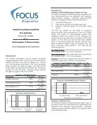
West Nile Virus IgG DxSelect⢠(English) - Focus Diagnostics
West Nile Virus IgG DxSelect⢠(English) - Focus Diagnostics
West Nile Virus IgG DxSelect⢠(English) - Focus Diagnostics
- No tags were found...

You also want an ePaper? Increase the reach of your titles
YUMPU automatically turns print PDFs into web optimized ePapers that Google loves.
<strong>West</strong> <strong>Nile</strong> <strong>Virus</strong> <strong>IgG</strong> DxSelect<br />
(<strong>English</strong>)<br />
Enzyme-linked Immunosorbent Assay (ELISA)<br />
Product Code EL0300G<br />
Rev. K<br />
Indirect Enzyme-linked immunosorbent assay for qualitatively detecting<br />
human serum <strong>IgG</strong> antibodies to <strong>West</strong> <strong>Nile</strong> virus<br />
For in vitro Diagnostic Use<br />
INTENDED USE<br />
The <strong>Focus</strong> <strong>Diagnostics</strong> <strong>West</strong> <strong>Nile</strong> <strong>Virus</strong> <strong>IgG</strong> DxSelect is intended for qualitatively detecting <strong>IgG</strong> antibodies to <strong>West</strong> <strong>Nile</strong> virus in human serum. In<br />
conjunction with the <strong>Focus</strong> <strong>Diagnostics</strong> <strong>West</strong> <strong>Nile</strong> <strong>Virus</strong> IgM Capture DxSelect, the test is indicated for testing persons having symptoms of<br />
meningioencephalitis, as an aid in the presumptive laboratory diagnosis of <strong>West</strong> <strong>Nile</strong> virus infection. Positive results must be confirmed by<br />
neutralization test, or by using the current CDC guidelines for diagnosing <strong>West</strong> <strong>Nile</strong> encephalitis. 1 This test is not intended for self-testing, and this<br />
test is not FDA cleared nor approved for testing blood or plasma donors. Assay performance characteristics have not been established for automated<br />
instruments.<br />
Caution: <strong>IgG</strong> assay cross-reactivity has been noted with some specimens containing antibody to cytomegalovirus (CMV). Reactive results<br />
must be reported with a caution statement regarding possible cross-reactivity with CMV and bunyaviruses, e.g., LaCrosse virus.<br />
SUMMARY AND EXPLANATION OF TEST<br />
Most people who are infected with <strong>West</strong> <strong>Nile</strong> virus (WNV) will not have any type of illness. Experts estimate that 20% of the people who become<br />
infected will develop <strong>West</strong> <strong>Nile</strong> fever: mild symptoms, including fever, headache, and body aches, occasionally with a skin rash on the trunk of the<br />
body and swollen lymph glands. 2 Symptoms of mild disease will generally last a few days. About 1 in 150 of <strong>West</strong> <strong>Nile</strong> virus infections (< 1%) result<br />
in meningitis or encephalitis. 2 Case fatality rates among patients hospitalized during recent outbreaks have ranged from 4% to 14%. 3 Advanced age is<br />
the most important risk factor for death, and patients older than 70 years of age are at particularly high risk. The case fatality rates for WNV are<br />
similar to St. Louis encephalitis virus (SLE) and <strong>West</strong>ern equine encephalitis (WEE) virus (5–15%), but much lower than Eastern equine encephalitis<br />
(EEE) virus (30–70%), and higher than La Crosse (LAC) virus (< 1%). 4,5<br />
WNV is an arbovirus. Arboviruses are zoonotic, and are transmitted through complex life cycles involving a vertebrate (e.g., birds) and an arthropod<br />
(e.g., mosquitoes). 3 Humans and domestic animals can develop clinical illness but usually are "dead-end" hosts because they do not produce<br />
significant viremia. Infection is most often not transmitted from person to person. Arbovirus infections can be prevented in two major ways: personal<br />
protective measures to reduce contact with mosquitoes and public health measures to reduce the population of infected mosquitoes in the<br />
environment. 2<br />
WNV can be detected by culturing the organism, by detecting viral antigen or by detecting viral nucleic acid in cerebrospinal fluid, tissue, blood, or<br />
other body fluid. Although a positive culture or a positive nucleic acid detection test are highly specific, experts do not recommend their use for<br />
screening because of their limited sensitivity. 2<br />
CDC reports that although the antibody response to human infection with WNV has not been thoroughly or systematically studied, the following are<br />
reasonable assumptions, based on extensive experience with other flaviviruses, or preliminary conclusions based on empirical observations made<br />
during the 1999 and 2000 epidemics:<br />
<strong>IgG</strong> antibody in serum: By three weeks post-infection (and often earlier), virtually all infected persons should demonstrate serum <strong>IgG</strong> antibody<br />
to WNV by enzymatic immunoassay (EIA) for 500 days or longer. 6<br />
IgM antibody in serum: By the eighth day of infection, a large majority of infected persons will have detectable serum IgM antibody to WNV;<br />
in most cases it will be detectable for at least 1–2 months after illness onset; and in some cases it will be detectable for 500 days or longer. 6<br />
Positive results are known to occur with persons vaccinated for flaviviruses (e.g., yellow fever, Japanese encephalitis, dengue), with persons infected<br />
with other flaviviruses, and with persons previously infected with WNV. Because closely related arboviruses exhibit serologic cross-reactivity,<br />
sometimes it may be epidemiologically important to attempt to pinpoint the infecting virus by conducting plaque reduction neutralization tests using<br />
an appropriate battery of closely related flaviviruses.<br />
TEST PRINCIPLE<br />
In the <strong>Focus</strong> <strong>Diagnostics</strong> <strong>West</strong> <strong>Nile</strong> <strong>Virus</strong> <strong>IgG</strong> DxSelect assay, the polystyrene microwells are coated with recombinant <strong>West</strong> <strong>Nile</strong> virus antigen.<br />
Diluted serum samples and controls are incubated in the wells to allow anti-WNV <strong>IgG</strong> antibody (if present in the sample) to react with the antigen.<br />
Nonspecific reactants are removed by washing and peroxidase-conjugated anti-human <strong>IgG</strong> is added that reacts with human <strong>IgG</strong> bound to the antigen.<br />
Excess conjugate is removed by washing. Enzyme substrate and chromogen are added, and the color is allowed to develop. After adding the Stop<br />
Reagent, the resultant color change is quantified by a spectrophotometric reading of optical density (OD). Sample optical density readings are<br />
compared with reference cut-off OD readings to determine results.
FOCUS <strong>Diagnostics</strong><br />
<strong>West</strong> <strong>Nile</strong> <strong>Virus</strong> <strong>IgG</strong> DxSelect<br />
Page 2<br />
MATERIALS SUPPLIED<br />
<strong>IgG</strong> Antigen Wells, 96 wells REF EL0351 Ag<br />
12 eight-well polystyrene microwell strips on a frame. Each well is coated with recombinant <strong>West</strong> <strong>Nile</strong> virus antigen. Each strip may be broken<br />
down into individual wells for cost effective use. To avoid condensation, allow the antigen strips to warm to room temperature before opening the<br />
sealed packets.<br />
<strong>IgG</strong> Conjugate, 16 mL REF EL0304 CONJ <strong>IgG</strong><br />
One vial of affinity-purified and peroxidase-conjugated goat anti-human <strong>IgG</strong> (Fc fragment specific). Contains protein, buffer, and preservatives.<br />
Positive Control, 0.3 mL REF EL0311 CONTROL <br />
One vial of human serum. Contains 0.1% sodium azide as a preservative. Requires dilution before use (see Specimen, Controls and Calibrator<br />
Preparation, below).<br />
Negative Control, 0.3 mL REF EL0312 CONTROL <br />
One vial of human serum. Contains 0.1% sodium azide as a preservative. Requires dilution before use (see Specimen, Controls and Calibrator<br />
Preparation, below).<br />
Cut-Off Calibrator, 0.3 mL REF EL0306 CONTROL CAL<br />
One vial of human serum. Contains 0.1% sodium azide as a preservative. Requires dilution before use (see Specimen, Controls and Calibrator<br />
Preparation, below).<br />
Sample Diluent, 100 mL REF EL1608 DIL SPE<br />
One vial of PBS containing protein, surfactant, and preservatives.<br />
10X Wash Buffer, 100 mL REF EL0405 BUF WASH<br />
One vial of surfactant in PBS with preservatives. Prepare a 1X wash buffer solution before use.<br />
To prepare a 1X wash buffer solution, mix 100 mL 10X Wash Buffer with 900 mL distilled (or deionized) water and rinse out any crystals. Use only<br />
the highest grade purified water for reconstitution of the wash buffer. It has been observed that some sources of deionized water contain materials<br />
that can interfere in the assay. Swirl until well mixed and all crystals are dissolved.<br />
Substrate Reagent, 16 mL REF EL0009 SUBS TMB<br />
One vial of tetramethylbenzidine (TMB) and hydrogen peroxide in buffer. The Substrate Reagent may turn slightly blue while stored cold, however,<br />
the color should return to slightly amber after warming to room temperature. A dark blue color indicates contamination with peroxidase; and, if this<br />
occurs, use a fresh bottle.<br />
Stop Reagent, 16 mL REF EL0105 SOLN STOP<br />
One vial 1 M sulfuric acid.<br />
Sealing Tape<br />
Two sheets of sealing tape.<br />
MATERIALS REQUIRED, BUT NOT SUPPLIED<br />
1. Distilled or deionized water<br />
2. 250 or 500 mL wash bottle or automated EIA plate washer<br />
3. 1 L graduated cylinder<br />
4. 12 x 75 mm borosilicate glass test tubes or equivalent<br />
5. 10 L and 100 L pipettors with disposable tips<br />
6. 1 mL pipet or dispenser<br />
7. 5 mL pipet<br />
8. Timer<br />
9. Paper towels or absorbant paper<br />
10. Sink<br />
11. Vortex mixer or equivalent<br />
12. ELISA plate spectrophotometer, wavelength = 450 nm<br />
SHELF LIFE AND HANDLING<br />
1. Kits and kit reagents are stable through the end of the month indicated by the label expiration date when stored at 2 to 8°C.<br />
2. Do not use test kit or reagents beyond their expiration dates.<br />
3. Do not expose reagents to strong light during storage or incubation.<br />
4. Allow reagents to warm to room temperature before use.
FOCUS <strong>Diagnostics</strong><br />
<strong>West</strong> <strong>Nile</strong> <strong>Virus</strong> <strong>IgG</strong> DxSelect<br />
Page 3<br />
WARNINGS AND PRECAUTIONS<br />
1. This kit is for in vitro diagnostic use only.<br />
2. All blood products should be treated as potentially infectious. Source materials from which this product (including controls) was derived have<br />
been screened for Hepatitis B surface antigen, Hepatitis C antigen and HIV-1/2 (AIDS) antibody by FDA-approved methods and found to be<br />
negative. However, as no known test methods can offer 100% assurance that products derived from human blood will not transmit these or other<br />
infectious agents, all controls, serum specimens and equipment coming into contact with these specimens should be considered potentially<br />
infectious and decontaminated or disposed of with proper biohazard precautions. CDC and the National Institutes of Health recommend that<br />
potentially infectious agents be handled at the Biosafety Level 2. 7<br />
3. The antigen wells are produced with recombinant <strong>West</strong> <strong>Nile</strong> virus antigens. After adding patient or control specimens, the strips should be<br />
considered potentially infectious and handled accordingly.<br />
4. Sodium azide at a concentration of 0.1% has been added to some reagents as an antibacterial agent. To prevent buildup of explosive metal<br />
azides in lead and copper plumbing, those reagents (see Materials Supplied, above) should be discarded into sewerage only if diluted and<br />
flushed with large volumes of water. Use copper-free and lead-free drain systems where possible. Occasionally decontaminate the drains with<br />
10% sodium hydroxide (CAUTION: caustic), allow to stand for 10 minutes, then flush with large volumes of water.<br />
5. Do not substitute or mix reagents from different kit lots or from other manufacturers.<br />
6. Use only protocols described in this insert. Incubation times or temperatures other than those specified may give erroneous results.<br />
7. Cross-contamination of patient specimens can cause erroneous results. Add patient specimens and handle strips carefully to avoid mixing of sera<br />
from adjoining incubation tray wells. Decant carefully.<br />
8. Bacterial contamination of serum specimens or reagents can produce erroneous results. Use aseptic techniques to avoid microbial<br />
contamination.<br />
9. Perform the assay at room temperature (approximate range 20 to 25C).<br />
10. Use proper pipetting techniques, maintaining the pipetting pattern throughout the procedure to ensure optimal and reproducible values.<br />
11. The stop reagent contains sulfuric acid. Do not allow to contact skin or eyes. If exposed, flush with copious amounts of water.<br />
12. This test must be performed on serum only. The use of whole blood, plasma or other specimen matrix has not been established.<br />
13. Sodium azide inhibits conjugate activity. Clean pipette tips must be used for the conjugate addition so that sodium azide is not carried over<br />
from other reagents.<br />
14. Hyper-lipemic, heat inactivated, hemolyzed, icteric, and contaminated sera must not be tested.<br />
SPECIMEN COLLECTION AND PREPARATION<br />
Serum is the specimen source. No attempt has been made to assess the assay's compatibility with other specimens. Performance characteristics have<br />
not been established with hyper-lipemic, heat inactivated, hemolyzed, icteric, or contaminated sera. It is unknown if such specimens will cause<br />
erroneous results.<br />
Specimen Collection and Handling<br />
Collect blood samples aseptically using approved venipuncture techniques by qualified personnel. Allow blood samples to clot at room temperature<br />
prior to centrifugation. Aseptically transfer serum to a tightly closing sterile container for storage. Separated serum should remain at 22C for no<br />
longer than 8 hours. If the assay will not be completed within 8 hours, refrigerate the sample at 2 to 8C. If the assay will not be completed within 48<br />
hours, or for shipment of samples, freeze at –20C or colder. 8 Thaw and mix samples well prior to use.<br />
Specimen, Controls and Calibrator Preparation<br />
Dilute each specimen, control and calibrator 1:101. For example, label tubes and dispense 1 mL of Sample Diluent into each labeled tube. Add 10 L<br />
of specimen, control or calibrator to each appropriate tube containing the 1 mL Sample Diluent and mix well by vortex mixing.<br />
TEST PROCEDURE<br />
Performance characteristics have not been established for procedures that are different from the procedure described below. Different procedures,<br />
e.g., different times, volumes, temperatures, or others, may produce invalid results.<br />
1. Bring all reagents to room temperature before use. Remove the Antigen Well packet from cold storage. To avoid condensation, allow microwell<br />
strips to reach room temperature before opening the foil packet. If less than a full plate is to be used, return unused strips to the foil packet<br />
with desiccant and reseal completely. Store unused antigen wells at 2 to 8C. (Note: At the end of the assay, retain the frame for use with the<br />
remaining strips.)<br />
2. Fill wells with 1X Wash Buffer solution (see Materials Supplied, above) and allow to soak for 5 minutes. Decant (or aspirate) the antigen wells<br />
and tap vigorously to remove Wash Buffer. Blot the emptied Antigen Wells face down on clean paper towels or absorbent paper to remove<br />
residual Wash Buffer.<br />
3. Dispense 100 µL of the Sample Diluent into the "blank" wells and 100 µL of each diluted specimen, control or calibrator (see Specimen,<br />
Controls, and Calibrator Preparation, above) into the appropriate wells. (Note: For runs with more than 48 wells it is recommended that 250 L<br />
of each diluted sample first be added to a blank microtiter plate in the location corresponding to that in the ELISA wells. The samples can then<br />
be efficiently transferred into the Antigen Wells with a 100 L 8 or 12-channel pipettor.)<br />
4. Cover plates with sealing tape (or place in a humid chamber), and incubate for 60 1 minute at room temperature (20 to 25C).<br />
5. Remove sealing tape (or remove wells from the humid chamber), and empty the contents of the wells into a sink or a discard basin.<br />
6. Fill each well with a gentle stream of 1X Wash Buffer solution from a wash bottle then empty contents into a sink or a discard basin.<br />
7. Repeat wash (step 6) an additional 2 times.<br />
8. Tap the antigen wells vigorously to remove 1X Wash Buffer. Blot the emptied Antigen Wells face down on clean paper towels or absorbent<br />
paper to remove residual 1X Wash Buffer.<br />
9. Dispense 100 L Conjugate to all wells, using a 100 L 8 or 12-channel pipettor.
FOCUS <strong>Diagnostics</strong><br />
<strong>West</strong> <strong>Nile</strong> <strong>Virus</strong> <strong>IgG</strong> DxSelect<br />
Page 4<br />
10. Cover plates with sealing tape (or place in a humid chamber) and incubate for 30 1 minutes at room temperature (20 to 25C).<br />
11. Repeat wash steps 5 through 8.<br />
12. Pipet 100 L of Substrate Reagent to all wells, using a 100 L 8 or 12-channel pipettor. Begin incubation timing with the addition of Substrate<br />
Reagent to the first well. (Note: Never pour the substrate reagent into the same trough as was used for the conjugate.)<br />
13. Incubate for 10 1 minutes at room temperature (20 to 25C).<br />
14. Stop the reaction by adding 100 L of Stop Reagent to all wells using a 100 L 8 or 12-channel pipettor. Add the Stop Reagent in the same<br />
sequence and at the same pace as the Substrate was added. In antibody-positive wells, color should change from blue to yellow.<br />
15. Gently blot the outside bottom of wells with a paper towel to remove droplets that may interfere with reading by the spectrophotometer. Do not<br />
rub with the paper towel as it may scratch the optical surface of the well. (Note: Large bubbles on the surface of the liquid may affect the OD<br />
readings.)<br />
16. Measure the absorbance of each well within 1 hour of stopping the assay. Set the microwell spectrophotometer at a wavelength of 450 nm. Zero<br />
the instrument on the blank wells, or correct all ODs by manually subtracting the blank ODs.<br />
ELISA <strong>IgG</strong> Procedure (condensed version)<br />
1. Dilute samples 1:101 in Sample Diluent (e.g., 10 L + 1000 L).<br />
2. Soak Wells for 5 minutes with 1X Wash, decant.<br />
3. 100 L of sample for 60 minutes, decant.<br />
4. Wash 3 times.<br />
5. 100 L of Conjugate for 30 minutes, decant.<br />
6. Wash 3 times.<br />
7. 100 L of Substrate Reagent for 10 minutes.<br />
8. 100 L of Stop Reagent, read at = 450 nm.<br />
Please see the PROCEDURE section for important details.<br />
QUALITY CONTROL<br />
Each plate run (or strips or wells from a single plate) must include the Cut-off Calibrator and the two Controls. If multiple plates are run, include the<br />
Cut-off Calibrator and both controls on each plate. It is recommended that until the user becomes familiar with the kit performance, all specimens,<br />
controls and the Cut-off Calibrator should be run in duplicate with the Cut-off Calibrator run twice for a total of four wells. If single wells are used,<br />
the Cut-off Calibrator should be run in triplicate. Include a minimum of 1 blank well (containing sample diluent only) for instrument calibration<br />
purposes.<br />
The Cut-off Calibrator has been formulated to give the optimum differentiation between negative and positive sera. Although the absorbance value<br />
may vary between runs and between laboratories, the mean value for the Cut-off Calibrator wells must be within 0.100 to 0.700 OD units. All<br />
replicate Cut-off Calibrator ODs should be within 0.10 absorbance units from the mean value.<br />
Report results as index values relative to the Cut-off Calibrator. To calculate index values, divide specimen and control optical density (OD) values<br />
by the mean of the Cut-off Calibrator OD values.<br />
1. The Positive Control index values should be between 1.5 and 3.5.<br />
2. The Negative Control index values should be less than 0.8.<br />
If the Calibrator or controls are not within these parameters, patient test results should be considered invalid and the assay repeated.<br />
The Positive and Negative Controls are intended to monitor for substantial reagent failure. The Positive Control should not be used as an indicator for<br />
Cut-off Calibrator precision and only ensures reagent functionality. Quality control requirements must be performed in conformance with local, state,<br />
and/or federal regulations or accreditation requirements and your laboratory's standard Quality Control procedures. In the US, regulatory authorities<br />
recommend that the user refer to NCCLS C24-A and 42 CFR 493.1256 for guidance on appropriate QC practices.<br />
INTERPRETATION OF TEST RESULTS<br />
To calculate index values, divide specimen optical density (OD) values by the mean of the Cut-off Calibrator absorbance values.<br />
The magnitude of the index value above the Cut-off Calibrator does not indicate the total amount of antibody present.<br />
Cut-off Development. In designing the assay, the assay Cut-off was established to slightly favor specificity over sensitivity by using 217 sera<br />
consisting of 3 different serum panels: 1) 136 sera submitted for <strong>West</strong> <strong>Nile</strong> testing and positive with an in-house WNV <strong>IgG</strong> ELISA (a native antigen<br />
<strong>West</strong> <strong>Nile</strong> ELISA <strong>IgG</strong> 8,9 ); 2) 61 sera submitted for <strong>West</strong> <strong>Nile</strong> testing and negative with an in-house WNV <strong>IgG</strong> ELISA; and 3) 20 blood donors. The<br />
<strong>Focus</strong> <strong>West</strong> <strong>Nile</strong> <strong>IgG</strong> was positive with 90.4% (122/135) of the in-house WNV <strong>IgG</strong> ELISA positive samples, negative with 100% (61/61) of the inhouse<br />
WNV <strong>IgG</strong> ELISA negative samples, and negative with 100% (20/20) of the blood donor samples.
FOCUS <strong>Diagnostics</strong><br />
<strong>West</strong> <strong>Nile</strong> <strong>Virus</strong> <strong>IgG</strong> DxSelect<br />
Page 5<br />
<strong>IgG</strong><br />
Index<br />
1.50<br />
< 1.50<br />
and<br />
1.30<br />
Interpretation (<strong>IgG</strong> Result Only)<br />
<strong>IgG</strong> Positive. An index value of 1.50 indicates <strong>IgG</strong> antibodies to <strong>West</strong> <strong>Nile</strong> virus were detected. The presence of <strong>IgG</strong> antibodies is presumptive evidence<br />
that the patient was or is currently infected with (or exposed to) <strong>West</strong> <strong>Nile</strong> virus or another flavivirus. A patient can be <strong>IgG</strong> positive because of crossreactivity<br />
to CMV and/or bunyaviruses, e.g., LAC. Positive results must be reported with a caution statement regarding possible cross-reactivity with CMV and/or<br />
bunyaviruses. These diseases must be excluded before confirming diagnosis. <strong>IgG</strong> positive results must be confirmed by plaque reduction neutralization test, or<br />
by using the recent CDC guidelines for diagnosis of <strong>West</strong> <strong>Nile</strong> encephalitis.<br />
<strong>IgG</strong> Equivocal. An index value of 1.30 and < 1.50 is considered an equivocal result. It is recommended that samples with equivocal results be tested<br />
using a different method; or the patient may be re-drawn two or more weeks later and re tested with this assay.<br />
< 1.30 <strong>IgG</strong> Negative. An index value of < 1.30 indicates <strong>IgG</strong> antibodies to <strong>West</strong> <strong>Nile</strong> virus were not detected. The absence of <strong>IgG</strong> antibodies is presumptive<br />
evidence that the patient was not infected with <strong>West</strong> <strong>Nile</strong> virus or another flavivirus.<br />
However, the sample may have been drawn before antibodies were detectable, or the patient may be immunosuppressed. If infection is suspected, then<br />
another sample should be drawn 7–14 days later and tested. Refer to the CDC guidelines for diagnosis of <strong>West</strong> <strong>Nile</strong> encephalitis.<br />
If the <strong>Focus</strong> <strong>West</strong> <strong>Nile</strong> ELISA IgM results are also available for the same specimen then use the following interpretation:<br />
IgM<br />
Result<br />
Pos<br />
Neg<br />
<strong>IgG</strong><br />
Result<br />
Pos<br />
Neg<br />
Pos<br />
Neg<br />
Combined Interpretation (Both IgM & <strong>IgG</strong> Results)<br />
Presume the patient was recently or is currently infected. The presence of IgM antibodies is presumptive evidence that the patient was<br />
recently or is currently infected with <strong>West</strong> <strong>Nile</strong> virus or another flavivirus. However, IgM anti-WNV has been shown to persist for ≥ 500 days.<br />
Refer to results above for IgM anti-WNV reactive results.<br />
Presume the patient was previously infected with (or exposed to) WNV. The presence of <strong>IgG</strong> antibodies without IgM antibodies is<br />
presumptive evidence that the patient was infected (or exposed to) <strong>West</strong> <strong>Nile</strong> virus or another flavivirus at a previous time.<br />
Presume the patient has not been infected with (or exposed to) WNV. The absence of IgM and <strong>IgG</strong> antibodies is presumptive evidence that<br />
the patient has not been recently infected with (or exposed to) <strong>West</strong> <strong>Nile</strong> virus or another flavivirus.<br />
LIMITATIONS<br />
1. The performance of this assay has not been established for screening the general population. Testing should only be performed on patients with<br />
clinical symptoms of meningioencephalitis.<br />
2. The performance of this assay has not been established for ruling out diseases with similar symptoms, e.g., herpes simplex virus encephalitis,<br />
enterovirus encephalitis, bacterial meningitis, causes of non-infectious encephalitis, or post-infectious encephalitis.<br />
3. The performance of this assay has not been established for matrices other than serum, or visual result determination(s), or monitoring <strong>West</strong> <strong>Nile</strong><br />
virus therapy.<br />
4. All results from this and other serologies must be correlated with clinical history, epidemiological data, and other data available to the attending<br />
physician in evaluating the patient. Positive results must be confirmed by neutralization test, or by using the current CDC guidelines for<br />
diagnosing <strong>West</strong> <strong>Nile</strong> encephalitis. 1<br />
5. The prevalence of infection will affect the assay’s predictive value.<br />
6. The test can be positive with persons that are not currently infected with <strong>West</strong> <strong>Nile</strong> virus. Because of the cross-reactivity between flaviviruses,<br />
persons vaccinated for other flaviviruses (e.g., Japanese encephalitis virus, dengue virus, yellow fever virus) or infected with other flaviviruses<br />
may be positive. These diseases and/or vaccinations must be excluded before confirmation of diagnosis.<br />
7. The test can be negative with persons that are currently infected with <strong>West</strong> <strong>Nile</strong> virus. Samples obtained very early in the infection may not have<br />
detectable antibodies.<br />
8. Cross-reactivity has been observed with some specimens containing antibody to cytomegalovirus (CMV) and bunyaviruses, e.g., LAC. Positive results must<br />
be reported with a caution statement regarding possible cross-reactivity with CMV and bunyaviruses, e.g., La Crosse virus.<br />
EXPECTED VALUES<br />
The prevalence of <strong>West</strong> <strong>Nile</strong> antibodies varies depending on age, geographic location, testing method used, and other factors. A community based<br />
serosurvey for <strong>West</strong> <strong>Nile</strong> infection conducted in New York in 2000 found that 0.2% (5/2433) of persons tested overall had antibodies indicating<br />
recent <strong>West</strong> <strong>Nile</strong> infection, and that 1.1% (2/176) of persons reporting a recent headache and fever had antibodies indicating a recent <strong>West</strong> <strong>Nile</strong><br />
infection. 11 Two serosurveys conducted in New York City (NYC) in 1999 and 2000 showed that approximately 1 in 150 infections (< 1%) resulted in<br />
meningitis or encephalitis. The NYC results are consistent with a 1996 Romanian serosurvey indicating that 1:140 to 1:320 infections resulted in<br />
meningitis or encephalitis. 2
FOCUS <strong>Diagnostics</strong><br />
<strong>West</strong> <strong>Nile</strong> <strong>Virus</strong> <strong>IgG</strong> DxSelect<br />
Page 6<br />
Prevalence in Samples Submitted for Non-Flavivirus Testing (n=476)<br />
<strong>Focus</strong> assessed reactivity with 476 samples prospectively collected from North America during August 2003. The samples had been submitted to a<br />
clinical laboratory located in Southern California for non-flavivirus tests (e.g., tests for other infectious diseases). The samples consisted of 63.7%<br />
females, 34.9% males, and 1.4% samples from persons of unspecified gender.<br />
<strong>IgG</strong> Prevalence with Samples Submitted for Non-Flavivirus Testing(n = 476)<br />
Age Neg Eqv Pos % Positive 95%CI<br />
0 to 9 20 0 4 16.7% (4/24) 4.7-37.4%<br />
10 to 19 25 2 2 6.9% (2/29) 0.9-22.8%<br />
20 to 29 63 4 3 4.3% (3/70) 0.9-12.0%<br />
30 to 39 76 1 5 6.1% (5/82) 2.0-13.7%<br />
40 to 49 69 1 8 10.3% (8/78) 4.5-19.2%<br />
50 to 59 47 1 3 5.9% (3/51) 1.2-16.2%<br />
60 to 69 32 1 6 15.4% (6/39) 5.9-30.5%<br />
70 to 79 28 1 5 14.7% (5/34) 5.0-31.1%<br />
80+ 15 1 2 11.1% (2/18) 1.4-34.7%<br />
Unknown 41 2 8 15.7% (8/51) 7.0-28.6%<br />
Overall 416 14 46 9.7% (46/476) 7.2-12.7%<br />
PERFORMANCE CHARACTERISTICS<br />
Study Site 1: <strong>Focus</strong> Reactivity with Encephalitis/Meningitis Patients (n = 300)<br />
A state department of health laboratory located in the northeastern U.S. assessed the device’s reactivity from encephalitis/meningitis patients (n =<br />
300). Patients were suspected of having either viral encephalitis or viral meningitis. Viral encephalitis criteria included: 1) fever; 2) altered mental<br />
status and/or other evidence of cortical involvement; and 3) CSF pleocytosis with predominant lymphocytes and/or elevated protein and a negative<br />
gram stain and culture. 12 Viral meningitis criteria included: 1) fever; 2) headache, stiff neck and/or other meningeal signs; and 3) CSF pleocytosis<br />
with predominant lymphocytes and/or elevated protein and a negative gram stain and culture). 12 The sera were sequentially submitted to the<br />
laboratory, archived, and masked. The reference methods were CDC WNV <strong>IgG</strong> ELISA, and plaque reduction neutralization test (PRNT) for <strong>West</strong><br />
<strong>Nile</strong> virus.<br />
Of 300 encephalitis/meningitis patients, 37 patients were classified as confirmed positive <strong>West</strong> <strong>Nile</strong> encephalitis patients (meningioencephalitis<br />
symptoms, CDC WNV <strong>IgG</strong> ELISA positive, and WNV PRNT positive), 210 patients had presumptive assay (CDC WNV <strong>IgG</strong> ELISA) results, and 53<br />
patients were unclassified because the CDC WNV <strong>IgG</strong> ELISA results were indeterminant or equivocal. The <strong>Focus</strong> <strong>IgG</strong> ELISA was positive with<br />
97.3% (36/37) of the confirmed positive WNV encephalitis patients (including 1 <strong>Focus</strong> equivocal calculated as negative). Of the 210 patients with<br />
presumptive assay results, 4 were classified as presumed positive flavivirus encephalitis patients (CDC WNV <strong>IgG</strong> positive, PRNT negative), one was<br />
classified as a confirmed dengue positive (CDC WNV <strong>IgG</strong> ELISA positive, dengue PRNT positive), and 205 were classified as presumed negative<br />
patients (CDC WNV <strong>IgG</strong> ELISA negative). The <strong>Focus</strong> <strong>IgG</strong> ELISA was positive with 100% (4/4) of the presumed positive WNV encephalitis<br />
patients, and positive with the one dengue positive patient. The <strong>Focus</strong> <strong>IgG</strong> ELISA was negative with 99.0% (203/205) of the presumed negative<br />
patients (including 1 <strong>Focus</strong> equivocal calculated as positive). The 53 unclassified patients were excluded from the calculations.<br />
Study Site 1: <strong>Focus</strong> Reactivity with Encephalitis/Meningitis Patients(n = 300)*<br />
Specimens Characterized by<br />
<strong>Focus</strong> WNV <strong>IgG</strong> ELISA Results<br />
Reference Assays Neg Eqv Pos Total %<br />
Clinical sensitivity (encephalitis or<br />
meningitis symptoms, CDC <strong>IgG</strong> ELISA<br />
positive and WNV PRNT positive)<br />
0 1 36 37 97.3% (36/37)<br />
95%CI 85.8-99.9%<br />
Agreement with the presumptive CDC<br />
<strong>IgG</strong> ELISA<br />
203 1 6 210 Positive†<br />
100% (5/5)<br />
95%CI 47.8-100%<br />
Negative<br />
99.0% (203/205)<br />
95%CI 96.5-99.9%<br />
* Excluding 53 CDC <strong>IgG</strong> ELISA results (49 indeterminant and 4 equivocal samples).<br />
† One of the presumptive positive samples was dengue PRNT positive and the other 4 presumptive<br />
positives were negative with WNV, dengue and SLE PRNT.<br />
Study Site 2: <strong>Focus</strong> Reactivity with WNV PRNT Positives (n = 75)<br />
A clinical laboratory located in the mid-western U.S. assessed the device’s reactivity with 75 retrospective sera that were pre-screened positive (by<br />
<strong>Focus</strong>) with a <strong>West</strong> <strong>Nile</strong> virus native antigen ELISA, and confirmed <strong>West</strong> <strong>Nile</strong> positive by plaque reduction neutralization test (PRNT). The sera<br />
were sequentially submitted to the laboratory, archived, and masked. The <strong>Focus</strong> <strong>IgG</strong> ELISA was positive with 36.0% (27/75) of the 75 PRNT<br />
positives (calculating 4 equivocals as negative), and negative with 44 samples.<br />
Study Site 2: <strong>Focus</strong> Reactivity with WNV PRNT Positives(n = 75)<br />
Specimens Characterized by<br />
<strong>Focus</strong> WNV <strong>IgG</strong> ELISA Results<br />
Reference Assays Neg Eqv Pos Total %<br />
Serological sensitivity positive (WNV<br />
PRNT positive)<br />
44 4 27 75 36.0% (27/75)<br />
95%CI 25.2-92.3%
FOCUS <strong>Diagnostics</strong><br />
<strong>West</strong> <strong>Nile</strong> <strong>Virus</strong> <strong>IgG</strong> DxSelect<br />
Page 7<br />
Study Site 3: <strong>Focus</strong> Reactivity with <strong>West</strong> <strong>Nile</strong> IFA Negatives (n=157)<br />
A clinical laboratory located in the southwestern U.S. assessed reactivity with 157 retrospective <strong>West</strong> <strong>Nile</strong> IFA negative samples. 13 The <strong>Focus</strong> <strong>IgG</strong><br />
ELISA was 96.8% (152/157) negative with WNV <strong>IgG</strong> IFA negative samples (including two equivocals calculated as positive), and positive with<br />
three samples.<br />
Study Site 3: <strong>Focus</strong> Reactivity with <strong>West</strong> <strong>Nile</strong> IFA Negatives (n=157)<br />
Specimens Characterized by<br />
<strong>Focus</strong> WNV <strong>IgG</strong> ELISA Results<br />
Reference Assays Neg Eqv Pos Total %<br />
Negative agreement with presumptive<br />
WNV IFA negative<br />
152 2 3 157 95.6% (152/157)<br />
95% CI 91.1-98.2%<br />
Study Site 4: <strong>Focus</strong> Reactivity with Suspected Encephalitis/Meningitis Patients (n = 50)<br />
<strong>Focus</strong> assessed the device’s reactivity with 50 sera from patients suspected of encephalitis/meningitis. A U.S. federal government laboratory<br />
provided the retrospective and masked sera. One sample was confirmed positive by <strong>West</strong> <strong>Nile</strong> PRNT, and the other 49 were presumptively negative<br />
(CDC ELISA) for arboviruses present in North America (LAC, EEE, SLE and WNV). The <strong>Focus</strong> <strong>IgG</strong> ELISA was negative with 95.9% (47/49) of<br />
the WNV negative samples, and positive with the one positive confirmed by <strong>West</strong> <strong>Nile</strong> PRNT.<br />
Study Site 4: Reactivity with Suspected Encephalitis/Meningitis Patients(n = 50)<br />
Specimens Characterized by<br />
<strong>Focus</strong> WNV <strong>IgG</strong> ELISA Results<br />
Reference Assays Neg Eqv Pos Total %<br />
Serological sensitivity (CDC <strong>IgG</strong> ELISA<br />
positive and WNV PRNT positive)<br />
0 0 1 1 100% (1/1)<br />
95% CI NA<br />
Negative agreement with presumptive<br />
CDC <strong>IgG</strong> ELISA negative<br />
47 0 2 49 95.9% (47/49)<br />
95% CI 86.0-99.5%<br />
Study Site 4: <strong>Focus</strong> Reactivity with Non-Flavivirus Test Samples (n = 476)<br />
<strong>Focus</strong> assessed the device's reactivity with 476 samples prospectively collected from North America during August 2003. The samples had been<br />
submitted to a clinical laboratory located in Southern California for non-flavivirus tests (e.g., test for other infectious diseases). Positive samples were<br />
tested with a CDC <strong>West</strong> <strong>Nile</strong> <strong>IgG</strong> ELISA. The <strong>Focus</strong> <strong>West</strong> <strong>Nile</strong> ELISA <strong>IgG</strong> was negative with 96.8% (426/440) of the CDC ELISA negative samples<br />
(including 14 <strong>Focus</strong> equivocals calculated as positive), and was positive with 100% (21/21) of the CDC ELISA positive samples. Fifteen CDC<br />
ELISA <strong>IgG</strong> indeterminant samples were excluded from performance calculations.<br />
Study Site 4: <strong>Focus</strong> Reactivity with Non-Flavivirus Test Samples (n = 476)*<br />
Specimens Characterized by<br />
<strong>Focus</strong> WNV <strong>IgG</strong> ELISA Results<br />
Reference Assays Neg Eqv Pos Total %<br />
Positive agreement with presumptive<br />
CDC <strong>IgG</strong> ELISA positive<br />
0 0 21 21 100% (21/21)<br />
95%CI 83.9-100%<br />
Negative agreement with presumptive<br />
CDC <strong>IgG</strong> ELISA negative<br />
426 14 0 440 96.8% (426/440)<br />
95% CI 94.7-98.3%<br />
* Excludes 15 CDC <strong>IgG</strong> ELISA indeterminant samples.<br />
Cross-reactivity<br />
<strong>Focus</strong> (Study Site 4) and a state department of health laboratory located in the northeastern U.S. (DOH) (Study Site 1) assessed the device’s crossreactivity<br />
with sera that were sero-positive to other potentially cross-reactive pathogens (n = 75). The DOH tested the SLE positives, and <strong>Focus</strong> tested<br />
the other sera. The sera were archived and masked. The results of the studies are summarized in the table below:<br />
<strong>Focus</strong> Cross-reactivity<br />
Specimens Characterized by<br />
<strong>Focus</strong> WNV <strong>IgG</strong> ELISA Results<br />
Reference Assays Site Neg Eqv Pos Total % Positive<br />
Dengue virus<br />
(secondary infections)<br />
4 1 0 19 20 95.0% (19/20)<br />
95%CI 75.1-99.9%<br />
Japanese encephalitis virus 4 14 3 3 20 30.0% (6/20)<br />
95%CI 11.9-54.3%<br />
St. Louis encephalitis virus 1 9 1 11 21 57.1% (12/21)<br />
95%CI 34.0-78.2%<br />
Yellow fever virus 4 11 4 5 20 45.0% (9/20)<br />
95%CI 23.1-68.5%<br />
Alphavirus (Sindbis & Eastern<br />
equine viruses)<br />
4 15 0 2 17 11.8% (2/17)<br />
95%CI 0.1-36.4%<br />
Bunyavirus (Jamestown Canyon<br />
& La Crosse)<br />
1 12 2 1 15 20.0% (3/15)<br />
95%CI 4.3-48.1%<br />
Herpes simplex virus type 1 4 55 0 5 60 8.3% (5/60)<br />
95%CI 2.8-18.4%<br />
Epstein-Barr virus 4 11 0 1 12 8.3% (1/12)<br />
95%CI 0.2-38.5%<br />
Cytomegalovirus 4 16 0 4 20 20.0% (4/20)<br />
95%CI 5.7-43.7%<br />
Echovirus/Poliovirus 4 18 1 1 20 10.0% (2/20)<br />
95%CI 1.2-31.7%<br />
Borrelia burgdorferi (Lyme<br />
disease)<br />
4 17 1 2 20 15.0% (3/20)<br />
95%CI 3.2-37.9%
FOCUS <strong>Diagnostics</strong><br />
<strong>West</strong> <strong>Nile</strong> <strong>Virus</strong> <strong>IgG</strong> DxSelect<br />
Page 8<br />
Reproducibility<br />
Reproducibility studies included Inter-lot Reproducibility, Inter/Intra-assay Reproducibility, and Inter-laboratory Reproducibility. In each study, two<br />
sets of samples were masked duplicates. <strong>Focus</strong> (Study Site 4) assessed the device's Inter-lot Reproducibility by testing five samples on three separate<br />
days with three separate lots. For one lot, the samples were run in triplicate, and run in duplicate with the other two lots. Each of the three lots had a<br />
different lot of Antigen and Capture Wells. <strong>Focus</strong> (Study Site 4) assessed the device’s Inter/Intra-assay Reproducibility by testing seven samples in<br />
triplicate, once a day, for three days, for a total of 63 data points. A state department of health laboratory located in the northeastern U.S. (Study Site<br />
1), and a clinical laboratory located in the mid-western U.S. (Study Site 2), <strong>Focus</strong> (Study Site 4), assessed the device's Inter-laboratory<br />
Reproducibility. Each of the three laboratories tested seven samples in triplicate on three different days.<br />
Sample<br />
Index<br />
Mean<br />
Reproducibility<br />
Inter- & Intra-assay Inter-lot Inter-Lab<br />
Intraassaassay<br />
Inter-<br />
Index Index Index<br />
Mean %CV Mean<br />
%CV %CV<br />
Index<br />
%CV<br />
G6* 0.23 12.2 18.2 0.30 13.1 0.32 11.6<br />
G2* 0.29 21.2 17.3 0.34 7.5 0.35 22.5<br />
G5 0.65 7.9 21.3 0.73 7.2 0.69 19.0<br />
G7* 1.14 3.5 18.2 1.30 5.7 1.21 14.1<br />
G1* 1.22 3.2 17.1 1.36 7.0 1.25 16.4<br />
G4 2.44 1.0 16.2 2.79 4.1 2.47 12.8<br />
G3 2.98 3.9 17.3 3.37 4.1 3.10 12.6<br />
* There were two sets of masked pairs (same sample, different labeled identity): G2 & G6 were<br />
one masked pair, and G1 & G7 were the second masked pair.<br />
REFERENCES<br />
1. Centers for Disease Control and Prevention (CDC). Epidemic/Epizootic <strong>West</strong> <strong>Nile</strong> <strong>Virus</strong> in the United States: Guidelines for Surveillance,<br />
Prevention and Control. 3rd Ed. 2003. p. 25.<br />
2. Petersen LR, Marfin AA.<strong>West</strong> <strong>Nile</strong> virus: a primer for the clinician. Ann Intern Med. 2002 Aug 6;137(3):173-9.<br />
3. Campbell GL, Marfin AA, Lanciotti RS, Gubler DJ. <strong>West</strong> <strong>Nile</strong> virus. Lancet Infect Dis 2002 Sep;2(9):519-29.<br />
4. CDC. Information on Arboviral Encephalitides [online], 2001 July 13,. Available from: http://www.cdc.gov/ncidod/dvbid<br />
5. CDC. Eastern Equine Encephalitis -- United States, 1989. Morb Mortal Wkly Rep. 1989 Sep 15;38(36):619-20, 625-6.<br />
6. Roehrig JT, Nash D, Maldin B, Labowitz A, Martin DA, Lanciotti RS, Campbell GL. Persistence of virus-reactive serum immunoglobulin m<br />
antibody in confirmed west nile virus encephalitis cases. Emerg Infect Dis 2003 Mar;9(3):376-9.<br />
7. CDC-NIH Manual. (1999) Biosafety in Microbiological and Biomedical Laboratories. 4 th ed. And National Committee for Clinical Laboratory<br />
Standards (NCCLS). Protection of Laboratory Workers from Instruments, Biohazards and Infectious Disease Transmitted by Blood, Body Fluids<br />
and Tissue (NCCLS M29-A).<br />
8. NCCLS. Procedures for the Handling and Processing of Blood Specimens; Approved Guideline (NCCLS H18-A2). 2 nd ed. (1999).<br />
9. Prince HE, Hogrefe WR. Detection of <strong>West</strong> <strong>Nile</strong> <strong>Virus</strong> (WNV)-Specific Immunoglobulin M in a Reference Laboratory Setting during the 2002<br />
WNV Season in the United States. Clin Diagn Lab Immunol. 2003 Sep;10(5):764-8.<br />
10. Prince HE, and Hogrefe WR, Performance Characteristics of an In-House Assay System Used To Detect <strong>West</strong> <strong>Nile</strong> <strong>Virus</strong> (WNV)-Specific<br />
Immunoglobulin M during the 2001 WNV Season in the United States. Clin Diagn Lab Immunol. 2003 Jan;10(1):177-9.<br />
11. Serosurveys for <strong>West</strong> <strong>Nile</strong> Infection - New York and Connecticut Counties, 2000. Morb Mortal Wkly Rep. 2001 Jan 26;50(3):37-9.<br />
12. New York State Department of Health. New York State <strong>West</strong> <strong>Nile</strong> <strong>Virus</strong> Response Plan. 2001 May. p. 44.<br />
13. Malan AK, Stipanovich PJ, Martins TB, Hill HR, Litwin CM Detection of <strong>IgG</strong> and IgM to <strong>West</strong> <strong>Nile</strong> virus. Development of an<br />
immunofluorescence assay. Am J Clin Pathol. 2003 Apr;119(4):508-15.<br />
This package insert is available in French, German, Italian, and Spanish at www.focusdx.com, and is available in other languages from your local<br />
distributor.<br />
AUTHORIZED REPRESENTATIVE<br />
mdi Europa GmbH, Langenhagener Str. 71, 30855 Langenhagen-Hannover, Germany<br />
ORDERING INFORMATION<br />
Telephone: (800) 838-4548 (U.S.A. only) (562) 240-6500 (International)<br />
Fax: (562) 240-6510<br />
TECHNICAL ASSISTANCE<br />
Telephone: (800) 838-4548 (U.S.A. only) (562) 240-6500 (International)<br />
Fax: (562) 240-6526<br />
Visit our web site at: www.focusdx.com<br />
PI.EL0300G<br />
Rev. K<br />
Date written 16 October 2012<br />
Cypress, California 90630 USA