How the Immune System Works - PDF - Arthritis Trust of America
How the Immune System Works - PDF - Arthritis Trust of America
How the Immune System Works - PDF - Arthritis Trust of America

Create successful ePaper yourself
Turn your PDF publications into a flip-book with our unique Google optimized e-Paper software.
Medical data is for informational purposes only. You should always consult your family physician, or one <strong>of</strong> our referral physicians prior to treatment<br />
Supplement to<br />
The Art <strong>of</strong> Getting Well<br />
<strong>How</strong> <strong>the</strong> <strong>Immune</strong> <strong>System</strong> <strong>Works</strong><br />
Anthony di Fabio<br />
Copyright 1998<br />
All rights reserved by <strong>the</strong> The Roger Wyburn-Mason and Jack<br />
M.Blount Foundation for Eradication <strong>of</strong> Rheumatoid Disease<br />
AKA The <strong>Arthritis</strong> <strong>Trust</strong> <strong>of</strong> <strong>America</strong> ®<br />
7376 Walker Road, Fairview, Tn 37062<br />
Throughout 4.5 billion years our cellular and multi-cellular<br />
ancestors struggled to survive, creatively developing and utilizing<br />
a marvelously complex immune system. Throughout <strong>the</strong>se aeons it<br />
has been eat or be eaten.<br />
Our bodies utilize an extremely diverse army <strong>of</strong> cells and o<strong>the</strong>r<br />
molecules designed especially to protect us from <strong>the</strong> strategies <strong>of</strong><br />
all <strong>of</strong> those would-be eaters. (See Diagram I.) These finely tuned<br />
protective cells also work well as a team.<br />
Researchers continue to uncover new and amazingly complex<br />
ways whereby protection from “outsiders” has developed. What is<br />
already known may sound complex, but please have patience.<br />
There’s a point <strong>of</strong> great understanding in what follows.<br />
There’s an “innate” immune protective system. We’re born<br />
with <strong>the</strong> ability to recognize certain microbes on sight, so to speak,<br />
and we can <strong>the</strong>n destroy <strong>the</strong>m.<br />
There is an “adaptive” immune protective system. The “receptors”<br />
(as a lock is to a key) activated in <strong>the</strong> adaptive immune<br />
response are formed by piecing toge<strong>the</strong>r gene segments, like piecing<br />
toge<strong>the</strong>r a jigsaw puzzle. The available pieces are used by each<br />
cell in a different way to make a unique receptor, enabling cells to<br />
collectively recognize infectious organisms confronted during our<br />
lifetimes.<br />
The end-point target <strong>of</strong> all immune processes is to destroy or<br />
o<strong>the</strong>rwise protect from an “antigen,” usually a foreign molecule<br />
from a microorganism.<br />
The end-object <strong>of</strong> vaccinations is to confront <strong>the</strong> immune system<br />
with an antigen forcing <strong>the</strong> immune system to adapt to <strong>the</strong><br />
foreign invader; that is, <strong>the</strong> immune system must learn to identify<br />
<strong>the</strong> invader and to retain <strong>the</strong> memory <strong>of</strong> this knowledge for <strong>the</strong><br />
purpose <strong>of</strong> destroying <strong>the</strong> invader now and in <strong>the</strong> future.<br />
One major end-result <strong>of</strong> such vaccinations is to develop a signal<br />
to specialized blood components, called “complement” (an enzyme<br />
substance) which is used to overwhelm and to destroy foreign<br />
invaders. (See Diagram II.)<br />
Specialized “antigen-presenting” cells, such as macrophages,<br />
roam throughout our bodies literally ingesting <strong>the</strong> antigens (invaders)<br />
and fragmenting <strong>the</strong>m. These fragments are called “antigenic<br />
peptides.”<br />
Pieces <strong>of</strong> <strong>the</strong>se peptides are layered on <strong>the</strong> surface <strong>of</strong> <strong>the</strong> cell<br />
called “major histocompatibility complex” (MHC). This joining<br />
produces a peptide-MHC combination.<br />
O<strong>the</strong>r specialized white cells called “T lymphocytes” have receptor<br />
cells that “recognize” different peptide-MHC. (The designation<br />
“T” means cells from <strong>the</strong> thymus gland.)<br />
®<br />
1<br />
The T cells that are activated by that “recognition” begin to<br />
divide, and <strong>the</strong>y also secrete a substance called “lymphokines.”<br />
Lymphokines are chemical signals that mobilize o<strong>the</strong>r components<br />
<strong>of</strong> <strong>the</strong> immune system.<br />
One set <strong>of</strong> those cells that responds to <strong>the</strong> lymphokine signals<br />
are <strong>the</strong> “B-lymphocytes” each <strong>of</strong> which also have receptor molecules<br />
<strong>of</strong> a single specificity on <strong>the</strong>ir surface. (See Diagram III.)<br />
(The designation “B” means cells from bone marrow.)<br />
<strong>How</strong>ever, unlike <strong>the</strong> receptors <strong>of</strong> T cells, those <strong>of</strong> B cells can<br />
recognize parts <strong>of</strong> antigens that are free in solution without <strong>the</strong><br />
MHC molecules attached.<br />
When <strong>the</strong> B-lymphocytes are activated, <strong>the</strong>y divide and differentiate<br />
into plasma cells that secrete “antibody” proteins, water<br />
soluble forms <strong>of</strong> <strong>the</strong>ir receptors.<br />
These antibodies bind to whatever antigens <strong>the</strong>y find, neutralizing<br />
<strong>the</strong>m or precipitating <strong>the</strong>ir destruction by enzymes derived<br />
from <strong>the</strong> molecule called “complement.” Complement is a blood<br />
protein which can destroy pathogens on first encounter.<br />
Complement activity -- <strong>the</strong> end point <strong>of</strong> adaptive immunization,<br />
to develop a “complement” to antibodies -- can be triggered<br />
in three ways. (See Diagram IV.)<br />
(1) One type <strong>of</strong> complement called C3 can bind to any protein.<br />
Once bound to <strong>the</strong> microbe <strong>the</strong> C3 molecule causes o<strong>the</strong>r<br />
complement molecules to bind to <strong>the</strong> bacterium. This is called <strong>the</strong><br />
“complement cascade.” Their joint action overwhelms <strong>the</strong> bacterium.<br />
Our body’s cells are protected from C3 by proteins that inactivate<br />
this molecule.<br />
(2) Antibodies produced as a result <strong>of</strong> infection can also activate<br />
complement. After detecting an infection, a macrophage secretes<br />
a substance called “interleukin-6.” As it’s carried through<br />
<strong>the</strong> blood stream, interleukin-6 reaches <strong>the</strong> liver, causing <strong>the</strong> secretion<br />
<strong>of</strong> a “mannose-binding protein.” (Mannose is a sugar formed<br />
by <strong>the</strong> oxidation <strong>of</strong> manitol.) Mannose-binding protein binds to<br />
<strong>the</strong> capsule <strong>of</strong> a bacterium, and this protein <strong>the</strong>n triggers <strong>the</strong> complement<br />
cascade, thus overwhelming <strong>the</strong> bacterium.<br />
(3) Antibodies produced as a result <strong>of</strong> infection also activate<br />
complement. B cells are activated if <strong>the</strong>y bind to <strong>the</strong> bacterium and<br />
are stimulated by a so-called helper T cell. The binding stimulates<br />
<strong>the</strong> B cell to proliferate and to secrete antibodies. The antibodies<br />
bind to <strong>the</strong> bacterium and activate complement protein called C1Q,<br />
which activates o<strong>the</strong>r complement molecules -- <strong>the</strong> “complement<br />
cascade” -- thus overwhelming and killing <strong>the</strong> bacterium.<br />
To make a ra<strong>the</strong>r long, complex story short, <strong>the</strong> many major<br />
immune defensive mechanisms we’ve inherited usually results<br />
in producing a complement cascade, which, working toge<strong>the</strong>r<br />
with antibodies, overwhelms an invading organism.
Medical data is for informational purposes only. You should always consult your family physician, or one <strong>of</strong> our referral physicians prior to treatment<br />
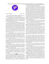
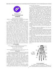
THE LYMPH SYSTEM IS A DECENTRALIZED IMMUNITY DEFENSE<br />
The lymph system consists <strong>of</strong> many organs and tissues that<br />
are scattered throughout <strong>the</strong> body, providing lymphocytes that<br />
are responsible for “specific immunity.” Specific immunity<br />
is <strong>the</strong> special affinity between antigen and its corresponding<br />
Adenoids<br />
Tonsils<br />
Lymph Nodes<br />
Thymus<br />
Spleen<br />
Small Intestine --<br />
Peyer’s Patch<br />
antibody. (1) Lymphocytes are born in <strong>the</strong> primary lymphoid<br />
organs -- <strong>the</strong> thymus making T cells and <strong>the</strong> bone marrow<br />
making B cells. (2) T and B cells leave <strong>the</strong>ir birthplace, circulating<br />
in <strong>the</strong> blood until <strong>the</strong>y reach one <strong>of</strong> <strong>the</strong> numerous<br />
secondary lymphoid organs. Lymph nodes, spleen and tonsils<br />
are examples. (3) T and B cells exit <strong>the</strong> blood stream<br />
through specialized blood vessels named high endo<strong>the</strong>lial<br />
venules. Each gram <strong>of</strong> lymph node contains 10 9 , or one billion,<br />
lymphocytes. Despite <strong>the</strong>ir density, <strong>the</strong> lymphocytes<br />
move about freely. (4) These two facts explain why <strong>the</strong> lymph<br />
nodes are excellent locations for <strong>the</strong> activation <strong>of</strong> antigens<br />
and antigen presenting cells which enter through afferent lymphatic<br />
vessels. Antigens in <strong>the</strong> paracortex generally activate<br />
T cells. The B cells, antibody producing cells, generally are<br />
activated in areas such as <strong>the</strong> germinal centers <strong>of</strong> <strong>the</strong> lymphoid<br />
follicles. (5) Lymphatic vessels carry activated<br />
lymphocytes from <strong>the</strong> nodes<br />
through efferent lymphatics, by<br />
means <strong>of</strong> fluid, until <strong>the</strong>y reach<br />
<strong>the</strong> blood stream where <strong>the</strong>y<br />
provide protection around <strong>the</strong><br />
body. (6) Eventually <strong>the</strong> lymphhocytes<br />
flow into o<strong>the</strong>r lymph<br />
nodes, whence <strong>the</strong> cycle repeats.<br />
Appendix<br />
Diagram I<br />
Lyphatic Vessel<br />
Bone Marrow<br />
Lymph Node<br />
Afferent Lymphatic<br />
High Endo<strong>the</strong>lial Venule<br />
Germinal Center<br />
Cortex<br />
Paracortex<br />
Efferent Lymphatic<br />
Adapted from Sir Gustav J.V. Nossal, “Life and Death <strong>of</strong> <strong>the</strong> <strong>Immune</strong> <strong>System</strong>,” Scientific <strong>America</strong>n, Sept. 1993, p. 56. Artwork originally by Dimitry Schildlovsky<br />
and Tomo Narashima, but readapted and drawn by France Watts.<br />
2
Medical data is for informational purposes only. You should always consult your family physician, or one <strong>of</strong> our referral physicians prior to treatment<br />
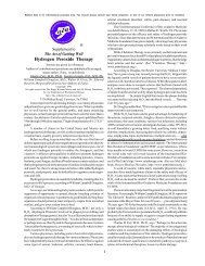
DEFENDING THE BODY WITH A WIDE VARIETY OF MOLECULES AND CELLS<br />
An antigen is a molecule usually from a foreign microorganism<br />
or o<strong>the</strong>r invader. (1) Macrophages, also called antigen-presenting<br />
cells, roam throughout <strong>the</strong> body ingesting antigens. (2) Macrophages<br />
fragment antigens into antigenic peptides. (3) These fragments<br />
are joined to major histocompatibility complexe (MHC) molecules,<br />
which are displayed on <strong>the</strong> surface <strong>of</strong> invading cells. (4) T lymphocytes,<br />
which are also white cells, have receptor sites that enable<br />
each one to recognize different peptide-MHC combinations. These<br />
activated T cells divide and secrete lymphokines, which are chemical<br />
signals that activate o<strong>the</strong>r components <strong>of</strong> <strong>the</strong> immune system.<br />
(5) Among cells that respond to lymphokines are B lymphocytes.<br />
These have specific receptor molecules on <strong>the</strong>ir surface, and unlike<br />
T cells can recognize parts <strong>of</strong> antigens free in a solution without<br />
MHC molecules. (6) Once activated, B cells divide, differentiating<br />
into plasma cells that secret antibody proteins which are also soluble<br />
forms <strong>of</strong> <strong>the</strong>ir receptors. (7) The antibodies can neutralize antigens<br />
by binding to <strong>the</strong>m or trigger <strong>the</strong>ir destruction by means <strong>of</strong> complement<br />
enzymes or even by scavenging cells. (8) Some T and B cells<br />
become memory cells that continue to circulate, boosting <strong>the</strong> immune<br />
system’s readiness if <strong>the</strong> same invader enters <strong>the</strong> body. (9) In<br />
B cells, antibody genes mutate frequently, <strong>the</strong> antibody response<br />
improves after each invasion <strong>of</strong> <strong>the</strong> same antigen.<br />
Antibodies<br />
Activated T Lymphocytes<br />
Activated B Lymphocytes<br />
Plasma Cells<br />
Lymphokines<br />
B Lymphocytes<br />
T Lymphocyte<br />
T cell Receptor<br />
Peptide<br />
Peptide<br />
MHC Protein<br />
(Major<br />
Histocompatibility<br />
Complex)<br />
Antigen<br />
MHC<br />
Protein<br />
Antigen Presenting Cell<br />
Diagram II<br />
Adapted from Sir Gustav J.V. Nossal, “Life and Death <strong>of</strong> <strong>the</strong> <strong>Immune</strong> <strong>System</strong>,” Scientific <strong>America</strong>n, Sept. 1993, p. 55. Artwork originally by Dimitry Schildlovsky,<br />
but readapted and drawn by France Watts.<br />
3
Medical data is for informational purposes only. You should always consult your family physician, or one <strong>of</strong> our referral physicians prior to treatment<br />
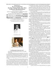
CLONAL SELECTION ENABLES SWIFT REACTION TO MANY POSSIBLE PATHOGENS<br />
Activated B Lymphocyte<br />
Mitosis<br />
Antibodies<br />
(1) Having millions <strong>of</strong> possible surface antibodies, lymphocytes<br />
constantly roam throughout <strong>the</strong> body. (2) When a<br />
matching antibody meets its matching antigen (bottom), <strong>the</strong><br />
lymphocyte swells and <strong>the</strong>n begins to rapidly divide. (3) Once<br />
maturity is reached, B cells secrete antibodies that attack an<br />
invader (top). (4) T cells produce lymphokines. Lymphokines<br />
are chemicals that increase <strong>the</strong> activity <strong>of</strong> o<strong>the</strong>r immune<br />
system cells.<br />
Diagram III<br />
Antibodies<br />
Antigen<br />
Adapted from Sir Gustav J.V. Nossal, “Life and Death <strong>of</strong> <strong>the</strong> <strong>Immune</strong> <strong>System</strong>,” Scientific <strong>America</strong>n, Sept. 1993, p. 56. Artwork originally by Dimitry<br />
Schildlovsky, but readapted and drawn by France Watts.<br />
4
Medical data is for informational purposes only. You should always consult your family physician, or one <strong>of</strong> our referral physicians prior to treatment<br />
THREE WAYS BY WHICH COMPLEMENT ACTIVITY CAN BE TRIGGERED<br />
COMPLEMENT EITHER KILLS BACTERIA OR RECRUITS OTHER IMMUNE SYSTEM CELLS, SUCH AS PHAGOCYTES<br />
Diagram IV<br />
Complement 3<br />
C1q<br />
2<br />
Antibodies<br />
Bacterium<br />
1<br />
Helper T<br />
Cell<br />
B Cell<br />
When antibodies bind to bacteria <strong>the</strong>y<br />
activate a complement protein called<br />
C1q, and this activates o<strong>the</strong>r complement<br />
molecules in turn.<br />
B cells proliferate and are<br />
stimulated to secrete<br />
antibodies once <strong>the</strong>y are<br />
bound<br />
If B cells are activated<br />
<strong>the</strong>y bind to bacteria and<br />
are stimulated by socalled<br />
helper T cells<br />
3<br />
Complement Produced as Result <strong>of</strong> Infection<br />
Mannose Binding Protein<br />
Interleukin-6<br />
2<br />
1<br />
Macrophage<br />
Complement<br />
Mannose-binding protein binds to<br />
<strong>the</strong> capsule <strong>of</strong> bacteria where it<br />
triggers <strong>the</strong> complement cascade.<br />
Bacterium<br />
Liver<br />
On detection <strong>of</strong> infection,<br />
macrophages secrete interleukin-6<br />
Interleukin-6 is carried through <strong>the</strong> blood<br />
stream, reaching <strong>the</strong> liver, and causing <strong>the</strong><br />
secretion <strong>of</strong> mannose-binding protein.<br />
2<br />
Complement Activated by Mannose-binding Protein<br />
1<br />
Bacterium<br />
Complement C3<br />
Once it is bound to<br />
<strong>the</strong> microbe, <strong>the</strong> C3 molecule causes<br />
o<strong>the</strong>r complement molecules to also bind<br />
The C3 complement molecule can bind to any protein<br />
including those on bacteria, but not on self-cells<br />
which are protected by proteins that inactivate <strong>the</strong> C3<br />
molecule.<br />
Complement Acting Directly on Bacteria<br />
Adapted from Charles A. J aneway, Jr., “<strong>How</strong> <strong>the</strong> <strong>Immune</strong> <strong>System</strong> Recognizes Invaders,” Scientific <strong>America</strong>n, Sept. 1993, p. 74. Artwork originally by Ian<br />
Warpole, but readapted and drawn by France Watts.<br />
5