From primordial germ cells to oogonia - eshre
From primordial germ cells to oogonia - eshre
From primordial germ cells to oogonia - eshre

Create successful ePaper yourself
Turn your PDF publications into a flip-book with our unique Google optimized e-Paper software.
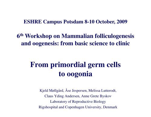
ESHRE Campus Potsdam 8-10 Oc<strong>to</strong>ber, 2009<br />
6 th Workshop on Mammalian folliculogenesis<br />
and oogenesis: from basic science <strong>to</strong> clinic<br />
<strong>From</strong> <strong>primordial</strong> <strong>germ</strong> <strong>cells</strong><br />
<strong>to</strong> <strong>oogonia</strong><br />
Kjeld Møllgård, Åse Jespersen, Melissa Lutterodt,<br />
Claus Yding Andersen, Anne Grete Byskov<br />
Labora<strong>to</strong>ry of Reproductive Biology<br />
Rigshospital and Copenhagen University, Denmark
Migration of the <strong>germ</strong> <strong>cells</strong> of human embryos from the yolk<br />
sac <strong>to</strong> the primitive gonadal folds (Witschi, 1948)<br />
The Carnegy Collection of human embryos and fetuses<br />
Whitschi concluded that the PGCs actively migrated<br />
from the yolk sac diverticle “allan<strong>to</strong>is” <strong>to</strong> the gonads<br />
i.e. PGCs should migrate 3.5 mm in 4 days - passing through the hindgut.<br />
24 days pc 28 days pc<br />
Umbilical cord<br />
PGCs<br />
stained<br />
With alkalic<br />
phosphatase<br />
Allan<strong>to</strong>is<br />
3.5mm<br />
Yolk sac<br />
Hindgut
Migration speed of the human PGCs<br />
If the PGCs should move 3.5 mm in 4<br />
days the speed would be 40mm per<br />
hour - if they go straight ahead<br />
<strong>to</strong>wards the gonadal ridges<br />
Mouse PGCs in vitro move 4 - 13mm<br />
per hour - but in random direction<br />
(Molyneaux et al., Development,<br />
2003)<br />
So, it seems unlikely that the human<br />
PGCs can migrate that fast without help.
Challenging Witschis concept of PGC migration<br />
1. How do the <strong>primordial</strong> <strong>germ</strong> <strong>cells</strong> (PGCs)<br />
of the yolk sac reach the hindgut <br />
2. How do the PGCs find their way from the<br />
hindgut <strong>to</strong> the gonadal-mesonephric area<br />
3. How do the PGCs actually enter the<br />
gonadal ridges
1: How do the PGCs reach the hind gut<br />
Since the <strong>germ</strong> <strong>cells</strong> are not “born” in<br />
the area where the gonads will<br />
develop they must – in some manner -<br />
find their way from the site where<br />
they arise.<br />
The PGCs must be able <strong>to</strong>:<br />
Migrate or be translocated<br />
Know where <strong>to</strong> go
”The active migration of <strong>germ</strong> <strong>cells</strong> in the<br />
embryos in mice and man is a myth”<br />
(Freeman, Reproduction, 2003)<br />
Somite no<br />
PGC<br />
Hindgut<br />
Cranial tip of Chorda<br />
used as fix point<br />
Somite no<br />
PGC<br />
Conclusion:<br />
The PGC are mainly<br />
”passively” translocated <strong>to</strong><br />
the hindgut as the body<br />
grows and curves<br />
(i.e.lateral folding)
Lateral foldings of the human embryo<br />
from day 24 pc <strong>to</strong> day 28 pc<br />
http://www.indiana.edu/%7eanat550/genanim/latfold/latfold.swf<br />
Indiana University Educational System
Lateral foldings of the human embryo<br />
from day 24 pc <strong>to</strong> day 28 pc<br />
http://www.indiana.edu/%7eanat550/genanim/latfold/latfold.swf<br />
<strong>From</strong>: Car<strong>to</strong>on of Indiana University Educational System<br />
The yolk sac containing the<br />
PGCs lines the developing gut<br />
during early development<br />
35 410<br />
2 13<br />
Thus, PGCs are part of the<br />
89 16<br />
developing gut at all times<br />
before and during the lateral<br />
folding.<br />
Therefore, PGCs just have <strong>to</strong><br />
be translocated from the gut<br />
through the dorsal mesentery<br />
<strong>to</strong> the gonadal ridges – not<br />
from the yolk-sac
Yolk sac<br />
Human embryo<br />
Stage 18, 5.2 weeks pc, CR: 14-15 mm<br />
Yolk sac<br />
cord<br />
Umbilical cord<br />
Legbud<br />
Gonadalmesonephric<br />
ridge
2. How do the PGCs find their way from the hindgut<br />
<strong>to</strong> the gonadal-mesonephric area<br />
Chemotaxis dependent recep<strong>to</strong>r- ligand interaction <br />
i.e. “directed migration”<br />
SDF1/CXCL12 (Stromal cell Derived Fac<strong>to</strong>r 1) and<br />
CXCR4 (its recep<strong>to</strong>r): Mouse (Molyneaux et al., 2003)<br />
Steel Fac<strong>to</strong>r (SCF): Mouse (deFelici et al.1994, Dolci et al.1991,<br />
Runyan et al. 2006; Gu et al. 2009)<br />
CKIT and SCF: Human (Høyer et al., Mol Cell Endocrinol, 2005)<br />
Phospholipids: Drosophila (Renault, Curr Op Gen Dev, 2006)
CKIT and SCF is expressed by <strong>oogonia</strong><br />
Human ovary 7.2 wpc (Hoyer et al., 2005)<br />
CKIT<br />
SCF<br />
Gu et al. proposed<br />
that Steel fac<strong>to</strong>r<br />
(SCF) is essential<br />
for survival and<br />
proliferation of<br />
PGCs during<br />
migration (2009)
Expression of SCF in PGCs of the mesentery<br />
Human female embryo 7,2 wpc stained for SCF (Hoyer et al., 2005)<br />
Adrenal gland<br />
Mesonephros<br />
Dorsal mesentery<br />
Aorta<br />
PGC ()<br />
stained for<br />
SCF within<br />
neurons () in<br />
the dorsal<br />
mesentery
Migration of PGCs in the dorsal mesentery<br />
Are the nerve-like structures of the dorsal mesentery in<br />
fact nerves<br />
Are the large CKIT-and SCF-positive <strong>cells</strong> of<br />
the nerve-like structures in fact PGCs
Staining of nerves and PGC<br />
Antigen<br />
β III Tubulin<br />
NSE<br />
PGP 9.5<br />
GFAP<br />
S100<br />
OCT 4<br />
C-Kit/CD117<br />
SCF<br />
MAGE-A4<br />
GAGE<br />
“neurotubuli”: Immature nerve <strong>cells</strong><br />
”neuron specific enolase”: perikaryon<br />
”protein gene product”: axons<br />
”glia fibrillary acetic protein”: glia and axons<br />
“Schwann-100”: Schwann-<strong>cells</strong>, glia<br />
Embryonic stem <strong>cells</strong><br />
Embryonic stem <strong>cells</strong><br />
Stem Cell Fac<strong>to</strong>r (KIT ligand)<br />
Cancer-testis antigen<br />
Cancer-testis antigen
Human aorta-gonadal-mesonephric region and<br />
the dorsal mesentery<br />
Mesonephrosgonadal<br />
complex<br />
Hind gut<br />
dorsal<br />
Age 7,3 wpc<br />
Adrenal gland<br />
Dorsal mesentery<br />
(Cut off from the hindgut)<br />
Mesonephrosgonadal<br />
complex<br />
Aorta<br />
ventral<br />
Adrenal gland<br />
Dorsal<br />
mesentery<br />
Kidney
Staining for PGCs in neurons of the mesentery<br />
The neuron-like<br />
structures stain for<br />
βIII tubulin<br />
βIII tubulin<br />
(1st section)<br />
CKIT-positive <strong>cells</strong> of<br />
the neurons also stain<br />
for OCT4<br />
CKIT<br />
(3 µm apart)<br />
OCT4<br />
(3 µm apart)
PGCs in neurons of the mesentery<br />
Human embryoes prepared for TEM<br />
Ovary<br />
Mesentery
TEM of PGC in<br />
neurons of the<br />
mesentery<br />
Human embryo 5.2 wpc<br />
Cross and<br />
longitudinal<br />
section of<br />
neurons with<br />
neurotubuli<br />
Cy<strong>to</strong>plasm of PGC
OCT4 expression<br />
Human female embryo 4.2 wpc stained for OCT4<br />
Somit no 16<br />
Axons<br />
Adrenal<br />
gland<br />
Mesonephros<br />
Gonadal ridge<br />
hindgut
CKIT expression<br />
Human female embryo 4.2 wpc stained for CKIT<br />
Somite no 16<br />
PGC in<br />
nerves<br />
Mesonephros<br />
Adrenal<br />
gland<br />
Gonadal ridge<br />
Hindgut
Au<strong>to</strong>nomic nerve fibres (stained for βIII tubulin) reach<br />
from the mesentery in<strong>to</strong> the ovarian anlage<br />
Human embryo 5.0 wpc<br />
Ovarian anlage<br />
Nerve fibres embracing PGCs<br />
reaching in<strong>to</strong> the ovarian anlage<br />
Somit no 16
PGCs in au<strong>to</strong>nomic nerve fibres of the mesentery of human<br />
embryos<br />
The arrows point at PGCs stained for SCF. Almost all are within nerve fibres<br />
7.5 WPC 9.2 WPC<br />
Nerve fibres<br />
The number of PGCs in the mesenterium decreases with age
Au<strong>to</strong>nomic nerve fibres (stained for βIII tubulin) reach<br />
from the mesonephric area in<strong>to</strong> the ovary<br />
Human embryo 9.0 wpc<br />
Ovarian-mesonephric<br />
complex<br />
Ovary<br />
Nerve fibres<br />
Surrounding PGCs
Ovary of a<br />
human embryo<br />
7.2 wpc - TEM<br />
Oogonium<br />
neurons<br />
Oogonium<br />
Axon with neurofilaments<br />
intimately connected <strong>to</strong><br />
an oogonium
Summary / Conclusion<br />
• PGCs in human are, at least<br />
partly, passively translocated<br />
from the yolk sac <strong>to</strong> the hind<br />
gut during the latteral folding<br />
• PGCs are almost exclusively<br />
present in nerve fibres while<br />
translocated from the hindgut <strong>to</strong><br />
the gonads<br />
Human ovarian mesonephric<br />
complex, 10 wpc<br />
(Lennart Nilsson, A.G.Byskov)<br />
• Within the gonads the PGCs<br />
remain in close connection <strong>to</strong><br />
neurons – until
Perhaps Leonardo da Vinci had a message when he<br />
proposed that the backbone is important for semen.<br />
In fact, the message goes right up in<strong>to</strong> the brain<br />
The drawing<br />
belongs <strong>to</strong> the<br />
British Queen<br />
and is located<br />
in Windsor<br />
Castle<br />
(- the red line<br />
is added)
Migration of PGCs in human embryos<br />
Melissa Lutterodt<br />
LRB RH<br />
Kjeld Møllgård<br />
Susie Forckhammer<br />
University of CPH<br />
Aase Jespersen<br />
University of CPH<br />
LRB/Rigshospital<br />
Inga Husum<br />
LRB RH<br />
Poul Erik Højer<br />
University of CPH<br />
Claus Yding Andersen<br />
LRB RH