Cells, Cell Division & Protoctista (Protista) Laboratory - Biosciweb.net
Cells, Cell Division & Protoctista (Protista) Laboratory - Biosciweb.net
Cells, Cell Division & Protoctista (Protista) Laboratory - Biosciweb.net

Create successful ePaper yourself
Turn your PDF publications into a flip-book with our unique Google optimized e-Paper software.
<strong><strong>Cell</strong>s</strong>, <strong>Cell</strong> <strong>Division</strong> & <strong>Protoctista</strong> (<strong>Protista</strong>) <strong>Laboratory</strong> 2.1<br />
Lab #2 - Biological Sciences 102 – Animal Biology<br />
This lab is designed to review some basic aspects of cell structure, function and division in the<br />
context of the group of single-celled organisms known as protozoa. The following is a basic<br />
review of cell theory. Many of the organelles (outside of the cell nucleus) are too small to see<br />
well even with a light microscope at high power, but you should think about the protozoa in the<br />
context that one cell is all they are, so literally these organelles are their “organs”. These<br />
organelles can only be clearly viewed using an electron microscope. Refer to the microscopy<br />
homework assignment regarding concepts related to electron microscopy.<br />
‣ A BRIEF HISTORY OF THE CELL THEORY<br />
• All living things are composed of cells and cells are in turn composed of large and small<br />
molecules which are in turn made up of atoms.<br />
• Early in the 17 th century, Galileo Galilei (of Astronomy fame) arranged two glass lenses<br />
within a cylinder and used it to look at an insect and later described the stunning<br />
geometric patterns of its tiny eyes. While Galileo was not a biologist, he was one of the first<br />
scientists to record a biological observation made through a microscope.<br />
• In the late 1600’s, Anton van Leeuwenhoek, a Dutch shopkeeper who had great skill in<br />
constructing lenses invents one of the first microscopes and uses it to look at scrapings of<br />
tartar from his own teeth, pond water and many other samples. In his studies (using a very<br />
primitive microscope by today’s standards), he observed “many very small animalcules, the<br />
motions of which were very pleasing to behold”. He observed diverse protoctistans, sperm<br />
and even bacteria – an organism so small it would not be seen again for another two<br />
centuries.<br />
• In 1665, the Curator of Instruments for the Royal Society in England, Robert Hooke, was<br />
at the forefront of microscopic study. When Hooke first turned a microscope to thinly<br />
sliced cork from a mature tree, he observed tiny compartments (formed from the plant cell<br />
walls). He gave them the latin name, cellulae, meaning small rooms and thus coined the<br />
term “cell”.<br />
• In 1820, Robert Brown, a botanist, using an improved microscope with better optics<br />
noticed an opaque spot in a variety of cells which he called a nucleus.<br />
• By 1839 Theodor Schwann (a German zoologist) and Matthias Schleiden (a German<br />
botanist) formulated the idea that tissues are composed of discrete units or cells which<br />
could divide.<br />
• In 1858, Rudolph Virchow, a german pathophysiologist states:<br />
"Every animal appears as a sum of vital units each of which bears in itself the complete<br />
characteristics of life"<br />
‣ The basic te<strong>net</strong>s of the cell theory are:<br />
1. All living things are made of cells.<br />
2. <strong><strong>Cell</strong>s</strong> only arise from pre-existing cells by division.<br />
(spontaneous generation does not occur)<br />
3. <strong><strong>Cell</strong>s</strong> are made of similar molecules with similar characteristics and biochemistry.<br />
• In single-celled organisms, nutrients, fluids, and other materials diffuse or are<br />
transported into and out of the cell membrane.
<strong><strong>Cell</strong>s</strong>, <strong>Cell</strong> <strong>Division</strong> & <strong>Protoctista</strong> (<strong>Protista</strong>) <strong>Laboratory</strong> 2.2<br />
Lab #2 - Biological Sciences 102 – Animal Biology<br />
• In order for a multi-cellular organism to communicate with itself and pass nutrients from<br />
one cell to another, materials and fluids need to be able to pass from one part of the body<br />
to another. Multi-cellular organisms may only take up nutrients, oxygen, and fluids and<br />
dispose of carbon dioxide and waste products through certain specialized tissues and<br />
organs.<br />
• In multi-cellular organisms, cells are further organized and integrated together to form<br />
tissues. In many organisms, tissues are organized into organs and organ systems.<br />
• For instance, humans are multi-cellular and made up of around 100 trillion cells<br />
(depending on their size) of over 200 different types.<br />
‣ CELL STRUCTURE AND FUNCTION<br />
• organelle = a compartmentalized structure with a specialized<br />
function in a cell.<br />
Note that most of these organelles are only found in eukaryotic cells.<br />
Prokaryotic cells (bacteria) lack a membrane bound cell nucleus and<br />
organelles. As noted below, each cell type (plant, animal, fungal, or<br />
protist) does not necessarily have every organelle. Some of these<br />
organelles would not be found in a typical animal cell, but they<br />
may be seen in some protozoa.<br />
A Eukaryotic <strong>Cell</strong><br />
Please refer to Chapter 3 in the Hickman text for illustrations and further information<br />
regarding the table below.<br />
ORGANELLE NAME<br />
<strong>Cell</strong> Membrane<br />
<strong>Cell</strong> Wall<br />
Cytoplasm<br />
Nucleus<br />
Rough Endoplasmic<br />
Reticulum<br />
Smooth Endoplasmic<br />
Reticulum<br />
Golgi Apparatus<br />
DESCRIPTION OF ORGANELLE<br />
separates cell from other cells and from the environment in which<br />
the cells exist; helps regulate what is transported into and out of the<br />
cell (for all cells); formed of a phospholipid bilayer, proteins,<br />
glycolipids, glycoproteins and cholesterol in animal cells<br />
maintains cell shape and provides skeletal support for the cell and<br />
the entire organism in the case of multicellular organisms such as<br />
plants; helps in the binding of the cell to other tissues;<br />
found in plants, fungi, and some protists only – not in animal cells<br />
the semifluid medium between the cell membrane and all of the<br />
organelles in a cell; found in all cells<br />
a membrane bound compartment with pores (small holes) where the<br />
eukaryotic cell's ge<strong>net</strong>ic material (DNA) is stored, copied and used to<br />
make RNA; found in all eukaryotic cells<br />
a <strong>net</strong>work of interconnected membranous sacs in a eukaryotic cell<br />
that is studded with ribosomes that make proteins which will<br />
become part of the cell membrane or proteins that will be secreted<br />
from the cell; found in all eukaryotic cells<br />
a <strong>net</strong>work of interconnected membranous tubules in a eukaryotic<br />
cell that contains specific enzymes; the smooth ER lacks ribosomes<br />
but it is important for the synthesis of special molecules such as the<br />
lipids which make up most of the cell membranes and steroids;<br />
found in all eukaryotic cells<br />
stacks of membranous sacs containing special enzymes that modify,<br />
store and ship products from the ER to the cell membrane or other<br />
parts of eukaryotic cells; found in all eukaryotic cells
<strong><strong>Cell</strong>s</strong>, <strong>Cell</strong> <strong>Division</strong> & <strong>Protoctista</strong> (<strong>Protista</strong>) <strong>Laboratory</strong> 2.3<br />
Lab #2 - Biological Sciences 102 – Animal Biology<br />
Lysosomes<br />
Vacuoles<br />
Chloroplasts<br />
Mitochondria (plural)<br />
Mitochondrion (singular)<br />
Cytoskeleton<br />
Microfilaments<br />
about 7-8 nanometers<br />
in diameter<br />
Intermediate filaments<br />
about 10-13 nanometers<br />
in diameter<br />
Microtubules<br />
about 25 nanometers<br />
in diameter<br />
Centriole<br />
Cilia (plural)<br />
Cilium (singular)<br />
Flagella (plural)<br />
Flagellum (singular)<br />
Pili<br />
an organelle that contains enzymes that digest food and wastes in<br />
eukaryotic cells; found in animal cells, not plant cells<br />
a membrane enclosed sac that has diverse functions in eukaryotic<br />
cells, but is often used to store substances inside the cell; the<br />
central vacuole in plant cells stores water and has diverse roles in<br />
reproduction, growth, and development of the plant (not animals)<br />
an organelle that is enclosed by two membranes (an inner and an<br />
outer membrane); chloroplasts absorb sunlight and use it to make<br />
food molecules (sugars) by photosynthesis<br />
(not found in animal cells)<br />
an organelle that is enclosed by two membranes (an inner and an<br />
outer membrane); a eukaryotic organelle that plays important roles<br />
in cellular respiration; mitochondria produce ATP which is the fuel<br />
the cell uses for its various activities; found in all eukaryotic cells<br />
a <strong>net</strong>work of small fibers that provides structural support and<br />
directs transport and movement of organelles within a eukaryotic<br />
cell; the cytoskeleton is formed of three different sized protein fibers:<br />
microfilaments, intermediate filaments and microtubules<br />
the thinnest of the three main kinds of protein fibers that make up<br />
the cytoskeleton; microfilaments are solid rods made of a the<br />
protein called actin; microfilaments help some cells change shape<br />
the middle sized of the three main kinds of protein fibers that make<br />
up the cytoskeleton; intermediate filaments are made of fibrous<br />
proteins<br />
the thickest of the three main kinds of protein fibers that make up<br />
the cytoskeleton; microtubules are hollow tubes made of proteins<br />
called tubulins; flagella, cilia and the spindle fibers used in cell<br />
division are made of microtubules.<br />
A structure in an animal cell, composed of cylinders of microtubule<br />
triplets arranged in a 9 + 0 pattern; an animal cell usually has a<br />
pair of centrioles which are involved in cell division (not in plants)<br />
a short cellular appendage that has a 9 + 2 arrangement of<br />
microtubules covered by the cell membrane; the cilia beat back and<br />
forth in synchrony to propel the cell or to move material outside the<br />
cell; not present in all eukaryotic cells<br />
a long cellular appendage that has a 9 + 2 arrangement of<br />
microtubules covered by the cell membrane; the flagellum moves<br />
back and forth to propel the cell forward; animal cells such as<br />
sperm and some protists have eukaryotic flagella, some bacteria<br />
have prokaryotic flagella of a different molecular structure;<br />
not found in plants<br />
short projections on the surface of bacterial cells that help the<br />
bacteria attach to other surfaces or other cells (bacteria only)<br />
Most animal cells (eukaryotes) contain: a nucleus, rough endoplasmic reticulum, ribosomes,<br />
smooth endoplasmic reticulum, peroxisomes, mitochondria, a cytoskeleton, a plasma (cell)<br />
membrane, Golgi apparatus, lysosomes, centrioles; some animal cells possess a flagella or cilia,<br />
but many do not possess a flagella or cilia.<br />
Most plant cells (eukaryotes) contain: a nucleus, a cell wall, a central vacuole, chloroplasts,<br />
peroxisomes, mitochondria, a plasma (cell) membrane, a cytoskeleton, Golgi apparatus, smooth<br />
endoplasmic reticulum, ribosomes, & rough endoplasmic reticulum.
<strong><strong>Cell</strong>s</strong>, <strong>Cell</strong> <strong>Division</strong> & <strong>Protoctista</strong> (<strong>Protista</strong>) <strong>Laboratory</strong> 2.4<br />
Lab #2 - Biological Sciences 102 – Animal Biology<br />
Most bacterial cells (prokaryotes) contain: a nucleoid region (where DNA is found),<br />
ribosomes, a plasma (cell) membrane, a capsule, some bacteria have a cell wall, pili or flagella,<br />
but many do not. Note that the chemical reactions that occur in a bacteria are not<br />
compartmentalized in different types of organelles, that is, bacteria (prokaryotes) lack<br />
organelles.<br />
• Organelles found in animal cells, but not found in plant cells = lysosomes, flagella,<br />
centrioles<br />
• Organelles found in plant cells, but not found in animal cells = cell wall, chloroplasts,<br />
central vacuole<br />
Etymology (word origin) of eukaryote and prokaryote<br />
“eu” = true (from Greek)<br />
“pro” = earlier than or before (from Greek)<br />
“kary” =a nut; the nucleus of a cell (from Greek for nut or kernel)<br />
eukaryote = “true nucleus”<br />
prokaryote = “before nucleus”<br />
• eukaryotic cell = a type of cell that has a membrane enclosed nucleus and other<br />
membrane enclosed organelles described above. All organisms except bacteria are<br />
composed of eukaryotic cells. All members of the Domain Eukarya which includes the<br />
Kingdoms <strong>Protoctista</strong> (<strong>Protista</strong>), Fungi, Plantae, and Animalia are comprised of eukaryotic<br />
cells. The first eukaryotic cells evolved around 1.7 billion years ago and are typically more<br />
than 10 times larger than prokaryotic cells with cell sizes of 10 to 30 micrometers (microns)<br />
for animal cells and 10 to 100 micrometers (microns) for plant cells.<br />
• prokaryotic cell = a bacterial cell; a type of cell lacking a membrane enclosed nucleus<br />
and other membrane-enclosed organelles; found only in members of the Domain Archaea<br />
and Prokarya (Bacteria) (Kingdom Monera in Whittaker’s 5 kingdom system); prokaryotic<br />
cells were the first type of cell to evolve 3.5 to 4 billion years ago with average sizes typically<br />
1 to 10 micrometers (microns).<br />
Kingdom <strong>Protoctista</strong> versus Kingdom <strong>Protista</strong><br />
General classification has seen marked changes since the mid-70s to the present. The term<br />
protista is still used in many schemes for classification. Protozoa was once considered a single<br />
phylum in the Kingdom <strong>Protista</strong>. Now considered to consist of at least 29 to 45 phyla within<br />
what used to be 3 different kingdoms (now the Kingdom <strong>Protoctista</strong> by some authors). Many<br />
taxonomists consider it best to group the different groups as different phyla.<br />
Margulis and Schwartz promote the term protoctista because protista denotes only organisms<br />
which are unicellular. The protoctista scheme contains many multi-cellular organisms with<br />
tissue specialization. The protista system places several of the protoctistan phyla into the<br />
plant and fungi kingdoms. For introductory students, protista and protoctista are<br />
basically interchangeable terms with both referring to the unicellular eukaryotic<br />
organisms (some of which form colonies)<br />
According to the system used for classification by Lynn Margulis and Karlene Schwartz,<br />
members of the protoctista are identified primarily by exclusion from all the other kingdoms.<br />
• Animals develop from a blastula.<br />
• Plants develop from an embryo.<br />
• Fungi develop from spores, and lack flagella and cilia<br />
• Monerans (prokaryotes) lack a membraned nucleus.
<strong><strong>Cell</strong>s</strong>, <strong>Cell</strong> <strong>Division</strong> & <strong>Protoctista</strong> (<strong>Protista</strong>) <strong>Laboratory</strong> 2.5<br />
Lab #2 - Biological Sciences 102 – Animal Biology<br />
All remaining organisms are placed into the Kingdom <strong>Protoctista</strong>. They are aquatic, living in<br />
saltwater, freshwater, and of the watery tissues of other organisms. There are many protozoan<br />
parasites of both invertebrates and vertebrates. Depending on the source the Kingdom<br />
<strong>Protoctista</strong> is now considered to consist of at least 29 to 45 phyla within what used to be 3<br />
different kingdoms.<br />
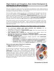
General Characteristics of <strong>Protoctista</strong>ns (<strong>Protista</strong>ns)<br />
1. unicellular eukaryotes (some multinucleate, a few loosely multicellular), not all have<br />
mitochondria (microspores, many flagellates).<br />
2. up to about 400 micrometer in size (some larger)<br />
3. all have at least one nucleus<br />
4. most are free living, but many parasitic forms including entire phyla<br />
5. motile by a variety of mechanisms but also several non-motile taxa<br />
6. many have cyst stages secreted by trophic or spore stages<br />
cysts/spores have four basic functions:<br />
• protect against unfavorable conditions<br />
• serve as sites for multiplication<br />
• assist in attachment to surfaces such as hosts<br />
• transmission stage from host to host<br />
7. all types of nutrition are exhibited by the Kingdom.<br />
• autotrophs: photosynthesis<br />
• heterotrophs (holozoic vs. saprozoic)<br />
• phagocytosis: ingestion of solid particles (e.g., bacteria)<br />
• pinocytosis: same as phagocytosis but intake of liquid<br />
• saprozoic or saprotrophy: diffusion or active transport across membrane<br />
Characteristics of “Protozoa” = <strong>Protoctista</strong>ns with animal cell characteristics<br />
The protozoa are a diverse assemblage of unicellular eukaryotic organisms having at<br />
least two animal-like properties: (1) absence of a cell wall and (2) presence of at least one<br />
motile stage in the life cycle. All functions of life are performed within the limits of a one cell<br />
membrane. Although protozoans have no organs or tissues, there is division of labor within the<br />
cytoplasm, where various complex organelles are specialized to carry out specific tasks. These<br />
organelles tend to be more specialized than those of an average cell of a multicellular organism,<br />
functioning as skeletons, locomotory systems, sensory systems, conduction mechanisms,<br />
defense mechanisms, and contractile systems.<br />
Protozoa are often called "simple" organisms. However, many are quite complex. Ciliates, for<br />
example, are not only the most complex of protozoans but also the most elaborately organized<br />
of all known cells. Protozoans are widespread ecologically, being found in fresh, marine, and<br />
brackish water and in moist soils. Some are free-living; others live as parasites or in some<br />
other symbiotic relationship.<br />
The protozoa are an artificial assemblage of organisms placed together for convenience.<br />
Traditionally, four main groups of protozoa have been recognized: flagellates, amebas, spore<br />
formers, and ciliates. These were assembled in a single phylum, Protozoa, within the kingdom<br />
Animalia. A revision adopted by the Society of Protozoologists in 1980 recognized seven<br />
separate phyla. However, more recent DNA sequence analyses of genes have shown that the<br />
protozoa represent numerous clades of varying evolutionary relationships. For example,<br />
ameboid organisms (formerly Sarcodina) fall into numerous lineages with undetermined<br />
associations. Nevertheless, the amebas comprise recognizable morphological groups, which we<br />
collect for convenience into the informal heading "Ameobas." (or “Amebas”).
<strong><strong>Cell</strong>s</strong>, <strong>Cell</strong> <strong>Division</strong> & <strong>Protoctista</strong> (<strong>Protista</strong>) <strong>Laboratory</strong> 2.6<br />
Lab #2 - Biological Sciences 102 – Animal Biology<br />
• Modern taxonomists now reject the term protozoa as an invalid<br />
taxonomic group.<br />
TAXONOMIC CLASSIFICATION OF THE “PROTOZOAN” PROTOCTISTANS<br />
The following is a condensed Linnaean classification of the protozoan groups as<br />
presently recognized.<br />
Amebas = about 12,000 different known species; locomotion by pseudopodia; body naked or<br />
with external or internal test or skeleton; asexual reproduction by fission; sexuality, if present,<br />
associated with flagellated (rarely ameboid) gametes; most free-living, some parasitic. Several<br />
groups of uncertain affinities.<br />
Rhizopodans Locomotion by lobopodia, filopodia (thin pseudopodia that may branch but<br />
do not rejoin), or by cytoplasmic flow without forming discrete pseudopodia. (The<br />
rhizopodans are divided among several clades.) Examples: Amoeba, Endamoeba, Difflugia,<br />
Arcella, Chlamydophrys.<br />
Granuloreticulosans Locomotion by reticulopodia (thin pseudopodia that branch and<br />
often rejoin). Includes foraminiferans. Examples: Globigerina, Vertebralima.<br />
Actinopodans Locomotion by axopodia (long, slender pseudopodia). Includes radiolarians.<br />
(The actinopodans are divided among several clades.) Examples: Actinophrys, Clathrulina.<br />
Phylum Euglenozoa (yu-glen-a-zo'a) (Gr. eu-, good, true, + glime, cavity, socket, + zoon,<br />
animal). Movement by flagella; cortical microtubules. Examples: Euglena,<br />
Trypanosoma. About 7500 known different species.<br />
Phylum Chlorophyta (klor-of'i-ta) (Gr. chloros, green, + phyton; plant). Unicellular and<br />
multicellular algae; photosynthetic chlorophyll pigments, flagella of equal length and<br />
smooth, mostly free-living photo autotrophs. Example: Volvox. About 7000 known species.<br />
Phylum Dinoflagellata (dy'no-fla-jel-at'a) (Gr. dinos, whirling, + flagellum, little whip).<br />
Typically with two flagella, one transverse, one trailing; body usually grooved<br />
transversely and longitudinally, each groove containing a flagellum; chromoplasts bearing<br />
chlorophyll; free-living, planktonic, parasitic, or mutualistic. Examples: Noctiluca, Ceratium,<br />
Gonyaulax. About 2000 known species.<br />
Phylum Apicomplexa (a'pi-com-plex'a) (1. apex, tip, + complex, twisted around, + a, suffix).<br />
Characteristic set of organelles (apical complex) at anterior end in some stages; cilia and<br />
flagella usually absent; all species parasitic; about 5500 known species.<br />
Class Gregarinea (gre-ga -ryn' e-a) (1. gregarius, belong to a herd or flock). Mature<br />
gamete-producing individuals large, extracellular; gametes usually alike in shape and<br />
size; parasites of digestive tract or body cavity of invertebrates; life cycle with one host.<br />
Examples: Monocystis, Gregarina.<br />
Class Coccidea (kok-sid'e-a) (Gr. kokkos, kernel, grain). Mature gamete-producing<br />
individuals small, typically intracellular; parasites mostly of vertebrates.<br />
Examples: Plasmodium, Toxoplasma, Eimeria.<br />
Phylum Ciliophora (sil-i-of' o-ra) (1. cilium, eyelash, + Gr. phora, bearing). Cilia or ciliary<br />
organelles present in at least one stage of life cycle; usually two types of nuclei; binary<br />
fission across rows of cilia; budding and multiple fission also occur; sexuality involving<br />
conjugation, autogamy, and cytogamy; heterotrophic nutrition; mostly free-living; contractile<br />
vacuole typically present. (This is a very large group, now divided into three classes and<br />
numerous orders.) Examples: Paramecium, Colpoda, Tetrahymena, Stentor, Blepharisma,<br />
Epidinium, Vorticella, Euplotes, Didinium; about 9000 different known species.
<strong><strong>Cell</strong>s</strong>, <strong>Cell</strong> <strong>Division</strong> & <strong>Protoctista</strong> (<strong>Protista</strong>) <strong>Laboratory</strong> 2.7<br />
Lab #2 - Biological Sciences 102 – Animal Biology<br />
LAB PROCEDURE<br />
NAME:<br />
LAB SCORE:<br />
Refer to chapter 3 in the Hickman text for illustrations and photos of<br />
Protozoa.<br />
Phylum Euglenozoa<br />
Your instructor will review and demonstrate how to properly prepare a wet mount.<br />
Obtain a compound microscope and prepare a wet-mount of Euglena sp. using Proto-Slo.<br />
(Make a thin ring of Proto-Slo on the slide, add a drop of the culture in the center of the ring,<br />
and carefully add the cover slip.) Examine the preparation under 10X magnification and<br />
then under 40X magnification.<br />
• After observing Euglena sp. for a few minutes, answer these questions:<br />
• What adjectives would you use to describe the movements<br />
• Can you see the flagellum Does the movement appear to correlate to the presence of<br />
flagella At which “end” (anterior or posterior) of the cell are the flagella found<br />
• Do Euglena push or pull themselves with their flagella<br />
Sketch a Euglena below and label it with the following structures (you may not clearly see all<br />
of these with your microscope so you can use a textbook or inter<strong>net</strong> picture to assist you):<br />
• cell nucleus<br />
• cell membrane<br />
• red eyespot (or stigma)<br />
• chloroplasts<br />
• contractile vacuole<br />
• flagella
<strong><strong>Cell</strong>s</strong>, <strong>Cell</strong> <strong>Division</strong> & <strong>Protoctista</strong> (<strong>Protista</strong>) <strong>Laboratory</strong> 2.8<br />
Lab #2 - Biological Sciences 102 – Animal Biology<br />
Hypermastids – the organisms in termites that actually “eat” wood….<br />
Historically, hypermastids were classified as flagellates in the Phylum Sarcomastigophora,<br />
Subphylum Mastigophora (flagellates), Order Hypermastigida (many flagella). Today many<br />
taxonomists do not recognize the Phylum Sarcomastigophora as a valid taxonomic group.<br />
Some hypermastids are now classified by taxonomists into the Phylum Axostylata due to the<br />
presence of an axostyle made of microtubules. These are symbiotic flagellates found in the<br />
intestines of termites, cockroaches and woodroaches. Hypermastids have many, many<br />
flagella and aid their hosts in the digestion of cellulose (wood).<br />
View the slide of hypermastids on the screen demo setup by your instructor at the front<br />
of the room.<br />
A termite<br />
A termite mound<br />
Answer these questions:<br />
• What type of symbiotic relationship occurs between hypermastids and a termite<br />
• How does the insect benefit these flagellates<br />
• How do the flagellates benefit the host insect<br />
• How does a young termite become infected (infaunation) with hypermastids
<strong><strong>Cell</strong>s</strong>, <strong>Cell</strong> <strong>Division</strong> & <strong>Protoctista</strong> (<strong>Protista</strong>) <strong>Laboratory</strong> 2.9<br />
Lab #2 - Biological Sciences 102 – Animal Biology<br />
Phylum Ciliophora<br />
Make a thin circle of Proto-Slo on a slide and add a drop of culture containing Paramecium<br />
caudatum. Carefully add the coverslip and examine the preparation under the 10X<br />
objective. Then view with the 40X objective.<br />
Sketch a Paramecium below and label it with the following structures (you may not clearly see<br />
all of these with your microscope so you can use a textbook or inter<strong>net</strong> picture to assist you):<br />
• cilia<br />
• oral groove<br />
• contractile vacuoles<br />
• cytostome (mouth)<br />
• cytopharynx<br />
• food vacuoles<br />
• macronucleus<br />
• micronucleus<br />
Answer these questions:<br />
• Do the paramecia swim in straight lines<br />
• Does it spin on an axis as it moves<br />
• Does it have a definite anterior (front) end<br />
• Can Paramecium swim backwards (reverse its direction)<br />
• How does Paramecium deal with a barrier (such as edge of coverslip or strand of hair or<br />
cotton) Is its body flexible enough to bend or to squeeze through tight places
<strong><strong>Cell</strong>s</strong>, <strong>Cell</strong> <strong>Division</strong> & <strong>Protoctista</strong> (<strong>Protista</strong>) <strong>Laboratory</strong> 2.10<br />
Lab #2 - Biological Sciences 102 – Animal Biology<br />
Amoebas<br />
Amebas may be naked or enclosed in a shell.<br />
Naked amebas, which include the genera Amoeba<br />
and Pelomyxa, live in both fresh water and<br />
seawater and in soil. They are bottom dwellers and<br />
must have a substratum on which to glide.<br />
Amoeba proteus is a freshwater species that is<br />
usually found in slow-moving or still-water<br />
ponds. They are often found on the underside<br />
of lily pads and other water plants. They feed<br />
on algae, bacteria, protozoa, rotifers, and other<br />
microscopic organisms.<br />
Prepare a wet mount of living Amoeba proteus and examine one under the 10X objective.<br />
Note the formation of the lobose pseudopodium (=Iobopodium) and the “protoplasmic<br />
streaming” leading to its extensions. Amoeba should be viewed in a thin depression slide<br />
with a coverslip or by placing short lengths of hair on each side of the coverslip, otherwise as<br />
the water evaporates the amebas will be crushed by the coverslip.<br />
• Regarding the endoplasm and ectoplasm, using the aid of your textbook or the<br />
inter<strong>net</strong>, write a brief description and draw a diagram to show the formation of a<br />
pseudopodium by an amoeba. This involves some important molecular biology<br />
that utilizes some of the same proteins found in animal muscle tissue that<br />
allows for muscle contraction. Be sure to include the names of these proteins in<br />
your description.<br />
• Your instructor will show you a living amoeba and video of amoeba on the screen<br />
at the front of the room.<br />
Observe the amebas and answer these questions:<br />
• Can you observe the change from endoplasm to ectoplasm Is one darker<br />
• Does an amoeba have an anterior (front) and posterior (back) side<br />
• Does the amoeba move steadily in one direction<br />
• Does more than one pseudopodium ever start to form at once<br />
• Tap the coverslip gently with the tip of a pencil. Does the amoeba respond<br />
• Do pseudopodia ever extend vertically as well as laterally
<strong><strong>Cell</strong>s</strong>, <strong>Cell</strong> <strong>Division</strong> & <strong>Protoctista</strong> (<strong>Protista</strong>) <strong>Laboratory</strong> 2.11<br />
Lab #2 - Biological Sciences 102 – Animal Biology<br />
Homework on <strong>Cell</strong> <strong>Division</strong> – due at the next lab meeting with this lab<br />
Using the inter<strong>net</strong>, textbook, lab manual and discussions with your classmates, answer<br />
the following questions.<br />
1. List four differences between mitosis and meiosis.<br />
1.<br />
2.<br />
3.<br />
4.<br />
2. In what organ(s) of a multicellular animal does meiosis take place.<br />
3. Meiosis forms sperm and egg (ova) cells. Regarding sexual reproduction in animals,<br />
why is it important that sperm and egg cells are formed by meiosis and not mitosis.<br />
4. Briefly list what occurs in a cell during each of the following phases of mitosis.<br />
PROPHASE<br />
METAPHASE<br />
ANAPHASE<br />
TELOPHASE
<strong><strong>Cell</strong>s</strong>, <strong>Cell</strong> <strong>Division</strong> & <strong>Protoctista</strong> (<strong>Protista</strong>) <strong>Laboratory</strong> 2.12<br />
Lab #2 - Biological Sciences 102 – Animal Biology<br />
LABORATORY NOTES:<br />
Mitosis