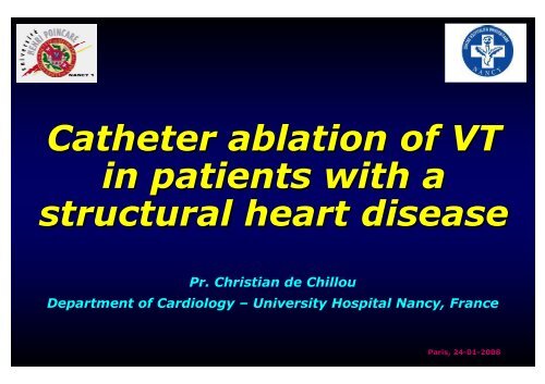
Catheter ablation of VT in patients with a structural heart disease
Catheter ablation of VT in patients with a structural heart disease
Catheter ablation of VT in patients with a structural heart disease
You also want an ePaper? Increase the reach of your titles
YUMPU automatically turns print PDFs into web optimized ePapers that Google loves.
<strong>Catheter</strong> <strong>ablation</strong> <strong>of</strong> <strong>VT</strong><br />
<strong>in</strong> <strong>patients</strong> <strong>with</strong> a<br />
<strong>structural</strong> <strong>heart</strong> <strong>disease</strong><br />
Pr. Christian de Chillou<br />
Department <strong>of</strong> Cardiology – University Hospital Nancy, France<br />
Paris, 24-01<br />
01-2008
Cl<strong>in</strong>ical <strong>VT</strong> <strong>in</strong>ducible
1<br />
GUIDELINES<br />
<strong>VT</strong> <strong>ablation</strong> <strong>in</strong> <strong>patients</strong> <strong>with</strong> a <strong>structural</strong> <strong>heart</strong> <strong>disease</strong>
Zipes DP, Camm AJ et al., J Am Coll CArdiol 2006;48:1064-1108
2<br />
GUIDELINES<br />
ICD implantation <strong>in</strong> <strong>patients</strong> <strong>with</strong> <strong>VT</strong> & <strong>heart</strong> <strong>disease</strong>
Indications de l’implantation d’un défibrillateur - Recommandations françaises<br />
Mise à jour: 21 janvier 2006<br />
Aliot E et al. Arch Mal Cœur 2006;99:141-54
Indications de l’implantation d’un défibrillateur - Recommandations françaises<br />
Mise à jour: 21 janvier 2006<br />
Aliot E et al. Arch Mal Cœur 2006;99:141-54
3<br />
CONVENTIONAL<br />
MAPPING
Activation Mapp<strong>in</strong>g<br />
Entra<strong>in</strong>ment & PPI<br />
Pace Mapp<strong>in</strong>g
Endocardial mapp<strong>in</strong>g dur<strong>in</strong>g <strong>VT</strong><br />
color-coded isochronal maps<br />
Isochronal steps = 5ms<br />
Focal <strong>VT</strong> pattern<br />
10mm<br />
Isochronal steps = 40ms<br />
Reentrant <strong>VT</strong> pattern<br />
10mm
What is the <strong>ablation</strong> target <br />
Isochronal steps = 5ms<br />
Focal <strong>VT</strong> pattern<br />
10mm<br />
Isochronal steps = 40ms<br />
Reentrant <strong>VT</strong> pattern<br />
10mm
Fibrosis & Slow Conduction<br />
AA<br />
Slow conduction perpendicular to the fiber direction <strong>in</strong> <strong>in</strong>farcted myocardial tissue is caused by a ‘’zigzag’’<br />
course <strong>of</strong> activation at high speed. Activation proceeds along pathways lengthened by branch<strong>in</strong>g and merg<strong>in</strong>g<br />
bundles <strong>of</strong> surviv<strong>in</strong>g myocytes unsheathed by collagenous septa.<br />
de Bakker JMT. et al. Circulation 1993;88:915-26
<strong>VT</strong> mechanism <strong>in</strong> relation <strong>with</strong><br />
the underly<strong>in</strong>g <strong>heart</strong> <strong>disease</strong><br />
Fibrosis <strong>VT</strong> substrate<br />
No Structural<br />
Heart Disease<br />
No<br />
Structural<br />
Heart Disease<br />
Yes<br />
Endocardial Mapp<strong>in</strong>g<br />
Focal <strong>VT</strong><br />
Reentrant <strong>VT</strong><br />
>90%
Actual significance <strong>of</strong> the <strong>VT</strong><br />
mechanism showed by mapp<strong>in</strong>g<br />
No Structural Heart Disease : focal is really focal !<br />
Structural Heart Disease : a ‘true’ focal mechanism<br />
is actually rare (10-15%) !
Activation Mapp<strong>in</strong>g<br />
Entra<strong>in</strong>ment & PPI<br />
Pace Mapp<strong>in</strong>g
Stevenson WG et al. J Am Coll Cardiol 1997;29:1180-9<br />
Waldo AL. Heart Rhythm 2004;1:94-106
Concealed entra<strong>in</strong>ment
Activation Mapp<strong>in</strong>g<br />
Entra<strong>in</strong>ment & PPI<br />
Pace Mapp<strong>in</strong>g
‘Conventional Electrophysiology’ Tools<br />
Pace mapp<strong>in</strong>g dur<strong>in</strong>g s<strong>in</strong>us rhythm
4<br />
‘HIGH-TECH’<br />
MAPPING
‘High-Tech’ Tools<br />
3D mapp<strong>in</strong>g systems
‘High-Tech’ Tools<br />
3D mapp<strong>in</strong>g systems
Without a 3D<br />
mapp<strong>in</strong>g system<br />
With a 3D<br />
mapp<strong>in</strong>g system<br />
Navigation<br />
Real-time<br />
position<strong>in</strong>g<br />
Mapp<strong>in</strong>g &<br />
Ablation
5<br />
CONTRIBUTION OF<br />
THE 12-LEAD ECG<br />
TO LOCALIZE <strong>VT</strong><br />
ORIGIN
Pour quoi faire <br />
Site d’orig<strong>in</strong>e de la TV # zone arythmogène<br />
Zone arythmogène = zone sa<strong>in</strong>e <br />
Corrélation avec cardiopathie sous-jacente
Comment faire <br />
Kuchar DL et al. J Am Coll Cardiol 1989;13:893-900<br />
de Chillou C et al. Arch Mal Coeur 2004;97(IV):13-24
Vue en OAG<br />
VR<br />
VL<br />
I<br />
III<br />
VF<br />
II
Coupe horizontale<br />
V1<br />
V2<br />
V6<br />
V5<br />
V4<br />
V3
Les sites de Josephson
TV <strong>in</strong>féro-apicale du VG post IDM
TV fasciculaire HBPG sur cœur sa<strong>in</strong>
TV morphologie BBG axe gauche méfiance !<br />
TV <strong>in</strong>féro-basale du VD sur DAVD
TV postéro-basale du VG post IDM
6<br />
TECHNICAL ASPECTS<br />
OF <strong>VT</strong> ABLATION IN<br />
PATIENTS WITH A<br />
STRUCTURAL HEART<br />
DISEASE
Bundle Branch Reentrant<br />
Ventricular Tachycardia
Bundle Branch Reentry (BBR) as<br />
the Mechanism <strong>of</strong> <strong>VT</strong><br />
Account<strong>in</strong>g for 5-6% <strong>of</strong> <strong>VT</strong> cases <strong>in</strong> CAD<br />
And up to 40% <strong>in</strong> non ischemic DCM *<br />
Diagnoses ** associated <strong>with</strong> BBR-<strong>VT</strong> (CAD excluded)<br />
- Non ischemic DCM (50-70% <strong>of</strong> all BBR-<strong>VT</strong>s)<br />
- Valvular <strong>heart</strong> <strong>disease</strong> : aortic valve replacement, mitral valve <strong>disease</strong><br />
- Muscular dystrophies, hypertrophic CM, Ebste<strong>in</strong>’s anomaly<br />
- No <strong>structural</strong> <strong>heart</strong> <strong>disease</strong> but His-Purk<strong>in</strong>je conduction delays<br />
Heart is dilated <strong>in</strong> the vast majority <strong>of</strong> cases<br />
Enlarged QRS, prolonged H to V <strong>in</strong>terval<br />
* Blanck Z et al. In Zipes DP, Jalife J Eds. Cardiac Electrophysiology: From cell to bedside, 2nd ed. Philadelphia Saunders, 1995, pp 878-85<br />
** Narasimhan C et al. Circulation 1997;96:4307-13<br />
Mer<strong>in</strong>o JL et al. Circulation 1998;98:541-6<br />
Blanck Z et al. J Cardiovasc Electrophysiol 1993;4:253-62<br />
Blanck Z et al. J Am Coll Cardiol 1993;22:1718-22<br />
Li YG et al. J Cardiovasc Electrophysiol 2002;13:1233-9
Ablation <strong>of</strong> Bundle Branch Reentrant <strong>VT</strong><br />
X
EP characteristics <strong>of</strong> BBR-<strong>VT</strong><br />
H and RB potentials<br />
preced<strong>in</strong>g V <strong>with</strong><br />
appropriate sequence<br />
accord<strong>in</strong>g BBR-<strong>VT</strong> type<br />
(A,B or C)<br />
HV <strong>in</strong>terval identical or<br />
10-30 ms longer than<br />
dur<strong>in</strong>g s<strong>in</strong>us rhythm<br />
(type B excepted)<br />
H-H variations preced<strong>in</strong>g<br />
V-V variations<br />
Induction depend<strong>in</strong>g<br />
upon a critical delay <strong>in</strong><br />
the HPS<br />
Term<strong>in</strong>ation by block <strong>in</strong><br />
the HPS<br />
Short-long-short<br />
sequences frequently<br />
required to <strong>in</strong>duce<br />
Non <strong>in</strong>ducibility after RBB<br />
<strong>ablation</strong>
RV apical stimulation to measure<br />
PPI <strong>in</strong> suspected BBR-<strong>VT</strong><br />
Mer<strong>in</strong>o JL et al. Circulation 2001;103:1102-8
Ventricular Tachycardia not<br />
Related to a Bundle Branch<br />
Reentry Mechanism
Substrate mapp<strong>in</strong>g<br />
<strong>VT</strong> mapp<strong>in</strong>g<br />
Ablation results
1<br />
Correlation between voltage mapp<strong>in</strong>g<br />
and histology <strong>in</strong> myocardial <strong>in</strong>farction<br />
Cut<strong>of</strong>f values:<br />
>1.5mV = healthy /
1<br />
University Hospital, Nancy<br />
Departments <strong>of</strong> Cardiology & Radiology
1<br />
Comparison between 3D MRI <strong>in</strong>farct<br />
reconstruction and 3D CARTOC<br />
ARTO mapp<strong>in</strong>g<br />
Improved <strong>in</strong>farct border<br />
del<strong>in</strong>eation <strong>in</strong> areas <strong>with</strong><br />
poor catheter contact<br />
Increased accuracy <strong>of</strong> LV<br />
geometry reconstruction<br />
Normal<br />
Subendocardial to transmural Subepicardial Intramural
2<br />
ARVD/C &<br />
Voltage mapp<strong>in</strong>g<br />
11/11 <strong>patients</strong> (100%)<br />
<strong>with</strong> ARVD / C & <strong>VT</strong><br />
showed RV areas <strong>with</strong><br />
low bipolar voltage .<br />
Latero-basal RV = 10/11<br />
(±<strong>in</strong>fero-basal)<br />
RVOT = 6/11<br />
RV apex = 0/11<br />
Miljoen H, de Chillou C et al.<br />
Europace 2005;7:516-24<br />
Boulos M et al. J Am Coll Cardiol 2001;38:2020-7<br />
Similar results: Marchl<strong>in</strong>ski FE et al. Circulation 2004;110:2293-8<br />
Satomi K et al. J Cardiovasc Electrophysiol 2006;17:469-76<br />
RV apex <strong>in</strong>volved <strong>in</strong> 2/7 <strong>patients</strong><br />
Apex always spared
3<br />
LV 3D map reconstruction<br />
<strong>with</strong> tissue characterization<br />
<strong>in</strong> idiopathic DCM
3<br />
Idiopathic DCM &<br />
Voltage mapp<strong>in</strong>g<br />
Hsia HH et al. Circulation 2003;108:704-10
3<br />
Idiopathic DCM &<br />
Voltage mapp<strong>in</strong>g<br />
6/6 <strong>patients</strong> (100%)<br />
<strong>with</strong> idiopathic DCM<br />
showed no low bipolar<br />
voltage areas (personnal data)
3<br />
Idiopathic DCM &<br />
Voltage mapp<strong>in</strong>g<br />
Cesario DA et al. Heart Rhythm 2006;3:1-10
Substrate mapp<strong>in</strong>g<br />
<strong>VT</strong> mapp<strong>in</strong>g<br />
Ablation results
1<br />
Endocardial reentrant circuit <strong>with</strong> a protected<br />
p<br />
isthmus <strong>in</strong> >90% <strong>of</strong> post-MI related mappable <strong>VT</strong>s
1<br />
Post-<strong>in</strong>farct<br />
mappable <strong>VT</strong> <strong>ablation</strong><br />
Step # 1 = substrate mapp<strong>in</strong>g
1<br />
Post-<strong>in</strong>farct<br />
mappable <strong>VT</strong> <strong>ablation</strong><br />
Step # 2 = <strong>VT</strong> <strong>in</strong>duction & <strong>VT</strong> mapp<strong>in</strong>g
1<br />
Post-<strong>in</strong>farct<br />
mappable <strong>VT</strong> <strong>ablation</strong><br />
Step # 2 = <strong>VT</strong> <strong>in</strong>duction & <strong>VT</strong> mapp<strong>in</strong>g
1<br />
Post-<strong>in</strong>farct<br />
mappable <strong>VT</strong> <strong>ablation</strong><br />
Step # 3 = <strong>VT</strong> circuit reconstruction
1<br />
Post-<strong>in</strong>farct<br />
mappable <strong>VT</strong> <strong>ablation</strong><br />
Step # 4 = <strong>VT</strong> protected isthmus del<strong>in</strong>eation<br />
Double potentials l<strong>in</strong>e<br />
Double potentials l<strong>in</strong>e<br />
MV annulus<br />
Protected <strong>VT</strong> isthmus
1<br />
Post-<strong>in</strong>farct<br />
mappable <strong>VT</strong> <strong>ablation</strong><br />
Step # 5 = Ablation isthmus ‘transection’<br />
Ablation l<strong>in</strong>es
1<br />
Coumel’s Triangle<br />
Trigger<br />
Substrate<br />
ANS
1<br />
Case Report<br />
• 70 year-old man<br />
• Chronic AFib rate control (severe hyperthyroïdism <strong>with</strong> amiodarone)<br />
• Acute <strong>in</strong>fero-lateral myocardial <strong>in</strong>farction (04/2000)<br />
• No acute revascularization procedure (late <strong>in</strong>-hospital admission)<br />
• LVEF = 0.55 - One vessel <strong>disease</strong> = Cx occluded delayed PTCA<br />
• Documented syncopal <strong>VT</strong> (220/mn) ICD implantation (03/2001)<br />
• Many fast <strong>VT</strong> (>200/mn) recurrences efficiently treated by ATP<br />
• Nadolol added: 80mg/d<br />
• July 2001 = fast <strong>VT</strong>s (240/mn) <strong>VT</strong> storms : 12 shocks / 5 days
A B C D<br />
A<br />
B<br />
C<br />
D<br />
15mm
1<br />
Pac<strong>in</strong>g Artifact
1<br />
RF onset
1<br />
RF <strong>of</strong>fset
1<br />
Myocardial <strong>in</strong>farction : late phase<br />
Mode <strong>of</strong> <strong>in</strong>itiation and <strong>ablation</strong> <strong>of</strong> VF storms <strong>in</strong> <strong>patients</strong> <strong>with</strong> ischemic cardiomyopathy<br />
Marrouche N. et al., J Am Coll Cardiol 2004;43:1715-20
1<br />
Post-<strong>in</strong>farct<br />
non mappable <strong>VT</strong> <strong>ablation</strong><br />
Different possible empiric approaches
1<br />
Post-<strong>in</strong>farct non mappable <strong>VT</strong> <strong>ablation</strong><br />
non contact mapp<strong>in</strong>g
1<br />
Post-<strong>in</strong>farct<br />
non mappable <strong>VT</strong> <strong>ablation</strong><br />
Step # 1 = substrate mapp<strong>in</strong>g & pace mapp<strong>in</strong>g
1<br />
Post-<strong>in</strong>farct<br />
non mappable <strong>VT</strong> <strong>ablation</strong><br />
Step # 2 = match<strong>in</strong>g w. cl<strong>in</strong>ical <strong>VT</strong> 12-lead ECG
1<br />
Post-<strong>in</strong>farct<br />
non mappable <strong>VT</strong> <strong>ablation</strong><br />
Step # 3 = Rank<strong>in</strong>g the match<strong>in</strong>g mapp<strong>in</strong>g sites
1<br />
Post-<strong>in</strong>farct<br />
non mappable <strong>VT</strong> <strong>ablation</strong><br />
Step # 4 = <strong>VT</strong> protected isthmus def<strong>in</strong>ition
2<br />
Endocardial reentrant circuit = 66% <strong>of</strong> ARVD/C<br />
related mappable <strong>VT</strong>s<br />
Miljoen H, de Chillou C et al. Europace 2005;7:516-24
2<br />
Peri-tricuspid reentrant <strong>VT</strong> : a frequent<br />
mechanism <strong>in</strong> ARVD/C reentrant <strong>VT</strong>s (5 <strong>of</strong> 12 <strong>VT</strong>s)<br />
Miljoen H, de Chillou C et al. Europace 2005;7:516-24
3<br />
Non BBR-related <strong>VT</strong> mapp<strong>in</strong>g <strong>in</strong><br />
<strong>patients</strong> <strong>with</strong> idiopathic DCM
3<br />
Non BBR-related <strong>VT</strong> mapp<strong>in</strong>g <strong>in</strong><br />
<strong>patients</strong> <strong>with</strong> idiopathic DCM<br />
Swarup V et al. J Cardiovasc Electrophysiol 2002;13:1164-8
3<br />
Non BBR-related <strong>VT</strong> mapp<strong>in</strong>g <strong>in</strong><br />
<strong>patients</strong> <strong>with</strong> idiopathic DCM<br />
Swarup V et al. J Cardiovasc Electrophysiol 2002;13:1164-8
3<br />
Non BBR-related <strong>VT</strong> mapp<strong>in</strong>g <strong>in</strong><br />
<strong>patients</strong> <strong>with</strong> idiopathic DCM<br />
Swarup V et al. J Cardiovasc Electrophysiol 2002;13:1164-8
Epicardial approach<br />
for <strong>VT</strong> <strong>ablation</strong><br />
Sosa E et al. J Cardiovasc Electrophysiol 2005;16:449-52
Substrate mapp<strong>in</strong>g<br />
<strong>VT</strong> mapp<strong>in</strong>g<br />
Ablation results
<strong>VT</strong> <strong>ablation</strong> <strong>in</strong> <strong>patients</strong> <strong>with</strong> a<br />
<strong>structural</strong> <strong>heart</strong> <strong>disease</strong><br />
Literature review (case reports excluded)<br />
Results for both <strong>ablation</strong> <strong>of</strong> stable and unstable <strong>VT</strong>s are shown<br />
Heart <strong>disease</strong><br />
Studies<br />
Years<br />
Patients<br />
Acute success<br />
Mean FU (mo)<br />
Recurrences *<br />
Post-MI<br />
29 1993 - 2007 1093<br />
76%<br />
19.2<br />
30%<br />
ARVD/C<br />
11 1998 - 2007 211<br />
72%<br />
31.5<br />
33%<br />
Idiopathic DCM<br />
6 1995 - 2006 77<br />
73%<br />
13<br />
41%<br />
* Global recurrence rate, mix<strong>in</strong>g <strong>patients</strong> <strong>with</strong> a successful <strong>ablation</strong> and those <strong>with</strong> a failed one
<strong>VT</strong> <strong>ablation</strong> : review <strong>of</strong> the literature<br />
(case reports excluded)<br />
Successful <strong>ablation</strong><br />
Failed <strong>ablation</strong><br />
O’Donnell D et al. Eur Heart J 2002;23:1699-1705
<strong>VT</strong> <strong>ablation</strong> : review <strong>of</strong> the literature<br />
(case reports excluded)<br />
Segal OR et al. Heart Rhythm 2005;2:474-82
1<br />
85%<br />
Antero-apical circuits<br />
LV lateral wall<br />
LV septum<br />
Apex<br />
A1 A2 A3 A4<br />
15%<br />
Septal circuits<br />
Anterior wall<br />
Septal circuits<br />
Anterior wall<br />
Mitral<br />
Aorta<br />
Mitral Mitral<br />
Aorta<br />
Mitral<br />
Aorta<br />
Mitral<br />
Aorta<br />
Inferior<br />
Wall<br />
Septum<br />
Apex<br />
S1 S2 S3 S4
1<br />
60%<br />
Peri-mitral circuits<br />
LV lateral wall<br />
Mitral<br />
Mitral<br />
Mitral<br />
Mitral<br />
Apex<br />
Inferior wall<br />
M1 M2 M3 M4<br />
40%<br />
Inferior<br />
Wall Mitral Septum<br />
Mitral<br />
Mitral<br />
Infero-lateral circuits<br />
Mitral<br />
Mitral<br />
Mitral<br />
Apex<br />
I1<br />
I2<br />
I3 I4 I5 I6
1<br />
160 ms<br />
152 ms<br />
-162 ms -170 ms<br />
Site RF<br />
Site RF<br />
15 mm 15 mm<br />
I<br />
II<br />
III<br />
VR<br />
VL<br />
VF<br />
V1<br />
V2<br />
V3<br />
V4<br />
V5<br />
V6<br />
RVA<br />
332 ms<br />
1 sec<br />
de Chillou C et al. Circulation 2002;105:726-31<br />
332 ms
2<br />
<strong>VT</strong> <strong>ablation</strong> <strong>in</strong> <strong>patients</strong> <strong>with</strong> ARVD/C
3<br />
<strong>VT</strong> <strong>ablation</strong> <strong>in</strong> <strong>patients</strong> <strong>with</strong> Idiopathic DCM
4<br />
Complications <strong>of</strong> <strong>VT</strong> Ablation <strong>in</strong> Patients<br />
<strong>with</strong> Structural Heart Disease<br />
Literature data = ma<strong>in</strong>ly post-<strong>in</strong>farct <strong>VT</strong><br />
Acute complications = 7.4% !!<br />
Death = 0.9%<br />
Transient Ischemic Attack or Stroke = 0.9%<br />
Pericardial Effusion or Tamponade = 1.8%


