C - Eureka Smile Center
C - Eureka Smile Center
C - Eureka Smile Center

You also want an ePaper? Increase the reach of your titles
YUMPU automatically turns print PDFs into web optimized ePapers that Google loves.
I technique_ giomers<br />
Esthetics, caries control &<br />
gingival health with a versatile<br />
giomer composite system<br />
Author_Jack D. Griffin Jr., DMD, MAGD<br />
Fig. 1_Patient concerned with<br />
“swollen gums” and holes in teeth.<br />
Fig. 2_He had braces removed the<br />
previous year and said he never<br />
smiled.<br />
Fig. 3_He had generalized sensitivity<br />
to sweets and cold. Composites were<br />
to be done on 23 teeth over over<br />
appointments.<br />
Fig. 4_He did not brush “very often”<br />
and really liked soda and energy<br />
drinks. He seemed committed to<br />
improving things in his mouth.<br />
Fig. 5_Diode laser gingivoplasty was<br />
done after local anesthetic with low<br />
wattage and brush strokes.<br />
Fig. 6_Preparations were done as<br />
conservatively as possible with a 330<br />
bur, finish diamond and slow-speed<br />
round bur. Caries indicator was used<br />
to verify decay removal.<br />
_There are many direct composite materials<br />
that have the strength to be used in the posterior<br />
and a level of esthetics acceptable in the anterior<br />
dentition. 1 Direct anterior composites must meet the<br />
minimum cosmetic demands of the patient while<br />
posterior restorations must provide resistance to<br />
mechanical forces. 2–5<br />
In patients with questionable hygiene, dietary<br />
habits and a history of caries, we must also choose<br />
materials that have properties such as fluoride release<br />
and high polishablility to decrease the effects<br />
of a less than ideal oral environment. 6<br />
A composite system has been developed with a<br />
surface pre-reacted glass ionomer (S-PRG) that has<br />
shown to have high esthetic properties as well as<br />
significant release of fluoride ions into the dentin and<br />
re-chargeability. 7,8<br />
Beautifil II and Beautifil Flow Plus (Shofu, San<br />
Marcos, Calif.) is a universal nano-hybrid composite<br />
with a glass ionomer component to meet the esthetic<br />
demands of the profession while giving long-term<br />
tissue health by sustained fluoride release from the<br />
S-PRG particles. 9 A potential reduction of secondary<br />
caries and maintenance of surface luster has been<br />
shown long-term with this material. 10,11<br />
In addition to the plaque and caries reducing<br />
properties of this material, the small nano-sized filler<br />
size of 10–20 nm, along with a filler load by weight<br />
of 83 percent, make it suitable for almost any clinical<br />
situation, including incisal edge replacement and<br />
posterior restorations in occlusion. 12 The versatility of<br />
these products, coupled with the giomer technology,<br />
make them unique in the dental marketplace and a<br />
strong choice in almost any clinical situation.<br />
The flowable comes in two viscosities with a varying<br />
degree of flowability, each with similar physical<br />
properties strong enough to withstand occlusal<br />
forces in posterior restorations. 13 The zero flow, F00,<br />
is a stackable flow with almost no movement or<br />
slump when syringing. The low flow, F03, is a great<br />
universal flow material with handling similar to other<br />
more viscous flowables on the market.<br />
Fig. 1 Fig. 2 Fig. 3<br />
Fig. 4 Fig. 5 Fig. 6<br />
32 I<br />
cosmetic<br />
dentistry 1_2012
technique_ giomers I<br />
Fig. 7 Fig. 8 Fig. 9<br />
Fig. 10 Fig. 11 Fig. 1<br />
Will these flowable materials replace each of the<br />
more viscous composite materials No. The sculptability<br />
and layering potential of conventional composites<br />
will always have a place in esthetic dentistry.<br />
Another advantage of a non-flowable material is void<br />
reduction in posterior composites as uncured flowable<br />
is followed by a more viscous material pushing<br />
out and displacing the flowable as the viscous compule<br />
material is injected.<br />
_Patient exam and planning<br />
A 15-year-old male came to the office with an<br />
“unpleasant” smile after having orthodontic treatment<br />
the previous year (Fig. 1). His primary concern<br />
was the hypertrophic tissue around his incisors and<br />
his cold and sweet sensitivity (Fig. 2). There was rampant<br />
decay, enlarged gingival tissue, poor hygiene<br />
and decalcification areas (Figs. 3, 4).<br />
A full series of radiographs and photographic images<br />
were taken for treatment planning, marketing<br />
and case documentation. These images were studied,<br />
along with clinical exam notes, before treatment so<br />
that a basic plan was formulated. 14<br />
A treatment plan was made to do 23 direct composite<br />
restorations over three appointments after a<br />
prophy, oral hygiene education and tray-delivered<br />
home topical fluoride delivery. The plan included<br />
laser gingivoplasty followed by restorations with<br />
Beautifil II because of its esthetics, ease of use, fluoride<br />
release and versatility.<br />
After several weeks of maintained oral hygiene<br />
improvement, the surface of the anterior teeth would<br />
be re-contoured and enhanced at no additional<br />
charge.<br />
_Soft-tissue enhancement<br />
Lasers have become a critical component of smile<br />
rehabilitations, and if done with respect to periodontal<br />
tissues and biologic width, results can be a<br />
great enhancement to cosmetic treatment. 15,16 Diode<br />
lasers offer excellent control of tissue sculpting with<br />
very predictable healing and tissue tolerance as long<br />
as sound biologic principles are followed. 17–19 These<br />
principles must be understood during treatment in<br />
order to prevent possible chronic periodontal inflammation<br />
and unwanted gingival responses such as<br />
redness, bleeding and irritation. 20,21<br />
On the first restorative appointment, a local anesthetic<br />
(Septocaine, Septodont USA, Lancaster, Pa.)<br />
was given and retractors (See More, Discus, Culver<br />
City, Calif.) were placed to keep the lips out of the way<br />
and to provide some isolation from saliva. An 810 mm<br />
diode laser (Odyssey, Ivolcar Vivadent, Amherst, N.Y.)<br />
was used on a relatively low wattage, 2.0, to sculpt<br />
the tissues and remove hyperplastic gingival tissue<br />
(Fig. 5). 22<br />
Clean up and removal of the charred tissue was<br />
done with a microbrush and hydrogen peroxide. It is<br />
expected that the new soft-tissue location would be<br />
maintained or even improve with properly contoured<br />
restorations, good surface polish and continued<br />
plaque control. 23<br />
_Giomer composite technique: maxillary<br />
The goal of this one-hour restorative appointment<br />
was to provide improved esthetics and an<br />
environment to promote tissue recovery on the<br />
maxillary anterior. Tooth preparation was done with<br />
Fig. 7_Contour Matrices were placed<br />
and a BAC containing 37 percent<br />
phosphoric acid etch was placed and<br />
rinsed.<br />
Fig. 8_Several coats of a universal<br />
bonding agent were applied and air<br />
thinned.<br />
Fig. 9_Beautifil Flow Plus was<br />
applied to cover all dentin and then<br />
cured.<br />
Fig. 10_The Contour Matricies form<br />
a good gingival seal keeping the<br />
crevicular fluids out while giving<br />
anatomical form to the material.<br />
Fig. 11_The remaining cavities were<br />
restored with the same flowable<br />
material. Notice how the material<br />
holds its shape without running even<br />
before curing.<br />
Fig. 12_Light polymerization was<br />
done from facial, lingual and incisal<br />
to insure a high level of conversion.<br />
(Images/Provided by Dr. Jack D.<br />
Griffin, Jr.)<br />
cosmetic<br />
dentistry 1_2012<br />
I 33
I technique_ giomers<br />
Fig. 13 Fig. 14<br />
Fig. 15<br />
Fig. 16 Fig. 17<br />
Fig. 18<br />
Fig. 13_Initial shaping was done<br />
with a finish diamond on high<br />
speed with water, making sure<br />
that the width and embrasures<br />
were adequate before restoring the<br />
adjacent teeth.<br />
Fig. 14_Other teeth were prepared<br />
and restored in the same way.<br />
Fig. 15_One-hundred percent of the<br />
restorations at this time were done<br />
with flowable material that has high<br />
fluoride release and re-chargeability.<br />
Fig. 16_At the end of the first<br />
appointment, contouring was done<br />
but polishing was minimal because<br />
of time.<br />
Fig. 17_Impressions were taken and<br />
bleach type trays were given with<br />
topical fluoride. The goal was to keep<br />
the giomer material recharged for<br />
optimal healing.<br />
Fig. 18_At the second appointment,<br />
two weeks after initial treatment,<br />
the gingiva showed marked<br />
improvement.<br />
a 330 bur, finish diamond and a #2 slow-speed round<br />
bur (Fig. 6). Caries indicator (Sable Seek, Ultradent,<br />
South Jordan, UT) was used to verify complete caries<br />
elimination and a long irregular bevel was placed on<br />
all enamel margins. Two teeth were done at a time.<br />
A contoured anatomical matrix (Contour Matrix,<br />
Ivoclar Vivadent, Amherst, N.Y.) was placed and<br />
wedged loosely. The matrix extends slightly, providing<br />
a “sulcular seal” aiding in marginal integrity.<br />
These matrices not only increase restoration longevity<br />
but also greatly increase placement efficiency. 24<br />
The teeth were etched with 37 percent phosphoric<br />
acid (Etch 37, Bisco, Schaumburg, Ill.) for 10 seconds,<br />
rinsed thoroughly and left damp (Fig. 7). This etch<br />
contains benzalkonium chloride (BAC) for a continued<br />
antimicrobial effect. A universal bonding agent<br />
(All Bond Universal, Bisco, Schaumburg, Ill.) was applied<br />
in several layers, air thinned and cured (Fig. 8). 25<br />
Beautifil Flow Plus, low flow, was placed into the<br />
preps covering all dentin and cured for 15 seconds<br />
(Fig. 10). The non-runny nature of this material and<br />
the great adaptability make this a great choice for<br />
dentin replacement.<br />
The remaining preparation was restored with the<br />
same material so that the entire restoration was done<br />
in this flowable nano-hybrid composite in a single<br />
shade of A2 (Fig. 11). 26<br />
Curing was then done for 20 seconds from the<br />
facial, lingual and incisal to insure complete polymerization<br />
(Fig. 12). Initial contouring was done quickly<br />
with a finishing diamond (Diatech Direct, Charleston,<br />
S.C.) to provide basic anatomical shaping. 27<br />
Tooth #8 was prepped, caries removed and restored<br />
in a similar fashion (Fig. 14). Because of the size<br />
and depth of this restoration, the flowable material<br />
was placed and cured in three different increments<br />
(Fig. 15). The remaining anterior teeth were restored<br />
and shaped (Fig. 16). Minimal polishing was done at<br />
this time because of time constraints.<br />
Alginate impressions were taken and flexible<br />
bleach-type trays were made. The high fluoride release<br />
and ability to be recharged make this giomer<br />
an ideal product in less than ideal oral environments<br />
such as this. 28<br />
The potential of recurrent decay and the plaque<br />
formation that may compromise the gingival recontouring<br />
may be lessened by the sustained antimicrobial<br />
effect of this material.<br />
0.4 percent stannous fluoride was given to the<br />
patient with instructions to place in the mouth overnight<br />
two to three times per week until all restorative<br />
work was completed. At that time, re-charging of the<br />
giomer material would continue between prophy<br />
appointments at once a week (Fig. 17).<br />
_Second appointment: mandibular<br />
restorations<br />
Two weeks later, a two-hour appointment was<br />
made to restore the mandibular teeth. The gingival<br />
health on the maxilla was consistent with better<br />
hygiene on restored teeth and bacterial control (Figs.<br />
18, 19). The patient claimed to use the fluoride trays<br />
two to three times a week and was brushing at least<br />
once a day.<br />
The patient’s right side was done first focusing on<br />
facial decay initially (Fig. 19). Cavity preparation, caries<br />
removal and enamel beveling as described above<br />
was completed with various burs. On the right bicuspid,<br />
a pinkish blush was noticed after decay removal<br />
34 I<br />
cosmetic<br />
dentistry 1_2012
xxxx_ giomers I<br />
Fig. 19 Fig. 20 Fig. 21<br />
Fig. 22 Fig. 23 Fig. 24<br />
with no obvious pulpal exposure (Fig. 20).<br />
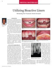
A bioactive liner (TheraCal, Bisco, Schaumburg,<br />
Ill.) was placed in a thin layer to stimulate secondary<br />
dentin formation and to seal deep pulpal dentin. This<br />
material was light cured and kept 2 mm away from<br />
restoration margins (Fig. 21). All facial lesions were<br />
restored using selective etching, a universal bonding<br />
agent and Beautifil Flow Plus as described above.<br />
The matrices used varied. In the anterior, Mylar<br />
strips were used and held in place with a plastic instrument<br />
while the flowable material was light cured.<br />
Where a strip had trouble going through the contact,<br />
a FenderWedge (Garrison Dental, Spring Lake, Mich.)<br />
was used (Fig. 22). Shaping was done with a finish<br />
diamond and flame-shaped finish bur.<br />
In the posterior, the teeth were isolated with a suction,<br />
light, bite-block system (Isolite, Santa Barbara,<br />
Calif.). Conservative preparations were done with a<br />
330 bur and sectional matrices placed with a wedge<br />
and 3-D ring system (Garrison Dental, Spring Lake,<br />
Mich.) to insure tight, broad contacts (Fig. 23).<br />
After etching and bonding, a 0.5 mm layer of the<br />
giomer flowable was placed on the pulpal floor and<br />
cured creating a good polymerized layer protecting<br />
the pulp. A small amount of flowable was added<br />
again, left un-polymerized and the more viscous<br />
Beautifil II forced the flowable into all areas of voids<br />
leaving a dense fill (Fig. 24).<br />
Shaping was done with a #6 round bur and a<br />
football-shaped finish bur leaving excellent margins<br />
and contacts (Fig. 25). All restorations were completed<br />
on the right and then the Isolite was moved<br />
to the left side and restorations done in the same<br />
manor (Fig. 26).<br />
_Final restorative appointment:<br />
maxillary enhancement<br />
Three months after the initial flowable placement<br />
on the maxillary teeth, the patient returned still<br />
showing soft-tissue and hygiene improvement. This<br />
1.5 hour appointment was completed to enhance<br />
the maxillary anterior teeth and complete several<br />
posterior maxillary restorations.<br />
Using a finish diamond with water on a highspeed<br />
handpiece, the surfaces were roughened in an<br />
irregular way to provide depth to the re-surfacing<br />
(Fig. 27). All preparation was done away from the<br />
gingiva about 3 mm and the surface left wavy (Fig.<br />
28). The materials used on the anterior were a universal<br />
bonding agent and etch, and both versions of<br />
the giomer material (Fig. 29).<br />
The surfaces were etched with 37 percent phosphoric<br />
acid for 10 seconds, rinsed well and dried. This<br />
left a roughened, frosty appearance to the Beautifil<br />
Flow (Fig. 31).<br />
The All Bond Universal has a high bond strength<br />
to polymerized composite without an air-inhibited<br />
layer and was applied in several coats and air thinned.<br />
(Fig. 32) Mylar strips were placed and the Beautifl<br />
Flow Plus low flow was placed in the center of the<br />
tooth and left uncured.<br />
This material helps to wet the surface and decrease<br />
void as the more viscous material was applied<br />
directly over it. A1 Beautifil from a compule was<br />
applied to each tooth, sculpted and cured using a<br />
free-handed technique. 29<br />
Most of the less viscous flowable was wiped<br />
away with the plastic instrument. The lighter surface<br />
Fig. 19_The lower quadrants were<br />
done at this appointment starting<br />
with the right side. Facials were done<br />
first, followed by interproximal to<br />
reduce the chance of bleeding.<br />
Fig. 20_After complete caries<br />
removal, pink blush was noticed.<br />
Fig. 21_Before bonding, an apetite<br />
promoting liner was placed to<br />
stimulate dentin bridge formation.<br />
Fig. 22_Various matrices were<br />
used, including mylar strips and a<br />
wedge-sectional combination called<br />
a FenderWedge where contacts were<br />
very tight.<br />
Fig. 23_Isolation in the posterior was<br />
done with an Isolite and the teeth<br />
were prepped. Sectional matrices<br />
were placed along with a 3-D ring.<br />
Fig. 24_Etching, rinsing and flowable<br />
liner were completed followed by<br />
Beautifil II from a compule.<br />
cosmetic<br />
dentistry 1_2012<br />
I 35
I technique_ giomers<br />
Fig. 25 Fig. 26 Fig. 27<br />
Fig. 28 Fig. 29 Fig. 30<br />
Fig. 25_The restorations show<br />
excellent margins with broad, deep,<br />
tight contacts.<br />
Fig. 26_Shaping was completed<br />
with a finish diamond, finish bur and<br />
disks.<br />
Fig. 27_At the third and final<br />
restorative appointment, the<br />
maxillary anteriors were enhanced.<br />
With water, a finish diamond was<br />
used to roughen the surface of the<br />
Beautifil Flow Plus from the first<br />
appointment.<br />
Fig. 28_The surface was irregular<br />
and kept 3 mm from the gingiva.<br />
Fig. 29_The materials used during<br />
this enhancement included 37<br />
percent etch, a universal bonding<br />
agent, flowable and more viscous<br />
composite.<br />
Fig. 30_The teeth were isolated with<br />
retractors, etched for 10 seconds<br />
and rinsed thoroughly.<br />
shade brightens the smile while giving vitality to<br />
the restorations that were placed over the darker A2<br />
shade from the first restorative appointment. Initial<br />
contouring was done with a finish diamond on high<br />
speed with water. 30–32<br />
Shaping and polishing was completed with a<br />
flexible disk system (Super Snap Rainbow, Shofu, San<br />
Marcos, Calif.). This system features a sequence of<br />
very thin, flexible disks that are very efficient at shaping<br />
embrasures, final shaping and high polish without<br />
metal in the center that may gouge or scratch the<br />
restoration surface (Fig. 34). The giomer material is<br />
easily polished and rivals many nano-hybrids on the<br />
market today. 33<br />
_Results<br />
Obviously, a great improvement was realized for<br />
a boy who said he “never smiled until now” (Fig. 37).<br />
It would be naïve to think that these restorations will<br />
last him his entire life without the need for more definitive<br />
porcelain restorations or other cosmetic procedure.<br />
However, the improvement in self-esteem,<br />
the decrease in sensitivity and the feeling of better<br />
oral health may help to stimulate him to be committed<br />
to better oral care.<br />
After 6 months, the improvement in soft- and<br />
hard-tissue health is undeniable (Fig. 38). The attached<br />
tissue stippling and lack of bleeding clearly<br />
shows how well the soft tissues tolerate these materials.<br />
The giomer materials have excellent esthetics<br />
and strength, which combined with the high longterm<br />
fluoride release make these materials a strong<br />
consideration in most all direct restorative cases. The<br />
patient has continued fluoride treatments at home<br />
on average about every one to two weeks, as often as<br />
he can remember. Now, if we can just keep him from<br />
losing his trays for the third time._<br />
_References<br />
1. Vargas M. Conservative aesthetic enhancement<br />
of the anterior dentition using a predictable<br />
direct resin protocol. Prac Proced Aesthet<br />
Dent.2006;18(8):501–507.<br />
2. Fahl, N. The direct/indirect composite resin<br />
veneers: a case report. Pract Periodont Aesthet<br />
Dent. 1996;8(7):627–638.<br />
3. Milnar F. A minimal intervention approach to the<br />
treatment of a class IV fratcture. J of Cosmet Dent<br />
21(4):106–112;2006.<br />
4. Christensen GJ. Bonding to dentin and enamel<br />
where does it stand in 2005 J Am Dent Assoc.<br />
136(9):1299–1302;2005<br />
5. Terry DA. Direct composite resin restoration of<br />
adolescent class IV tooth fracture: a case report.<br />
Prac Perio Aesthet Dent. 12(1):23–29;2000.<br />
6. Wiegand A, Buchalla W, Attin T. Review on fluoride-releasing<br />
restorative materials—fluoride<br />
release and uptake characteristics, antibacterial<br />
activity and influence on caries formation. Dent<br />
Mater. 2007 Mar;23(3):343–362. Epub 2006<br />
Apr 17.<br />
7. Ikemura K, Tay FR, Endo T, Pashley DH. A review<br />
of chemical-approach and ultramorphological<br />
studies on the development of fluoride-releasing<br />
dental adhesives comprising new prereacted<br />
glass ionomer (PRG) fillers. Dent Mater J.<br />
2008 May;27(3):315–339.<br />
36 I<br />
cosmetic<br />
dentistry 1_2012
LVI GLOBAL
I technique_ giomers<br />
Fig. 31 Fig. 32 Fig. 33<br />
Fig. 34 Fig. 35<br />
Fig. 36<br />
Fig. 31_The surface was rough and<br />
frosty in appearance.<br />
Fig. 32_The universal bonding<br />
agent bonds very well to a previously<br />
polymerized composite surface,<br />
is clear and has a very low film<br />
thickness.<br />
Fig. 33_Mylar strips were placed<br />
and the A1 Beautifil II was injected<br />
onto uncured flowable on each tooth.<br />
Sculpting was done with a plastic<br />
instrument and cured.<br />
Fig. 34_Initial contouring was done<br />
with a finish diamond, which was<br />
followed by Super Snap disks.<br />
Fig. 35_The black disk was used<br />
with light pressure to improve form.<br />
The embrasures were primarily<br />
formed with the purple disk.<br />
Fig. 36_The green and red disks<br />
complete the polish.<br />
Fig. 37_Excellent hard- and<br />
soft-tissue result with a great<br />
improvement in oral health.<br />
Fig. 38_The smile is much improved<br />
as the patient continues at home<br />
fluoride use and brushing.<br />
Fig. 37<br />
8. Itota T, Carrick TE, Yoshiyama M, McCabe JF.<br />
Fluoride release and recharge in giomer, compomer<br />
and resin composite. Dent Mater. 2004<br />
Nov;20(9):789–795.<br />
9. Gordan VV, Mondragon E, Watson RE, Garvan C,<br />
Mjör IA. A clinical evaluation of a self-etching<br />
primer and a giomer restorative material: results<br />
at eight years. J Am Dent Assoc. 2007<br />
May;138(5):621–627.<br />
10. Valeria V. Gordan, DDS, MS; Eduardo Mondragon;<br />
Ronald E. Watson, DDS, MAE; Cyndi Garvan, PhD;<br />
Ivar A.Mjör, BDS, MSD, MS, Dr.odont. JADA, Vol.<br />
138, May 2007.<br />
11. Jyothi K, Annapurna S, Kumar AS, Venugopal P,<br />
Jayashankara C. Clinical evaluation of giomerand<br />
resin-modified glass ionomer cement in<br />
class V noncarious cervical lesions: An in vivo<br />
study. J Conserv Dent. 2011 Oct;14(4):409–413.<br />
12. Company data, www.shofu.com.<br />
13. Beautifil Flow Plus. Inside Dentistry. Tech<br />
profile;February, 2011(108).<br />
14. Griffin JD Jr. Assessing aesthetic composite<br />
veneer placement via digital photography. Pract<br />
Proced Aesthet Dent. 19(5):2889–2894;2007.<br />
15. Adams TC, Pang PK. Lasers in aesthetic dentistry.<br />
Dent Clin North Am. 2004;48(4):833–860.<br />
Fig. 38<br />
16. Rice JH. Laser use in fixed, removable, and implant<br />
dentistry. Dent Clin North Am. 2000;44(4):767–<br />
777.<br />
17. Coluzzi DJ. Fundamentals of dental lasers: science<br />
and instruments. Dent Clin North Am.<br />
2004;48:751–770.<br />
18. Kokich VG, et al. Gigival contour and clinical<br />
crown length: their effect on the esthetic appearance<br />
of maxillary anterior teeth. Amer J<br />
Orthod. 1994;86(2):89–94.<br />
19. Yeh S, Andreana S. Crown lengthening: basic<br />
principles, indications, techniques and clinical<br />
case reports. NY State Dental J. 2004;70(8):30–<br />
36.<br />
20. Padbury A Jr, Eber R, Wang HL. Interactions<br />
between the gingival and the margin of restorations.<br />
J Clin Peridontol. 2003;30(5):379–385.<br />
21. Nemcovsky CE, Artzi Z, Moses O. Preprosthetic<br />
clinical crown lengthening procedures in the<br />
anterior maxilla. Prac Proced Aesthetic Dent.<br />
2001;13(7):581–588.<br />
22. Press J. Effective use of the 810 nm diode laser<br />
within the wellness model. Prac Proced Asthet<br />
Dent, 2006. Oct;18(9):suppl, l8–21.<br />
23. Cunliffe J, Grey N. Crown lengthening surgery-indications<br />
and techniques. Dent Update.<br />
38 I<br />
cosmetic<br />
dentistry 1_2012
technique_ giomers I<br />
2008;35(1):29–35.<br />
24. Belvedere, PC. Direct bulk placement for posterior<br />
composites using an anatomically shaped clear<br />
matris creating true anatomic interproximal surfaces.<br />
J Indiana Dent Assoc. 2006;85(1):14–18.<br />
25. Vargas M. Conservative aesthetic enhancement<br />
of the anterior dentition using a predictable<br />
direct resin protocol. Prac Proced Aesthet<br />
Dent.2006;18(8):501–507.<br />
26. Chyz G. Postorthodontic restoration of worn incisal<br />
edges. Contemp Esthet.10(4):36–39;2006<br />
27. Rosenthal L. The art of tooth shaping and recontouring.<br />
Dent Today. 16(4):1997.<br />
28. Dhull KS, Nandlal B. Effect of low-concentration<br />
daily topical fluoride application on fluoride<br />
release of giomer and compomer: an in vitro<br />
study. J Indian Soc Pedod Prev Dent. 2011 Jan–<br />
Mar;29(1):39–45.<br />
29. Dietschi D. Free-hand composite resin restorations:<br />
a key to anterior aesthetics. Pract Periodontics<br />
Aesthet Dent. 7(7):15–25;1995.<br />
30. Jackson RD. Understanding the characteristics<br />
of naturally shaded composite resins. Pract<br />
Proced Aesthet Dent. 15(8):577–585;2003.<br />
Christensen GJ. Bonding to dentin and enamel<br />
where does it stand in 2005 J Am Dent Assoc.<br />
136(9):1299–1302;2005<br />
31. Terry DA. Direct composite resin restoration of<br />
adolescent class IV tooth fracture: a case report.<br />
Prac Perio Aesthet Dent. 12(1):23–29;2000.<br />
32. Fahl, N. The direct/indirect composite resin<br />
veneers: a case report. Pract Periodont Aesthet<br />
Dent. 1996;8(7):627–638.<br />
_about the author<br />
cosmetic<br />
dentistry<br />
Jack D. Griffin Jr., DMD, MAGD, has practice in St.<br />
Louis county, Miss., where he and his staff have<br />
maintained a 50 to 55 percent overhead for 20 years<br />
while doing all phases of general dentistry from highend<br />
cosmetic procedures to every day restorative and<br />
preventive care. Griffin has a passion for sharing what<br />
he has learned was awarded diplomat status with the<br />
American Board of Aesthetic Dentistry (ABAD), accreditation<br />
with the American Academy of Cosmetic<br />
Dentistry (AACD) and mastership in the Academy<br />
of General Dentistry (AGD). He has published many<br />
articles in professional journals lectured for a variety<br />
of dental groups. You may reach him at esmilecenter@<br />
aol.com or online at www.eurekasmile.com.<br />
AD<br />
AO 1/2 AD<br />
cosmetic<br />
dentistry 1_2012<br />
I 39