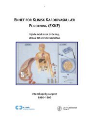
Preface - Ous-research.no
Preface - Ous-research.no
Preface - Ous-research.no

You also want an ePaper? Increase the reach of your titles
YUMPU automatically turns print PDFs into web optimized ePapers that Google loves.
Plastic and reconstructive surgery<br />
ence, our group is performing investigations to optimize<br />
the reconstruction technique and to minimize the do<strong>no</strong>r<br />
morbidity.<br />
Until <strong>no</strong>w little attention has been paid to reinnervation of<br />
the flap. We have investigated the spontaneous reinnervation<br />
of the DIEAP flap after breast reconstruction and at the<br />
do<strong>no</strong>r site at the abdomen. Pressure thresholds have been<br />
analysed on the skin using Semmes-Weinstein mo<strong>no</strong>filaments.<br />
Histological studies to evaluate the reinnervation in<br />
skin are planned both for the perforator flaps and for the<br />
do<strong>no</strong>r site.<br />
Through better understanding of flap anatomy, physiology<br />
and better surgical technique the complication rate has decreased<br />
and the cosmetic outcome has improved. However,<br />
partial flap necrosis is still a recurrent complication that can<br />
affect the final cosmetic result and the patient satisfaction.<br />
In most cases this can be avoided by discarding parts with<br />
unreliable capillary refilling after transferring the flap to the<br />
recipient site. The abdominal flap is divided into four equal<br />
vertical perfusion zones based on clinical observations. The<br />
zone with the best perfusion was designated zone I represented<br />
by the quarter part of the flap where the vascular<br />
pedicle entered. Zone II was represented by the kontralaterale<br />
neighbor zone and zone III the ipsilaterale neighbor<br />
zone. Zone IV was the remaining part most distant from<br />
zone I. Since the introduction of this perfusion model it has<br />
been widely accepted. Today it is common clinical practice<br />
to discard zone IV to avoid partial flap necrosis using the<br />
DIEAP flap for unilateral breast reconstruction. Occasionally<br />
further trimming is necessary to obtain an optimal cosmetic<br />
result because the flap is still too full. In order to preserve<br />
tissue with better vascularity the next zone to be sacrificed<br />
would be zone III. However, we have little scientific data that<br />
prove the validity of these perfusion zones. In other words<br />
trimming zone III before zone II could be wrong. We are<br />
performing quantitative evaluation of the perfusion zones<br />
with laser Doppler perfusion imaging (LDPI) in order to get<br />
a more exact picture of the microcirculatory differences in<br />
the DIEAP flap (fig 5). Our results showed that the perfusion<br />
of zone II was significantly lower than zone III in the period<br />
between 2 hours and 3 days after surgery. This suggest<br />
that it may be right to convert zone II to III and zone III to II,<br />
which will have major clinical impact on all surgical procedures<br />
involving DIEAP-flaps.<br />
Figure 5.<br />
Colour map of blood flow for the DIEAP flap processed by laser<br />
Doppler perfusion imaging. The flap has been divided into<br />
perfusion zones. The colour scale red-yellow-green-blue-black<br />
represents perfusion values where red is the highest and black the<br />
lowest value<br />
Measurements of microcirculation with laser Doppler<br />
perfusion imaging (LDPI)<br />
Measurements of microcirculation are a central part of all<br />
our animal and human experiments. It is performed with<br />
a PIM 3.0 LDPI from Perimed, Stocholm, Sweden. The LDPI<br />
generates, processes and displays colour-coded images of<br />
tissue perfusion. An optical scanner guides a low power laser<br />
beam stepwise to the tissue surface. The LDPI measures<br />
microcirculation to a depth of a few hundred micrometers.<br />
When the laser beam hit moving erytrocytes in the subepidermal<br />
plexus the light is backscattered and detected by a<br />
photodetector, this convert the light intensity to electrical<br />
signals and colour-coded images.<br />
49