Preface - Ous-research.no
Preface - Ous-research.no
Preface - Ous-research.no

Create successful ePaper yourself
Turn your PDF publications into a flip-book with our unique Google optimized e-Paper software.
Vilhelm Magnus Laboratory for Neurosurgical Research<br />
and exposure to differentiation cues (mainly withdrawal<br />
of growth factors and addition of serum) these cells went<br />
through characteristic steps of morphological and electrophysiological<br />
development and developed<br />
into the three principal building blocks of the brain:<br />
1. Astrocytes<br />
2. Oligodendrocytes<br />
3. Neurons<br />
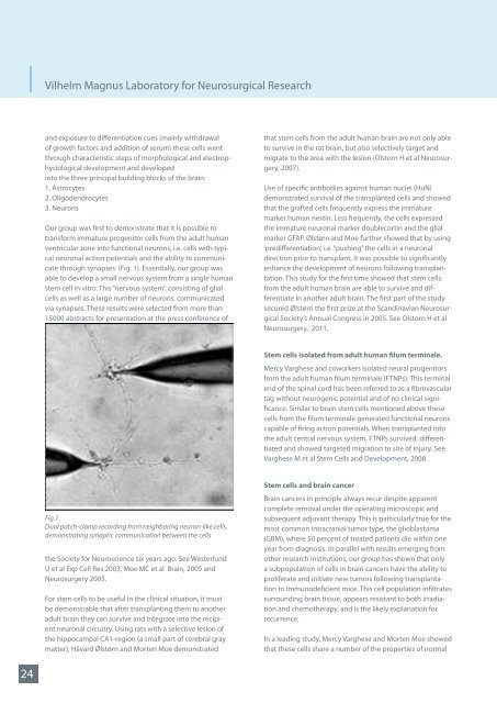
Our group was first to demonstrate that it is possible to<br />
transform immature progenitor cells from the adult human<br />
ventricular zone into functional neurons, i.e. cells with typical<br />
neuronal action potentials and the ability to communicate<br />
through synapses (Fig. 1). Essentially, our group was<br />
able to develop a small nervous system from a single human<br />
stem cell in vitro. This “nervous system”, consisting of glial<br />
cells as well as a large number of neurons, communicated<br />
via synapses. These results were selected from more than<br />
15000 abstracts for presentation at the press conference of<br />
that stem cells from the adult human brain are <strong>no</strong>t only able<br />
to survive in the rat brain, but also selectively target and<br />
migrate to the area with the lesion (Olstorn H et al Neurosurgery,<br />
2007).<br />
Use of specific antibodies against human nuclei (HuN)<br />
demonstrated survival of the transplanted cells and showed<br />
that the grafted cells frequently express the immature<br />
marker human nestin. Less frequently, the cells expressed<br />
the immature neuronal marker doublecortin and the glial<br />
marker GFAP. Ølstørn and Moe further showed that by using<br />
‘predifferentiation’, i.e. “pushing” the cells in a neuronal<br />
direction prior to transplant, it was possible to significantly<br />
enhance the development of neurons following transplantation.<br />
This study for the first time showed that stem cells<br />
from the adult human brain are able to survive and differentiate<br />
in a<strong>no</strong>ther adult brain. The first part of the study<br />
secured Ølstørn the first prize at the Scandinavian Neurosurgical<br />
Society’s Annual Congress in 2005. See Olstorn H et al<br />
Neurosurgery, 2011.<br />
Stem cells isolated from adult human filum terminale.<br />
Mercy Varghese and coworkers isolated neural progenitors<br />
from the adult human filum terminale (FTNPs). This terminal<br />
end of the spinal cord has been referred to as a fibrovascular<br />
tag without neurogenic potential and of <strong>no</strong> clinical significance.<br />
Similar to brain stem cells mentioned above these<br />
cells from the filum terminale generated functional neurons<br />
capable of firing action potentials. When transplanted into<br />
the adult central nervous system, FTNPs survived, differentiated<br />
and showed targeted migration to site of injury. See<br />
Varghese M et al Stem Cells and Development, 2008.<br />
Fig.1<br />
Dual patch-clamp recording from neighboring neuron-like cells,<br />
demonstrating synaptic communication between the cells<br />
the Society for Neuroscience six years ago. See Westerlund<br />
U et al Exp Cell Res 2003, Moe MC et al Brain, 2005 and<br />
Neurosurgery 2005.<br />
For stem cells to be useful in the clinical situation, it must<br />
be demonstrable that after transplanting them to a<strong>no</strong>ther<br />
adult brain they can survive and integrate into the recipient<br />
neuronal circuitry. Using rats with a selective lesion of<br />
the hippocampal CA1-region (a small part of cerebral gray<br />
matter), Håvard Ølstørn and Morten Moe demonstrated<br />
Stem cells and brain cancer<br />
Brain cancers in principle always recur despite apparent<br />
complete removal under the operating microscopic and<br />
subsequent adjuvant therapy. This is particularly true for the<br />
most common intracranial tumor type, the glioblastoma<br />
(GBM), where 50 percent of treated patients die within one<br />
year from diag<strong>no</strong>sis. In parallel with results emerging from<br />
other <strong>research</strong> institutions, our group has shown that only<br />
a subpopulation of cells in brain cancers have the ability to<br />
proliferate and initiate new tumors following transplantation<br />
to immu<strong>no</strong>deficient mice. This cell population infiltrates<br />
surrounding brain tissue, appears resistant to both irradiation<br />
and chemotherapy, and is the likely explanation for<br />
recurrence.<br />
In a leading study, Mercy Varghese and Morten Moe showed<br />
that these cells share a number of the properties of <strong>no</strong>rmal<br />
24