Maxillofacial Prosthetic Rehabilitation of a Cancer Patient
Maxillofacial Prosthetic Rehabilitation of a Cancer Patient Maxillofacial Prosthetic Rehabilitation of a Cancer Patient
Maxillofacial Prosthetic Rehabilitation of a Cancer Patient at Terminal Stage Maxillofacial Prosthetic Rehabilitation of a Cancer Patient at Terminal Stage Dr. Arun Dupare*, Dr. Roshni Dupare**, Dr. Lata Dupare *** * Dean and Professor, Prosthodontics, College of Dental Science and Hospital, Amargadh, Gujarat, ** Department of Community Dentistry, ITS Dental College, Murad Nagar, Ghaziabad, ***Senior Staff Surgeon, Periodontology, CGHS Polyclinic, Nagpur Abstracts: Face is the forefront of aesthetics. Surgical resection of the maxillae and facial structures for treatment of cancer, trauma, congenital deformities or infection causes maxillofacial defect that has serious impact on the individual’s esthetic and has great psychological trauma of social outcast. When the presented defect is extensive and supporting structures is lacking, the rehabilitation and restoring functions is a challenging task. Rehabilitation becomes even more difficult when the problems at defect site are associated with the complications. An attempt has been made to rehabilitate of a patient at terminal stage having extensive maxillofacial defect with inoperable carcinoma, infections, and restricted mouth opening. [ Dupare A et al NJIRM 2012; 3(2) : 173-176] Key words: facial prosthesis, heat cured acrylic resin, obturator prosthesis, rare earth magnet, self cured acrylic resin. Author for correspondence: Dr Dr. Arun Dupare, Dean, College of Dental Science & Hospital, Amargadh, Taluka Sihor, Dist. Bhavnagar, Gujarat India Pin: 364210, Email: drarundupare@yahoo.com Introduction Maxillofacial prosthetic rehabilitation and restoring function is important as impairments have detrimental effect on the quality of life and self-esteem. 1 When the defect presented is extensive and the supporting bone is lacking, the rehabilitation is a challenging task. Rehabilitation of the defect becomes even more difficult when the defect area is associated with the complications. An attempt has been made to rehabilitate a patient at terminal stage having extensive maxillofacial defect with inoperable cancer, restricted mouth opening, and other complications. Two component light weight prosthesis was fabricated to cover the defect. 2, 3 The obturator and facial prosthesis was fabricated with heat cured acrylic resin lighter in weight retained by rare earth magnets and spectacle was delivered to make the remaining life of the patient tolerable. Case report : A 38 year-old male patient was referred from the department of ENT to the Department of Prosthetic Dentistry, at Government Dental College and Hospital Nagpur, for rehabilitation of right side of face. The patient’s medical history revealed that he was diagnosed as squamous cell carcinoma of the right maxillary antrum involving the floor of the orbit. To remove the carcinoma maxillectomy and enucleating right eye was done. It was followed by frequent recurrence and surgical procedures. In the frequent surgical procedures he lost his more than half of the nose including the medial septum, right eye, supraorbital structures, right maxilla, part of zygomatic bone and associated structures. The recurrence was again diagnosed at defect site at lateral supraorbital and zygomatic areas. It was reported that any further surgical attempt to remove the carcinoma can cause serious complications. The defect site was inflamed, infective and extremely sensitive with bleeding spot. Right side corner of lips and part of alae of the nose was intact. (Fig.1) He was reported to be at terminal stage due to faster growth of malignancy near vital structures. There was no history of radiation therapy. He was under cover of chemotherapy. Fig.1 Extraoral photograph- before prosthesis Patient had limited vertical opening. Thorough intraoral examination revealed a total maxillectomy and the muscles of mastication were detached from NJIRM 2012; Vol. 3(2). April-June eISSN: 0975-9840 pISSN: 2230 - 9969 173
- Page 2 and 3: Maxillofacial Prosthetic Rehabilita
- Page 4: Maxillofacial Prosthetic Rehabilita
<strong>Maxill<strong>of</strong>acial</strong> <strong>Prosthetic</strong> <strong>Rehabilitation</strong> <strong>of</strong> a <strong>Cancer</strong> <strong>Patient</strong> at Terminal Stage<br />
<strong>Maxill<strong>of</strong>acial</strong> <strong>Prosthetic</strong> <strong>Rehabilitation</strong> <strong>of</strong> a <strong>Cancer</strong> <strong>Patient</strong> at Terminal Stage<br />
Dr. Arun Dupare*, Dr. Roshni Dupare**, Dr. Lata Dupare ***<br />
* Dean and Pr<strong>of</strong>essor, Prosthodontics, College <strong>of</strong> Dental Science and Hospital, Amargadh, Gujarat,<br />
** Department <strong>of</strong> Community Dentistry, ITS Dental College, Murad Nagar, Ghaziabad, ***Senior Staff Surgeon, Periodontology, CGHS<br />
Polyclinic, Nagpur<br />
Abstracts: Face is the forefront <strong>of</strong> aesthetics. Surgical resection <strong>of</strong> the maxillae and facial structures for<br />
treatment <strong>of</strong> cancer, trauma, congenital deformities or infection causes maxill<strong>of</strong>acial defect that has serious<br />
impact on the individual’s esthetic and has great psychological trauma <strong>of</strong> social outcast. When the presented<br />
defect is extensive and supporting structures is lacking, the rehabilitation and restoring functions is a<br />
challenging task. <strong>Rehabilitation</strong> becomes even more difficult when the problems at defect site are associated<br />
with the complications. An attempt has been made to rehabilitate <strong>of</strong> a patient at terminal stage having<br />
extensive maxill<strong>of</strong>acial defect with inoperable carcinoma, infections, and restricted mouth opening. [ Dupare A<br />
et al NJIRM 2012; 3(2) : 173-176]<br />
Key words: facial prosthesis, heat cured acrylic resin, obturator prosthesis, rare earth magnet, self cured<br />
acrylic resin.<br />
Author for correspondence: Dr Dr. Arun Dupare, Dean, College <strong>of</strong> Dental Science & Hospital, Amargadh,<br />
Taluka Sihor, Dist. Bhavnagar, Gujarat India Pin: 364210, Email: drarundupare@yahoo.com<br />
Introduction <strong>Maxill<strong>of</strong>acial</strong> prosthetic rehabilitation<br />
and restoring function is important as impairments<br />
have detrimental effect on the quality <strong>of</strong> life and<br />
self-esteem. 1 When the defect presented is<br />
extensive and the supporting bone is lacking, the<br />
rehabilitation is a challenging task. <strong>Rehabilitation</strong> <strong>of</strong><br />
the defect becomes even more difficult when the<br />
defect area is associated with the complications. An<br />
attempt has been made to rehabilitate a patient at<br />
terminal stage having extensive maxill<strong>of</strong>acial defect<br />
with inoperable cancer, restricted mouth opening,<br />
and other complications. Two component light<br />
weight prosthesis was fabricated to cover the<br />
defect. 2, 3 The obturator and facial prosthesis was<br />
fabricated with heat cured acrylic resin lighter in<br />
weight retained by rare earth magnets and<br />
spectacle was delivered to make the remaining life<br />
<strong>of</strong> the patient tolerable.<br />
Case report : A 38 year-old male patient was<br />
referred from the department <strong>of</strong> ENT to the<br />
Department <strong>of</strong> <strong>Prosthetic</strong> Dentistry, at Government<br />
Dental College and Hospital Nagpur, for<br />
rehabilitation <strong>of</strong> right side <strong>of</strong> face. The patient’s<br />
medical history revealed that he was diagnosed as<br />
squamous cell carcinoma <strong>of</strong> the right maxillary<br />
antrum involving the floor <strong>of</strong> the orbit. To remove<br />
the carcinoma maxillectomy and enucleating right<br />
eye was done. It was followed by frequent<br />
recurrence and surgical procedures. In the frequent<br />
surgical procedures he lost his more than half <strong>of</strong> the<br />
nose including the medial septum, right eye,<br />
supraorbital structures, right maxilla, part <strong>of</strong><br />
zygomatic bone and associated structures. The<br />
recurrence was again diagnosed at defect site at<br />
lateral supraorbital and zygomatic areas. It was<br />
reported that any further surgical attempt to<br />
remove the carcinoma can cause serious<br />
complications. The defect site was inflamed,<br />
infective and extremely sensitive with bleeding<br />
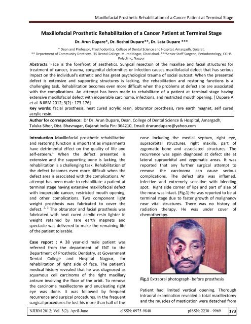
spot. Right side corner <strong>of</strong> lips and part <strong>of</strong> alae <strong>of</strong><br />
the nose was intact. (Fig.1) He was reported to be at<br />
terminal stage due to faster growth <strong>of</strong> malignancy<br />
near vital structures. There was no history <strong>of</strong><br />
radiation therapy. He was under cover <strong>of</strong><br />
chemotherapy.<br />
Fig.1 Extraoral photograph- before prosthesis<br />
<strong>Patient</strong> had limited vertical opening. Thorough<br />
intraoral examination revealed a total maxillectomy<br />
and the muscles <strong>of</strong> mastication were detached from<br />
NJIRM 2012; Vol. 3(2). April-June eISSN: 0975-9840 pISSN: 2230 - 9969 173
<strong>Maxill<strong>of</strong>acial</strong> <strong>Prosthetic</strong> <strong>Rehabilitation</strong> <strong>of</strong> a <strong>Cancer</strong> <strong>Patient</strong> at Terminal Stage<br />
maxilla <strong>of</strong> the right side. The presented defect<br />
situation corresponded to a class I situation<br />
(resected performed along the palatal midline)<br />
according to the Aramany classification <strong>of</strong> defects. 4<br />
He had difficulty in speech, mastication, swallowing<br />
and to maintain oral hygiene. A left side maxilla and<br />
the mandibular arch were completely dentulous<br />
with healthy teeth and normal occlusion. Tongue<br />
size and function was normal, but speech was<br />
altered.<br />
Treatment plan: A patient was at terminal stage <strong>of</strong><br />
life. The defect area was extensive with restricted<br />
mouth opening. Moreover, intimate contact <strong>of</strong> the<br />
prosthesis to tissue for support and retention<br />
cannot be utilized due to presence <strong>of</strong> malignancy<br />
and infected sensitive areas. The bone or teeth on<br />
right side to support the large and bulky prosthesis<br />
was lacking. Hence prosthetic rehabilitation <strong>of</strong> a<br />
case was challenging. He was very much distressed<br />
because <strong>of</strong> the extensive facial disfigurement and<br />
open surgical cavity with bleeding spots. He wanted<br />
us to give him some option to cover the defect till<br />
his survival.<br />
A case was considered to fabricate two -piece<br />
interim prosthesis separately to cover intraoral<br />
defects by obturator prosthesis and extraoral<br />
defects by facial prosthesis. This two-piece<br />
construction makes the insertion <strong>of</strong> the prosthesis<br />
easy and is done piece by piece making it less a<br />
struggle for the patient with limited mouth opening.<br />
The fabrication <strong>of</strong> intraoral obturator prosthesis<br />
was essential to prevent communication <strong>of</strong> food<br />
between the oral and nasal cavities. Moreover, the<br />
most important function this obturator served in<br />
the case was to support the facial prosthesis<br />
through the healthy teeth and bone present on left<br />
side. The extraoral facial prosthesis to fabricate was<br />
consisting <strong>of</strong> cheek, part <strong>of</strong> nose and eye. The heat<br />
cured acrylic resin was used to fabricate these two<br />
prostheses. Rare earth magnets were used to link<br />
the two portions. The case option <strong>of</strong> prosthesis was<br />
given to the patient that he readily accepted. He<br />
was ready to wear spectacle to hold the facial<br />
prosthesis.<br />
Clinical procedure: Fabrication <strong>of</strong> obturator<br />
prosthesis: A sterilized wet cotton gauze was<br />
placed in the operated cavity to prevent flow <strong>of</strong> the<br />
impression material in throat. An impression <strong>of</strong> the<br />
dentulous and supporting area to prepare obturator<br />
was then made with an alginate impression<br />
material. Working cast was prepared from this<br />
impression. The undercuts present in the defect<br />
were blocked out. To reduce weight simple plate<br />
type obturator prosthesis was fabricated and was<br />
extended on defect side so as to reach to the facial<br />
prosthesis. Healthy teeth and bone present on left<br />
side were used for retention and support to<br />
obturator and also to the facial prosthesis. The<br />
extension <strong>of</strong> the flange <strong>of</strong> obturator was done on<br />
left side in buccal sulcus area and on buccal surface<br />
<strong>of</strong> the teeth.<br />
Fabrication <strong>of</strong> facial prosthesis: Facial moulage was<br />
done to obtain a working cast to orient the<br />
prosthesis properly to the rest <strong>of</strong> the face. The<br />
obturator prosthesis was inserted in the mouth and<br />
the nasal opening was blocked with gauze. Plastic<br />
tube was placed in the mouth for air intake. The<br />
operated cavity was lined with sterilized cotton<br />
gauge. The face was coated with petroleum jelly. An<br />
impression for facial defect was made with alginate<br />
impression material. Alginate was mixed and<br />
applied to skin surface. Gauze squares were placed<br />
all over the alginate surface to provide mechanical<br />
retention for rigid plaster backing. Dental plaster<br />
was then spread over the entire surface to a<br />
sufficient thickness. After setting <strong>of</strong> plaster set<br />
impression was removed, grasping it on the sides<br />
and lifting it gently. The impression was inspected<br />
for any void or distortion. The impression was<br />
poured in type-III dental stone <strong>of</strong> about 5 millimeter<br />
thickness and after setting it was retrieved (Fig.2).<br />
Fig.2 Facial moulage<br />
All the undercuts on affected area <strong>of</strong> the cast was<br />
blocked. A wax pattern for facial prosthesis was<br />
NJIRM 2012; Vol. 3(2). April-June eISSN: 0975-9840 pISSN: 2230 - 9969 174
<strong>Maxill<strong>of</strong>acial</strong> <strong>Prosthetic</strong> <strong>Rehabilitation</strong> <strong>of</strong> a <strong>Cancer</strong> <strong>Patient</strong> at Terminal Stage<br />
sculpted with required thickness. Over extension on<br />
lower side <strong>of</strong> mandible was given to avoid visibility<br />
<strong>of</strong> the defect while opening <strong>of</strong> mouth. A<br />
prefabricated acrylic eye shell was selected <strong>of</strong><br />
shape, size and color after matching with left eye.<br />
The position <strong>of</strong> the pupil and the sclera was<br />
adjusted to mimic the left eye position while the<br />
patient gazes forward. A wax pattern was carved<br />
around the acrylic eye shell on the model. Upper<br />
and lower eyelids were carved such that the area<br />
covered by them on sclera mimicked the left eye<br />
(Fig.3).<br />
means <strong>of</strong> securing a facial prosthesis. Hooks were<br />
placed in the frame <strong>of</strong> spectacle and the loops in<br />
the facial prosthesis for stability and in position.<br />
Fig.4 Tissue surface <strong>of</strong> obturator & facial prosthesis<br />
with magnet<br />
Fig.3 Waxed-up facial prosthesis<br />
For orientation <strong>of</strong> the eye, a wax pattern was<br />
checked on patient face. This pattern without the<br />
eye shell was then invested. Clear heat cured acrylic<br />
resin was used for facial prosthesis. Skin matched<br />
colored stains were added in heat cured acrylic<br />
resin to fabricate facial surface <strong>of</strong> the prosthesis.<br />
Self cured acrylic resin was used to stabilize the eye<br />
shell section. The inner surface <strong>of</strong> the prosthesis<br />
after curing was further hollowed and carved to<br />
reduce thickness and weight and also to eliminate<br />
any contact with infected area. The eye shell was<br />
then attached to its position with self cured acrylic<br />
resin. Stains were mixed and painted in<br />
predetermined sequence and quantity to achieve<br />
staining to create lifelike appearance.<br />
Attachment <strong>of</strong> retentive devises: A projection on<br />
inner surface <strong>of</strong> facial prosthesis and to cover the<br />
obturator prosthesis was made with self cured<br />
acrylic resin. For attaching the rare earth magnets<br />
to this final prosthesis the obturator was first placed<br />
in patient’s mouth and second magnet was<br />
attached to the first magnet on the obturator using<br />
self cured acrylic resin (Fig.4). A spectacle with<br />
broad frame was used that provided an excellent<br />
Delivery <strong>of</strong> prosthesis and instructions: Both the<br />
parts <strong>of</strong> the prosthesis were decided to wear<br />
separately for convenience. <strong>Patient</strong> was trained to<br />
insert, to stabilize in position (Fig.5) and also to<br />
remove the facial and obturator prosthesis for<br />
cleaning and during sleep.<br />
Fig.5 Extraoral photograph with prosthesis<br />
Discussion : The treatment <strong>of</strong> maxillary defects is<br />
always limited by difficulties with retention,<br />
movable tissue beds and lack <strong>of</strong> sufficient bone<br />
support. 5 When the defect is large then the solid<br />
bulky prosthesis is heavy in weight and to maintain<br />
its position is difficult. In case trismus is present,<br />
making execution <strong>of</strong> the procedures extremely<br />
cumbersome and for the patient, manipulation <strong>of</strong><br />
the prosthesis into and out <strong>of</strong> the mouth becomes a<br />
Herculean task, as it requires multiple paths <strong>of</strong><br />
insertion and removal. To overcome these problems<br />
<strong>of</strong> retention skin adhesives, spectacles,<br />
engagements <strong>of</strong> undercuts using flexible materials<br />
and implants have been advocated. 6<br />
NJIRM 2012; Vol. 3(2). April-June eISSN: 0975-9840 pISSN: 2230 - 9969 175
<strong>Maxill<strong>of</strong>acial</strong> <strong>Prosthetic</strong> <strong>Rehabilitation</strong> <strong>of</strong> a <strong>Cancer</strong> <strong>Patient</strong> at Terminal Stage<br />
The use <strong>of</strong> frames by attaching eye prosthesis has<br />
advantageous when the patients has undergone<br />
maxillectomy in combination with an orbital<br />
exenterations as reported by Beumer J et al. 2<br />
The rare earth magnets have been used in dentistry<br />
3<br />
since 1960 and Federick. in the year 1976<br />
presented a technique for fabrication <strong>of</strong> a sectional<br />
interim maxillary obturator with retention<br />
augmented by magnet. The use <strong>of</strong> rare earth<br />
magnet achieves a more life like appearance and<br />
keeps facial prosthesis independent to external<br />
support.<br />
The fabrication <strong>of</strong> flexible obturator for patients<br />
with severely limited mouth opening using either<br />
silicone or even a flexible vinyl resin mouth guard<br />
material can be the choice according to Lauciello` et<br />
al. 7 However, these materials are far from ideal and<br />
in an average maxillectomy case it would be<br />
inadequate.<br />
<strong>Prosthetic</strong> rehabilitation for facial defects has<br />
several advantages over surgical reconstruction as it<br />
is quite inexpensive, allows for periodic examination<br />
and cleaning and is also an alternative to surgery in<br />
unsuitable candidates. Acrylic resin was introduced<br />
to dental pr<strong>of</strong>ession in 1937 for both intra and extra<br />
oral prosthesis. The fabrication process <strong>of</strong> acrylic<br />
resin is relatively short and the clinician has a lot <strong>of</strong><br />
control over the color, shape and size <strong>of</strong> prostheses.<br />
Heat cured acrylic resins are routinely used for<br />
maxill<strong>of</strong>acial prosthesis.<br />
References :<br />
1. Kreissl ME. Zygoma implant supported<br />
prosthetic rehabilitation after partial<br />
maxillectomy using surgical navigation. A<br />
clinical report. J Prosthet Dent 2007;97:121-28<br />
2. Beumer J III, Curtis TA, Marunick MT.<br />
<strong>Maxill<strong>of</strong>acial</strong> rehabilitation: Prosthodontic and<br />
surgical consideration. 2 nd edition St. Louis<br />
Ishiyaku euro America Inc. 1996 ;408-16.<br />
3. Federick DR. A magnetically retained interim<br />
maxillary obturator. J Prosthet Dent<br />
1976;36:671-75.<br />
4. Aramany MA. Basic principles <strong>of</strong> obturator<br />
design for partially edentulous patients Part I:<br />
Classification. J Prosthet Dent 1978; 40:554-57.<br />
5. Taylor TD, Flyer A, La Ville WE. Alternate<br />
obturation for the maxillectomy patient with<br />
severely limited mandibular opening. J Prosthet<br />
Dent 1985;53:83-85.<br />
6. Chalian VA, DraneJB, Standish SM. <strong>Maxill<strong>of</strong>acial</strong><br />
prosthetics- A Multidisciplinary Practice,4 th<br />
edition Williams & Wilkin Co. page: 1972;142-46<br />
7. Chen MS, Udagama A, Drane JB. Evaluation <strong>of</strong><br />
facial prostheses for head and neck cancer<br />
patients. J Prosthet Dent 1981; 46:538-44.<br />
In the case heat cured acrylic resin was used to<br />
fabricate facial and simple obturator prosthesis.<br />
Polymeric coated rare earth magnets (Neodeniumiron-boron)<br />
and broad frame spectacle was used for<br />
retention and support <strong>of</strong> this maxill<strong>of</strong>acial<br />
prosthesis.<br />
Conclusion: An attempt has been made to<br />
rehabilitate a patient at terminal stage associated<br />
with extensive maxill<strong>of</strong>acial defect and<br />
complications. A two components, obturator and<br />
facial prosthesis fabricated with heat cured acrylic<br />
resin lighter in weight retained by rare earth<br />
magnets and spectacle was delivered to make the<br />
remaining life <strong>of</strong> the patient tolerable.<br />
NJIRM 2012; Vol. 3(2). April-June eISSN: 0975-9840 pISSN: 2230 - 9969 176


