Marchevsky Meniscal Injury
Marchevsky Meniscal Injury
Marchevsky Meniscal Injury

You also want an ePaper? Increase the reach of your titles
YUMPU automatically turns print PDFs into web optimized ePapers that Google loves.
<strong>Meniscal</strong> <strong>Injury</strong> <br />
<strong>Meniscal</strong> injury is most commonly occurs associated with cranial cruciate ligament disease. <br />
Incidence of meniscal injury a at the time of initial surgery for 1000 cases of cranial cruciate <br />
ligament disease was approximately 33% (1) whilst in a series of 100 cases 59% had medial <br />
meniscal tears. (2) <br />
<strong>Meniscal</strong> anatomy <br />
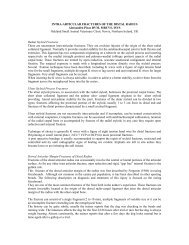
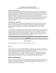
The menisci are crescent shaped discs of fibrocartilage. The abaxial border of each meniscus is <br />
thick tapering to a thin axial edge. The distal surface of each meniscus is flat whereas the <br />
proximal surface is convex allowing it to “hug” <br />
the femoral condyles and improve joint <br />
congruity. In the abaxial two thirds of the <br />
menisci, The collagen fibres that make up the <br />
menisci are largely circumferential “tied” <br />
together by radial fibres. Centrally the fibres <br />
are generally radial. Because there are <br />
relatively few radial fibres, tears tend to be <br />
circumferential-‐ the radial fibres tear and the <br />
circumferential fibres separate abaxially and <br />
more likely to be radial in the thinner central <br />
portions. <br />
Figure 1 From Veterinary Surgery Small Animal <br />
Vol 1 Ed. Tobias and Johnston 2012 <br />
The medial meniscus is firmly attached to the <br />
tibia via the caudal and cranial meniscal <br />
ligaments and to the lateral meniscus via the <br />
intermeniscal ligament. The lateral meniscus <br />
is similarly but less firmly attached to the tibia. <br />
In addition the lateral meniscofemoral <br />
ligament firmly attaches it to the <br />
intercondyloid fossa of the femur. It is also <br />
attached to the popliteal tendon. These two <br />
attachments cause it to move with the femoral <br />
condyle. This explains why it is less likely to be <br />
damaged after cranial cruciate ligament <br />
rupture compared with the medial side. <br />
<strong>Meniscal</strong> function <br />
The concave proximal surfaces of the menisci increase the congruity of the femoral condyles and <br />
the tibial plateau. They contribute in a minor way to joint stability if the cranial cruciate ligament <br />
is intact but if ruptured the menisci adopts a more primary role. The caudal pole acts as a wedge <br />
and stops cranial translation of the meniscus. However this predisposes to meniscal tearing (3)
Procedures that eliminate cranial tibial thrust may not address rotational forces and may explain <br />
why meniscal injury can still occur even after surgery is performed (4) <br />
During weight bearing the contact between the condyles and the menisci increases and they bear <br />
between 40 and 70% of the load. During weight bearing the menisci expand and the <br />
circumferential fibres elongate resulting in hoop stress. This stress is transmitted to the tibia <br />
through the cranial and caudal tibio-‐meniscal ligaments. Transection of any one of these <br />
ligaments destroys the ability to generate the stress within the meniscus and causes a 140% <br />
increase in peak contact pressure and 50% decrease in contact area on the femoral condyles and <br />
tibial plateau (5). <br />
<strong>Meniscal</strong> pathology <br />
As noted from the meniscal anatomy, most meniscal tears are circumferential and occur in the <br />
abaxial two thirds of the menisci. The medial meniscus is most affected because it is tightly <br />
connected to the tibia and is wedged between the tibial plateau and the femoral condyles when <br />
the tibia translates cranially leading to tears in the caudal pole. The tears are usually <br />
circumferential vertical tears. They can be incomplete in which case they may not be visible as <br />
the tear son the tibial surfaces. Only careful probing (using a small blunt right angled probe) will <br />
detect this. This may possibly be a cause of meniscal tears that apparently occur soon after <br />
surgery. Vertical tears may be full thickness and in which case will be seen on the femoral <br />
meniscal surfaces and may be non displaced where they appear as a longitudinal tear or <br />
displaced to form the classic bucket handle tears. An extension of the latter is when the torn <br />
meniscal portion tears away at either the cranial or caudal extent. The meniscus can also detach <br />
from the joint capsule (peripheral detachment) <br />
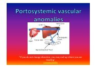
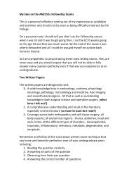
Figure 2 <strong>Meniscal</strong> tear classification Modified from Thieman et al (6). A, Normal; B, <br />
Non displaced horizontal tear; C, Bucket handle tear; D Flap tear
The lateral meniscal tends to ride with the femoral <br />
condyles and its caudal extent is largely protected. <br />
One arthroscopic study demonstrated meniscal tears <br />
in 77 0f 100 stifles with cranial cruciate ligament <br />
ruptures. These tears were radial, in the cranial pole <br />
and of unknown clinical significance. The actual <br />
mechanism of injury is unknown but was postulated <br />
to occur as a result of the cranial pole of the meniscus <br />
being pinched between the lateral femoral condyle <br />
and the lateral condylar eminence of the tibia. <br />
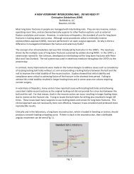
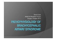
Figure 3 From Ralphs SC, Whitney WO J Am <br />
Vet Med Assoc 2002 (2) A, Radial lateral <br />
meniscal tear <br />
<strong>Meniscal</strong> surgery <br />
Approach: The medial meniscus can be accessed from either a lateral or medial <br />
arthrotomy. Laterally it is probably necessary to luxate the patella to get adequate <br />
visualisation of the medial meniscus. Medially a mini arthrotomy can be performed <br />
extending from the mid to distal portion of the patella to the tibial crest. Gelpi retractors <br />
are used to open the joint in a medial to lateral direction. Visualisation is facilitated using <br />
either a Hohmann retractor with the point placed at the back of the tibial plateau and <br />
levered forward using the trochlear as the fulcrum. Alternatively a stifle distractor can be <br />
used and is especially useful for non-‐assisted arthrotomy. <br />
Detection: A small blunt right-‐angled probe is invaluable tool for detecting meniscal tears. <br />
It is used to probe the tibial surface of the meniscus. This should be smooth. If the probe <br />
catches then this is likely to be an incomplete longitudinal tear. Additionally non-displaced<br />
full thickness vertical tears can also be difficult to see and can be retracted <br />
cranially. Subluxating the tibia cranial will also help detect non-‐displaced caudal pole <br />
tears and peripheral detachments. <br />
Resection: The torn portion should be grasped with hemostats (toothed or otherwise) <br />
and retracted. The attachments should be sharply transected with a number 11 blade <br />
(partial meniscectomy). It is important to examine the remaining meniscus once the <br />
resection has been performed to ensure that another tear is not present. If this is present <br />
or if there is caudal detachment of the remaining portion, then a caudal pole <br />
meniscectomy is performed. I have found very few indications for complete
meniscectomy as the cranial pole is rarely affected and there is a strong attachment to the <br />
medial collateral ligament. <br />
Prevention: <strong>Meniscal</strong> release has been shown to reduce the incidence of late meniscal <br />
tears to between 1 and 3.7% (6,7,8) However it is not a benign procedure as it alters the <br />
pressure distributions on the medial plateau, decreases the contact area and increases <br />
peak contact pressure. (3,11) Additionally dogs that have undergone extracapsular <br />
stabilisation and meniscectomy developed more arthritis long term than dogs in which <br />
the meniscal remained intact. (10) Therefore there needs to be some rationale behind <br />
performing a meniscal release. The true incidence (using only those cases that had no <br />
meniscal pathology or resection/release at the first surgery) of post-‐operative meniscal <br />
tears in cases treated with TTA has been variously reported as 10% (12,13), 16% (14), <br />
22% (13), 27% (15) and most recently a staggering 56% (16). (Interestingly this latter <br />
paper also demonstrated a 31% incidence of post-‐operative tears in dogs treated with <br />
TPLO and Tightrope extracapsular stabilisation (16)) The true incidence of late meniscal <br />
tears in dogs that have had a TPLO performed is reported at 3.7% (17), 3.8% (18), 8.2 <br />
(19) and 31% (15). In studies of dogs where extracapsular repair was performed the <br />
incidence of late meniscal tears was 6.1% (20) 31% (15) This may suggest that a medial <br />
meniscal release is warranted when a TTA is performed but is less indicated in cases <br />
treated with TPLO <br />
<strong>Meniscal</strong> tears area common problem associated with cranial cruciate ligament disease. <br />
There is a high incidence of concurrent meniscal injury at the time of first surgery and a <br />
significant incidence post operatively as well. Examination and careful inspection (visual <br />
and probe) to ensure meniscal injuries are correctly diagnosed and treated. Medial <br />
meniscal release at the time of surgery does reduce the incidence of post operative late <br />
meniscal injury but may not be necessary in cases that have been treated with a TPLO.
1) Fitzpatrick N, Solano M: Predictive variables for complications after tibial plateau <br />
leveling osteotomy with stifle inspection by arthrotomy in 1000 consecutive dogs. Vet <br />
Surg 39:460, 2010 <br />
2) Ralphs SC, Whitney WO: Arthroscopic evaluation of menisci in dogs with cranial <br />
cruciate ligament injuries: 100 cases (1999–2000). J Am Vet Med Assoc 221:1601, 2002. <br />
3) Pozzi A, Kowaleski MP, Apelt D, et al: Effect of medial meniscal release on tibial translation <br />
after tibial plateau leveling osteotomy. Vet Surg 35:486, 2006 <br />
4) Pacchiana PD, Morris E, Gillings SL, et al: Surgical and postoperative complications associated <br />
with tibial plateau leveling osteotomy in dogs with cranial cruciate ligament rupture: 397 cases <br />
(1998–2001). J Am Vet Med Assoc 222:184, 2003. <br />
5) Pozzi A, Kim S, Lewis D: Effect of transection of the caudal menisco-‐tibial ligament on <br />
femorotibial contact mechanics. Vet Surg 39:489, 2010 <br />
6) Wolf RE, Scavelli TD, Hoelzler MG et al: Surgical and postoperative complications associated <br />
with tibial tuberosity advancement for cranial cruciate ligament rupture in dogs: 458 cases <br />
(2007–2009) J Am Vet Med Assoc 240:1481 2012 <br />
7) Fitzpatrick N, Solano M: Predictive variables for complications after tibial plateau <br />
leveling osteotomy with stifle inspection by arthrotomy in 1000 consecutive dogs. Vet <br />
Surg 39:460, 2010 <br />
8) Gatineau M, Dupuis J, Planté J; Moreau M: Retrospective study of 476 tibial plateau <br />
levelling osteotomy procedures: Rate of subsequent ‘pivot shift’, meniscal tear and other <br />
complications VCOT 24: 333 2011 <br />
9) Thieman K, Pozzi A, Ling C, et al: The contact mechanics of simulated meniscal tears in <br />
cadaveric dog stifles. Vet Surg 38:803, 2009. <br />
10) Innes JF, Bacon D, Lynch C, et al: Long-‐term outcome of surgery for dogs with cranial <br />
cruciate ligament deficiency. Vet Rec 147:325, 2000. <br />
11) Pozzi A, Litsky AS, Field J, et al: Pressure distributions on the medial tibial plateau <br />
after medial meniscal surgery and tibial plateau levelling osteotomy in dogs. Vet Comp <br />
Orthop Traumatol 21:1, 2008. <br />
12) Hoffmann DE, Miller JM, Ober CP, et al Tibial tuberosity advancement in 65 canine <br />
stifles VCOT 19 219 2006
13)Dymond NL Goldsmid SE Simpson DJ: Tibial tuberosity advancement in 92 canine <br />
stifles: initial results, clinical outcome and owner evaluation. Aust Vet J 88, 281 2010 <br />
14) Stein S, Schmoekel H: Short-‐term and eight to 12 months results of a tibial tuberosity <br />
advancement as treatment of canine cranial cruciate ligament damage. J Small Anim Pract <br />
49:398, 2008 <br />
15) Lafaver S, Miller NA, Stubbs WP, et al: Tibial tuberosity advancement for stabilization of the <br />
canine cranial cruciate ligament-‐deficient stifle joint: surgical technique, early results, and <br />
complications in 101 dogs. Vet Surg 36:573, 2007. <br />
16) Christopher SA Beetem J, Cook JL: Comparison of Long‐Term Outcomes Associated <br />
With Three Surgical Techniques for Treatment of Cranial Cruciate Ligament Disease in <br />
Dogs Vet Surg 432 329 2013 <br />
16) Priddy NH, Tomlinson JL, Dodam JR. Complications with and owner assess-‐ ment of <br />
the outcome of tibial plateau leveling osteotomy for treatment of cranial cruciate ligament <br />
rupture in dogs: 193 cases (1997–2001). J Am Vet Med Assoc 222:1726 2003 <br />
19) Kalff S, Meachem S, Preston C, Vet Surg 40 952 2011 Incidence of medial meniscal <br />
tears after arthroscopic assisted tibial plateau leveling osteotomy.<br />
20 Casale SA, McCarthy RJ: Complications associated with lateral fabellotibial suture <br />
surgery for cranial cruciate ligament injury in dogs: 363 cases (1997–2005). J Am Vet Med <br />
Assoc 234:229, 2009. <br />
14—6 <br />
17-‐7 <br />
18-‐8 <br />
6-‐9 <br />
7-‐10 <br />
9-‐11 <br />
10-‐12 <br />
11-‐13 <br />
12-‐14