Leiomyoma of Esophagus - Atcs.jp
Leiomyoma of Esophagus - Atcs.jp
Leiomyoma of Esophagus - Atcs.jp
You also want an ePaper? Increase the reach of your titles
YUMPU automatically turns print PDFs into web optimized ePapers that Google loves.
Original<br />
Article<br />
<strong>Leiomyoma</strong> <strong>of</strong> <strong>Esophagus</strong><br />
Ajay Punpale, MS, 1 Ashvin Rangole, 1 Navin Bhambhani, 1 George Karimundackal, 1<br />
Nandini Desai, 1 Ashwin de Souza, 1 C. S. Pramesh, MS, 1 Nirmala Jambhekar, MD, 2<br />
and Rajesh C. Mistry, MS 1<br />
<strong>Leiomyoma</strong>s are rare benign esophageal neoplasms with an indolent clinical course. Symptoms<br />
mimic that <strong>of</strong> esophageal cancer. Esophagoscopy and endoscopic ultrasonography are the main<br />
diagnostic methods. Symptomatic and large leiomyomas should be treated surgically while small,<br />
asymptomatic lesions may be managed by regular follow up and repeated endoscopies. (Ann<br />
Thorac Cardiovasc Surg 2007; 13: 78–81)<br />
Key words: leiomyoma, esophagus<br />
Introduction<br />
Benign tumors <strong>of</strong> the esophagus are rare lesions that constitute<br />
less than 1% <strong>of</strong> esophageal neoplasms. Nearly two<br />
thirds <strong>of</strong> benign esophageal tumors are leiomyomas; the<br />
others are mostly polyps and cysts. 1,2) <strong>Leiomyoma</strong>s are<br />
the commonest benign mesenchymal tumors <strong>of</strong> the<br />
esophagus. Most tumors described as leiomyomas and<br />
leiomyosarcomas in the older medical literature actually<br />
refer to gastrointestinal stromal tumors (GISTs). 3) <strong>Leiomyoma</strong><br />
is a term used only in relation to tumors <strong>of</strong> the<br />
esophagus.The significance <strong>of</strong> knowing about leiomyomas<br />
is due to the fact that it can mimic esophageal cancer and<br />
lead to diagnostic confusion. In this article, we review<br />
our experience with esophageal leiomyomas at our institute.<br />
Patients and Methods<br />
We retrospectively reviewed the medical records <strong>of</strong> patients<br />
diagnosed with esophageal tumors at the Tata<br />
Memorial Hospital from January 2003 to January 2005.<br />
About 1,500 patients were registered with esophageal<br />
From Division <strong>of</strong> Thoracic Surgery, Departments <strong>of</strong> 1 Surgical<br />
Oncology and 2 Pathology, Tata Memorial Hospital, Mumbai, India<br />
Received April 24, 2006; accepted for publication July 28, 2006.<br />
Address reprint requests to Rajesh C. Mistry, MS: Division <strong>of</strong><br />
Thoracic Surgery, Department <strong>of</strong> Surgical Oncology, Tata<br />
Memorial Hospital, Parel, Mumbai – 400 012, India.<br />
tumor during the study period. Of these, 290 patients<br />
underwent some types <strong>of</strong> esophageal surgery. We found<br />
six patients with esophageal leiomyomas. The esophageal<br />
leiomyomas thus constituted 0.4% <strong>of</strong> all the esophageal<br />
tumors in this study. We recorded the presenting symptoms,<br />
investigations, management and outcome <strong>of</strong> these<br />
patients. The diagnostic workup included barium swallow,<br />
upper gastrointestinal endoscopy (UGI scopy), endoscopic<br />
ultrasound (EUS) and computed tomography<br />
(CT). Fine needle aspiration cytology (FNAC) or biopsy<br />
was done in cases where there was suspicion <strong>of</strong> malignancy.<br />
Of the six patients, we had histological diagnosis<br />
<strong>of</strong> leiomyoma in five patients. The sixth patient had classical<br />
features <strong>of</strong> leiomyoma on EUS and CT scan and<br />
was planned for regular follow up. We had no tissue diagnosis<br />
for this patient.<br />
Results<br />
Six patients were diagnosed with esophageal leiomyomas<br />
<strong>of</strong> which five were female. Their age [TK1] ranged from<br />
34 to 65 years. Of the six patients, tissue diagnosis <strong>of</strong><br />
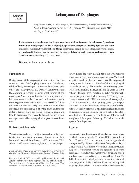
leiomyoma (Fig. 1) was available for five patients. Dysphagia<br />
was the commonest presentation though nonulcer<br />
dyspepsia, retrosternal burning and epigastric pain were<br />
associated. One patient had symptoms <strong>of</strong> dyspnoea, cough<br />
and hoarseness related to a large mass (Figs. 2 and 3).<br />
Table 1 shows the clinical presentation and the details <strong>of</strong><br />
the management <strong>of</strong> all the patients. Three patients required<br />
esophageal resection, while two patients underwent lo-<br />
78 Ann Thorac Cardiovasc Surg Vol. 13, No. 2 (2007)
<strong>Leiomyoma</strong> <strong>of</strong> <strong>Esophagus</strong><br />
Fig. 1. Esophageal leiomyoma showing a submucosal fasciculated<br />
spindle cell tumor with cigar-shaped nuclei. (H&E ×<br />
200)<br />
Fig. 2. Barium swallow showing smooth convex mass and distinct<br />
margins at the junction <strong>of</strong> mass and normal mucosa.<br />
cal enucleation and one patient with a small, relatively<br />
asymptomatic leiomyoma was kept under close observation.<br />
All the operated patients had uneventful postoperative<br />
recoveries. One patient who had undergone total transthoracic<br />
esophagectomy (Fig. 4) had a minor anastomotic<br />
leak in the neck which healed with conservative management.<br />
All patients are asymptomatic and recurrence-free<br />
at last follow up.<br />
Discussion<br />
Fig. 3. Computed tomography (transverse section) <strong>of</strong> one <strong>of</strong> our<br />
patients showing large leiomyoma in the esophagus occupying<br />
the posterior mediastinum.<br />
<strong>Leiomyoma</strong>s are the commonest benign mesenchymal tumors<br />
<strong>of</strong> the esophagus contributing about two-thirds <strong>of</strong><br />
all benign lesions <strong>of</strong> the esophagus. <strong>Leiomyoma</strong>s usually<br />
arise as intramural growths, most commonly along the<br />
distal two thirds <strong>of</strong> the esophagus. They are multiple in<br />
approximately 5% <strong>of</strong> patients. 1,2,4,5)<br />
Esophageal leiomyomas rarely cause symptoms when<br />
they are smaller than 5 cm in diameter. Large tumors can<br />
cause dysphagia, vague retrosternal discomfort, chest<br />
pain, esophageal obstruction, and regurgitation. Rarely,<br />
they can cause gastrointestinal bleeding, with erosion<br />
through the mucosa. Other than the nonspecific symptoms<br />
associated with esophageal leiomyomas, very few<br />
physical findings are usually noted. In extremely rare cases<br />
where severe esophageal obstruction is caused by a leiomyoma,<br />
weight loss and muscle wasting may be observed.<br />
6)<br />
Histologically, leiomyomas comprise <strong>of</strong> bundles <strong>of</strong><br />
interlacing smooth muscle cells, well-demarcated by adjacent<br />
tissue or by a definitive connective tissue capsule.<br />
Ann Thorac Cardiovasc Surg Vol. 13, No. 2 (2007) 79
Punpale et al.<br />
Table 1. Clinical presentation and management <strong>of</strong> patients<br />
Age/Sex Symptoms UGI scopy EUS CT scan Treatment<br />
55/F Dysphagia Gr III, Extrinsic Not done Well-defined mass Left thoracotomy-TTE<br />
dyspnoea, hoarseness, compression<br />
in tracheo-esophageal<br />
cough, mass in lower on left side groove extending from<br />
neck from cricopharynx left lobe <strong>of</strong> thyroid<br />
to 24 cm<br />
to aortopulmonary<br />
window, size: 9×5 cm<br />
34/F Dysphagia Gr II Submucosal tumor Well-defined Well-defined s<strong>of</strong>t tissue Right thoracotomy-<br />
22–27 cm hypoechoic mass in M/3 esophagus myotomy with<br />
tumor with causing obliteration enucleation<br />
calcification <strong>of</strong> lumen with proximal<br />
dilatation, size: 5×3cm<br />
65/F Dysphagia Gr II Submucosal lesion Large Mass in M/3 esophagus THE<br />
at 22–28 cm encapsulated with luminal narrowing<br />
tumor pushing and proximal dilatation,<br />
tracheobronchial size: 6×3.5 cm<br />
tree<br />
62/M Nonulcer dyspepsia, Submucosal tumor Not done Mass in L/3 esophagus, Left thoraco-<br />
Dysphagia Gr I at cardia and hiatal size: 6×3 cm abdominal approach<br />
sac with ulceration<br />
—Extramucosal<br />
excision<br />
48/F Dysphagia Gr III, Submucosal nodule Submucosal Mass in L/3 esophagus Esophagogastrectomy<br />
epigastric pain nodule involving posterior<br />
wall, size: 5×3 cm<br />
40/F Retrosternal pain Polypoidal Well-defined Mass lesion in lower Observation with<br />
related to meal submucosal encapsulated esophagus, regular endoscopy<br />
lesion at 30 cm lesion from size: 3×3 cm<br />
muscularis<br />
mucosa<br />
TTE, total transthoracic esophagectomy; THE, transhiatal esophagectomy; UGI scopy, upper gastrointestinal endoscopy; EUS,<br />
endoscopic ultrasound; CT, computed tomography.<br />
Fig. 4. Specimen <strong>of</strong> total esophagogastrectomy done for one <strong>of</strong><br />
our patients showing a large leiomyoma in upper and middle<br />
third <strong>of</strong> esophagus.<br />
They are composed <strong>of</strong> fascicles <strong>of</strong> spindle cells that tend<br />
to intersect with each other at varying angles. The tumor<br />
cells have blunt-ended elongated nuclei and show minimal<br />
atypia with few mitotic figures. 7)<br />
On barium swallow, the classic appearance is a smooth<br />
concave mass underlying the intact mucosa. Distinct sharp<br />
angles are seen at the junction <strong>of</strong> the tumor and normal<br />
tissue. At endoscopy, the lesions are identified as mobile<br />
submucosal masses. If a leiomyoma is suspected at<br />
esophagoscopy, a biopsy should not be performed as it<br />
would cause scarring at the biopsy site, which would hamper<br />
definitive extramucosal resection at surgery. 4) However,<br />
an ulcerated growth should be biopsied to rule out<br />
malignancy. Endoesophageal ultrasonography is important<br />
in the diagnosis <strong>of</strong> leiomyomas, which demonstrate<br />
a homogeneous region <strong>of</strong> hypoechogenicity juxtaposed<br />
80<br />
Ann Thorac Cardiovasc Surg Vol. 13, No. 2 (2007)
<strong>Leiomyoma</strong> <strong>of</strong> <strong>Esophagus</strong><br />
with the overlying mucosa. 8,9)<br />
Asymptomatic or smaller leiomyomas may be followed<br />
up periodically with regular barium swallow and endoscopy<br />
as they have a characteristic radiographic appearance,<br />
slow growth rate, and negligible risk <strong>of</strong> malignant<br />
transformation. 2) Resection may be required to confirm<br />
the histopathological diagnosis in some cases. Surgical<br />
excision is recommended for symptomatic leiomyomas<br />
and those greater than 5 cm. Although a formal esophageal<br />
resection is not mandatory for leiomyomas. Frequently<br />
the large size and extent <strong>of</strong> the tumor (as in three<br />
<strong>of</strong> our patients) may preclude local enucleation.<br />
Tumors <strong>of</strong> the middle third <strong>of</strong> the esophagus may be<br />
approached using a right thoracotomy. Tumors in the distal<br />
third <strong>of</strong> the esophagus may be resected through a left<br />
thoracoabdominal approach, transhiatally or by a left thoracotomy.<br />
For extramucosal excision or enucleation, the<br />
outer esophageal muscle is incised longitudinally. Careful<br />
dissection is done to separate and remove the leiomyoma<br />
from the underlying submucosa. If the mucosa<br />
is inadvertently opened during dissection, the underlying<br />
mucosa is reapproximated, followed by closure <strong>of</strong> the<br />
longitudinal muscle. 10) Some authors have shown that<br />
large extramucosal defects may be left open without subsequent<br />
complications developing.<br />
Segmental esophageal resection (esophagogastrectomy)<br />
may be indicated for giant leiomyomas <strong>of</strong> the<br />
cardia. Total transthoracic esophagectomy (as for one<br />
patient in our series) is rarely needed especially for very<br />
large leiomyomas involving long segment <strong>of</strong> the esophagus.<br />
While open surgery is the mainstay <strong>of</strong> therapy for<br />
leiomyomas, combined esophagoscopy and video-assisted<br />
resection (thoracoscopy) have been performed recently<br />
in six patients, without complication. 11)<br />
References<br />
1. Seremetis MG, Lyons WS, de Guzman VC, Peabody<br />
JW Jr. <strong>Leiomyoma</strong>ta <strong>of</strong> the esophagus: an analysis <strong>of</strong><br />
838 cases. Cancer 1976; 38: 2166–77.<br />
2. Mutrie CJ, Donahue DM, Wain JC, et al. Esophageal<br />
leiomyoma: a 40-year experience. Ann Thorac Surg<br />
2005; 79: 1122–5.<br />
3. Miettinen M, Lasota J. Gastrointestinal stromal tumors—definition,<br />
clinical, histological, immunohistochemical,<br />
and molecular genetic features and differential<br />
diagnosis. Virchows Arch 2001; 438: 1–12.<br />
4. Wang Y, Zhang R, Ouyang Z, Zhang D, Wang L, Zhang<br />
D. Diagnosis and surgical treatment <strong>of</strong> esophageal leiomyoma.<br />
Zhonghua Zhong Liu Za Zhi 2002; 24: 394–<br />
6.<br />
5. Levine MS, Buck JL, Pantongrag-Brown L, Buetow<br />
PC, Lowry MA, Sobin LH. Esophageal leiomyomatosis.<br />
Radiology 1996; 199: 533–36.<br />
6. Rendina EA, Venuta F, Pescarmona EO, et al. <strong>Leiomyoma</strong><br />
<strong>of</strong> the esophagus. Scand J Thorac Cardiovasc<br />
Surg 1990; 24: 79–82.<br />
7. Evans HL. Smooth muscle tumors <strong>of</strong> the gastrointestinal<br />
tract: a study <strong>of</strong> 56 cases followed for a minimum<br />
<strong>of</strong> 10 years. Cancer 1985; 56: 2242–50.<br />
8. Massari M, De Simone M, Ci<strong>of</strong>fi U, Gabrielli F,<br />
Boccasanta P, Bonavina L. Endoscopic ultrasonography<br />
in the evaluation <strong>of</strong> leiomyoma and extramucosal<br />
cysts <strong>of</strong> the esophagus. Hepatogastroenterology 1998;<br />
45: 938–43.<br />
9. Rösch T, Lorenz R, Dancygier H, von Wickert A,<br />
Classen M. Endosonographic diagnosis <strong>of</strong> submucosal<br />
upper gastrointestinal tract tumors. Scand J<br />
Gastroenterol 1992; 27: 1–8.<br />
10. Bonavina L, Segalin A, Rosati R, Pavanello M,<br />
Peracchia A. Surgical therapy <strong>of</strong> esophageal leiomyoma.<br />
J Am Coll Surg 1995, 181: 257–62.<br />
11. Roviaro GC, Maciocco M, Varoli F, Rebuffat C, Vergani<br />
C, Scarduelli A. Videothoracoscopic treatment <strong>of</strong> oesophageal<br />
leiomyoma. Thorax 1998; 53: 190–92.<br />
Ann Thorac Cardiovasc Surg Vol. 13, No. 2 (2007) 81


