Lipid peroxidation and metallothionein induction by chromium and ...
Lipid peroxidation and metallothionein induction by chromium and ...
Lipid peroxidation and metallothionein induction by chromium and ...
You also want an ePaper? Increase the reach of your titles
YUMPU automatically turns print PDFs into web optimized ePapers that Google loves.
Hidrobiológica 2010, 20 (1): 31-40<br />
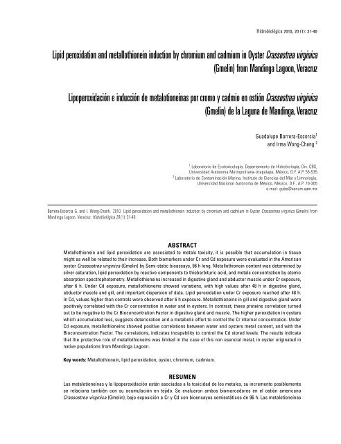
<strong>Lipid</strong> <strong>peroxidation</strong> <strong>and</strong> <strong>metallothionein</strong> <strong>induction</strong> <strong>by</strong> <strong>chromium</strong> <strong>and</strong> cadmium in Oyster Crassostrea virginica<br />
(Gmelin) from M<strong>and</strong>inga Lagoon, Veracruz<br />
Lipoperoxidación e inducción de metalotioneínas por cromo y cadmio en ostión Crassostrea virginica<br />
(Gmelin) de la Laguna de M<strong>and</strong>inga, Veracruz<br />
Guadalupe Barrera-Escorcia 1<br />
<strong>and</strong> Irma Wong-Chang 2<br />
1 Laboratorio de Ecotoxicología, Departamento de Hidrobiología, Div. CBS,<br />
Universidad Autónoma Metropolitana-Iztapalapa, México, D.F. A.P. 55-535<br />
2 Laboratorio de Contaminación Marina, Instituto de Ciencias del Mar y Limnología,<br />
Universidad Nacional Autónoma de México, México, D.F., A.P. 70-305<br />
e-mail: gube@xanum.uam.mx<br />
Barrera-Escorcia G. <strong>and</strong> I. Wong-Chanh. 2010. <strong>Lipid</strong> <strong>peroxidation</strong> <strong>and</strong> <strong>metallothionein</strong> <strong>induction</strong> <strong>by</strong> <strong>chromium</strong> <strong>and</strong> cadmium in Oyster Crassostrea virginica (Gmelin) from<br />
M<strong>and</strong>inga Lagoon, Veracruz. Hidrobiológica 20 (1): 31-40.<br />
Abstract<br />
Metallothionein <strong>and</strong> lipid <strong>peroxidation</strong> are associated to metals toxicity, it is possible that accumulation in tissue<br />
might as well be related to their increase. Both biomarkers under Cr <strong>and</strong> Cd exposure were evaluated in the American<br />
oyster Crassostrea virginica (Gmelin) <strong>by</strong> Semi-static bioassays, 96 h long. Metallothionein content was determined <strong>by</strong><br />
silver saturation, lipid <strong>peroxidation</strong> <strong>by</strong> reactive components to thiobarbituric acid, <strong>and</strong> metals concentration <strong>by</strong> atomic<br />
absorption spectrophotometry. Metallothioneins increased in digestive gl<strong>and</strong> <strong>and</strong> abductor muscle under Cr exposure,<br />
after 6 h. Under Cd exposure, <strong>metallothionein</strong>s showed variations, with high values after 48 h in digestive gl<strong>and</strong>,<br />
abductor muscle <strong>and</strong> gill, <strong>and</strong> important dispersion of data. <strong>Lipid</strong> <strong>peroxidation</strong> under Cr exposure reached after 48 h.<br />
In Cd, values higher than controls were observed after 6 h exposure. Metallothioneins in gill <strong>and</strong> digestive gl<strong>and</strong> were<br />
positively correlated with the Cr concentration in water <strong>and</strong> in oysters. In contrast, these proteins correlation turned<br />
out to be negative to the Cr Bioconcentration Factor in digestive gl<strong>and</strong> <strong>and</strong> muscle. The higher <strong>peroxidation</strong> in oysters<br />
which accumulated less, suggests deterioration <strong>and</strong> a metabolic effort to control the Cr internal concentration. Under<br />
Cd exposure, <strong>metallothionein</strong>s showed positive correlations between water <strong>and</strong> oysters metal content, <strong>and</strong> with the<br />
Bioconcentration Factor. The correlations, indicates incapability to control the Cd stored levels. The results indicate<br />
that the protective role of <strong>metallothionein</strong>s was limited in the case of this non esencial metal, in oyster originated in<br />
native populations from M<strong>and</strong>inga Lagoon.<br />
Key words: Metallothionein, lipid <strong>peroxidation</strong>, oyster, <strong>chromium</strong>, cadmium.<br />
Resumen<br />
Las metalotioneínas y la lipoperoxidación están asociadas a la toxicidad de los metales, su incremento posiblemente<br />
se relaciona también con su acumulación en tejido. Se evaluaron ambos biomarcadores en el ostión americano<br />
Crassostrea virginica (Gmelin), bajo exposición a Cr y Cd con bioensayos semiestáticos de 96 h. Las metalotioneínas
32<br />
Barrera-Escorcia <strong>and</strong> Wong-Chang<br />
se determinaron por saturación de plata, la lipoperoxidación por los componentes reactivos al ácido tiobarbitúrico,<br />
y la concentración de metales por espectrofotomería de absorción atómica. Las metalotioneínas se incrementaron<br />
en la glándula digestiva y músculo abductor, después de 6 h de exposición a Cr. En el caso de exposición a Cd las<br />
metalotioneínas mostraron variaciones, con valores elevados e importante dispersión de datos en glándula digestiva,<br />
músculo y branquia, posteriores a 48 h. La lipoperoxidación se incrementó después de 48 h bajo exposición a Cr.<br />
Valores superiores al control fueron observados después de 6 h de exposición a Cd. Las metalotioneínas en branquia<br />
y glándula digestiva se correlacionaron positivamente con la concentración de Cr en agua y ostión. En contraste,<br />
esta correlación fue negativa respecto al Factor de Bioconcentración de Cr en glándula digestiva y músculo. La<br />
mayor peroxidación en ostiones que acumularon menos Cr, sugiere deterioro y un esfuerzo metabólico para controlar<br />
la concentración interna. Las correlaciones positivas de las metalotioneínas con el Cd en agua y ostión, así como<br />
con el Factor de Bioconcentración, indica la incapacidad para controlar el Cd almacenado. Los resultados indican<br />
que el papel protector de las metalotioneínas fue limitado para este metal no esencial, en ostiones provenientes de<br />
poblaciones nativas de la Laguna de M<strong>and</strong>inga.<br />
Palabras clave: Metalotioneína, lipoperoxidación, ostión, cromo, cadmio.<br />
Introduction<br />
Metals, such as Cr <strong>and</strong> Cd, tend to increase in diverse coastal<br />
environments of the Mexican Gulf, hoarding in the sediment<br />
(Paez, 2005) <strong>and</strong> in filtering organisms, like oysters, where critical<br />
levels for consumption can be exceeded (Guzman et al.,<br />
2005). The oyster Crassostrea virginica (Gmelin, 1791) is found<br />
in the Atlantic coast from North America <strong>and</strong> literature about<br />
its physiology is profuse (Roesijadi, 1996); it is considered an<br />
adequate indicator to assess the contaminants contributions,<br />
since the metals concentrations incorporated in tissue fluctuates<br />
reflecting the environmental concentration. In Mexico, they<br />
are also distributed in the Gulf coastal lagoons <strong>and</strong> there are<br />
some studies about Cr <strong>and</strong> Cd levels accumulated in organisms<br />
(Contreras & Castañeda, 1995; Botello, 1994; Villanueva & Botello,<br />
1998). This specie represents a regionally important exploitable<br />
resource <strong>and</strong> it is extracted directly from the lagoons. It has<br />
been considered that metals concentrations collected in tissue,<br />
constitute potential biological impact indicator of environmental<br />
concentration, since the metals bioavailability is automatically<br />
considered (Borgmann, 2000). Wood et al. (1997) state that this<br />
kind of approach can be useful to predict the metals toxicity<br />
induced from water to benthonic organisms. In filtering organisms,<br />
such as C. virginica, tissue hoarding is particularly important<br />
since, even though there are several access routes for both<br />
metals into the animals, the main route is the water (Neff, 2002;<br />
Rebouças et al., 2005).<br />
Besides the accumulated metal, it is feasible to assess<br />
different biomarkers as specific reflections of its presence, under<br />
chronic or sublethal exposure conditions. Biomarkers are identifiable<br />
signs that provide early damage warnings (Koeman, 1991),<br />
<strong>and</strong> generate quantifiable responses. For instance, the <strong>metallothionein</strong><br />
synthesis associated with the essential metals requirements<br />
(Simkiss et al., 1982; Klaasen & Eaton, 1991). In C. virginica<br />
play an important role in the regulation of Zn, a metal which enters<br />
in large amounts <strong>and</strong> is promptly cleared out from the organism,<br />
which is related with the Ca fixation in valves <strong>and</strong>, therefore,<br />
with their resistance <strong>and</strong>, consequently, with the organism development,<br />
moreover, <strong>metallothionein</strong> regulate Cu levels (Savva,<br />
1999). In presence of excessive Zn amounts, or other essential or<br />
non-essential metals, such as a Cd, the <strong>metallothionein</strong> synthesis<br />
increase (Roesijadi, 1996). Due to its regulatory activity with<br />
essential-metals, its production has also been correlated with<br />
detoxifying mechanisms linked with toxic metals, principally nonessential<br />
ones, in several species, including C. virginica (Köhler &<br />
Riisgard 1982; Stegeman et al., 1992; Roesijadi et al., 1996).<br />
Another important biomarker is the oxidative stress damage,<br />
a common phenomenon under physiological stress conditions.<br />
The production of unbound radicals manifests, amongst other<br />
effects, increase in the membrane lipids oxidation, event also<br />
known as lipid <strong>peroxidation</strong>. The peroxidative damage can also<br />
result from the action of electrophilic intermediaries which interact<br />
with nucleophilic sites in the cell, including glutathione <strong>and</strong><br />
the thiol-group proteins, producing oxidative stress in the entire<br />
cell. Moreover, due to the presence of nucleophilic sites in the<br />
nucleic acids, the production of unbound radicals is associated<br />
with mutagenesis <strong>and</strong> carcinogenesis (Goyer, 1991). The effect<br />
of Cd has been demonstrated in lipid <strong>peroxidation</strong>, however, the<br />
mechanisms are not entirely acknowledged (Souza et al., 1996),<br />
on the other h<strong>and</strong>, albeit scant information about <strong>chromium</strong> exists,<br />
it is necessary to consider that Cr 6+ is an oxidant <strong>and</strong> corrosive<br />
agent therefore, it can be associated with oxidative stress.<br />
The purpose of this work was to determine the effects over<br />
<strong>metallothionein</strong> <strong>and</strong> lipid <strong>peroxidation</strong> in Crassostrea virginica<br />
native from a mexican coastal tropical lagoon under sublethal Cr<br />
<strong>and</strong> Cd exposure.<br />
MATERIALS AND METHODS<br />
Adult commercial-size oysters (longer than 5 cm), extracted from<br />
M<strong>and</strong>inga lagoon, located in Veracruz state, Mexico (19º 03’ N<br />
Hidrobiológica
Oyster lipid <strong>peroxidation</strong> <strong>and</strong> <strong>metallothionein</strong> <strong>by</strong> metals<br />
<strong>and</strong> 96º 05’ W) were selected. Measures in situ of: dissolved oxygen<br />
(± 0.005 mg/L), temperature (± 0.05 ºC), pH (± 0.005), salinity<br />
(± 0.05‰) <strong>and</strong> turbidity (0.5 UT), were taken with a Horiba U-10<br />
multianalyzer. Simultaneously, three water samples were taken<br />
for further Cd <strong>and</strong> Cr concentration analysis in the laboratory.<br />
These samples were placed in one-litter plastic containers <strong>and</strong><br />
titrated to pH < 2 with nitric acid 0.5 mL as indicates the American<br />
Public Health Association (APHA, 1995), placed in ice chests for<br />
transportation <strong>and</strong> frozen at - 20 ºC, until metal-content analysis<br />
performance.<br />
The oysters were transported at 4ºC temperature in plastic<br />
bags to the laboratory. After arrival, the organisms were washed<br />
according to the APHA method (1995) using plastic brushes, with<br />
abundant water, eliminating epibionts <strong>and</strong> mud that could interfere<br />
with results. Oysters were washed with alcohol 70% <strong>and</strong><br />
ten minutes later with water again. Homogenous organisms (69<br />
± 8 mm length, mean st<strong>and</strong>ard deviation) were selected. Twelve<br />
specimens were used for metals determination.<br />
A total of 144 oysters were introduced in a 600 L capacity<br />
maintenance system with closed water circulation, continuous<br />
air flow (> 5 mg/L of dissolved oxygen), a carbon <strong>and</strong> anthracite<br />
particle filter <strong>and</strong> a wet/dry trickle biological filter. The salinity<br />
was similar to that registered in the field <strong>and</strong> it was adjusted <strong>by</strong><br />
0.5‰ per day, up to the bioassays selected value (22‰). Artificial<br />
salted water Instant Ocean (Shumway & Köehn, 1982), prepared<br />
<strong>and</strong> filtered a month earlier, was used. The temperature was<br />
maintained at 25 ºC <strong>and</strong> the air flow was continuous (> 5 mg/L of<br />
dissolved oxygen). During the 23 days the organisms remained in<br />
the system, they were fed with Tetraselmis suecica Kylin (15 to<br />
20 X 10 6 cells per organism per day, according to Castrejon et al.,<br />
1994) <strong>and</strong> fasted 24 h prior the experimental phase.<br />
Semi-static bioassays, 96 h long, were carried out in these<br />
filtering organisms to provide an adequate water renewal in<br />
a relatively little space as recommends Buikema et al. (1982).<br />
System fixed conditions were 22‰ salinity <strong>and</strong> 25 ºC temperature.<br />
The physicochemical parameters were monitored daily <strong>and</strong> 25%<br />
of the water volume was replaced, with reposition of metal in<br />
each concentration.<br />
The metal concentrations chosen for the bioassays were<br />
lower than the LC 1 obtained for C. virginica of the M<strong>and</strong>inga<br />
Lagoon (Barrera, 2006), lower than the Mexican legislation<br />
accepted limits for residual water (DOF, 1997), similar to those<br />
concentrations used in other investigations in sublethal bioassays<br />
with this species (Conners & Ringwood, 1997) but higher<br />
than those detected in the lagoon. Five conditions were selected:<br />
control (without metals), 88 μgCr/L, 144 μgCr/L, 110 μgCd/L <strong>and</strong> 210<br />
μgCd/L. A total of 140 specimens were used (120 for the selected<br />
biomarkers <strong>and</strong> 20 for metal accumulated), 28 per concentration,<br />
placed in 40 L glass water-tanks, at the rate of 8 oysters per tank.<br />
After 6, 48 <strong>and</strong> 96 h of exposure, 8 organisms were extracted<br />
from each concentration, including controls. The remaining four<br />
animals were used to determine metals concentrations in gill,<br />
abductor muscle <strong>and</strong> digestive gl<strong>and</strong>.<br />
Metallothionein production (μg/g of tissue) was determined<br />
<strong>by</strong> the silver saturation method as described <strong>by</strong> Scheuhammer<br />
& Cherian (1991) in gill, abductor muscle <strong>and</strong> digestive gl<strong>and</strong>.<br />
Oyster tissue samples were homogenized individually (360 samples)<br />
with four 0.25 M sucrose volumes <strong>and</strong> froze to - 85 ºC for<br />
further processing. Metallothionein coupled to metal was replaced<br />
<strong>by</strong> silver in glycine buffer; a blood haemolyzed of lamb was<br />
used in order to drag unbound metal. Subsequently, unwanted<br />
molecules were cleared out through water bath (2 min) <strong>and</strong> 4,000<br />
rpm centrifugation, recovering the over floating remanent, which<br />
was centrifuged at 13,000 rpm for 5 min. The <strong>metallothionein</strong><br />
concentration (μg/g of wet tissue) was determined through the<br />
remnant silver concentration.<br />
<strong>Lipid</strong> <strong>peroxidation</strong> mediated damage was analysed only in<br />
the digestive gl<strong>and</strong> (120 samples). This procedure was carried<br />
out straight after extracting the organisms from the water tanks.<br />
The thiobarbituric acid reactive components were expressed<br />
as malondialdehyde concentration equivalents (MDA nmol/mg<br />
protein), which represent more than 80% of the components<br />
associated to lipid <strong>peroxidation</strong> (Bucio et al., 1995). A part of<br />
the sample was separated for protein determination. Tissue<br />
was immediately frozen at - 20 ºC, in order to perform further<br />
analysis through the Lowry’s technique (Cooper, 1997), based on<br />
colorimetric readings, indicated <strong>by</strong> the Folin’s reactive, <strong>and</strong> on a<br />
bovine-albumin curve-pattern.<br />
Metal levels were analysed in water <strong>and</strong> the organisms<br />
arriving at the end of the bioassay. The dissected tissues were<br />
dehydrated <strong>and</strong> subsequently grinded. Metals concentrations in<br />
the water were also determined at the beginning, the middle <strong>and</strong><br />
the end of the assay. Tissue <strong>and</strong> water samples were titrated to<br />
pH < 2 with nitric acid for further analysis. Samples digestion was<br />
carried out in a CEM-MDS-81D microwave oven for 10 minutes<br />
at 80% oven’s power. The method proposed <strong>by</strong> Huan (1994) was<br />
used for tissue processing, using 0.25 g of dry weight (DW).<br />
Metals were determined with an atomic absorption spectrophotometer<br />
Varian AA20, equipped with acetylene-air flame (three<br />
readings per sample). Reference samples from the Metrology<br />
National Centre (Centro Nacional de Metrología) were used,<br />
with 4.0 ± 0.12 mgCr/L (where our reading was 4.02 mgCr/L) <strong>and</strong><br />
with 1.45 mgCd/L ± 0.059 (where our reeding was 1.40 mgCd/L).<br />
The bioconcentration factor (BCF) of the analysed samples was<br />
determined (Buikema et al., 1982).<br />
For statistical analysis, the software used was: Windows<br />
environment Excel 95 <strong>and</strong> XP2000 version <strong>and</strong> Statistica software<br />
<strong>by</strong> Statsoft Ser. (1997). The Kruskal-Wallis test <strong>and</strong> the<br />
33<br />
Vol. 20 No. 1 • 2010
34<br />
Barrera-Escorcia <strong>and</strong> Wong-Chang<br />
Spearman’s rank correlation coefficient (Marques de Cantú,<br />
1991) were applied. The tests significance was p < 0.05.<br />
RESULTS<br />
Figura 2. <strong>Lipid</strong> <strong>peroxidation</strong> (MDA nmol/mg of protein) in the<br />
digestive gl<strong>and</strong> of oysters exposed to Cr <strong>and</strong> Cd (mean ± st<strong>and</strong>ard<br />
deviation).<br />
Metallothionein. Metallothionein levels were higher in digestive<br />
gl<strong>and</strong>, than gill <strong>and</strong> abductor muscle. Significant statistical differences<br />
were confirmed, after 6 h the test began (Fig. 1), in the<br />
control oysters digestive gl<strong>and</strong> (33.0 ± 37.2 μg/g, mean ± st<strong>and</strong>ard<br />
deviation) compared to Cr exposed animals (211.5 ± 132.8 μg/g).<br />
Similarly, the control oysters abductor muscle (34.5 ± 21.3 μg/g)<br />
had lower values than the exposed animals (137.3 ± 73.1 μg/g).<br />
The gill did not show differences among control <strong>and</strong> organisms<br />
under both metals exposure. Metallothionein levels demonstrated<br />
variations amongst the analysed days in Cd exposed oysters,<br />
with high values after 48 h, nevertheless, no differences between<br />
groups were found.<br />
<strong>Lipid</strong> <strong>peroxidation</strong>. During the assay, the mean value in the<br />
digestive gl<strong>and</strong> of control organisms was 155.8 ± 42.2 MDA nmol/<br />
mg of protein. The oysters under 88 μgCr/L exposure reached<br />
significantly higher concentrations after 48 h (271.7 ± 98.3 MDA<br />
nmol/mg of protein). In 144 μgCr/L exposure, values lower than<br />
controls were observed after 96 h (97.0 ± 14.5 MDA nmol/mg of<br />
protein). In Cd exposure oysters presented higher values than<br />
controls after 6 h exposure, 213.0 ± 111.6 <strong>and</strong> 262.6 ± 109.1 MDA<br />
nmol/mg of protein (Fig. 2).<br />
Metal content. Oysters from M<strong>and</strong>inga lagoon presented<br />
1.20 ± 0.63 μgCr/g DW <strong>and</strong> 2.33 ± 1.11 μgCd/g DW. In the water the<br />
concentrations were 80 ± 44 μgCr/L <strong>and</strong> 77 ± 8 μgCd/L. During the<br />
bioassay control oysters showed similar concentrations in the<br />
analysed tissues, with mean values of 6.3 ± 4.92 μgCr/g DW <strong>and</strong><br />
1.7 ± 2.36 μgCd/g DW (Fig. 3). These basal values implied high Cr<br />
BCF in control oysters.<br />
In Cr exposed animals, the abductor muscle had similar Cr<br />
concentration to control specimens (10.76 ± 3.42 μgCr/g DW). The<br />
rest of the tissues reported higher values: in gills of organisms exposed<br />
to 88 μgCr/L <strong>and</strong> 144 μgCr/L the values were 27.47 ± 18.65 μgCr/g<br />
DW <strong>and</strong> 19.41 ± 2.87 μgCr/g DW, respectively. The levels reached<br />
in the digestive gl<strong>and</strong> in oysters exposed to 144 μgCr/L were 28.19<br />
± 12.85 μgCr/g DW. These values 2-folded the accumulation value<br />
under exposure to 88 μgCr/L of 13.89 ± 5.69 μgCr/g DW.<br />
Figura 1. Metallothionein (μg/g of wet tissue) in three Cr <strong>and</strong> Cd<br />
exposure time periods (mean ± st<strong>and</strong>ard deviation). *Significative<br />
differences from control.<br />
The gill <strong>and</strong> the digestive gl<strong>and</strong> hoarded Cd in similar proportions.<br />
The registered values in gill were 34.54 ± 10.42 μgCd/g<br />
DW <strong>and</strong> 47.51 ± 52.11 μgCd/g DW, <strong>and</strong> the digestive gl<strong>and</strong> were<br />
39.84 ± 5.66 μgCd/g DW <strong>and</strong> 48.69 ± 49.38 μgCd/g DW. These<br />
concentrations contrasted with the ones registered in muscle<br />
(10.80 ± 2.93 μgCd/g DW), once more, the lowest concentrations<br />
amongst the studied tissues.<br />
Hidrobiológica
Oyster lipid <strong>peroxidation</strong> <strong>and</strong> <strong>metallothionein</strong> <strong>by</strong> metals<br />
35<br />
(µg/gDW)<br />
Figura 3. Cr <strong>and</strong> Cd concentrations in the oysters tissues at the end of the assay (mean ± st<strong>and</strong>ard deviation).<br />
The Cr BCF in gill <strong>and</strong> abductor muscle was higher under 88<br />
μgCr/L than in 144 μg/L exposure. The Cd BCF was higher than Cr<br />
BCF <strong>and</strong> it was associated to the 110 μgCd/L concentration. In the<br />
abductor muscle it implied lower accumulation compared to the<br />
rest of the tissues (Fig. 4).<br />
Biomarkers behaviour regarding the accumulated metal.<br />
Significantly negative correlations between Cr water concentrations<br />
<strong>and</strong> BCF in the three tissues were found (table 1). In<br />
gill <strong>and</strong> digestive gl<strong>and</strong>, the <strong>metallothionein</strong> production was<br />
directly correlated with the Cr concentration in water <strong>and</strong> in<br />
oysters. In contrast, the correlation turned out to be negative<br />
to the BCF in digestive gl<strong>and</strong> <strong>and</strong> muscle. In other words, there<br />
was a higher <strong>metallothionein</strong> concentration in organisms with<br />
lower BCF. High levels of lipid <strong>peroxidation</strong> revealed a negative<br />
correlation (r = - 0.75) with the oysters Cr concentration. This<br />
implies larger peroxidative damage in oysters with lower metal<br />
concentration.<br />
Metallothionein production in organisms exposed to Cd<br />
showed positive metal content correlations between water <strong>and</strong><br />
oysters, however, in contrast with Cr exposure, the BCF showed<br />
also a positive <strong>metallothionein</strong> production correlation in the<br />
digestive gl<strong>and</strong> <strong>and</strong> in the gill. There were no relevant correlations<br />
observed in the abductor muscle. There was a positive correlation<br />
between the lipid <strong>peroxidation</strong> <strong>and</strong> the Cd concentration<br />
in water (r = 0.99), that is, there was larger peroxidative damage<br />
associated with the Cd exposure concentration.<br />
DISCUSSION<br />
A larger <strong>induction</strong> of <strong>metallothionein</strong> in the digestive gl<strong>and</strong>,<br />
compared to other tissues, has been demonstrated in Mytilus<br />
edulis Linnaeus, 1758, M. galloprovincialis Lamark, 1819, C. gigas<br />
Thunberg, 1793 <strong>and</strong> other molluscs (Geret et al., 1997; Geret &<br />
Cosson, 2000; Mourgaud et al., 2002). Our results were consistent<br />
Figura 4. Bioconcentration factor associated to the metals concentration registered in water (μg/L).<br />
Vol. 20 No. 1 • 2010
36<br />
Barrera-Escorcia <strong>and</strong> Wong-Chang<br />
Table 1. Significative correlations among <strong>metallothionein</strong> <strong>and</strong> lipid <strong>peroxidation</strong>, <strong>and</strong> the metals in water <strong>and</strong> tissues.<br />
Water Cr Tissue Cr Cr BCF Water Cd Tissue Cd Cd BCF<br />
Gill Metallothionein 0.71 0.99 0.48 0.77 0.99<br />
Metal in water 0.64 - 0.92 0.93 0.51<br />
Metal in tissue 0.80<br />
Abductor muscle Metallothionein 0.84 - 0.99<br />
Metal in water 0.72 - 0.89 0.99 0.96<br />
Metal in tissue 0.95<br />
Digestive gl<strong>and</strong> Metallothionein 0.94 0.73 - 0.98 0.99 0.94 0.60<br />
Metal in water 0.92 - 0.85 0.88 0.47<br />
Metal in tissue - 0.58 0.83<br />
<strong>Lipid</strong> <strong>peroxidation</strong> - 0.75 0.99<br />
BCF = Bioconcentration factor.<br />
with those reported <strong>by</strong> Köhler & Riisgard (1982), where higher<br />
concentrations in the digestive gl<strong>and</strong>, then in the gill <strong>and</strong> finally in<br />
the abductor muscle of Mytilus edulis exposed to Cd, were found.<br />
The hoarding in digestive gl<strong>and</strong>, as consequence of Cd exposure,<br />
has been attributed to the fact that metal-bound proteins are not<br />
functional <strong>and</strong>, consequently, they are stored in the tissue. The<br />
cells involved in metals transportation, such as digestive, hepatic<br />
<strong>and</strong> renal cells, are more affected (Goyer, 1991), therefore, deterioration<br />
in these tissues is expected. A relationship has been<br />
found between high levels of body Cd <strong>and</strong> atrophy of digestive<br />
gl<strong>and</strong> in C. virginica (Gold-Bouchot et al., 1995).<br />
Metallothionein <strong>induction</strong> in digestive gl<strong>and</strong> <strong>and</strong> abductor<br />
muscle, as consequence of Cr exposure, was confirmed after 6<br />
h with values 7.4- <strong>and</strong> 5.4-fold higher than controls, respectively.<br />
Cd exposure showed a different behavior, even though high concentrations<br />
were observed in Cd exposed specimens (810.1 μg/g,<br />
in the digestive gl<strong>and</strong>) after 48 h, the difference against the controls<br />
was not confirmed. Data dispersion was important <strong>and</strong> time<br />
variation was present in Cd exposed oysters. Apparently, in natural<br />
environments, <strong>and</strong> even in the contaminated ones, previous<br />
exposure to low cadmium concentrations are the underlying<br />
basis of raise in <strong>metallothionein</strong>, more than <strong>induction</strong> itself, due to<br />
Cd stabilization <strong>and</strong> accretion, while basal levels are synthesized<br />
(Roesijadi, 1999). The increase in <strong>metallothionein</strong> concentrations<br />
during the bioassay can be interpreted as a consequence of<br />
the presence of metals in the exposed organisms. Experiments<br />
with Crassostrea gigas have shown increases in the expression<br />
of <strong>metallothionein</strong>s mRNA in time, up eleven days (Choi et al.,<br />
2008). But increase in control organisms can only be associated<br />
to a stress related to laboratory conditions. The <strong>metallothionein</strong><br />
expression is regulated <strong>by</strong> a complex behaviour associated to<br />
essential metals, as Zn <strong>and</strong> Ca. Since the organisms were not<br />
fed during the experiment, it is possible that lack of food could<br />
have generated a metabolic unbalance (Roesijadi, 1999). This<br />
behaviour could be expected in all the organisms <strong>and</strong> prevents<br />
the possibility of demonstrating differences among controls <strong>and</strong><br />
experimental animals after 48 <strong>and</strong> 96 h.<br />
There are several works reporting <strong>metallothionein</strong> <strong>induction</strong><br />
for Cd exposure in molluscs; as described in the Asiatic<br />
clamp Corbicula fluminea, (O. F. Muller, 1774), were concentrations<br />
2.5 times higher than in controls were found (Baudrimont<br />
et al.,1997). Langston & Zhou (1987), reported in Macoma baltica<br />
(Lannaeus, 1758) the increase from 35 μg/g in controls, to 450 μg/g<br />
in organisms under 100 μgCd/L exposure. Viarengo et al. (1997),<br />
<strong>and</strong> Mourgaud et al. (2002) determined increase of <strong>metallothionein</strong>s<br />
associated to metals exposure including Cd in Mytilus<br />
galloprovincialis. In Mytilus edulis, exposed to levels from 200<br />
μgCd/L to 500 μgCd/L the <strong>metallothionein</strong>s increased approximately<br />
up to 500 μg/g (Bebiano & Langston, 1991; Köhler & Riisgard,<br />
1982). Moreover, an increase has been observed in C.gigas<br />
larvae exposed to 200 μgCd/L, approximately 2.5 times higher<br />
than in controls (Gautier et al., 2006). Other studies have reported<br />
increases in <strong>metallothionein</strong>s in gill, mantle <strong>and</strong> abductor muscle<br />
of C. virginica after Cd exposure (Carpene, 1993; Roesijadi, 1994,<br />
1996; Roesijadi & Klerks, 1989; Roesijadi et al., 1996). Although in<br />
these studies the <strong>metallothionein</strong> raise derived from Cd exposure<br />
is similar to the obtained values, the data can only be compared,<br />
strictly speaking, when it has been obtained through the same<br />
technique (Amiard et al., 2006).<br />
Metallothionein <strong>induction</strong> in Cr (an essential metal) exposed<br />
specimens was observed after 6 h. However, the values<br />
were low in further time periods. This finding might be the result<br />
of a toxic effect previous to a detoxifying process, as stated <strong>by</strong><br />
Amiard et al. (2006).<br />
Several results presented high data dispersion, heterocedasticity<br />
<strong>and</strong> the presence of extreme cases. The absence of<br />
normality <strong>and</strong> homocedasticity can influence the interpretation<br />
Hidrobiológica
Oyster lipid <strong>peroxidation</strong> <strong>and</strong> <strong>metallothionein</strong> <strong>by</strong> metals<br />
with parametrical techniques, therefore Kruskal-Wallis test, a<br />
robust technique, was used (Tukey, 1977). The analysis must<br />
consider the biological specific features of this specie too, for<br />
instance, the valves closure, in C. virginica can last for up to 7 h<br />
under normal conditions (Shumway, 1982), <strong>and</strong> the valves closure<br />
duration proportionally increases according to the environment<br />
toxics concentration. This closure might produce the responses<br />
delay <strong>and</strong>, therefore, the broad dispersion of data. Furthermore,<br />
if closure is proportional to the toxic concentration, it is possible<br />
that the greater effect observed occasionally in lower concentrations<br />
in <strong>metallothionein</strong>, as well as in lipid <strong>peroxidation</strong>, could be<br />
related to this behavior. A narcotic effect has also been attributed<br />
to metals (Smith, 1985; Lin et al., 1992), which might affect the<br />
organism activity, the slow valves closure could be an effect of<br />
metals exposure.<br />
The dispersion of data could be a consequence of the origin<br />
of organisms too. The oysters came from the M<strong>and</strong>inga lagoon,<br />
they were selected with commercial size <strong>and</strong> morphologically<br />
homogenous, but there is natural variability in organisms from<br />
native populations. The variability in results of bioassays using<br />
organisms from natural environments could be expected, but the<br />
evaluation of their responses is desirable because they can be<br />
related to a real situation. On the other h<strong>and</strong>, the oyster culture in<br />
Mexico does not h<strong>and</strong>le the entire life cycle <strong>and</strong> it is not possible<br />
to obtain organisms from controlled laboratories or cultures.<br />
Besides, the previous exposure must to be considered<br />
because, in natural environments as M<strong>and</strong>inga, several pollutants<br />
could be present. It has been demonstrated that Perna<br />
viridis (Linnaeus 1758), is more sensitive <strong>and</strong> reacts faster to Cd,<br />
after it has been previously exposed to this metal. These findings<br />
are relevant due to the high levels detected in the M<strong>and</strong>inga<br />
lagoon (77 μgCd/L), which are far beyond those established <strong>by</strong><br />
the Mexican regulations intended for the environment protection<br />
(0.2 μgCd/L) as indicates Comisión Nacional del Agua (2003).<br />
Due to the essential metals regulation activity in bivalve<br />
organisms, the <strong>metallothionein</strong> has been associated with cellular<br />
detoxification <strong>and</strong> protection mechanisms; furthermore, in some<br />
studies it is considered to play the same roles in presence of<br />
metals without known biological function, such as Cd (Köhler<br />
& Riisgard, 1982; Stegeman et al., 1992; Roesijadi et al., 1996;<br />
Viarengo et al., 1997; Romeo et al., 1997). However, other studies<br />
specify that <strong>metallothionein</strong>-Cd complexes are metabolized slower<br />
than complexes with essential metals (Simkiss & Masson,<br />
1983). Other protective roles of these proteins have been described,<br />
such as general antioxidative defence; e.g. it is considered<br />
that previously exposed organisms to Cd, might resist more<br />
effectively the oxidative stress catching the hydroxyl <strong>and</strong> superoxide<br />
radicals (Amiard et al., 2006). However, <strong>metallothionein</strong>s<br />
are not able to intercept all of the molecules, therefore, ionicform<br />
metals are also able to bind to other sensitive cellular sites,<br />
namely enzymes <strong>and</strong> membranes, stimulating the production of<br />
free radicals, resulting in membrane destabilization (Roesijadi,<br />
1996). Consequently, the protective role of these proteins seems<br />
to be limited.<br />
Metallothionein <strong>induction</strong>, as result of Cr exposure in C.<br />
virginica, is a narrowly explored area. This metal is associated<br />
to a greater extent with oxidative stress, since it is involved in<br />
several enzymatic activations, for it bounds to the enzymes<br />
active sites in redox reactions with Fe <strong>and</strong> Cu; Cr 3+ promotes<br />
insulin action <strong>and</strong> is an essential nutrient for the metabolism<br />
of sugars <strong>and</strong> lipids, performing several functions in vital<br />
processes. The organisms exposed to Cr 6+ must transform it<br />
into Cr 3+ within the cell. Cr 6+ entry occurs through the sulphate<br />
transport system, <strong>and</strong> once inside, it is transformed through<br />
two routes: an enzymatic route, involving the cytochrome P450<br />
<strong>and</strong> glutathione reductase, where Cr 3+ is the final product;<br />
<strong>and</strong> another non-enzymatic route, which transforms Cr 6+ in<br />
Cr 5+ <strong>and</strong> Cr 4+ <strong>by</strong> means of the ascorbic acid. These Cr-forms<br />
produce reactive-oxygen species (Moreno, 2003). The different<br />
Cr transformation routes could explain the reason why<br />
it accumulates less than Cd, <strong>and</strong> why the lipid <strong>peroxidation</strong> is<br />
larger with lower BCF values.<br />
Nevertheless, the increase in lipid <strong>peroxidation</strong> could also<br />
be expected under Cd exposure, since this metal promotes<br />
equally the reactive-oxygen species production. Other studies<br />
state that Cd exposure induces <strong>metallothionein</strong> production <strong>and</strong><br />
peroxidative damage in the freshwater Asiatic clam Corbicula<br />
fluminea (Legeay et al., 2005). Considering the time of expression<br />
of the cellular-protection processes in response to exposure <strong>and</strong><br />
the role played <strong>by</strong> lysosomes in metals detoxification (Amiard et<br />
al., 2006), it is expected that <strong>metallothionein</strong> <strong>induction</strong> occurs first<br />
<strong>and</strong> the lipid <strong>peroxidation</strong> takes place later. The results obtained<br />
in C. virginica specimens exposed to Cr, sustain this theory, since<br />
the lipid <strong>peroxidation</strong> in the digestive gl<strong>and</strong> occurred after 48 h<br />
exposure <strong>and</strong> the <strong>metallothionein</strong> <strong>induction</strong> ensued after 6 h. In<br />
contrast, the lipid <strong>peroxidation</strong> became evident after 6 h to Cd<br />
exposure; however, there was no evident association with the<br />
<strong>metallothionein</strong> increase. In other studies, significative correlations<br />
between Cd <strong>and</strong> Cu sedimentary levels <strong>and</strong> <strong>metallothionein</strong>,<br />
in juvenile C. virginica specimens, were found (Ringwood et al.,<br />
1999); however, there was no correlation between these meals<br />
levels <strong>and</strong> peroxidative damage. The difficulty to incorporate the<br />
Cd into different metabolic routes could explain why the larger<br />
the lipid <strong>peroxidation</strong> is, the higher the Cd BCF becomes.<br />
The <strong>metallothionein</strong>, as metals exposure biomarker, is considered<br />
highly useful for environmental protection (Goyer, 1991;<br />
Viarengo et al., 1999); nonetheless, in order to corroborate if lipid<br />
<strong>peroxidation</strong> is associated to <strong>metallothionein</strong> <strong>induction</strong>, as result<br />
of Cd exposure, it would be suitable to consider the possible<br />
influence of previous exposure.<br />
37<br />
Vol. 20 No. 1 • 2010
38<br />
Water is the main route of exposure (Neff, 2002; Rebouças<br />
et al., 2005), but not only water content is important, levels<br />
in sediment must be considered, due the possibility of metal<br />
transference to the water column under different environmental<br />
conditions associated to changes in pH, temperature <strong>and</strong> salinity.<br />
M<strong>and</strong>inga sediment concentrations determined <strong>by</strong> Botello (1994)<br />
indicated values of 45.74 μgCr/sDW <strong>and</strong> 1.22 μgCd/gDW. Those<br />
values represent an increase to previous analysis (Rosas, et al.,<br />
1983) <strong>and</strong> represent a risk to native organisms.<br />
In that manner, the selected trial concentrations, slightly<br />
higher than those detected in water of the M<strong>and</strong>inga lagoon,<br />
where the organisms were obtained, revealed answers in the<br />
analysed biomarkers. There was <strong>induction</strong> of <strong>metallothionein</strong><br />
due to Cr exposure after 6 h in the digestive gl<strong>and</strong> (where<br />
the highest concentrations were found) <strong>and</strong> in the abductor<br />
muscle, <strong>and</strong> lipid <strong>peroxidation</strong> in the digestive gl<strong>and</strong> after 48<br />
h exposure. The negative correlation amongst the <strong>metallothionein</strong><br />
levels <strong>and</strong> the digestive gl<strong>and</strong> BCF, suggests a metabolic<br />
effort to control the internal Cr concentration, however, higher<br />
<strong>peroxidation</strong> in oysters which accumulated less, suggests<br />
deterioration.<br />
The higher Cd toxicity, a non essential metal, became evident<br />
due to lipid <strong>peroxidation</strong> after 6 h of exposure. The direct<br />
correlation amongst <strong>metallothionein</strong>s <strong>and</strong> the Cd BCF in the three<br />
analysed tissues, points out the organisms incapability to control<br />
the stored levels, therefore, the protective role of these proteins<br />
from this metal is very limited, which can be supported <strong>by</strong> the<br />
lipid <strong>peroxidation</strong>, which was proportional to the metal water<br />
concentration.<br />
M<strong>and</strong>inga native oysters had Cd pre-exposure, this fact<br />
could involve a lower resistance to the posterior Cd exposure,<br />
<strong>and</strong> could explain the limited protective role of <strong>metallothionein</strong>s<br />
in oyster from M<strong>and</strong>inga Lagoon.<br />
Acknowledgements<br />
To Ricardo Rosas Cedillo for his consultancy in the assemblage<br />
of techniques for the analysis of metals in water <strong>and</strong> tissues,<br />
<strong>and</strong> the access to the atomic absorption equipment. We thank<br />
to Cecilia Vanegas Pérez <strong>and</strong> Concepción Gutiérrez Ruíz for their<br />
valuable comments <strong>and</strong> advices.<br />
References<br />
American Public Health Association (APHA). 1995. St<strong>and</strong>ard methods for<br />
the examination of water <strong>and</strong> wastewater. Eaton, A.D., L.S. Clesceri<br />
& A.E. Greenberg (Eds.). American Public Health Association,<br />
American Waterworks Association, Water Environment Federation.<br />
Washington, D.C.: 8-1 – 8-47.<br />
Barrera-Escorcia <strong>and</strong> Wong-Chang<br />
Am i a r d, J. C., C. Am i a r d-Triquet, S. Barka, J. Pellerin & P. S. Rainbow.<br />
2006. Metallothioneins in aquatic invertebrates: Their role in metal<br />
detoxification <strong>and</strong> their use as biomarkers. Aquatic Toxicology 76:<br />
160-202.<br />
Barrera, E.G. 2006. Toxicidad del cromo y cadmio en ostión Crassostrea<br />
virginica de la Laguna de M<strong>and</strong>inga, Veracruz. Tesis Doctorado en<br />
Ciencias Biológicas, UAM, México, 229 p.<br />
Baudrimont, M, J. Metivaud, R. F. Maury-Brachet & A. Boudou. 1997.<br />
Bioaccumulation <strong>and</strong> <strong>metallothionein</strong> response in the asiatic clam<br />
(Corbicula fluminea) after experimental exposure to cadmium <strong>and</strong><br />
inorganic mercury. Environmental Toxicology <strong>and</strong> Chemistry 16:<br />
2096-2105.<br />
Bebianno; M. J. & W. J. Langston. 1991. Metallothionein <strong>induction</strong> in<br />
Mytilus edulis exposed to cadmium. Marine Biology 108: 91-96.<br />
Borgmann, U. 2000. Methods for assessing the toxicological significance<br />
of metals in aquatic ecosystems: Bioaccumulation-toxicity relationships<br />
water concentrations <strong>and</strong> sediment spiking approaches.<br />
Aquatic Ecosystems Health Management 3: 277-289.<br />
Botello, A. V. 1994. Estudio geoquímico y diagnóstico ambiental de las<br />
lagunas de los alrededores de la Central Nucleoelectrica de Laguna<br />
Verde, Veracruz. Informe Final 1992-1993. Comisión Federal de<br />
Electricidad/ICMyL/UNAM, México. 128 p.<br />
Bucio, L., V. Souza, A. Albores, E. Chávez & M. C. Gutiérrez. 1995. Cadmium<br />
<strong>and</strong> mercury toxicity in the WRL-68 cells. Toxicology 99: 153-167.<br />
Bu i k e m a, Jr. A.L., B. R. Niederlehner & J. Cairns. 1982. Biological monitoring.<br />
Part IV Toxicity testing. Water Research 16: 239-262.<br />
Carpené, E. 1993. Metallothionein in marine molluscs. In: Dallinger, R. &<br />
P. Dainbow (Eds.). Ecotoxicology of metals in invertebrates proceedings.<br />
Lewis Publishers, Boca Raton. 461 p.<br />
Castrejón, O. L., D .D. Porras, & S. Ch. B<strong>and</strong>. 1994. Cultivo de alimento<br />
vivo para acuicultura. Instituto Nacional Indigenista. Universidad<br />
del Mar, Oaxaca. 118 p.<br />
Comisión Nacional del Agua. 2003. Ley federal de derechos normas<br />
aplicables en materia de Aguas Nacionales y sus bienes públicos<br />
inherentes 2003. Diario Oficial de la Federación, México, D.F. diciembre<br />
31. 259 p.<br />
Conners, D. E. & A. H. Ringwood. 1997. The effecs of glutathione depletion<br />
on copper cytotoxicity on oysters (Crassostrea virginica). 18 th<br />
Annual Meeting Society of Environmental Toxicology <strong>and</strong> Chemistry,<br />
San Francisco, Ca.: p. 327.<br />
Contreras, E. F. & C. O. Castañeda. 1995. Los ecosistemas costeros del Estado<br />
de Veracruz. Gobierno Edo. De Veracruz/SEDAFP, México: 81-88.<br />
Cooper, T. G. 1977. Biochemistry. John Wiley <strong>and</strong> Sons, New York: 53-55.<br />
Choi, Y. K., P.G. Jo & Ch.Y. Choi. 2008. Cadmium affects the expression of<br />
heat shock protein 90 <strong>and</strong> <strong>metallothionein</strong> mRNA in the pacific oys-<br />
Hidrobiológica
Oyster lipid <strong>peroxidation</strong> <strong>and</strong> <strong>metallothionein</strong> <strong>by</strong> metals<br />
ter, Crassostrea gigas. Comparative Biochemistry <strong>and</strong> Physiology,<br />
Part C 147: 286-292.<br />
Diario Oficial de la Federación (DOF). 1997. Norma Oficial Mexicana<br />
NOM-001-ECOL-1996 que establece los límites máximos permisibles<br />
de contaminantes en las descargas de aguas residuales en aguas<br />
y bienes nacionales. Secretaría de Medio Ambiente Recursos naturales<br />
y Pesca, México, enero 6: 68-86.<br />
Gautier, D., C. Mouneyrac, F. Quiniou, E. His, M. Gnassia-Barelli & M. Ro m é o.<br />
2006. Metal bioaccumulation <strong>and</strong> <strong>metallothionein</strong> concentrations in<br />
larvae of Crassostrea gigas. Environmental Pollution 140: 492-499.<br />
Geret, F. & R.P. Cosson. 2000. Utilisation des métallothionéines comme<br />
biomarqueur de la contamination metallique: variabilité entre sites<br />
et organes chez l´huître Crassostrea gigas. Oceanology Acta 23:<br />
261-271.<br />
Geret, F., F. Rainglet & R. P. Cosson. 1997. Comparison between two protocols<br />
for the asolation of <strong>metallothionein</strong>s within mussel Mytilus<br />
edulis. Journal de la Recherce Oceanographique 22: 151-156.<br />
Gold-Bouchot, G., R. Si m á-Álvarez, O. Zapata-Pérez, J. Güémez–Ricalde.<br />
1995. Histopatological effects of petroleum hydrocarbons <strong>and</strong> heavy<br />
metal on the American Oyster (Crassostrea virginica) from Tabasco,<br />
México. Marine Pollution Bulletin 31: 439-445.<br />
Goyer, R. A. 1991. Toxic effects of metals. In: Amdur, M.O., J. Doull & C.D.<br />
Klaassen (Eds.). Casarett <strong>and</strong> Doull´s Toxicology. Pergamon Press,<br />
New Cork, pp. 623-680.<br />
Guzmán, A. P., F. S. Villanueva & A. V. Botello. 2005. Metales en tres<br />
lagunas costeras del estado de Veracruz. In: Botello, A.V., J. Rendón<br />
von Osten, G. Gold-Bouchot & C. Agraz-Hernández (Eds.). Golfo de<br />
México, contaminación e impacto ambiental: diagnóstico y tendencias.<br />
Universidad Autónoma de Campeche, Universidad Nacional<br />
Autonoma de México, INE, Campeche, pp. 361-372.<br />
Huan, S.Ch.T. 1994. General guidelines for the acid digestion of fish <strong>and</strong><br />
shellfish. Reference 83. Method number NIEA C303.01T. Executive<br />
Yuan Environmental Protection Agency Republic of China Official<br />
Gazette 7: 1-6.<br />
Klaassen, C.D. & D.L. Eaton. 1991. Principles of toxicology. In: Amdur,<br />
M.O., J. Doull & C.D. Klaassen (Eds.). Casarett <strong>and</strong> Doull´s Toxicology.<br />
Pergamon Press, Toronto, pp. 12-49.<br />
Koeman, J.H. 1991. From comparative physiology to toxicological risk<br />
assessment. Comparative Biochemical Physiology 100C: 7-10.<br />
Köhler, K. & H. U. Riisgard. 1982. Formation of <strong>metallothionein</strong>s in relation<br />
to accumulation of cadmium in the common mussel Mytilus edulis.<br />
Marine Biology 66: 53-58.<br />
Langston, W. J. & M. J. Zhou. 1987. Cadmium accumulation distribution<br />
<strong>and</strong> elimination in the bivalve Macoma balthica: neither <strong>metallothionein</strong><br />
nor <strong>metallothionein</strong>-like proteins are involved. Marine<br />
Environmental Research 21: 225-237.<br />
Legeay, A., M. Achard-Joris, M. Baudrimont, J.Ch. Massabuau & J.P.<br />
Bourdineaud. 2005. Impact of cadmium contamination <strong>and</strong> oxygenation<br />
levels on biochemical responses in the Asiatic clam Corbicula<br />
fluminea. Aquatic Toxicology 74: 242-253.<br />
Lin, W., A. R. Mitchel & P.K. Chien. 1992. The effects of copper, cadmium<br />
<strong>and</strong> zinc on particle filtration <strong>and</strong> uptake of glycine in the Pacific<br />
oyster Crassostrea gigas. Comparative Biochemical Physiology<br />
103C: 181-187.<br />
Marques de Cantú, M.J. 1991. Probabilidad y estadística para ciencias<br />
químico-biológicas. McGraw-Hill, México. 657 p.<br />
Moreno, G.M. D. 2003. Toxicología ambiental. McGraw Hill, México. 370 p.<br />
Mourgaud, Y., E. Martinez, A. Geffard, B. Andral, J. Y. Stanisiere & J.C.<br />
Am i a r d. 2002. Metallothionein concentration in the mussel Mytilus<br />
galloprovincialis as a biomarker of response to metal contamination:<br />
validation in the field. Biomarkers 7: 479-490.<br />
Neff, J. M. 2002. Bioaccumulation in marine organisms. Elsevier, New<br />
York. 452 p.<br />
Páez-Osuna, F. 2005. Efectos de los metales. In: Botello, A.V., J. Rendón<br />
von Osten, G. Gold-Bouchot, C. Agraz-Hernández (Eds.). Golfo de<br />
México, contaminación e impacto ambiental: diagnóstico y tendencias.<br />
Univ. Autón. de Campeche, Univ. Nal. Auton. de México, INE,<br />
Campeche, p.p. 343-360.<br />
Rebouças do Amaral, M. C., M. Freitas-Rebelo, J. P. Machado-Torres &<br />
W.Ch. Pfeiffer. 2005. Bioaccumulation <strong>and</strong> depuration of Zn <strong>and</strong><br />
Cd in mangrove oysters (Crassostrea rhizophorae, Guilding, 1828)<br />
Transplanted to <strong>and</strong> from a contaminated tropical coastal lagoon.<br />
Marine Environmental Research 59: 277-285.<br />
Ringwood, A. H., D. E. Co n n e r s, Ch. J. Keppler & A. A. Dinovo. 1999.<br />
Biomarker studies with juvenile oysters (Crassostrea virginica)<br />
deployed in-situ. Biomarkers 4: 400-414.<br />
Roesijadi, G. 1994. Behavior of <strong>metallothionein</strong>-bound metals in a natural<br />
population of an estuarine mollusc. Marine Environmental Research<br />
38: 147-168.<br />
Roesijadi, G. 1996. Environmental Factors: Response to Metals. In:<br />
Kennedy, V. S., R. I. E. Newell & A.F. Eble (Eds.). The eastern oyster<br />
Crassostrea virginica. A Maryl<strong>and</strong> Sea Grant Book College Park,<br />
Maryl<strong>and</strong>, pp. 515-537.<br />
Roesijadi, G. 1999. The basis for increased <strong>metallothionein</strong> in a natural<br />
population of Crassostrea virginica. Biomarkers 4: 467-472.<br />
Roesijadi, G. & P.L. Klerks. 1989. Kinetic analysis of Cd binding to <strong>metallothionein</strong><br />
<strong>and</strong> other intracellular lig<strong>and</strong>s in oyster gills. Journal of<br />
Experimental Zoology 251: 1-12.<br />
Roesijadi, G., K. M. Han s e n & E. Un ge r. 1996. Cadmium-induced <strong>metallothionein</strong><br />
expression during embrionic <strong>and</strong> early larval development<br />
of the mollusc Crassostrea virginica. Toxicological Applied<br />
Pharmacology 140: 356-363.<br />
39<br />
Vol. 20 No. 1 • 2010
40<br />
Ro m e o, M. R., P. Cosson, M. Gnassia-Barelli, Ch. Risso, X Stein & M.<br />
Jafaurie. 1997. Metallothionein determination in the liver of the sea<br />
bass Dicentrarchus labrax treated with copper <strong>and</strong> B(a)P. Marine<br />
Environmental Research 44: 275-284.<br />
Rosas, I., A. Baez, R. Be l m o n t. 1983. Oyster (Crassostrea virginica) as an<br />
indicator of heavy metal pollution in some lagoons of the Gulf of<br />
Mexico. Water Air <strong>and</strong> Soil Pollution 20: 127-135.<br />
Savva, D. 1999. Cloning <strong>and</strong> analysis of the <strong>metallothionein</strong> gene of the<br />
shore crab Carcinus maenas. Consiglio Nazionale della Recherche<br />
Universitá degli Studi di Siena, Italia, Siena, p. p. 92-94.<br />
Scheuhammer, A. M. & M. G. Cherian. 1991. Quantification of <strong>metallothionein</strong><br />
<strong>by</strong> silver saturation. Methods in Enzimology 205: 78-82.<br />
Shumway, S.E. 1982. Oxygen Consumption in Oysters: An Overwiew.<br />
Marine Biology Letters 3: 1-23.<br />
Shumway, S. E. & R. K. Köehn. 1982. Oxygen consumption in the American<br />
oyster Crassostrea virginica. Marine Ecology Progress Series 9: 59-68.<br />
Simkiss, K. & Masson. 1983. Metal ions: Metabolic <strong>and</strong> toxic effects.<br />
In: Hochachka, P. W. J K. M. Wilbur (Eds.) 1. The Mollusca. 2.<br />
Environmental Biochemistry <strong>and</strong> Physiology. Academic Press, San<br />
Diego, pp. 101-164.<br />
Simkiss, K., M. Taylor & A.Z. Mason. 1982. Metal detoxification <strong>and</strong> bioaccumulation<br />
in molluscs. Marine Biology Letters 3: 187-201.<br />
Sm i t h, J.R. 1985. Copper exposure <strong>and</strong> ciliary function in gill tissue of<br />
Mytilus californianus. Bulletin of Environmental Contamination <strong>and</strong><br />
Toxicology 35: 556-563.<br />
Souza, V., L. Bucio, M. C. Gutiérrez & E.Ch. Cossio. 1996. Acumulación de<br />
cadmio y su efecto en el transporte de calcio en las células WRL-<br />
Barrera-Escorcia <strong>and</strong> Wong-Chang<br />
68. In: Albert, A. L. & L. A. Osorio (Eds.). La toxicología en México.<br />
Estado actual y perspectivas. Sociedad Mexicana de Toxicología,<br />
México, pp. 63-66.<br />
Stegeman, J. J., M. Br o w n e r, R. T. Giulio, L. Förlin, B. A. Fo w l e r, B. M. S<strong>and</strong>ers<br />
& P. A. Van Veld. 1992. Molecular responses to environmental contamination:<br />
Enzime <strong>and</strong> protein systems as indicators of chemical Exposure<br />
effect. In: Hugget, R. J., R .A. Kimerle, P. M. Mehrle & H. L. Bergman<br />
(Eds.). Biomarkers, biochemical, physiological <strong>and</strong> histological markers<br />
of antropogenic stress. Lewis Publishers, Chelsea, pp. 235-336.<br />
Tuckey, J. W. 1977. Exploratory data analysis. Addison-Wesley Co.<br />
Massachusetts, 688 p.<br />
Viarengo, A., B. Burl<strong>and</strong>o, F. Dondero, A. Marro & R. Fabbri. 1999.<br />
Metallothionein as a tool in biomonitoring programmes. Biomarkers<br />
4: 455-466.<br />
Viarengo, A., E. Pon zan o, F. Dondero & R. Fabbri. 1997. A simple spectrophotometic<br />
method for <strong>metallothionein</strong> evaluation in marine<br />
organisms: An application to mediterranean <strong>and</strong> antartic molluscs.<br />
Marine Environmental Research 44: 69-84.<br />
Villanueva, F.S. & A. V. Botello. 1998. Metal pollution in coastal areas of<br />
Mexico. Bulletin of Environmental Contamination <strong>and</strong> Toxicology<br />
157: 53-94.<br />
Wood, Ch. M., W. J. Adams, D.R. DiBo na, S.N. Luoma, R.C. Playle, W.A.<br />
Stubblefield, H. L. Bergman, R. J. Erickson, J. S. Mattice & Ch. E.<br />
Schlekat. 1997. Environmental toxicology of metals. In: Bergman, H.<br />
L. & E. J. Dorward-King. (Eds.). Reassessment of metals criteria for<br />
aquatic life protection. SETAC Press, Pensacola, pp. 31-56.<br />
Recibido: 26 de julio de 2009.<br />
Aceptado: 06 de enero de 2010.<br />
Hidrobiológica


