
You also want an ePaper? Increase the reach of your titles
YUMPU automatically turns print PDFs into web optimized ePapers that Google loves.
Latta<br />
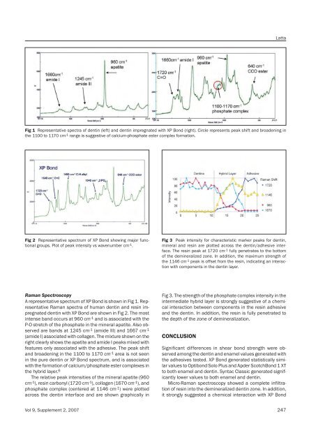
Fig 1 Representative spectra of dentin (left) and dentin impregnated with XP Bond (right). Circle represents peak shift and broadening in<br />
the 1100 to 1170 cm -1 range is suggestive of calcium-phosphate ester complex formation.<br />
Fig 2 Representative spectrum of XP Bond showing major functional<br />
groups. Plot of peak intensity vs wavenumber cm -1 .<br />
Fig 3 Peak intensity for characteristic marker peaks for dentin,<br />
mineral and resin are plotted across the dentin/adhesive interface.<br />
The resin peak at 1720 cm -1 fully penetrates to the bottom<br />
of the demineralized zone. In addition, the maximum strength of<br />
the 1146 cm -1 peak is offset from the resin, indicating an interaction<br />
with components in the dentin layer.<br />
Raman Spectroscopy<br />
A representative spectrum of XP Bond is shown in Fig 1. Representative<br />
Raman spectra of human dentin and resin impregnated<br />
dentin with XP Bond are shown in Fig 2. The most<br />
intense band occurs at 960 cm -1 and is associated with the<br />
P-O stretch of the phosphate in the mineral apatite. Also observed<br />
are bands at 1245 cm -1 (amide III) and 1667 cm -1<br />
(amide I) associated with collagen. The mixture shown on the<br />
right clearly shows the apatite and amide I peaks mixed with<br />
features only associated with the adhesive. The peak shift<br />
and broadening in the 1100 to 1170 cm -1 area is not seen<br />
in the pure dentin or XP Bond spectrum, and is associated<br />
with the formation of calcium/phosphate ester complexes in<br />
the hybrid layer. 6<br />
The relative peak intensities of the mineral apatite (960<br />
cm -1 ), resin carbonyl (1720 cm -1 ), collagen (1670 cm -1 ), and<br />
phosphate complex (centered at 1146 cm -1 ) were plotted<br />
across the dentin interface and are shown graphically in<br />
Fig 3. The strength of the phosphate complex intensity in the<br />
intermediate hybrid layer is strongly suggestive of a chemical<br />
interaction between components in the resin adhesive<br />
and the dentin. In addition, the resin is fully penetrated to<br />
the depth of the zone of demineralization.<br />
CONCLUSION<br />
Significant differences in shear bond strength were observed<br />
among the dentin and enamel values generated with<br />
the adhesives tested. XP Bond generated statistically similar<br />
values to Optibond Solo Plus and Apder ScotchBond 1 XT<br />
to both enamel and dentin. Syntac Classic generated significantly<br />
lower values to both enamel and dentin.<br />
Micro-Raman spectroscopy showed a complete infiltration<br />
of resin into the demineralized dentin zone. In addition,<br />
it strongly suggested a chemical interaction with XP Bond<br />
Vol 9, Supplement 2, 2007 247