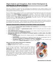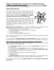
Phyla Cnidaria and Ctenophora, Basic Animal ... - Biosciweb.net
Phyla Cnidaria and Ctenophora, Basic Animal ... - Biosciweb.net
Phyla Cnidaria and Ctenophora, Basic Animal ... - Biosciweb.net

Create successful ePaper yourself
Turn your PDF publications into a flip-book with our unique Google optimized e-Paper software.
<strong>Phyla</strong> <strong>Cnidaria</strong> <strong>and</strong> <strong>Ctenophora</strong>, <strong>Basic</strong> <strong>Animal</strong> Development &<br />
Introduction to Nervous Systems 6.1<br />
Lab #6 - Biological Sciences 102 – <strong>Animal</strong> Biology<br />
This lab is designed to explore the basic physiology <strong>and</strong> adaptations of the radiate animals in<br />
the <strong>Phyla</strong> <strong>Cnidaria</strong> <strong>and</strong> <strong>Ctenophora</strong>. We will also introduce nervous tissues <strong>and</strong> basic animal<br />
development with particular reference to the radiates.<br />
The two radiate phyla, <strong>Cnidaria</strong> (ny-dar'e-a) (= Coelenterata) <strong>and</strong> <strong>Ctenophora</strong> (te-nof'o-ra)<br />
are the most primitive of the eumetazoans (true multicellular animals). They are all<br />
radially (or sometimes biradially) symmetrical. Radially symmetrical animals have a body<br />
plan in which the parts are arranged concentrically around an oral-aboral (central) axis.<br />
These are the simplest animals that actually possess a true tissue level of organization.<br />
Generally, however, the tissues are not organized into organs with specialized functions.<br />
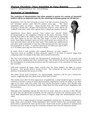
These phyla include several successful groups such as sea anemones, jellyfish, <strong>and</strong> corals,<br />
as well as the hydroids <strong>and</strong> comb jellies. Many cnidarians are brilliantly colored with<br />
many species forming the great tropical coral reefs that harbor the greatest diversity of life<br />
observed in the ocean<br />
Important Characteristics of Members of the <strong>Phyla</strong> <strong>Cnidaria</strong> <strong>and</strong> <strong>Ctenophora</strong><br />
‣ two embryological primary germ layers (ectoderm <strong>and</strong> endoderm) that are<br />
homologous to those of more complex metazoans<br />
‣ internal space for digestion, the gastrovascular cavity, which lies along the polar axis<br />
<strong>and</strong> opens to the outside by a mouth<br />
‣ some cnidarians have a skeleton (eg. coral), but in most radiates, fluid in the<br />
gastrovascular cavity serves as a simple form of hydrostatic skeleton.<br />
‣ Although both cnidarians <strong>and</strong> ctenophores are grouped together as radiate phyla,<br />
they differ in important ways:<br />
o cnidarians have characteristic stinging organelles called nematocysts,<br />
usually absent in ctenophores.<br />
o polymorphism- the presence in a species of more than one morphological<br />
kind of individual- is common in cnidarians but absent in ctenophores.<br />
o ctenophores have distinctive adhesive cells called colloblasts on their<br />
tentacles <strong>and</strong> unique rows of ciliated comb plates not found in other phyla<br />
There are two main types of body form in cnidarians:<br />
1. polyp (hydroid) form, often sessile.<br />
2. medusa (“jellyfish”) form, which is free-swimming.<br />
In some groups of cnidarians, both polyp <strong>and</strong> medusa stages are<br />
found in their life cycle. These animals are therefore<br />
polymorphic. In others, such as sea anemones <strong>and</strong> corals, there<br />
is no medusa; in still others, such as the scyphozoans, or "true"<br />
jellyfish, the polyp stage is reduced or absent. In life cycles<br />
having both polyps <strong>and</strong> medusae, the juvenile polyp stage gives<br />
rise asexually to a medusa, which reproduces sexually. Both<br />
polyp <strong>and</strong> medusa have the diploid number of chromosomes, but<br />
the gametes are haploid.<br />
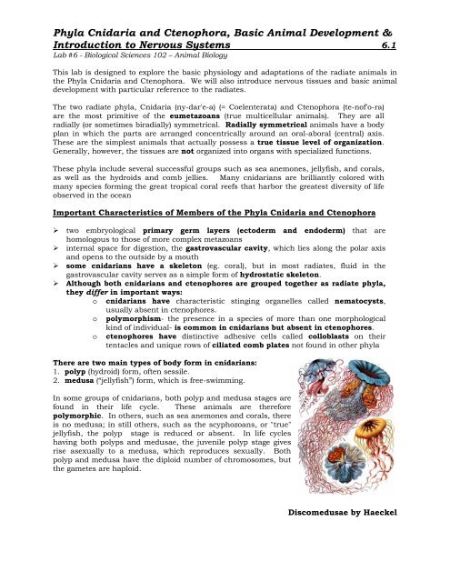
Discomedusae by Haeckel
<strong>Phyla</strong> <strong>Cnidaria</strong> <strong>and</strong> <strong>Ctenophora</strong>, <strong>Basic</strong> <strong>Animal</strong> Development &<br />
Introduction to Nervous Systems 6.2<br />
Lab #6 - Biological Sciences 102 – <strong>Animal</strong> Biology<br />
Classification of the Radiate <strong>Animal</strong>s<br />
Phylum <strong>Cnidaria</strong><br />
Class Hydrozoa (hy-dro-zo'a) (Gr. hydra, water serpent, + zoon, animal).<br />
Both polyp <strong>and</strong> medusa stages represented, although one type may be<br />
suppressed; medusa with a velum; found in fresh <strong>and</strong> marine water. The<br />
hydroids. Examples: Hydra, Obelia, Gonionemus,<br />
Tubularia, Physalia.<br />
Hydra<br />
Class Scyphozoa (sy-fo-zo'a) (Gr. skyphos, cup, + zoon, animal). Solitary; medusa stage<br />
emphasized; polyp reduced or absent; enlarged mesoglea; medusa without a velum. The true<br />
jellyfish. Examples: Aurelia, Rhizostoma, Cassiopeia.<br />
Class Cubozoa (ku'bo-zo'a) (Gr. kybos, a cube, + zQon, animal). Solitary; polyp stage reduced;<br />
bellshaped medusae square in cross section, with a tentacle or group of tentacles at each<br />
corner; margin without velum but with velarium; all marine. Examples: Carybdea, Chironex.<br />
Class Anthozoa (an-tho-zo'a) (Gr. anthos, flower, + zQon, animal). All polyps, no medusae;<br />
gastrovascular cavity subdivided by mesenteries (septa).<br />
Subclass Hexacorallia (hek-sa-ko-ral'e-a) (Gr. hex, six, +<br />
korallion, coral) (Zoantharia). Polyp with simple, unbranched<br />
tentacles; septal arrangement hexamerous; skeleton, when<br />
present, external. Sea anemones <strong>and</strong> stony corals. Examples:<br />
Metridium, Tealia, Astrangia.<br />
Subclass Ceriantipatharia (se-re-an-tip' a-tha' ri-a) (N. 1.<br />
combination of Ceriantharia <strong>and</strong> Antipatharia, from type<br />
genera). With simple, unbranched tentacles; mesenteries<br />
unpaired. Tube anemones <strong>and</strong> black or thorny corals.<br />
Examples: Cerianthus, Antipathes.<br />
Cross section through a<br />
hexacorallian<br />
Subclass Octocorallia (ok'to-ko-ral'e-a) (1. octo, + Gr. korallion, coral) (Alcyonaria). Polyp<br />
with eight pinnate tentacles; septal arrangement octamerous. Soft <strong>and</strong> horny corals.<br />
Examples: Gorgonia, Renilla, Alcyonium.<br />
Phylum <strong>Ctenophora</strong><br />
Class Tentaculata (ten-tak'yu-la'ta) (1. tentaculum, feeler, + ata, group suffix).<br />
With tentacles; tentacles may have sheaths into which they retract; some types<br />
flattened in oral-aboral axis for creeping; others compressed in tentacular<br />
plane to a b<strong>and</strong>-like form; in some the comb plates may be confined to the<br />
larva. Examples: Pleurobrachia, Cestum.<br />
Class Nuda (nu-da) (1. nudus, naked). Without tentacles, but flattened in<br />
tentacular plane; wide mouth <strong>and</strong> pharynx; gastrovascular canals much<br />
branched. Example: Beroe.<br />
A Ctenophore
<strong>Phyla</strong> <strong>Cnidaria</strong> <strong>and</strong> <strong>Ctenophora</strong>, <strong>Basic</strong> <strong>Animal</strong> Development &<br />
Introduction to Nervous Systems 6.3<br />
Lab #6 - Biological Sciences 102 – <strong>Animal</strong> Biology<br />
<strong>Basic</strong> <strong>Animal</strong> Life Cycles & Development (Embryology)<br />
On the chalkboard, your instructor will diagram a basic, generalized animal lifecycle to<br />
introduce the following terms related to animal life cycles <strong>and</strong> development. Your<br />
instructor will then review with you the basic life cycle of the cnidarians, Obelia sp. <strong>and</strong><br />
Aurelia aurita as specific examples.<br />
EARLY ANIMAL DEVELOPMENT (see diagrams from board)<br />
‣ sperm = the male gamete (haploid = N)<br />
‣ ovum (egg) = the female gamete (haploid = N)<br />
‣ fertilization = the species specific binding of the sperm to the egg <strong>and</strong> fusion of the<br />
sperm cell nucleus with the egg cell nucleus to create a diploid zygote<br />
‣ zygote = fertilized egg (diploid = 2N)<br />
‣ cleavage = the mitotic cell divisions of an animal zygote; the first cell divisions that<br />
occur to create a morula from the zygote<br />
‣ morula = a solid balls of cells that froms from the mitotic divisions of the zygote<br />
‣ blastula = the next embryonic stage that marks the end of early cell division during<br />
animal development; an embryo that look like a hollow ball of cells<br />
‣ gastrula = the next embryonic stage resulting from the division of the early cells into<br />
three major tissue types during early animal development. The gastrula may have two<br />
(endoderm & ectoderm only) or three tissue layers (ectoderm, mesoderm, & endoderm)<br />
‣ gastrulation = the process that leads to the formation of a gastrula<br />
‣ ectoderm = the outer layer of three embryonic cell layers in a gastrula; forms the skin<br />
of the gastrula <strong>and</strong> gives rise to the epidermis <strong>and</strong> nervous system of the adult<br />
‣ mesoderm = the middle layer of the three embryonic cell layers in a gastrula; gives rise<br />
to muscles, bones, the dermis of the skin, <strong>and</strong> many other organs in the adult<br />
‣ endoderm = the innermost of three embryonic cell layers in a gastrula; forms the gut of<br />
the gastrula <strong>and</strong> gives rise to the innermost linings of the digestive tract <strong>and</strong> other<br />
hollow organs (e.g. lungs) in the adult<br />
‣ larvae = an immature animal life stage that is significantly different from the adult <strong>and</strong><br />
usually incapable of sexual reproduction<br />
‣ metamorphosis = a drastic change in form during postembryonic development from<br />
the larval stage to another form, usually the adult<br />
‣ adult = the sexually mature form of an animal<br />
Cleavage, the earliest stage in embryonic development, consists of a succession of regular<br />
mitotic cell divisions that partition the egg into a multitude of small cells clustered together. In<br />
lower animals, cleavage is so rapid that hundreds, sometimes thous<strong>and</strong>s, of cells are produced<br />
in a matter of hours.
<strong>Phyla</strong> <strong>Cnidaria</strong> <strong>and</strong> <strong>Ctenophora</strong>, <strong>Basic</strong> <strong>Animal</strong> Development &<br />
Introduction to Nervous Systems 6.4<br />
Lab #6 - Biological Sciences 102 – <strong>Animal</strong> Biology<br />
In animals such as sponges <strong>and</strong> cnidarians, cleavage is irregular <strong>and</strong> seemingly disorganized;<br />
egg cytoplasm is partitioned r<strong>and</strong>omly into daughter cells of highly variable size <strong>and</strong> shape with<br />
no apparent relevance to future cell fates. As the metazoa evolved, however, cleavage began to<br />
follow precise patterns <strong>and</strong> rhythms. In virtually all animal groups above the cnidarians,<br />
cleavage is regular; the egg cytoplasm is segregated into specific cells called blastomeres<br />
(Gr. blastos, bud, + meros, part) occupying discrete positions <strong>and</strong> having specific developmental<br />
fates.<br />
Patterns of regular cleavage depend greatly on amount <strong>and</strong> distribution of yolk in the egg. In<br />
eggs having a large amount of yolk, cleavage may be either complete (= holoblastic), as in<br />
amphibians, or incomplete (= meroblastic), as in birds <strong>and</strong> reptiles. In birds <strong>and</strong> reptiles with<br />
extreme telolecithal (Gr. telos, end, + lekithos, yolk) eggs, cleavage is restricted to a small disc<br />
of cytoplasm on the animal pole; this type of cleavage is called discoidal. The eggs of most<br />
insects follow another pattern of cleavage called superficial. In these the nuclei divide<br />
mitotically into hundreds or thous<strong>and</strong>s of "free" nuclei, which later migrate to the egg surface.<br />
Only then do cleavage furrows form, rapidly partitioning the cytoplasm into a superficial layer<br />
of cells.<br />
In most invertebrates, eggs have little yolk (= isolecithal ["equal-yolk"]), <strong>and</strong> cleavage is<br />
complete (holoblastic) <strong>and</strong> equal. Two major kinds of holoblastic cleavage exist: spiral <strong>and</strong><br />
radial (see text page 156, fig 8-7). The first two cleavages are the same in both kinds of eggs:<br />
the cleavage planes are along the animal-vegetal axis, producing a quartet of cells. At the third<br />
cleavage, however, these two patterns-spiral <strong>and</strong> radial-can be distinguished from each other<br />
by the geometric positioning of the cells.<br />
In radial cleavage, the third cleavage is perpendicular to the first two, yielding two quartets of<br />
cells, with the upper quartet lying directly on top of the lower. In spiral cleavage, the third<br />
cleavage planes are oblique to the polar axis <strong>and</strong> typically produce an upper quartet of smaller<br />
cells that come to lie between the furrows of the lower quartet of larger cells.<br />
There are other important differences between these two cleavage patterns. Spiral cleavage is<br />
typically mosaic, meaning that the embryo is constructed as a mosaic, with each cell fitting<br />
into its predetermined location in the larval body. If cells of the embryo are experimentally<br />
separated at this early stage, each cell will develop into partial or defective larvae because the<br />
developmental fate of each cell has already been determined. Spiral cleavage is found in<br />
several phyla, including annelids, many molluscs, some flatworms, <strong>and</strong> ribbon worms<br />
(nemerteans). All groups showing spiral cleavage belong to the grouping of animal phyla called<br />
the Protostomia, in which the embryonic blastopore forms the mouth.<br />
Early Embryonic Development - Cell Cleavages (mitosis)<br />
Radial cleavage is characteristically regulative<br />
because cell fate does not become fixed until after<br />
the first few cleavages. Radial cleavage is found in<br />
eggs of echinoderms <strong>and</strong> many chordates,<br />
especially protochordates, amphibians, <strong>and</strong><br />
mammals. (As mentioned earlier, eggs of birds <strong>and</strong><br />
reptiles, as well as many fishes, show discoidal<br />
cleavage.) All of these belong to the<br />
Deuterostomia, a group of phyla in which the<br />
mouth is formed from a secondary embryonic<br />
opening.
<strong>Phyla</strong> <strong>Cnidaria</strong> <strong>and</strong> <strong>Ctenophora</strong>, <strong>Basic</strong> <strong>Animal</strong> Development &<br />
Introduction to Nervous Systems 6.5<br />
Lab #6 - Biological Sciences 102 – <strong>Animal</strong> Biology<br />
Introduction to Nervous Systems: Nerve Nets in <strong>Cnidaria</strong>ns<br />
The cnidarian nerve <strong>net</strong> is an excellent example of a simple, diffuse nervous system. The<br />
nerve cells form a plexus (a <strong>net</strong>work) at the base of the epidermis <strong>and</strong> the gastrodermis.<br />
So, there are two interconnected nerve <strong>net</strong>s, one in each tissue layer. Axons (nerve<br />
processes) end on other nerve cells at synapses or neural junctions with sensory cells or<br />
effector organs. As seen in most animals, nerve impulses are transmitted from one cell to<br />
another by release of a neurotransmitter via synaptic vesicles in the presynaptic neuron.<br />
One way transmission between neurons in higher animals occurs because of the relative<br />
activity of the protein channels for sodium <strong>and</strong> potassium in the axons that generate the<br />
electrical signals (action potentials). In addition, synaptic vesicles are only found in the<br />
nerve cell terminal (synaptic buton). <strong>Cnidaria</strong>n nerve <strong>net</strong>s are comprised of cells that<br />
have synaptic vesicles on both sides <strong>and</strong> different ion channel properties allowing<br />
transmission across the synapse in either direction. Most nerve cells in the epidermis are<br />
multipolar (many processes) although some have bipolar neurons (two processes). In<br />
cnidarians, there can be both one-way <strong>and</strong> two-way synapses with other neurons.<br />
<strong>Cnidaria</strong>n neurons lack the myelin sheaths which we will see in other animals. This<br />
myelin is a fatty acid membrane (in fact, a cell membrane) that will increase the speed of<br />
action potentials in larger, more complex animals.<br />
There is no “central nervous system” in cnidarians where a concentration <strong>and</strong><br />
integration between many neurons occurs as seen in other animals. In other animals there<br />
is a cerebral ganglion or brain <strong>and</strong> dorsal or ventral nerve cord which controls the activity of<br />
other peripheral nerve cells. In cnidarians, there is some organization to the nerve <strong>net</strong>.<br />
The nerves are grouped in ring nerves in the medusae of hydrozoans <strong>and</strong> in the marginal<br />
sense organs of scyphozoan medusae. In schyphozoans there is a fast conducting nervous<br />
system to coordinate swimming movements <strong>and</strong> a slower conducting system that<br />
coordinates the activity of the tentacles.<br />
Sensory cells synapse with nerve cells that have junctions with epitheliomuscular cells <strong>and</strong><br />
nematocysts. Collectively this combination of cells forms a neuromuscular system which<br />
is an important milestone in the evolution of nervous systems. In fact, versions of nerve<br />
<strong>net</strong>s (plexuses) coordinate the rhythmic contractions of the digestive systems of many other<br />
more complex invertebrate <strong>and</strong> vertebrate animals.<br />
Some types of sensory cells found scattered in the<br />
cnidarian epidermis:<br />
chemoreceptors = detect molecules/chemicals in the<br />
environment (eg. prey)<br />
mechanoreceptors or tactile receptors = responds to touch or<br />
water movement<br />
Some types of effector cells found scattered in the<br />
cnidarian epidermis:<br />
cnidocytes = have nematocysts<br />
epitheliomuscular cells = for covering <strong>and</strong> muscular<br />
contraction<br />
‣ Near the end of the lab, your instructor will describe the<br />
basic structure of a multipolar nerve cell (neuron).<br />
‣ Be sure to copy <strong>and</strong> label the board diagram in your<br />
notes.
<strong>Phyla</strong> <strong>Cnidaria</strong> <strong>and</strong> <strong>Ctenophora</strong>, <strong>Basic</strong> <strong>Animal</strong> Development &<br />
Introduction to Nervous Systems 6.6<br />
Lab #6 - Biological Sciences 102 – <strong>Animal</strong> Biology<br />
LAB PROCEDURE<br />
NAME:<br />
LAB SCORE:<br />
Class Hydrozoa<br />
<strong>Basic</strong> Body Plan<br />
While the dominant life stage of both Physalia <strong>and</strong> Velella exist as pneustonic (living at the airwater<br />
interface) polyp stage, Physalia is a floating colony of polyps exhibiting extensive<br />
polymorphism while the polyp stage of Velella is actually solitary.<br />
‣ Observe the specimens <strong>and</strong>/or diagrams of the species listed below.<br />
‣ Record the descriptive information requested at the end of the lab for each species.<br />
Aglaophenia latirostris (common name = Ostrich Plume Hydroid)<br />
Physalia physalia (common name = Portugeuese Man-of-War)<br />
Vellela velella (common name = By-the-wind Sailor)
<strong>Phyla</strong> <strong>Cnidaria</strong> <strong>and</strong> <strong>Ctenophora</strong>, <strong>Basic</strong> <strong>Animal</strong> Development &<br />
Introduction to Nervous Systems 6.7<br />
Lab #6 - Biological Sciences 102 – <strong>Animal</strong> Biology<br />
Using the preserved, prepared slides identify the following structures of Obelia sp.<br />
‣ Below, draw <strong>and</strong> label the Obelia sp. (polyp colony) with hydranths <strong>and</strong> gonangia<br />
with clear labels for each of the structures listed below.<br />
‣ Use the inter<strong>net</strong> <strong>and</strong> textbook to assist you in your drawings.<br />
‣ coenosarc<br />
‣ perisarc<br />
‣ gonangium<br />
o medusae in gonangium<br />
‣ hydranth<br />
o hydrotheca<br />
o mouth<br />
o tentacles
<strong>Phyla</strong> <strong>Cnidaria</strong> <strong>and</strong> <strong>Ctenophora</strong>, <strong>Basic</strong> <strong>Animal</strong> Development &<br />
Introduction to Nervous Systems 6.8<br />
Lab #6 - Biological Sciences 102 – <strong>Animal</strong> Biology<br />
‣ Draw <strong>and</strong> CLEARLY label the Obelia sp. medusa from a prepared slide.<br />
‣ Use the inter<strong>net</strong> <strong>and</strong> textbook to assist you in your drawing.<br />
‣ tentacles<br />
‣ radial canals<br />
‣ manubrium<br />
‣ gonads<br />
‣ How many gonads are observed in this species<br />
Feeding Demonstration in a Hydrozoan<br />
Hydra, a Solitary Hydroid<br />
Phylum <strong>Cnidaria</strong><br />
Class Hydrozoa<br />
Order Hydroida<br />
Suborder Anthomedusae<br />
Genus Hydra, Pelmatohydra, or Chlorohydra<br />
Hydras are found in pools, quiet streams <strong>and</strong> ponds often on the underside of aquatic<br />
vegetation. You instructor will prepare a slide of hydra for your observation. The hydra may<br />
be contracted at first <strong>and</strong> then extend. Small ciliated protozoans which are symbionts of<br />
hydras are sometimes seen gliding over the body <strong>and</strong> tentacles.<br />
Your instructor will add a drop of Daphnia or similar food to the slide. Some extracellular<br />
digestion occurs within the gastrovascular cavity via enzymes released by gl<strong>and</strong> cells. Food<br />
particles are then engulfed by cells of the gastrodermis <strong>and</strong> digestion is completed<br />
intracellularly. Undigested food is regurgitated as there is no anus.<br />
‣ Observe <strong>and</strong> briefly describe the feeding reaction of the hydra below:
<strong>Phyla</strong> <strong>Cnidaria</strong> <strong>and</strong> <strong>Ctenophora</strong>, <strong>Basic</strong> <strong>Animal</strong> Development &<br />
Introduction to Nervous Systems 6.9<br />
Lab #6 - Biological Sciences 102 – <strong>Animal</strong> Biology<br />
Class Scyphozoa<br />
<strong>Basic</strong> Body Plans & Life Cycles<br />
‣ Clearly label the stages of the life cycle of Aurelia aurita shown below.<br />
‣ Use the inter<strong>net</strong> <strong>and</strong> textbook to assist you in with the labels.
<strong>Phyla</strong> <strong>Cnidaria</strong> <strong>and</strong> <strong>Ctenophora</strong>, <strong>Basic</strong> <strong>Animal</strong> Development &<br />
Introduction to Nervous Systems 6.10<br />
Lab #6 - Biological Sciences 102 – <strong>Animal</strong> Biology<br />
‣ Look at the prepared slides of the following life stages of a life cycle for Aurelia aurita.<br />
‣ Make drawings <strong>and</strong> clearly label them to complete the diagram below.<br />
‣ Use the inter<strong>net</strong> <strong>and</strong> textbook to assist you in your drawings.<br />
‣ scyphistoma<br />
‣ strobila<br />
‣ ephyra
<strong>Phyla</strong> <strong>Cnidaria</strong> <strong>and</strong> <strong>Ctenophora</strong>, <strong>Basic</strong> <strong>Animal</strong> Development &<br />
Introduction to Nervous Systems 6.11<br />
Lab #6 - Biological Sciences 102 – <strong>Animal</strong> Biology<br />
Class Cubozoa<br />
‣ Observe the specimen <strong>and</strong>/or diagram of Carybdea sp.<br />
‣ Record the descriptive information requested at the end of the lab for this species.<br />
Class Anthozoa<br />
Subclass Hexacorallia<br />
‣ Observe the specimens <strong>and</strong>/or diagrams of the following species.<br />
‣ Record the descriptive information requested at the end of the lab for these species.<br />
‣ Balanophyllia elegans<br />
Note that the scleractinian hexacorallians (stony corals) synthesize a consolidated calcium<br />
carbonate skeleton while anemones do not.<br />
‣ Anthopleura sola<br />
‣ What accounts for the green color often present in this species<br />
Feeding Demonstration in an Anthozoan<br />
As available, your instructor may feed a small piece of mussel to one of the anemones on<br />
display in the lab.<br />
‣ Briefly describe the feeding behavior of the anemone:<br />
Cnidocytes <strong>and</strong> Examination of Nematocyst Discharge<br />
Cnidocytes are cells found in the epidermis between epitheliomuscular cells of<br />
cnidarians. Cnidocytes have nematocysts which are tiny capsules composed of material<br />
similar to chitin <strong>and</strong> contain a coiled tubular filament which is a continuation of the<br />
narrowed end of the capsule. This end of the cnidocyte is covered by the operculum. The<br />
base of the filament may have spines are barbs. The filament <strong>and</strong> barbs may contain a<br />
poison. The cnidocytes in anthozoans do not have the trigger-like cnidocil. The cnidocil is<br />
a modified cilium. In anthozoans a modified ciliary-like mechanoreceptor is involved in<br />
triggering nematocyst discharge.
<strong>Phyla</strong> <strong>Cnidaria</strong> <strong>and</strong> <strong>Ctenophora</strong>, <strong>Basic</strong> <strong>Animal</strong> Development &<br />
Introduction to Nervous Systems 6.12<br />
Lab #6 - Biological Sciences 102 – <strong>Animal</strong> Biology<br />
Discharge of nematocysts involves the following events:<br />
1. molecules released from prey may sensitize the cnidocytes, but tactile stimulation causes<br />
the nematocysts to discharge<br />
2. discharge is due to tensional forces created by a very high osmotic pressure within the<br />
nematocyst<br />
3. when triggered the high osmotic pressure causes water to rush in to the capsule.<br />
3. the build of water (increased hydrostatic pressure) forces the filament out <strong>and</strong> it turns<br />
inside out at the same time<br />
4. the barbs are then exposed <strong>and</strong>, if present, poison is injected into the prey<br />
Nematocysts of most cnidarians are not harmful to humans, but the stings of Physalia <strong>and</strong><br />
some true jellies are painful <strong>and</strong> can be dangerous – particularly if the individual is allergic<br />
to the organic poison.<br />
The instructor will remove the tip of a tentacle from Corynactis californica or similar species<br />
<strong>and</strong> prepare a slide by squashing the tip under a cover slip.<br />
‣ Label the undischarged <strong>and</strong> sketch a discharged nematocyst below.<br />
UNDISCHARGED NEMATOCYST<br />
DISCHARGED NEMATOCYST<br />
Subclass Ceriantipatharia<br />
Order Ceriantharia (tube anemones)<br />
‣ Observe the specimens <strong>and</strong>/or diagrams of the following species.<br />
‣ Record the descriptive information requested at the end of the lab for these species.<br />
‣ The available ceriantharian such as Pachycerianthus fimbriatus<br />
Subclass Octocorallia<br />
Under a dissecting scope, observe the polyp morphology of Lophogorgia chilensis or Renilla<br />
kollikeri to identify the unique features of an octocorallian polyp.<br />
‣ Record the descriptive information requested at the end of the lab for these species.
<strong>Phyla</strong> <strong>Cnidaria</strong> <strong>and</strong> <strong>Ctenophora</strong>, <strong>Basic</strong> <strong>Animal</strong> Development &<br />
Introduction to Nervous Systems 6.13<br />
Lab #6 - Biological Sciences 102 – <strong>Animal</strong> Biology<br />
For the live specimens available for observation in the lab, record the requested<br />
information.<br />
Be sure to include descriptions of any live members of the Phylum <strong>Ctenophora</strong> if<br />
they are available.<br />
Phylum <strong>Cnidaria</strong><br />
Class Hydrozoa<br />
Scientific name: Aglaophenia latirostris<br />
Common name:<br />
General dimensions of specimen:<br />
Color of specimen:<br />
Life stage present (observed):<br />
Solitary or colonial:<br />
Benthic, planktonic or pneustonic:<br />
Unique structures or features:<br />
Draw a simple sketch to remind you of the basic structure of this species <strong>and</strong> any unique<br />
characteristics observed.<br />
Notes & observations to help you remember <strong>and</strong> distinguish this group/species:
<strong>Phyla</strong> <strong>Cnidaria</strong> <strong>and</strong> <strong>Ctenophora</strong>, <strong>Basic</strong> <strong>Animal</strong> Development &<br />
Introduction to Nervous Systems 6.14<br />
Lab #6 - Biological Sciences 102 – <strong>Animal</strong> Biology<br />
Phylum <strong>Cnidaria</strong><br />
Class Hydrozoa<br />
Scientific name: Physalia physalia<br />
Common name:<br />
General dimensions of specimen:<br />
Color of specimen:<br />
Life stage present (observed):<br />
Solitary or colonial:<br />
Benthic, planktonic or pneustonic:<br />
Unique structures or features:<br />
Draw a simple sketch to remind you of the basic structure of this species <strong>and</strong> any unique<br />
characteristics observed.<br />
Notes & observations to help you remember <strong>and</strong> distinguish this group/species:
<strong>Phyla</strong> <strong>Cnidaria</strong> <strong>and</strong> <strong>Ctenophora</strong>, <strong>Basic</strong> <strong>Animal</strong> Development &<br />
Introduction to Nervous Systems 6.15<br />
Lab #6 - Biological Sciences 102 – <strong>Animal</strong> Biology<br />
Phylum <strong>Cnidaria</strong><br />
Class Hydrozoa<br />
Scientific name: Vellela velella<br />
Common name:<br />
General dimensions of specimen:<br />
Color of specimen:<br />
Life stage present (observed):<br />
Solitary or colonial:<br />
Benthic, planktonic or pneustonic:<br />
Unique structures or features:<br />
Draw a simple sketch to remind you of the basic structure of this species <strong>and</strong> any unique<br />
characteristics observed.<br />
Notes & observations to help you remember <strong>and</strong> distinguish this group/species:
<strong>Phyla</strong> <strong>Cnidaria</strong> <strong>and</strong> <strong>Ctenophora</strong>, <strong>Basic</strong> <strong>Animal</strong> Development &<br />
Introduction to Nervous Systems 6.16<br />
Lab #6 - Biological Sciences 102 – <strong>Animal</strong> Biology<br />
Phylum <strong>Cnidaria</strong><br />
Class Scyphozoa<br />
Scientific name: Aurelia aurita<br />
Common name:<br />
General dimensions of specimen:<br />
Color of specimen:<br />
Life stage present (observed):<br />
Solitary or colonial:<br />
Benthic, planktonic or pneustonic:<br />
Unique structures or features:<br />
Draw a simple sketch to remind you of the basic structure of this species <strong>and</strong> any unique<br />
characteristics observed.<br />
Notes & observations to help you remember <strong>and</strong> distinguish this group/species:
<strong>Phyla</strong> <strong>Cnidaria</strong> <strong>and</strong> <strong>Ctenophora</strong>, <strong>Basic</strong> <strong>Animal</strong> Development &<br />
Introduction to Nervous Systems 6.17<br />
Lab #6 - Biological Sciences 102 – <strong>Animal</strong> Biology<br />
Phylum <strong>Cnidaria</strong><br />
Class Cubozoa<br />
Scientific name: Carybdea sp.<br />
Common name:<br />
General dimensions of specimen:<br />
Color of specimen:<br />
Life stage present (observed):<br />
Solitary or colonial:<br />
Benthic, planktonic or pneustonic:<br />
Unique structures or features:<br />
Draw a simple sketch to remind you of the basic structure of this species <strong>and</strong> any unique<br />
characteristics observed.<br />
Notes & observations to help you remember <strong>and</strong> distinguish this group/species:
<strong>Phyla</strong> <strong>Cnidaria</strong> <strong>and</strong> <strong>Ctenophora</strong>, <strong>Basic</strong> <strong>Animal</strong> Development &<br />
Introduction to Nervous Systems 6.18<br />
Lab #6 - Biological Sciences 102 – <strong>Animal</strong> Biology<br />
Phylum <strong>Cnidaria</strong><br />
Class Anthozoa<br />
Subclass Hexacorallia<br />
Scientific name: Balanophyllia elegans or other hexacorallian<br />
Common name:<br />
General dimensions of specimen:<br />
Color of specimen:<br />
Life stage present (observed):<br />
Solitary or colonial:<br />
Benthic, planktonic or pneustonic:<br />
Unique structures or features:<br />
Draw a simple sketch to remind you of the basic structure of this species <strong>and</strong> any unique<br />
characteristics observed.<br />
Notes & observations to help you remember <strong>and</strong> distinguish this group/species:
<strong>Phyla</strong> <strong>Cnidaria</strong> <strong>and</strong> <strong>Ctenophora</strong>, <strong>Basic</strong> <strong>Animal</strong> Development &<br />
Introduction to Nervous Systems 6.19<br />
Lab #6 - Biological Sciences 102 – <strong>Animal</strong> Biology<br />
Phylum <strong>Cnidaria</strong><br />
Class Anthozoa<br />
Subclass Hexacorallia<br />
Scientific name: Anthopleura sola<br />
Common name:<br />
General dimensions of specimen:<br />
Color of specimen:<br />
Life stage present (observed):<br />
Solitary or colonial:<br />
Benthic, planktonic or pneustonic:<br />
Unique structures or features:<br />
Draw a simple sketch to remind you of the basic structure of this species <strong>and</strong> any unique<br />
characteristics observed.<br />
Notes & observations to help you remember <strong>and</strong> distinguish this group/species:
<strong>Phyla</strong> <strong>Cnidaria</strong> <strong>and</strong> <strong>Ctenophora</strong>, <strong>Basic</strong> <strong>Animal</strong> Development &<br />
Introduction to Nervous Systems 6.20<br />
Lab #6 - Biological Sciences 102 – <strong>Animal</strong> Biology<br />
Phylum <strong>Cnidaria</strong><br />
Class Anthozoa<br />
Subclass Ceriantipatharia<br />
Order Ceriantharia<br />
Scientific name: Pachycerianthus fimbriatus<br />
Common name:<br />
General dimensions of specimen:<br />
Color of specimen:<br />
Life stage present (observed):<br />
Solitary or colonial:<br />
Benthic, planktonic or pneustonic:<br />
Unique structures or features:<br />
Draw a simple sketch to remind you of the basic structure of this species <strong>and</strong> any unique<br />
characteristics observed.<br />
Notes & observations to help you remember <strong>and</strong> distinguish this group/species:
<strong>Phyla</strong> <strong>Cnidaria</strong> <strong>and</strong> <strong>Ctenophora</strong>, <strong>Basic</strong> <strong>Animal</strong> Development &<br />
Introduction to Nervous Systems 6.21<br />
Lab #6 - Biological Sciences 102 – <strong>Animal</strong> Biology<br />
Phylum <strong>Cnidaria</strong><br />
Class Anthozoa<br />
Subclass Octocorallia<br />
Scientific name: Lophogorgia chilensis<br />
Common name:<br />
General dimensions of specimen:<br />
Color of specimen:<br />
Life stage present (observed):<br />
Solitary or colonial:<br />
Benthic, planktonic or pneustonic:<br />
Unique structures or features:<br />
Draw a simple sketch to remind you of the basic structure of this species <strong>and</strong> any unique<br />
characteristics observed.<br />
Notes & observations to help you remember <strong>and</strong> distinguish this group/species:
<strong>Phyla</strong> <strong>Cnidaria</strong> <strong>and</strong> <strong>Ctenophora</strong>, <strong>Basic</strong> <strong>Animal</strong> Development &<br />
Introduction to Nervous Systems 6.22<br />
Lab #6 - Biological Sciences 102 – <strong>Animal</strong> Biology<br />
Phylum <strong>Cnidaria</strong><br />
Class Anthozoa<br />
Subclass Octocorallia<br />
Scientific name: Renilla kollikeri or other octocorallian<br />
Common name:<br />
General dimensions of specimen:<br />
Color of specimen:<br />
Life stage present (observed):<br />
Solitary or colonial:<br />
Benthic, planktonic or pneustonic:<br />
Unique structures or features:<br />
Draw a simple sketch to remind you of the basic structure of this species <strong>and</strong> any unique<br />
characteristics observed.<br />
Notes & observations to help you remember <strong>and</strong> distinguish this group/species:
<strong>Phyla</strong> <strong>Cnidaria</strong> <strong>and</strong> <strong>Ctenophora</strong>, <strong>Basic</strong> <strong>Animal</strong> Development &<br />
Introduction to Nervous Systems 6.23<br />
Lab #6 - Biological Sciences 102 – <strong>Animal</strong> Biology<br />
Phylum <strong>Ctenophora</strong> (if available)<br />
Class:<br />
Scientific name:<br />
Common name:<br />
General dimensions of specimen:<br />
Color of specimen:<br />
Life stage present (observed):<br />
Solitary or colonial:<br />
Benthic, planktonic or pneustonic:<br />
Unique structures or features:<br />
Draw a simple sketch to remind you of the basic structure of this species <strong>and</strong> any unique<br />
characteristics observed.<br />
Notes & observations to help you remember <strong>and</strong> distinguish this group/species:
<strong>Phyla</strong> <strong>Cnidaria</strong> <strong>and</strong> <strong>Ctenophora</strong>, <strong>Basic</strong> <strong>Animal</strong> Development &<br />
Introduction to Nervous Systems 6.24<br />
Lab #6 - Biological Sciences 102 – <strong>Animal</strong> Biology<br />
LABORATORY NOTES:<br />
Actiniae by Haeckel<br />
Ernst Haeckel (1834 – 1919) was an eminent German biologist, naturalist, philosopher,<br />
physician, professor <strong>and</strong> artist who discovered, described <strong>and</strong> named thous<strong>and</strong>s of new<br />
species, mapped a genealogical tree relating all life forms, <strong>and</strong> coined many terms in biology,<br />
including phylum, phylogeny, ecology <strong>and</strong> the kingdom Protista. Haeckel promoted <strong>and</strong><br />
popularized Charles Darwin's work in Germany <strong>and</strong> developed the controversial recapitulation<br />
theory ("ontogeny recapitulates phylogeny") claiming that an individual organism's biological<br />
development, or ontogeny, parallels <strong>and</strong> summarizes its species' entire evolutionary<br />
development, or phylogeny.
<strong>Phyla</strong> <strong>Cnidaria</strong> <strong>and</strong> <strong>Ctenophora</strong>, <strong>Basic</strong> <strong>Animal</strong> Development &<br />
Introduction to Nervous Systems 6.25<br />
Lab #6 - Biological Sciences 102 – <strong>Animal</strong> Biology<br />
LABORATORY NOTES:<br />
Peromedusae by Haeckel<br />
Discomedusae by Haeckel<br />
Trachomedusae by Haeckel
<strong>Phyla</strong> <strong>Cnidaria</strong> <strong>and</strong> <strong>Ctenophora</strong>, <strong>Basic</strong> <strong>Animal</strong> Development &<br />
Introduction to Nervous Systems 6.26<br />
Lab #6 - Biological Sciences 102 – <strong>Animal</strong> Biology<br />
LABORATORY NOTES: