3D Ultrasound in Gynaecology - isuog
3D Ultrasound in Gynaecology - isuog
3D Ultrasound in Gynaecology - isuog
You also want an ePaper? Increase the reach of your titles
YUMPU automatically turns print PDFs into web optimized ePapers that Google loves.
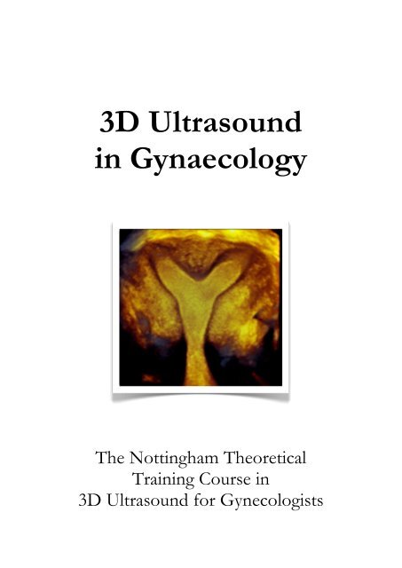
<strong>3D</strong> <strong>Ultrasound</strong><br />
<strong>in</strong> <strong>Gynaecology</strong><br />
The Nott<strong>in</strong>gham Theoretical<br />
Tra<strong>in</strong><strong>in</strong>g Course <strong>in</strong><br />
<strong>3D</strong> <strong>Ultrasound</strong> for Gynecologists
Dear Friends,<br />
I would like to <strong>in</strong>vite you to Nott<strong>in</strong>gham to jo<strong>in</strong> us for this unique and excit<strong>in</strong>g course<br />
dedicated to <strong>3D</strong> gynaecological ultrasound. The course, which has now been runn<strong>in</strong>g for<br />
five years, has received excellent feedback and comes highly recommended. I promise it<br />
will prove to be <strong>in</strong>tellectually stimulat<strong>in</strong>g and reward<strong>in</strong>g for each and every one of you<br />
regardless of your expertise and knowledge. The course has been designed specifically for<br />
those of you who practice gynaecological ultrasound and want to learn more about<br />
advanced scann<strong>in</strong>g techniques particularly <strong>3D</strong> and Doppler ultrasound but is also suitable<br />
for beg<strong>in</strong>ners and improvers. Its’ bespoke nature allows us to personalise the course for all<br />
who attend so do let us know what you hope to achieve either before or when you arrive.<br />
The aims of the course, which has dist<strong>in</strong>ct practical and cl<strong>in</strong>ical components, <strong>in</strong>clude:<br />
Establish<strong>in</strong>g the basic pr<strong>in</strong>ciples of gynaecological ultrasound:<br />
Ø gett<strong>in</strong>g the best out of your mach<strong>in</strong>e<br />
Ø assessment of the pelvis with 2D and Doppler ultrasound<br />
Qualitative and quantitative <strong>3D</strong> ultrasound:<br />
Ø <strong>3D</strong> data acquisition and image display<br />
Ø manual and automatic quantification of <strong>3D</strong> data<br />
Ø the practical applications and cl<strong>in</strong>ical relevance of <strong>3D</strong><br />
Hands-on practical sessions:<br />
Ø live scann<strong>in</strong>g (us<strong>in</strong>g a gynecology phantom)<br />
Ø 4D View: basic and advanced applications<br />
Ø 4D View: cl<strong>in</strong>ical cases and worked examples<br />
A large proportion of the course <strong>in</strong>volves work<strong>in</strong>g with 4D View. Each delegate will be<br />
given a USB or a CD, which conta<strong>in</strong> a series of <strong>3D</strong> datasets that we will look through<br />
together as a group dur<strong>in</strong>g the hands-on practical sessions to provide work<strong>in</strong>g examples<br />
of the topics discussed. You will become confident with the software and leave be<strong>in</strong>g<br />
able to use the different display options and perform reliable measurements of volume<br />
and vascularity. You can assess your progress <strong>in</strong> an <strong>in</strong>formal quiz on the f<strong>in</strong>al day! We<br />
also have Voluson mach<strong>in</strong>es and a gynaecological phantom to practice ‘live’ scann<strong>in</strong>g.<br />
The course is held at the Park Plaza Hotel, a modern, four-star hotel <strong>in</strong> the heart of the<br />
city with<strong>in</strong> walk<strong>in</strong>g distance of the ma<strong>in</strong> shopp<strong>in</strong>g and bus<strong>in</strong>ess districts. Accommodation<br />
and cater<strong>in</strong>g dur<strong>in</strong>g the day are provided <strong>in</strong> the course fee. The course d<strong>in</strong>ner is held on<br />
the Friday leav<strong>in</strong>g Thursday even<strong>in</strong>g free for you to do as you please. Please note that<br />
whilst we can offer advice and help arrange your transport to and from the venue this is<br />
not covered <strong>in</strong> the course fee and will be added to your <strong>in</strong>voice.<br />
Venue:<br />
Park Plaza Nott<strong>in</strong>gham<br />
41 Maid Marian Way<br />
Nott<strong>in</strong>gham NG1 6GD<br />
For further <strong>in</strong>formation:<br />
E-mail: adm<strong>in</strong>@tealefenn<strong>in</strong>g.co.uk<br />
Telephone: +44 (0) 115 9418152<br />
Mobile: +44 (0) 774 9134750 The legendary Rob<strong>in</strong> Hood
Timetable<br />
Thu Session Learn<strong>in</strong>g objective<br />
GETTING THE BASICS RIGHT<br />
13.00 Introduction & Welcome An Overview of the Course<br />
13.15 Sett<strong>in</strong>g up Your Mach<strong>in</strong>e How to Get the Best 2D Image<br />
13.45 2D Assessment of the Pelvis Def<strong>in</strong><strong>in</strong>g Standards <strong>in</strong> Gynecological Scann<strong>in</strong>g<br />
14.15 <strong>3D</strong> / 4D Data Acquisition How to Acquire Your <strong>3D</strong> datasets<br />
14.30 Live ‘Phantom’ Demonstration Putt<strong>in</strong>g this Altogether on the E8 / Voluson-i<br />
15.15 COFFEE<br />
15.30<br />
4D View:<br />
An Introduction<br />
Dataset Management, Image Sett<strong>in</strong>gs,<br />
Options for Image Display and Standardization<br />
16.30 Practical Session 1 How to Get the Best Image<br />
Even<strong>in</strong>g: free (please ask for a list of recommended restaurants)<br />
Fri Lecture Learn<strong>in</strong>g objective<br />
ADVANCED <strong>3D</strong> IMAGING<br />
09.00<br />
4D View:<br />
Advanced Options<br />
Tomographic <strong>Ultrasound</strong> Imag<strong>in</strong>g, Omni View,<br />
Image Render<strong>in</strong>g, and C<strong>in</strong>e Mode<br />
09.45 Practical Session 2 Advanced Image Displays and Render<strong>in</strong>g<br />
11.00 COFFEE<br />
11.15<br />
Cl<strong>in</strong>ical Applications of<br />
<strong>3D</strong> <strong>Ultrasound</strong><br />
Diagnostic Advantages of <strong>3D</strong>:<br />
The Uterus and Ovaries<br />
12.00 Practical Session 3 Cl<strong>in</strong>ical Cases<br />
13.00 LUNCH
Fri Lecture Learn<strong>in</strong>g objective<br />
QUANTITATIVE <strong>3D</strong> IMAGING<br />
13:45<br />
Volume<br />
Analysis<br />
VOCAL and SonoAVC:<br />
Techniques and Cl<strong>in</strong>ical Applications<br />
14.30 Practical Session 4 Manual and Automatic Volume Calculation<br />
15.30 COFFEE<br />
15.45<br />
Doppler<br />
<strong>Ultrasound</strong><br />
Qualitative and Quantitative 2D and <strong>3D</strong> Doppler:<br />
Techniques and Cl<strong>in</strong>ical Applications<br />
16.30 Practical Session 5 Qualitative and Quantitative <strong>3D</strong> power Doppler<br />
Even<strong>in</strong>g: Course D<strong>in</strong>ner<br />
Sat Lecture Learn<strong>in</strong>g objective<br />
CLINICAL APPLICATIONS<br />
09.00 Early Pregnancy Normal and Abnormal Pregnancy<br />
10.00 Urogynecology Urogynecology and the Pelvic Floor<br />
10.30 Gyne-oncology Ovarian and Endometrial Cancer<br />
11.00 COFFEE<br />
11.15 Practical Session 6 Cl<strong>in</strong>ical Cases & Measurements - Quiz<br />
12.30 The F<strong>in</strong>al Word Answers and an Overview of the Course<br />
13.00 LUNCH<br />
Meet<strong>in</strong>g closes
Important<br />
Please note that the practical sessions us<strong>in</strong>g 4D View require that it is essential to:<br />
1. br<strong>in</strong>g your own laptop, and<br />
2. have a work<strong>in</strong>g version of 4D View <strong>in</strong>stalled on it<br />
Previous courses have shown this ensures you get the most out of the course.<br />
There are 3 ways to do this:<br />
1. Purchase 4D View Option from GE - & <strong>in</strong>stall it on your computer. It is essential to<br />
remember the Dongle to allow the 4D View programme to open and work the datasets.<br />
2. Install the 60 day demo version - this option does not require a dongle and lasts for 60<br />
days from when you first open the application (please note this version does not offer<br />
sonoAVC and cannot currently be <strong>in</strong>stalled on PCs runn<strong>in</strong>g W<strong>in</strong>dows Vista)<br />
3. Install a “free unlimited de-featured version” - this option does not require a dongle<br />
and does not expire over time but has reduced <strong>3D</strong> / 4D data capability and several<br />
restrictions as it does not offer:<br />
⊗ volume data storage / archiv<strong>in</strong>g<br />
⊗ VOCAL<br />
⊗ SRI filter<br />
⊗ <strong>in</strong>version mode<br />
⊗ static VCI<br />
⊗ sonoAVC<br />
Whilst you would still be able to open and manipulate the data files supplied dur<strong>in</strong>g the<br />
course the absence of these facilities may reduce your enjoyment and learn<strong>in</strong>g<br />
experience. We would only advise you to use this version if you are unable to load the<br />
demo version (i.e. you have W<strong>in</strong>dows Vista or have previously used the demo version<br />
and the 60 day free trial period has expired)<br />
For further <strong>in</strong>formation, and to download 4D View, please visit the Voluson Club<br />
website: http://www.volusonclub.net/emea/4dview<br />
Please note – 4D View does not work with Apple Mac<strong>in</strong>tosh computers unless you have<br />
<strong>in</strong>stalled ‘Parallels’ or ‘WMware Fusion’ but we recommend you check this before your arrival.<br />
We do have a limited number of spare laptops but would strongly advise you to br<strong>in</strong>g your own<br />
to avoid disappo<strong>in</strong>tment. We also recommend that you check your version works before you<br />
leave by physically open<strong>in</strong>g and work<strong>in</strong>g on one or two datasets. Please feel free to br<strong>in</strong>g any<br />
<strong>in</strong>terest<strong>in</strong>g cases along with you. We can look at these as a group.<br />
I s<strong>in</strong>cerely look forward to welcom<strong>in</strong>g you to Nott<strong>in</strong>gham.<br />
Yours s<strong>in</strong>cerely,<br />
Nick Ra<strong>in</strong>e-Fenn<strong>in</strong>g<br />
N J Ra<strong>in</strong>e-Fenn<strong>in</strong>g<br />
MBChB MRCOG PhD<br />
Course Director & Convenor
Biography<br />
Nick is a Consultant Gynecologist and Reader / Associate Professor of Reproductive Medic<strong>in</strong>e<br />
& Surgery <strong>in</strong> Nott<strong>in</strong>gham. He is based with<strong>in</strong> the University of Nott<strong>in</strong>gham’s assisted conception<br />
unit, NURTURE (Nott<strong>in</strong>gham University Research and Treatment Unit <strong>in</strong> Reproduction), at<br />
Queen’s Medical Centre where he is the lead for Academic Imag<strong>in</strong>g and Director of Research.<br />
Nick has a special <strong>in</strong>terest <strong>in</strong> both gynaecological ultrasound and reproductive medic<strong>in</strong>e. He is an<br />
<strong>in</strong>ternationally recognised expert <strong>in</strong> three-dimensional ultrasound and was awarded a PhD <strong>in</strong><br />
2004 for work relat<strong>in</strong>g to the quantification of pelvic blood flow us<strong>in</strong>g quantitative <strong>3D</strong> power<br />
Doppler angiography. He is Deputy Editor-<strong>in</strong>-Chief of <strong>Ultrasound</strong> <strong>in</strong> Obstetrics & Gynecology,<br />
the lead journal <strong>in</strong> acoustics, and Board member of the International Society for <strong>Ultrasound</strong> <strong>in</strong><br />
Obstetrics and Gynecology (ISUOG) where he sits on the Cl<strong>in</strong>ical Standards, Web, and<br />
Education Committees. He has been an <strong>in</strong>vited speaker on the VISUS Course s<strong>in</strong>ce 2004 and has<br />
made a DVD about volume ultrasound <strong>in</strong> gynecology that is available through the Voluson Club.<br />
Selected publications relevant to the Course:<br />
Jayaprakasan K, Chan Y, Islam R, Haoula Z, Hopkisson J, Coomarasamy A, Ra<strong>in</strong>e-Fenn<strong>in</strong>g N. Prediction of <strong>in</strong> vitro fertilization<br />
outcome at different antral follicle count thresholds <strong>in</strong> a prospective cohort of 1,012 women. Fertility and Sterility. 2012<br />
Sur SD, Clewes JS, Campbell BK, Ra<strong>in</strong>e-Fenn<strong>in</strong>g NJ. Embryo volume measurement: An <strong>in</strong>traobserver, <strong>in</strong>termethod comparative<br />
study of semi-automated and manual <strong>3D</strong> ultrasound techniques. <strong>Ultrasound</strong> <strong>in</strong> Obstetrics & Gynecology. 2011;38:516-523<br />
Ra<strong>in</strong>e-Fenn<strong>in</strong>g N. What's <strong>in</strong> a number The polycystic ovary revisited. Hum Reprod. 2011;26:3118-3122<br />
Mart<strong>in</strong>s WP, Welsh AW, Lima JC, Nastri CO, Ra<strong>in</strong>e-Fenn<strong>in</strong>g NJ. The "volumetric" pulsatility <strong>in</strong>dex as evaluated by spatiotemporal<br />
imag<strong>in</strong>g correlation (STIC). <strong>Ultrasound</strong> <strong>in</strong> Medic<strong>in</strong>e & Biology. 2011;37:2160-2168<br />
Mart<strong>in</strong>s WP, Ra<strong>in</strong>e-Fenn<strong>in</strong>g NJ, Leite SP, Ferriani RA, Nastri CO. A standardized measurement technique may improve the reliability<br />
of measurements of endometrial thickness and volume. <strong>Ultrasound</strong> <strong>in</strong> Obstetrics & Gynecology. 2011;38:107-115<br />
Jayaprakasan K, Chan YY, Sur S, Deb S, Clewes JS, Ra<strong>in</strong>e-Fenn<strong>in</strong>g NJ. Prevalence of uter<strong>in</strong>e anomalies and their impact on early<br />
pregnancy <strong>in</strong> women conceiv<strong>in</strong>g after assisted reproduction treatment. <strong>Ultrasound</strong> <strong>in</strong> Obstetrics & Gynecology. 2011;37:727-732<br />
Chan YY, Jayaprakasan K, Zamora J, Thornton JG, Ra<strong>in</strong>e-Fenn<strong>in</strong>g N, Coomarasamy A. The prevalence of congenital uter<strong>in</strong>e<br />
anomalies <strong>in</strong> unselected and high-risk populations: a systematic review. Human Reproduction Update. 2011;17:761-771<br />
Sur SD, Jayaprakasan K, Jones NW, Clewes J, W<strong>in</strong>ter B, Cash N, Campbell B, Ra<strong>in</strong>e-Fenn<strong>in</strong>g NJ. A novel technique for the semiautomated<br />
measurement of embryo volume: An <strong>in</strong>traobserver reliability study. <strong>Ultrasound</strong> <strong>in</strong> Medic<strong>in</strong>e & Biology. 2010;36:719-725<br />
Deb S, Campbell BK, Clewes JS, Ra<strong>in</strong>e-Fenn<strong>in</strong>g NJ. Quantitative analysis of antral follicle number and size: a comparison of twodimensional<br />
and automated three-dimensional ultrasound. <strong>Ultrasound</strong> Obstet Gynecol. 2010 Mar; 35(3): 354-60.<br />
Ra<strong>in</strong>e-Fenn<strong>in</strong>g N, Jayaprakasan K, Chamberla<strong>in</strong> S, Devl<strong>in</strong> L, Priddle H, Johnson I. Automated measurements of follicle diameter: a<br />
chance to standardize Fertil Steril. Apr 2009;91(4):1469-1472.<br />
Ra<strong>in</strong>e-Fenn<strong>in</strong>g N, Deb S, Jayaprakasan K, Campbell B. Tim<strong>in</strong>g of oocyte maturation and egg collection dur<strong>in</strong>g controlled ovarian<br />
stimulation: RCT evaluat<strong>in</strong>g manual and automated measurements of follicle diameter. Fertil Steril. Mar 2009.<br />
Ra<strong>in</strong>e-Fenn<strong>in</strong>g N, Nord<strong>in</strong> N, Ramnar<strong>in</strong>e K, Campbell B, Clewes J, Perk<strong>in</strong>s A, Johnson I. Evaluation of the effect of mach<strong>in</strong>e sett<strong>in</strong>gs<br />
on quantitative <strong>3D</strong> power Doppler angiography. <strong>Ultrasound</strong> Obstet Gynecol. Sep 2008;32(4):551-559.<br />
Ra<strong>in</strong>e-Fenn<strong>in</strong>g N, Nord<strong>in</strong> N, Ramnar<strong>in</strong>e K, Campbell B, Clewes J, Perk<strong>in</strong>s A, Johnson I. Determ<strong>in</strong><strong>in</strong>g the relationship between <strong>3D</strong><br />
power Doppler data and true blood flow characteristics. <strong>Ultrasound</strong> Obstet Gynecol. Sep 2008;32(4):540-550.<br />
Ra<strong>in</strong>e-Fenn<strong>in</strong>g N, Jayaprakasan K, Deb S. <strong>3D</strong> characteristics of endometriomata. <strong>Ultrasound</strong> Obstet Gynecol. 2008;31(6):718-724.<br />
Lam P, Ra<strong>in</strong>e-Fenn<strong>in</strong>g N. The role of <strong>3D</strong> ultrasonography <strong>in</strong> PCO. Hum Reprod. Sep 2006;21(9):2209-2215.<br />
Ra<strong>in</strong>e-Fenn<strong>in</strong>g N, Fleischer A. Clarify<strong>in</strong>g the role of three-dimensional transvag<strong>in</strong>al sonography <strong>in</strong> reproductive medic<strong>in</strong>e: an<br />
evidenced-based appraisal. J Exp Cl<strong>in</strong> Assist Reprod. Aug 2005;2:10.<br />
Ra<strong>in</strong>e-Fenn<strong>in</strong>g N, Campbell B, Johnson I. The reliability of VOCAL for the semiquantification of ovarian, endometrial and<br />
subendometrial perfusion. <strong>Ultrasound</strong> Obstet Gynecol. Dec 2003;22(6):633-639.<br />
Ra<strong>in</strong>e-Fenn<strong>in</strong>g N, Kendall N, Campbell B, Johnson I. The <strong>in</strong>terobserver reliability and validity of volume calculation from threedimensional<br />
ultrasound datasets <strong>in</strong> the <strong>in</strong> vitro sett<strong>in</strong>g. <strong>Ultrasound</strong> Obstet Gynecol. Mar 2003;21(3):283-291.<br />
Ra<strong>in</strong>e-Fenn<strong>in</strong>g N, Campbell B, Collier J, Br<strong>in</strong>cat M, Johnson I. The reproducibility of endometrial volume acquisition and<br />
measurement with the VOCAL-imag<strong>in</strong>g program. <strong>Ultrasound</strong> Obstet Gynecol. Jan 2002;19(1):69-75.


