Lab #7: Introduction to the Kingdom Animalia
Lab #7: Introduction to the Kingdom Animalia
Lab #7: Introduction to the Kingdom Animalia

You also want an ePaper? Increase the reach of your titles
YUMPU automatically turns print PDFs into web optimized ePapers that Google loves.
General Biology II <strong>Lab</strong><br />
<strong>Lab</strong> #5: <strong>Introduction</strong> <strong>to</strong> <strong>the</strong> <strong>Kingdom</strong> <strong>Animalia</strong><br />
______________________________________________________________________________<br />
OBJECTIVES:<br />
1. Understand hierarchical organization of animal complexity.<br />
2. Learn <strong>the</strong> differences between acoelomate, pseudocoelomate and coelomate organisms.<br />
3. Learn <strong>the</strong> advantages of cellular specialization <strong>to</strong> form tissues and organs.<br />
4. Learn how <strong>to</strong> classify organisms based on body symmetry.<br />
5. Understand <strong>the</strong> major differences between pro<strong>to</strong>s<strong>to</strong>mes and deuteros<strong>to</strong>mes.<br />
6. Learn and employ <strong>the</strong> directional terms used <strong>to</strong> identify body positions on different types<br />
of organisms.<br />
______________________________________________________________________________<br />
INTRODUCTION:<br />
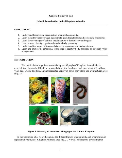
The multicellular organisms that make up <strong>the</strong> 32 phyla of <strong>Kingdom</strong> <strong>Animalia</strong> have<br />
evolved from <strong>the</strong> nearly 100 phyla produced during <strong>the</strong> Cambrian explosion about 600 million<br />
years ago. During this time, an unprecedented variety of novel body plans and architectures arose<br />
(Fig. 1).<br />
Figure 1. Diversity of members belonging <strong>to</strong> <strong>the</strong> Animal <strong>Kingdom</strong><br />
In <strong>the</strong> upcoming labs, we will examine <strong>the</strong> different levels of complexity and organization in<br />
representative phyla of <strong>Kingdom</strong> <strong>Animalia</strong> (See Fig. 2). We will consider <strong>the</strong> environmental<br />
1
constraints that led <strong>to</strong> <strong>the</strong> evolution of particular body plans and <strong>the</strong> adaptations that certain<br />
animals evolved in order <strong>to</strong> survive in <strong>the</strong>ir respective environments.<br />
In general, members of <strong>Kingdom</strong> <strong>Animalia</strong> are eukaryotic, multicellular, motile (at least<br />
during certain developmental stages), heterotrophic and unlike plants, lack a cell wall.<br />
Additionally, most animals reproduce sexually and have a characteristic pattern of embryonic<br />
development. Similar <strong>to</strong> alternation of generations observed in previous phyla, organisms in <strong>the</strong><br />
Animal kingdom undergo stages of development, starting from <strong>the</strong> fusion of an egg and a sperm<br />
and ending with a multicellular adult phase. While <strong>the</strong> morphology of <strong>the</strong> adult organism is<br />
highly species-specific, <strong>the</strong> genes that regulate organismal development are often conserved<br />
across species. In addition, <strong>the</strong> life cycles of members of <strong>Kingdom</strong> <strong>Animalia</strong> vary considerably,<br />
i.e., <strong>the</strong> stages may look completely different from each o<strong>the</strong>r (metamorphosis), <strong>the</strong>y may last<br />
for different periods of time (hours vs. years) and can occur in different habitats (e.g. dragonflies<br />
- adults live in air while larvae are aquatic).<br />
Figure 2. Phylogenetic tree of members of <strong>Kingdom</strong> <strong>Animalia</strong><br />
NOTE: Make sure that you fully understand EVERY term used <strong>to</strong><br />
characterize animals because <strong>the</strong>se terms will appear again in <strong>the</strong><br />
upcoming labs.<br />
2
______________________________________________________________________________<br />
Task 1: Understanding <strong>the</strong> hierarchical organization of animal complexity<br />
The common descent of animals within <strong>Kingdom</strong> <strong>Animalia</strong> can be observed in <strong>the</strong><br />
organization of body plans and <strong>the</strong> fundamental building blocks that all animals share.<br />
Unicellular pro<strong>to</strong>zoans, one of <strong>the</strong> simplest and most ancient groups, limit all <strong>the</strong>ir metabolic,<br />
sensory, and reproductive functions <strong>to</strong> one cell. By varying <strong>the</strong> organization and specialization of<br />
organelles within this cell, <strong>the</strong>y are able <strong>to</strong> achieve all <strong>the</strong> same functions as more structurally<br />
complex organisms.<br />
Pro<strong>to</strong>zoans, which display cellular organization, are described as pro<strong>to</strong>plasmic while<br />
multicellular animals (e.g. sponges) characterized by <strong>the</strong> same cellular level of organization are<br />
collectively referred <strong>to</strong> as parazoans. In this simplest level of <strong>the</strong> hierarchy, cells may be<br />
functionally differentiated, i.e. certain sets of cells are devoted <strong>to</strong> perform a specialized role<br />
within <strong>the</strong> body. Over time, cellular organization led <strong>to</strong> <strong>the</strong> evolution of a cell-tissue level of<br />
organization, where groups of similar cells aggregated in<strong>to</strong> layers (tissues) enabling <strong>the</strong>m <strong>to</strong><br />
perform a common function(s). The nerve net in jellyfish (Fig. 14.7 in your dissection atlas) is a<br />
good example of this level of organization.<br />
Following in complexity is <strong>the</strong> tissue-organ level of organization, produced when<br />
different types of tissues combine <strong>to</strong> form organs. In general, organs perform more specialized<br />
functions than tissues and can be composed of different tissue types (e.g. <strong>the</strong> heart, which is<br />
composed of cardiac muscle, epi<strong>the</strong>lial, connective and nervous tissues). This level of<br />
organization is observed exclusively in metazoans, most of which also exhibit an organ-system<br />
level of organization, where multiple organs operate <strong>to</strong>ge<strong>the</strong>r, forming a system that has a<br />
specific function (Fig. 3). In metazoans, <strong>the</strong>re are eleven organ systems: skeletal, muscular,<br />
integumentary, digestive, respira<strong>to</strong>ry, circula<strong>to</strong>ry, excre<strong>to</strong>ry, nervous, endocrine, immune and<br />
reproductive. We will examine some of <strong>the</strong>se systems in greater depth during <strong>Lab</strong>s 8-11.<br />
Figure 3. Hierarchical organization<br />
3
The major patterns of organization of animal complexity are described below in Table 1.<br />
As you examine <strong>the</strong> organisms <strong>to</strong>day, note which level of organization is present in each. Make<br />
sure <strong>to</strong> sketch <strong>the</strong> organisms listed for each level of organization, noting <strong>the</strong> phylum, genus and<br />
species of each.<br />
Table 1<br />
Level of<br />
organization<br />
Pro<strong>to</strong>plasmic Cellular Cell-tissue Tissue-organ Organsystem<br />
Description<br />
Representative<br />
group<br />
Example:<br />
All functions<br />
are confined<br />
<strong>to</strong> a cell<br />
Protista<br />
**not a part<br />
of <strong>Kingdom</strong><br />
<strong>Animalia</strong>.<br />
We will<br />
NOT examine<br />
<strong>the</strong>m <strong>to</strong>day**<br />
Aggregation<br />
of cells that<br />
are<br />
functionally<br />
differentiated.<br />
Cells are<br />
aggregated in<strong>to</strong><br />
patters/layers =<br />
tissues.<br />
Different tissues<br />
are organized<br />
in<strong>to</strong> organs;<br />
more<br />
specialized than<br />
tissues.<br />
Organs work<br />
<strong>to</strong>ge<strong>the</strong>r as a<br />
system <strong>to</strong><br />
perform a<br />
coordinated<br />
function<br />
Parazoa Radiata Bilateria Bilateria<br />
a. phylum<br />
b. genus<br />
c. common name<br />
a. Porifera<br />
b. Grantia<br />
c. Sponges<br />
a. Cnidaria<br />
b. Metridium<br />
c. Sea anemone<br />
a. Platyhelmin<strong>the</strong>s<br />
b. Dugesia<br />
c. Planarian<br />
a. Chordata<br />
b. Perca<br />
c. Perch<br />
Drawing of<br />
whole organism<br />
Questions:<br />
1. Can you suggest why, during <strong>the</strong> evolution of separate animal lineages, <strong>the</strong>re has been a<br />
tendency for complexity <strong>to</strong> increase when body size increases<br />
4
2. Sponges have folded walls. What advantage could this trait have for <strong>the</strong> sponge<br />
3. Could you think of o<strong>the</strong>r organisms or organ systems that also have similar folded<br />
structures<br />
a. What advantages does folding provide for <strong>the</strong>se organisms<br />
______________________________________________________________________________<br />
Task 2: Differentiating between acoelomate and coelomate organisms<br />
A major developmental event in bilaterally symmetrical organisms (see Task 3) was <strong>the</strong><br />
development of a fluid filled cavity (coelom) between <strong>the</strong> outer body wall and <strong>the</strong> gut (Fig. 14.46<br />
in your dissection atlas). The coelom created a tube-within-tube arrangement allowing space for<br />
visceral organs and an increase in overall body size (Why). This structure also provides support<br />
and aids in movement/burrowing in some animals. However, not all organisms are coelomates;<br />
some lack a coelom al<strong>to</strong>ge<strong>the</strong>r and are called acoelomate (a = without, see Fig. 14.22-14.24 in<br />
your dissection atlas), while o<strong>the</strong>rs are characterized by a pseudocoelom (pseudo = false, see<br />
Fig. 14.36 and 14.37 in your dissection atlas). All three types of body cavities are illustrated<br />
below in Figure 4.<br />
Figure 4. Types of body cavities<br />
5
Examine <strong>the</strong> organisms listed in Table 2 and complete <strong>the</strong> missing sections.<br />
Table 2<br />
Sample Organism Acoelomate Pseudocoelomate Coelomate<br />
Phylum Platyhelmin<strong>the</strong>s Nema<strong>to</strong>da Annelida<br />
Genus Dugesia Ascaris Lumbricus<br />
Common name Flatworms, planaria Roundworms Segmented worms,<br />
Earthworms<br />
Drawing of<br />
Cross section<br />
(slide)<br />
If specimens are<br />
available, dissect<br />
<strong>the</strong>m<br />
longitudinally.<br />
Sketch your<br />
observations in <strong>the</strong><br />
space provided.<br />
Questions:<br />
1. Looking at <strong>the</strong> three representative specimens, what is <strong>the</strong> main difference between<br />
coelomate, pseudocoelomate and acoelomate organisms<br />
6
2. How are <strong>the</strong> organs and tissues organized differently in coelomates and acoelomates<br />
______________________________________________________________________________<br />
Task 3: Body plans and symmetry<br />
While <strong>the</strong> diversity of animal forms is great, <strong>the</strong> basic body plans can be categorized by<br />
<strong>the</strong> presence and type of body symmetry (Fig. 5). Symmetry refers <strong>to</strong> <strong>the</strong> correspondence in size<br />
and shape between opposite sides of an organism’s body. Sponges, which lack body symmetry,<br />
are considered asymmetrical whereas animals whose bodies are arranged around a central axis<br />
and can be divided by more than two planes along <strong>the</strong> longitudinal axis exhibit radial symmetry.<br />
This primitive type of symmetry evolved amongst members of phylum Cnidaria (sea anemones,<br />
box jellies, jellyfish and hydra, see Fig 14.7 and 14.16 in your dissection atlas) and Ctenophora<br />
(comb jellies, see Fig. 14.21 in your dissecting atlas). The bodies of <strong>the</strong> more evolutionarily<br />
advanced bilaterians, in contrast, can be divided in<strong>to</strong> right and left halves along a sagittal plane.<br />
Make sure you understand <strong>the</strong> basic differences between <strong>the</strong> three types of symmetry.<br />
Figure 5. Types of symmetry<br />
Compare and contrast <strong>the</strong> different types of symmetry by examining <strong>the</strong> animals listed for each<br />
type in Table 3. Answer <strong>the</strong> questions that follow.<br />
7
Table 3<br />
Symmetry type Description Example Phyla/Species<br />
Spherical<br />
This symmetry is found in<br />
pro<strong>to</strong>zoa. Any plane passing through<br />
<strong>the</strong> center divides <strong>the</strong> body in<strong>to</strong><br />
equivalent/mirrored halves. Best suited<br />
for floating and rolling.<br />
Radiolaria (amoeboid pro<strong>to</strong>zoa)<br />
WE WILL NOT EXAMINE THIS<br />
TYPE OF SYMMETRY IN THIS<br />
LAB<br />
Asymmetrical<br />
Sponge<br />
Radial<br />
Sea anemone<br />
Bilateral<br />
Perch<br />
Questions:<br />
1. In what kind of environment would each type of body symmetry would be most efficient<br />
8
2. What is <strong>the</strong> advantage of having bilateral symmetry Can any particular task be achieved<br />
more efficiently<br />
a. Why would this type of symmetry lead <strong>to</strong> cephalization<br />
3. Out of all <strong>the</strong> organisms you examined, is <strong>the</strong>re a particular pattern between <strong>the</strong><br />
organisms that have bilateral symmetry Radial symmetry Make sure <strong>to</strong> consider<br />
morphology.<br />
______________________________________________________________________________<br />
Task 4: Developmental patterns in bilateral animals: Pro<strong>to</strong>s<strong>to</strong>mes vs. Deuteros<strong>to</strong>mes<br />
Bilateral animals follow two major patterns of embryonic development. Based on <strong>the</strong>se<br />
patterns, <strong>the</strong>y are classified as ei<strong>the</strong>r deuteros<strong>to</strong>mes or pro<strong>to</strong>s<strong>to</strong>mes. In deuteros<strong>to</strong>mes, <strong>the</strong><br />
blas<strong>to</strong>pore (first embryonic opening) becomes <strong>the</strong> anus, while in pro<strong>to</strong>s<strong>to</strong>mes <strong>the</strong> blas<strong>to</strong>pore<br />
becomes <strong>the</strong> mouth. Also, cleavage, <strong>the</strong> initial process of cell division after a zygote is formed,<br />
differs in <strong>the</strong> two lineages; in pro<strong>to</strong>s<strong>to</strong>mes, cleavage is spiral while in deuteros<strong>to</strong>mes, it is radial<br />
(Fig 6).<br />
The separation of <strong>the</strong> metazoans (multicellular animals) in<strong>to</strong> two separate lineages,<br />
suggests an evolutionary divergence of <strong>the</strong> bilateral body plan. This suggests that deuteros<strong>to</strong>mes<br />
and pro<strong>to</strong>s<strong>to</strong>mes are separate, monophyletic lineages (See Fig 2).<br />
9
PROTOSTOMES<br />
Mouth<br />
Mesoderm<br />
Mouth<br />
Coelom<br />
Spiral<br />
Determinate<br />
Gut<br />
Anus<br />
DEUTEROSTOMES<br />
Mesoderm<br />
Mouth<br />
Radial<br />
Anus<br />
Gut<br />
Coelom<br />
Anus<br />
Figure 6. Comparison of pro<strong>to</strong>s<strong>to</strong>mes and deuteros<strong>to</strong>mes<br />
Examine <strong>the</strong> animals noted under <strong>the</strong> ―Example species‖ row in Table 4. Answer <strong>the</strong> questions<br />
that follow.<br />
10
Table 4<br />
Pro<strong>to</strong>s<strong>to</strong>mes<br />
Deuteros<strong>to</strong>mes<br />
Cleavage type Spiral Radial<br />
Blas<strong>to</strong>pore<br />
Mouth<br />
Anus<br />
becomes<br />
Representative<br />
Phyla<br />
Platyhelmin<strong>the</strong>s, Arthropoda,<br />
Annelida, Mollusca, Nema<strong>to</strong>da, and<br />
smaller phyla<br />
Chordata, Echinodermata, and<br />
smaller phyla<br />
Example species Nema<strong>to</strong>da - Ascaris Sea star – Asterias<br />
Drawing<br />
______________________________________________________________________________<br />
Task 5: Describing positions in bilaterally symmetrical animals<br />
For a large portion of this course you will be examining bilaterally symmetrical animals<br />
from various phyla. To be able <strong>to</strong> locate and refer <strong>to</strong> specific regions of animal bodies, we will<br />
use terminology listed in Table 5.<br />
Table 5<br />
Term<br />
dorsal<br />
ventral<br />
anterior; cranial<br />
posterior; caudal<br />
medial<br />
proximal<br />
lateral<br />
distal<br />
frontal plane<br />
transverse plane<br />
sagittal plane<br />
Meaning<br />
<strong>to</strong>ward <strong>the</strong> upper surface (back)<br />
<strong>to</strong>ward <strong>the</strong> lower surface (belly)<br />
<strong>to</strong>ward <strong>the</strong> head<br />
<strong>to</strong>ward <strong>the</strong> tail<br />
<strong>to</strong>ward <strong>the</strong> midline of <strong>the</strong> body<br />
<strong>to</strong>ward <strong>the</strong> end of <strong>the</strong> appendage nearest <strong>the</strong> body<br />
<strong>to</strong>ward <strong>the</strong> side; away from <strong>the</strong> midline of <strong>the</strong> body<br />
<strong>to</strong>ward <strong>the</strong> end of <strong>the</strong> appendage far<strong>the</strong>st away from <strong>the</strong> body<br />
divides <strong>the</strong> body in<strong>to</strong> dorsal and ventral halves<br />
divides <strong>the</strong> body in<strong>to</strong> anterior and posterior halves<br />
divides <strong>the</strong> body in<strong>to</strong> left and right halves<br />
11
transverse<br />
plane<br />
sagittal<br />
plane<br />
frontal<br />
plane<br />
Figure 7. Planes of sections in a crayfish<br />
In addition <strong>to</strong> <strong>the</strong> terms listed in Table 5, different terminology is used <strong>to</strong> describe<br />
radially symmetrical vs. bilaterally symmetrical animals. These terms are listed in Table 6.<br />
Table 6<br />
Radial<br />
Bilateral<br />
Direction Synonyms Direction Synonyms<br />
oral apical anterior rostral, cranial,<br />
cephalic<br />
aboral basal posterior caudal<br />
peripheral — dorsal —<br />
peripheral — ventral —<br />
peripheral — left (lateral) sinister<br />
peripheral — right (lateral) dexter<br />
medial<br />
medial<br />
proximal<br />
proximal<br />
distal<br />
distal<br />
As a group, practice using <strong>the</strong>se directional terms <strong>to</strong> refer <strong>to</strong> a particular part/portion of<br />
<strong>the</strong> body. Make sure <strong>to</strong> use available specimens <strong>to</strong> practice and <strong>to</strong> include both radially and<br />
bilaterally symmetrical animals during this exercise.<br />
______________________________________________________________________________<br />
12
Task 6: Body axes charades – Run by your TA<br />
To practice using <strong>the</strong> correct terminology when referring <strong>to</strong> different locations on <strong>the</strong><br />
body, you will play a game of charades. Your TA will divide <strong>the</strong> whole class in<strong>to</strong> two groups,<br />
each of which will be given a list of organs/body parts. Each group’s list will be different<br />
<strong>the</strong>refore make sure that you do not <strong>to</strong> share your list with members from <strong>the</strong> o<strong>the</strong>r group.<br />
Your group will choose a student from ano<strong>the</strong>r group <strong>to</strong> describe one of <strong>the</strong> words on<br />
your list <strong>to</strong> his/her group. The student will have 2 minutes <strong>to</strong> describe <strong>the</strong> word, using only <strong>the</strong><br />
words from <strong>the</strong> bilateral body axes (see Tables 5 and 6). Note that you cannot use words that<br />
describe <strong>the</strong> function of <strong>the</strong> organ/body part. For example, if <strong>the</strong> organ <strong>to</strong> be described is <strong>the</strong><br />
heart, you are not allowed <strong>to</strong> say that it pumps blood. Instead, you can say that it is posterior <strong>to</strong><br />
<strong>the</strong> head and is anterior <strong>to</strong> <strong>the</strong> belly but<strong>to</strong>n. If his/her group can guess <strong>the</strong> right answer, <strong>the</strong>n that<br />
team gets a point but if <strong>the</strong>y don’t guess correctly, <strong>the</strong>n your team gets <strong>the</strong> point. Make sure <strong>to</strong><br />
alternate <strong>the</strong> order of <strong>the</strong> teams guessing.<br />
______________________________________________________________________________<br />
LOOK AHEAD:<br />
Before coming <strong>to</strong> lab next week, make sure <strong>to</strong> read <strong>the</strong> Development task sheet.<br />
13