

A Simplified Approach to Midface Aging - VGbras
A Simplified Approach to Midface Aging - VGbras
A Simplified Approach to Midface Aging - VGbras

You also want an ePaper? Increase the reach of your titles
YUMPU automatically turns print PDFs into web optimized ePapers that Google loves.
SURGICAL TECHNIQUE<br />
A <strong>Simplified</strong> <strong>Approach</strong> <strong>to</strong> <strong>Midface</strong> <strong>Aging</strong><br />
Ryan N. Heffelfinger, MD; Keith E. Blackwell, MD; Jeffrey Rawnsley, MD; Gregory S. Keller, MD<br />
We review herein our experience with subperiosteal midface-lifting under direct<br />
vision with a simple fixation technique. The technical aspects of the procedure<br />
are described in detail. A <strong>to</strong>tal of 121 patients underwent midface-lifting and<br />
meloplication with the 82/18 L-lactide/glycolide device (Coapt Endotine <strong>Midface</strong><br />
ST 4.5; Coapt Technologies, Palo Al<strong>to</strong>, Calif) by both the senior (G.S.K.) and junior (R.N.H.)<br />
authors. The senior author’s experience included 110 patients over a 26-month period. Thirtytwo<br />
of these cases were isolated procedures. The other 78 were performed in conjunction with<br />
various procedures, most commonly rhytidec<strong>to</strong>my. There were no revisions during this period.<br />
Two cases of “puckering” were noted. Both were immediately corrected, one with fat injection and<br />
one with poly-L-lactic acid injection (Sculptra; Dermik Aesthetics, distributed by Besse Medical<br />
Supply, West Chester, Ohio). The junior author’s experience included 11 cases over an 8-month<br />
period. Two cases of asymmetry were noted. One was corrected with fat injection, and the other<br />
required revision. Subperiosteal midface-lifting and meloplication using the Coapt Endotine <strong>Midface</strong><br />
ST 4.5 device is a simple, effective technique that can be quickly learned and applied.<br />
Arch Facial Plast Surg. 2007;9:48-55<br />
Fullness of the cheek represents youthfulness.<br />
In the process of aging, a constant<br />
hollowness of the midface develops.<br />
As a result, a patient may display an<br />
appearance that is tired, old, or sad.<br />
Weakening of the malar and orbital ligaments<br />
is a major component of the aging<br />
process. The result is a downward and medial<br />
displacement of the malar fat pad or<br />
panniculus adiposus over the fixed ligaments<br />
of the nasolabial fold. A gap or<br />
crease, angled posteroinferiorly, forms between<br />
the malar eminence and the fallen<br />
fat pad. This is referred <strong>to</strong> as the “cheek<br />
hollow.” The fat over the malar emi-<br />
Author Affiliations: Division of Head and Neck Surgery, UCLA Medical Center,<br />
University of California, Los Angeles.<br />
(REPRINTED) ARCH FACIAL PLAST SURG/ VOL 9, JAN/FEB 2007 WWW.ARCHFACIAL.COM<br />
48<br />
Downloaded from<br />
www.archfacial.com at Coapt Systems, Inc., on January 16, 2007<br />
©2007 American Medical Association. All rights reserved.<br />
nence is left standing, accentuating the malar<br />
bag (Figure 1).<br />
Another ana<strong>to</strong>mical change that occurs<br />
is the weakening of the orbital ligaments,<br />
which contributes <strong>to</strong> hollowness<br />
under the orbit. This is referred <strong>to</strong> as the<br />
“orbital hollow.” The malar fat pad, which<br />
in youth was at the level of the orbital rim,<br />
falls downward and medially, producing<br />
this hollowness. This concavity, which is<br />
below the convexity of the ocular globe and<br />
orbital fat, accentuates and no longer covers<br />
the bulge of herniated orbital fat. This<br />
produces the “double con<strong>to</strong>ur” deformity<br />
characteristic of the aging orbital/<br />
midface complex.<br />
A volume loss of the midface also contributes<br />
<strong>to</strong> the aging process. Thus, many
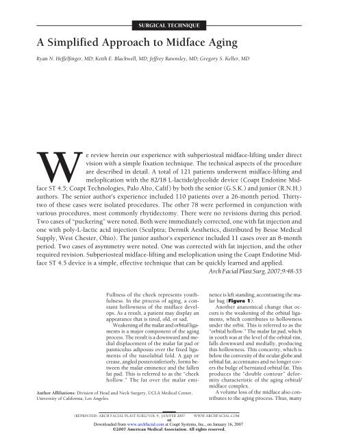
Tear Trough<br />
Descent of Eyelid<br />
Skin Below Orbital<br />
Rim<br />
Nasolabial Fold<br />
Malar Fat Pad<br />
Figure 1. Illustration demonstrates the process of midface aging. As the<br />
malar fat pad descends, the nasolabial fold is accentuated. The orbital rim is<br />
left covered only by thin eyelid skin, and the tear-trough deformity becomes<br />
apparent.<br />
physicians use fat injections or malar/submalar implants<br />
in an attempt <strong>to</strong> correct this aspect of aging. These<br />
have their own set of problems and complications. 1,2<br />
Finally, the zygomaticus major and minor muscles suspendtheligamen<strong>to</strong>usandmuscularconnectionsofthemidface.<br />
These connections weaken and fall, producing an “oral<br />
frown.” The collapse of the muscular structures, with their<br />
associated ligaments and fat pad, results in a droop of the<br />
corner of the mouth and deepens the labiomental fold.<br />
The senior author (G.S.K.) has lifted and augmented<br />
the midface using different techniques over the last quarter<br />
century. 3-6 These techniques, while all useful, required<br />
either wide dissections (open or endoscopic) or<br />
treated only a portion of the aging problems of the midface.<br />
Suture suspension techniques have proven minimally<br />
invasive and are associated with low morbidity, but<br />
we continue <strong>to</strong> search for a solution that more completely<br />
addresses all of the problems of midface aging (eg,<br />
volume augmentation, fat pad and ligament suspension,<br />
and muscle elevation). 7<br />
Recently, a new fixation technology with a spiked suspension<br />
and fixation device (Endotine ST 4.5, Coapt Technologies,<br />
Palo Al<strong>to</strong>, Calif) has allowed us <strong>to</strong> develop a<br />
single incision technique that requires only 10 <strong>to</strong> 15 minutes<br />
per side after surgeon familiarization. While the recovery<br />
period is compatible with string or loop procedures,<br />
this technique more completely addresses the<br />
midface aging process.<br />
TECHNIQUE<br />
Subperiosteal meloplication is performed under direct observation.<br />
An incision is planned 2 cm behind the tem-<br />
DTF<br />
Temporalis<br />
Muscle<br />
Plane of Dissection<br />
Figure 2. Illustration shows the location of incision and plane of dissection.<br />
This plane is between the deep temporal fascia (DTF) and the<br />
temporoparietal fascia (TPF).<br />
poral hairline perpendicular <strong>to</strong> an imaginary line drawn<br />
from the nasoalar junction <strong>to</strong> the lateral canthus and carried<br />
in<strong>to</strong> the hairline. This imaginary line bisects the incision<br />
at its midpoint. In an attempt <strong>to</strong> camouflage the<br />
scar, we originally made this incision as small as possible.<br />
We soon realized that a larger incision is equally<br />
well hidden behind the hairline and significantly improves<br />
exposure. The length of the incision we currently<br />
use is approximately 4 cm. This incision is carried<br />
down <strong>to</strong> the superficial layer of deep temporal fascia<br />
(Figure 2).<br />
Using a gently curved eleva<strong>to</strong>r, the physician carries<br />
the dissection medially and inferiorly, taking care <strong>to</strong> stay<br />
directly on the superficial layer of deep temporal fascia.<br />
A lighted Aufricht retrac<strong>to</strong>r or a headlight and a standard<br />
Aufricht retrac<strong>to</strong>r are used <strong>to</strong> illuminate the dissection<br />
field. The sentinel vein is identified and cauterized.<br />
Lateral widening of this dissection above the zygomatic<br />
arch is carried out <strong>to</strong> the second branch of the zygomaticotemporal<br />
nerve and vein, which are left intact<br />
(Figure 3A).<br />
A periosteal eleva<strong>to</strong>r is then placed on the malar eminence.<br />
Using tactile sensation under direct observation,<br />
the physician elevates the periosteum of the malar eminence<br />
and the medial one third of the zygomatic arch. It<br />
is important <strong>to</strong> use careful, short sweeping motions of<br />
the eleva<strong>to</strong>r. A protective finger of the left hand (if the<br />
physician is right handed) palpates and guards the foramen<br />
of the infraorbital nerve. Elevation near the infraorbital<br />
neurovascular bundle is performed with upward<br />
“prying” motions of the periosteum that do not<br />
endanger the nerve.<br />
Dissection is then carried downward via palpation and<br />
direct observation over the masseteric fascia, with gently<br />
sweeping downward motions. The dissection can<br />
proceed as far medially as necessary, usually elevating<br />
the nasolabial fold. The arcus marginalis is freed insofar<br />
as possible, while avoiding the infraorbital nerve<br />
(Figure 3B).<br />
The Aufricht eleva<strong>to</strong>r is then used <strong>to</strong> visualize the dissection.<br />
Usually, at this point, the precanthal ligament<br />
is released with scissors dissection in the supraperi-<br />
(REPRINTED) ARCH FACIAL PLAST SURG/ VOL 9, JAN/FEB 2007 WWW.ARCHFACIAL.COM<br />
49<br />
Downloaded from<br />
www.archfacial.com at Coapt Systems, Inc., on January 16, 2007<br />
©2007 American Medical Association. All rights reserved.<br />
TPF<br />
Fat<br />
Skin
A B<br />
Extent of<br />
Dissection<br />
Massateric Fascia<br />
(Over Masseter Muscle)<br />
LZTN<br />
MZTN<br />
osteal plane. This maneuver widens the dissection cavity<br />
<strong>to</strong> accommodate the spiked device.<br />
The device is encased in a protective plastic sheath.<br />
The entire complex is placed in<strong>to</strong> the dissection cavity.<br />
The spiked portion of the device is positioned immediately<br />
lateral <strong>to</strong> the nasolabial fold. The sheath trigger is<br />
then squeezed, the spikes are engaged in<strong>to</strong> periosteum<br />
with the aid of digital pressure, and the sheath is<br />
removed.<br />
The device, with the sheath removed, has a “leash”<br />
that extends from the device upwards and backward in<strong>to</strong><br />
the temporal incision. This leash is pulled posterosuperiorly<br />
until the spiked portion is in approximate vertical<br />
alignment with the midpupillary line. If it appears that<br />
<strong>to</strong>o much or <strong>to</strong>o little elevation has occurred, the device<br />
is disengaged by pinching and lifting the cheek fat. Repositioning<br />
can proceed as necessary. Generally, as the<br />
device is relocated in a more lateral direction, the less<br />
elevation will be seen. Conversely, the more medial the<br />
original position of the spikes, greater vertical movement<br />
is possible.<br />
If necessary, a gingivobuccal incision is made <strong>to</strong> improve<br />
exposure of the midface and ensure the proper plane<br />
of dissection. It is also useful if the device is disengaged<br />
initially in an improper position, since it is difficult <strong>to</strong><br />
reposition the spiked apparatus from the temporal incision<br />
alone. We used gingivobuccal incisions in all of our<br />
initial cases. With more experience, we found that we<br />
ZFN<br />
Zygomaticus<br />
Major and Minor<br />
Muscles<br />
Extent of<br />
Dissection<br />
MZTV MZTV<br />
MZTN<br />
Massateric Fascia<br />
(Over Masseter Muscle)<br />
LZTN<br />
could perform adequate dissection and place the device<br />
properly through the temporal incision alone.<br />
With posterosuperior tension on the leash, a large<br />
absorbable suture is placed <strong>to</strong> fix the device <strong>to</strong> the<br />
superficial layer of deep temporal fascia. The cut margin<br />
of temporoparietal fascia is grasped, pulled superiorly,<br />
and sutured <strong>to</strong> the superficial layer of deep temporal<br />
fascia. The incision is closed with staples. Application<br />
of a pressure dressing is optional. Patients are given<br />
antibiotics for 7 <strong>to</strong> 10 days following the procedure;<br />
oral flora coverage is crucial if a gingivobuccal sulcus<br />
incision was used. A soft diet is followed for 2 days<br />
pos<strong>to</strong>peratively.<br />
RESULTS<br />
From December 2003 <strong>to</strong> February 2006, the senior<br />
author performed 110 of the described procedures: 32<br />
were independent procedures, and 78 were performed<br />
with ancillary procedures. The most common ancillary<br />
procedure was rhytidec<strong>to</strong>my (59 patients), followed<br />
by blepharoplasty (28 patients) and endoscopic<br />
browlift (17 patients). There were 2 cases of “puckering”<br />
overlying the spiked portion of the device. One<br />
patient was treated with au<strong>to</strong>logous fat injection (approximately<br />
3 mL), the other with serial injections of<br />
poly-L-lactic acid (Sculptra; Dermik Aesthetics, dis-<br />
ZFN<br />
Zygomaticus<br />
Major and Minor<br />
Muscles<br />
Figure 3. Illustration depicts the upper (A) and lower (B) dissection. The subperiosteal plane is entered over the malar eminence, remaining medial <strong>to</strong> and<br />
preserving the medial zygomaticotemporal nerve (MZTN). The medial zygomaticotemporal vein (MZTV) is often encountered and is cauterized at its deep aspect<br />
and transected. LZTN indicates lateral zygomaticotemporal nerve; ZFN, zygomaticofacial nerve.<br />
(REPRINTED) ARCH FACIAL PLAST SURG/ VOL 9, JAN/FEB 2007 WWW.ARCHFACIAL.COM<br />
50<br />
Downloaded from<br />
www.archfacial.com at Coapt Systems, Inc., on January 16, 2007<br />
©2007 American Medical Association. All rights reserved.
A B<br />
C D<br />
tributed by Besse Medical Supply, West Chester,<br />
Ohio). No patients required revision or implant<br />
removal and/or replacement. Complaints of lip hypesthesia,<br />
suture irritation, and pain related <strong>to</strong> the gingivobuccal<br />
incision were frequent early in the development<br />
of our technique. As we evolved <strong>to</strong> omit this<br />
incision, this short-term complication was eliminated.<br />
In the group that had gingivobuccal incisions, these<br />
problems were transient and resolved during the period<br />
of observation.<br />
From July 2005 <strong>to</strong> February 2006, the junior<br />
author (R.N.H.) performed the procedure on 10<br />
patients. It was performed as an isolated procedure in<br />
1 patient and in conjunction with rhytidec<strong>to</strong>my in the<br />
other 9 patients. The second patient in this series<br />
developed an asymmetry that was corrected with<br />
au<strong>to</strong>logous fat injection. The third patient had an<br />
asymmetry that was due <strong>to</strong> either improper placement<br />
or incomplete engagement of the spikes with the periosteum<br />
and/or malar mound. This required revision<br />
Figure 4. Preoperative (A and C) and<br />
10-month pos<strong>to</strong>perative (B and D)<br />
pho<strong>to</strong>graphs of a female patient who<br />
underwent subperiosteal midface-lift<br />
and laser skin tightening of the lower<br />
face and neck (3 treatment sessions).<br />
and repositioning of the implant. This was performed<br />
with the patient under local anesthesia using both the<br />
temporal and gingivobuccal incisions. Through the<br />
temporal incision, the leash was released from the<br />
deep temporalis fascia and freed from the surrounding<br />
tissue. Intraorally, the malar mound was elevated from<br />
the spikes and repositioned under direct vision. The<br />
leash was sutured <strong>to</strong> the superficial layer of the deep<br />
temporal fascia, and the wounds were closed as usual.<br />
Ten days pos<strong>to</strong>peratively, this patient developed a cellulitis,<br />
which responded <strong>to</strong> treatment with oral antibiotics.<br />
The most common pos<strong>to</strong>perative complaint was<br />
transient lip numbness (4 patients). The last 3 patients<br />
in the junior author’s series did not require gingivobuccal<br />
incisions, thus eliminating this complication.<br />
The longest follow up is 26 months, which involved<br />
a male patient with the device inserted as an independent<br />
procedure. The range of follow-up is 1 <strong>to</strong> 26 months.<br />
Representative patients are seen in Figures 4, 5, 6, 7,<br />
and 8. Results are stable over the follow-up period.<br />
(REPRINTED) ARCH FACIAL PLAST SURG/ VOL 9, JAN/FEB 2007 WWW.ARCHFACIAL.COM<br />
51<br />
Downloaded from<br />
www.archfacial.com at Coapt Systems, Inc., on January 16, 2007<br />
©2007 American Medical Association. All rights reserved.
A B<br />
C D<br />
COMMENT<br />
As the mechanisms of midface aging have been elucidated,<br />
techniques and devices that make elevation of the<br />
midface easier <strong>to</strong> perform were developed. 3-9 Specifically,<br />
we believe that no single solution addresses volume<br />
augmentation, fat pad and ligament suspension, and<br />
muscle elevation in a simple procedure. Our technique<br />
of subperiosteal midface-lifting and suspension using the<br />
Coapt Endotine ST 4.5 device is a potential step <strong>to</strong>ward<br />
this direction.<br />
Our observations indicate that the following occur<br />
after the procedure:<br />
1. The cheek mound advances upward and backward.<br />
A tremendous amount of vertical lift is produced.<br />
The fat pad is repositioned, diminishing the orbital hollow<br />
and the “double con<strong>to</strong>ur deformity.”<br />
2. A volume augmentation is enhanced by meloplication<br />
that fills in both the “orbital hollow” and the “cheek<br />
hollow.”<br />
Figure 5. Preoperative (A and C) and<br />
pos<strong>to</strong>perative (B and D) pho<strong>to</strong>graphs<br />
of a female patient who underwent a<br />
midface-lift as an isolated procedure<br />
(4-month follow-up period).<br />
3. The nasolabial fold is diminished.<br />
4. The “oral frown” is diminished, <strong>to</strong> a degree.<br />
5. Malar bags are diminished, <strong>to</strong> a degree.<br />
Standard superficial musculoaponeurotic system<br />
rhytidec<strong>to</strong>my techniques are inadequate at correcting midface<br />
aging. 10 Deep-plane lifting addresses the midface <strong>to</strong><br />
a greater degree, although the technique is challenging,<br />
and the long-term rewards in the midface are modest. 11<br />
In an attempt <strong>to</strong> specifically address the midface, techniques<br />
have been reported that similarly require advanced<br />
training and technical expertise. 5,6 We (and others)<br />
have reported experience with a simple technique,<br />
the percutaneous suspension lift. 7-9,12 While this remains<br />
a good technique in selected patients, the issue of<br />
foreign body reaction and longevity of elevation remain.<br />
In addition, it does not address all of the components<br />
of midface aging.<br />
The long-term results of the senior author show<br />
this <strong>to</strong> be a reproducible technique with little risk of<br />
complications. As with any procedure, there is a learn-<br />
(REPRINTED) ARCH FACIAL PLAST SURG/ VOL 9, JAN/FEB 2007 WWW.ARCHFACIAL.COM<br />
52<br />
Downloaded from<br />
www.archfacial.com at Coapt Systems, Inc., on January 16, 2007<br />
©2007 American Medical Association. All rights reserved.
A B<br />
C D<br />
ing curve <strong>to</strong> overcome when first performing this technique.<br />
This is demonstrated by the junior author’s<br />
experience.<br />
Specifics of the Coapt device can be found on their<br />
Web site (http://www.coaptsystems.com). The device<br />
is bioabsorbable, with a reported reduction in strength<br />
and mass of 95% and 50%, respectively, in 5 months time.<br />
In our experience, the device is rarely palpable after<br />
8 <strong>to</strong> 10 months. As an isolated procedure, we found that<br />
patients may experience edema and ecchymosis that<br />
usually resolves enough for public appearance after<br />
approximately 5 days. The addition of a gingivobuccal<br />
incision seems <strong>to</strong> add 2 <strong>to</strong> 3 days <strong>to</strong> this recovery<br />
period.<br />
Several groups of patients can benefit from this procedure.<br />
Younger patients with isolated midface aging<br />
are frequently unwilling <strong>to</strong> undergo face-lift surgery.<br />
These patients often present with isolated aging of the<br />
midface. Patients in the middle <strong>to</strong> older age group can<br />
benefit from this procedure in conjunction with a facelift<br />
or other procedures that are invasive, noninvasive,<br />
or minimally invasive. In addition, midface elevation is<br />
Figure 6. Preoperative (A) and<br />
pos<strong>to</strong>perative (B) pho<strong>to</strong>graphs of a<br />
female patient who underwent<br />
subperiosteal midface-lift, upper<br />
and lower blepharoplasty, face-lift,<br />
and a skin rejuvenation program<br />
(6-month follow-up period);<br />
preoperative (C) and 2-month<br />
pos<strong>to</strong>perative (D) views of a male<br />
patient after subperiosteal<br />
midface-lift in conjunction with<br />
face-lift and upper blepharoplasty.<br />
often useful as a face-lift “<strong>to</strong>uch up” in the properly selected<br />
patient.<br />
All age groups can benefit from ancillary procedures<br />
that are performed in conjunction with midface elevation.<br />
Fat and/or poly-L-lactic acid injections, radiofrequency,<br />
or laser skin-tightening procedures (THERMAGE,<br />
Hayward, Calif; Syneron, Yokneam, Israel; and Alma,<br />
Caesarea, Israel), fillers (RADIESSE; Bioform Medical, San<br />
Mateo, Calif; and RESTYLANE; Medicis Aesthetics, Scottsdale,<br />
Ariz), and laser resurfacing are all examples of these<br />
ancillary procedures. In addition, blepharoplasty and browlift<br />
are 2 surgical procedures that produce a nice result in<br />
conjunction with midface-lifting.<br />
In conclusion, we believe that subperiosteal midfacelifting<br />
under direct vision and meloplication of the malar<br />
fat pad with the 82/18 L-lactide/glycolide device is a safe,<br />
effective procedure with a short learning period. It can be<br />
considered a minimally invasive procedure with powerful<br />
results. We have inserted these devices under local and<br />
general anesthesia. Patients have been uniformly happy<br />
and the results stable over the follow-up period.<br />
(REPRINTED) ARCH FACIAL PLAST SURG/ VOL 9, JAN/FEB 2007 WWW.ARCHFACIAL.COM<br />
53<br />
Downloaded from<br />
www.archfacial.com at Coapt Systems, Inc., on January 16, 2007<br />
©2007 American Medical Association. All rights reserved.
A B<br />
C D<br />
A B<br />
Figure 7. Preoperative (A and C)<br />
and 12-month pos<strong>to</strong>perative (B and<br />
D) views of a female patient after<br />
subperiosteal midface-lift in<br />
conjunction with face-lift,<br />
endoscopic brow-lift, and<br />
blepharoplasty.<br />
Figure 8. Preoperative (A) and 9-month pos<strong>to</strong>perative (B) pho<strong>to</strong>graphs of a female patient after subperiosteal midface-lift in conjunction with an endoscopic brow-lift<br />
and face-lift.<br />
(REPRINTED) ARCH FACIAL PLAST SURG/ VOL 9, JAN/FEB 2007 WWW.ARCHFACIAL.COM<br />
54<br />
Downloaded from<br />
www.archfacial.com at Coapt Systems, Inc., on January 16, 2007<br />
©2007 American Medical Association. All rights reserved.
Accepted for Publication: September 9, 2006.<br />
Correspondence: Gregory S. Keller, MD, 221 West Pueblo<br />
St, Suite A, Santa Barbara, CA 93105 (faclftr@aol.com).<br />
Author Contributions: Study concept and design: Rawnsley<br />
and Keller. Acquisition of data: Heffelfinger and Keller.<br />
Analysis and interpretation of data: Heffelfinger and Keller.<br />
Drafting of the manuscript: Heffelfinger and Keller. Critical<br />
revision of the manuscript for important intellectual<br />
content: Heffelfinger, Blackwell, Rawnsley, and Keller.<br />
Statistical analysis: Heffelfinger. Administrative, technical,<br />
and material support: Heffelfinger, Blackwell, Rawnsley,<br />
and Keller. Study supervision: Blackwell, Rawnsley,<br />
and Keller.<br />
Financial Disclosure: Dr Keller serves on the Medical<br />
Board of Coapt Technologies, Palo Al<strong>to</strong>, Calif.<br />
REFERENCES<br />
1. Wilkinson TS. Complications in aesthetic malar augmentation. Plast Reconstr<br />
Surg. 1983;71:643-649.<br />
2. Toledo LS, Mauad R. Fat Injection: a 20-year revision. Clin Plast Surg. 2006;33:<br />
47-53.<br />
3. Freeman MS. Endoscopic malar pad lift and subperiosteal midface lift. In: Keller<br />
GS, ed. Endoscopic Facial Plastic Surgery. St Louis, Mo: Mosby-Year Book Inc;<br />
1997:109-136.<br />
4. Owsley JQ. Lifting the malar fat pad for correction of prominent nasolabial folds.<br />
Plast Reconstr Surg. 1993;91:463-474.<br />
5. Anderson RD, Lo MW. Endoscopic malar/midface suspension procedure. Plast<br />
Reconstr Surg. 1998;102:2196-2208.<br />
6. Namazie A, Alum D, Keller GS. Current techniques in midface lifting. Facial Plast<br />
Surg Clin North Am. 2002;10:53-62.<br />
7. Keller GS, Namazie A, Blackwell K, Rawnsley J, Khan S. Elevation of the malar<br />
fat pad with a percutaneous technique. Arch Facial Plast Surg. 2002;4:20-25.<br />
8. Su NN. Closed suspension mini-cheek lift <strong>to</strong> reduce the nasolabial fold: a preliminary<br />
report. Am J Cosmetic Surg. 1995;12:31-34.<br />
9. Sasaki GH, Cohen AT. Meloplication of the malar fat pads by percutaneous cablesuture<br />
technique for midface rejuvenation: outcome study (392 cases, 6 years’<br />
experience). Plast Reconstr Surg. 2002;110:635-654.<br />
10. Stuzin JM, Baker TJ, Gordon HL, Baker TM. Extended SMAS dissection as an<br />
approach <strong>to</strong> midface rejuvenation. Clin Plast Surg. 1995;22:295-311.<br />
11. Hamra ST. A study of the long-term effect of malar fat repositioning in face lift<br />
surgery: short-term success but long-term failure. Plast Reconstr Surg. 2002;<br />
110:940-951.<br />
12. Laferriere KA, Castellano RD. Experience with percutaneous suspension of the<br />
malar fat pad for midface rejuvenation. Facial Plast Surg Clin North Am. 2005;<br />
13:393-399.<br />
(REPRINTED) ARCH FACIAL PLAST SURG/ VOL 9, JAN/FEB 2007 WWW.ARCHFACIAL.COM<br />
55<br />
Downloaded from<br />
www.archfacial.com at Coapt Systems, Inc., on January 16, 2007<br />
©2007 American Medical Association. All rights reserved.
