PCR + - Central Institute of Brackishwater Aquaculture
PCR + - Central Institute of Brackishwater Aquaculture
PCR + - Central Institute of Brackishwater Aquaculture

You also want an ePaper? Increase the reach of your titles
YUMPU automatically turns print PDFs into web optimized ePapers that Google loves.
,I<br />
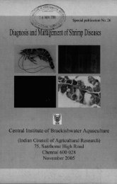
Expert Consultation on Rapid Diagnosis <strong>of</strong><br />
Shrimp Viral Diseases<br />
12" - 14" June 2002, Chennai<br />
Workshop Programhe and Powerpoint Presentations
Expert Consultation on Rapid D i e hf ~<br />
Shrimp Viral Diseases<br />
12* - 14* June 2002, Chennai<br />
urn ICAR<br />
Programme<br />
12"' June 2002<br />
1 OOOh Inauguration ceremony<br />
1 lOOh Tea break<br />
1130h Session 1 Indian scenario and perspectives on I'CR and shrimp<br />
health management<br />
Session Chair.$: Dr T.C S~intingo and F'r<strong>of</strong>: Tint Flegel<br />
Brief introductory remarks on workshop organization. object~ves and outputs<br />
I)r Michael Phillips, NACA<br />
Application <strong>of</strong> <strong>PCR</strong> technique in the detection <strong>of</strong> shrimp pathogenic viruses ir<br />
lndia - Ittyn Karunnsagar and Indrani Kurunmagar, College o f Fisheries,<br />
Mangulore<br />
. Shrimp health management in lndia with special reference to viral diseases an<br />
<strong>PCR</strong> - K.K. Vjuyan and T.C Santiagu, CIIjA, Clzennrri<br />
Role and activities <strong>of</strong> MPEDA in the Rapid Diagnosis <strong>of</strong> shrimp d~seases<br />
through <strong>PCR</strong> - B. Vishnu Bhrrt, MPEDA<br />
Discussion session<br />
1300h<br />
1400h<br />
Lunch break<br />
Session I (continued)<br />
Issues and implications <strong>of</strong> <strong>PCR</strong> results to management <strong>of</strong> WSSV Lessons<br />
from DFID white spot syndrome epidemiology project C. I'. Mohnn, College<br />
<strong>of</strong> Fisheries, Mangalore
Epidemiological study <strong>of</strong> viral diseases <strong>of</strong> shrimps from culture<br />
ponds <strong>of</strong> North Coastal Andhra Pradesh Application <strong>of</strong><br />
histological and molecular diagnostic techniques -R Madhavi, Andhra<br />
Unhversity. Visakhnpainam<br />
Private sector comments and presentations - D.Ramraj, Padmanabha Labs.<br />
M..Surlarshan Swanry, Andhro Pradesh Shrimp Hatcheries Association, Ms<br />
Subhnshni, Bangalore tienn'<br />
Discussion session<br />
1530h<br />
Tea break<br />
1600h Session 2 Regional experiences<br />
Sess~ori C'hnrrs: Pr<strong>of</strong> R Madhatpi and Dr Richard Hodgson<br />
The development <strong>of</strong> <strong>PCR</strong> and other diagnostic tests for control <strong>of</strong><br />
viral diseases <strong>of</strong> shrimp in Thailand - Tim Flegel, Mahiilol University<br />
Practical application <strong>of</strong> <strong>PCR</strong> testing in Thailand and the Impact<br />
on shrimp disease management on farms - Roonsirn~ Withycltunnnrnkul,<br />
Mnhirlol University<br />
Discussion sesslon<br />
1700h<br />
Close for day<br />
13"' June 2002<br />
1900h<br />
Session 2 (continued)<br />
<strong>PCR</strong> test formats and the future <strong>of</strong><strong>PCR</strong> technology for shrimp v~ral<br />
detection -Richnr~l Hodgson, CSIRO, Australia<br />
The appl~catton <strong>of</strong><strong>PCR</strong> for control <strong>of</strong> shrimp viral disease -<br />
strategies for rtsk management - Peter Walker, CSIRO, Awralin<br />
- Nattonal and rey~onal harmonisation <strong>of</strong> WSSV <strong>PCR</strong> testlns protocols<br />
- Peter Walker, CSIRO, Australia<br />
Recent NACA experiences, including lessons on shrtmp d~sease dtagnost~cs<br />
from the MPEDA-NACA technical assistance -Micltrrcl Phillips, Vishnu<br />
Hhat and Arun Parliyar<br />
Discusston sesston<br />
1030h<br />
Tea break
1100h Session 2 (continued)<br />
Application <strong>of</strong> <strong>PCR</strong> diagnosis in WSSV - K. K Rnjendrnn, CIFE. Munthni<br />
Use <strong>of</strong> dot blot tests for white spot diagnosis - K.M. Shnnhnr, College <strong>of</strong><br />
Fisheries, Mangnlore<br />
Some emerging shrimp viruses - Peter Walher, CSIRO. Austrnlin<br />
1230h Session 3<br />
Working groups<br />
.Ycssion Chnirs: Peter Walker nnd Indrani Knrunacagnr<br />
Introduction to working group sessions -Michnel Phillips<br />
. Working group 1. Practical application <strong>of</strong> rapid diagnostics (lihrrry)<br />
Working group 2 <strong>PCR</strong> and rapid diagnostic technologies (confererrce roonr)<br />
o Working group 3. Researchable issues (G'BII lahorntory - roont 206)<br />
(see separate document on working groups)<br />
1300h<br />
Lunch break<br />
Working groups continued<br />
1700h<br />
1930h<br />
Close for day<br />
Dinner at Vedika Hall, Savera Hotel<br />
14"' June 2002<br />
0900h Session 3 Presentation <strong>of</strong> working group findings and<br />
recommendat~ons by Workrng Ciroup Charrs<br />
1030h<br />
l lO0h<br />
Tea break<br />
Summary <strong>of</strong> workshop recommendat~ons followed by clos~ng<br />
ceremony
Introductory remarks<br />
Network <strong>of</strong> <strong>Aquaculture</strong> Centres in Asia-Pacif<br />
aquatic animal health programme<br />
Support to diagnostic capacity build~ng is an important<br />
part <strong>of</strong> the programme<br />
m <strong>PCR</strong> has been promoted extensively to detect shrimp<br />
viruses, in India and elsewhere, but problems noted<br />
AClAR supported project "Diagnostic tests and<br />
epidemiological probes for prawn viruses In Thailand
Cooperation with NACA<br />
Financial support provided by AClAR<br />
Cooperation with MPEDA, India and CSIRO, Australra<br />
Working group to assist in technical aspects <strong>of</strong><br />
programme development<br />
ClBA organisatlon committee<br />
Thanks to all concerned!<br />
procedures In lndla<br />
To ~ntroduce recent regional development In <strong>PCR</strong> and rapld<br />
diagnostic techniques and their application In shrimp health<br />
management elsewhere In Asla<br />
To develop practrcal recommendations for effective use <strong>of</strong> <strong>PCR</strong><br />
and rapid diagnostic techniques in shrimp health management<br />
procedures with~n lnd~a<br />
To initiate a process <strong>of</strong> identifying re<br />
disease diagnosis and shrimp health
information on current <strong>PCR</strong> method<br />
techniques) in use in shrimp culture in lnd~<br />
diagnostic) techniques as part <strong>of</strong> shrimp health management<br />
procedures in India.<br />
m Participants exposed to recent regional development in <strong>PCR</strong><br />
and rapid diagnostic techniques and their application in shrimp<br />
health management elsewhere in Asia.<br />
Consensus on a set <strong>of</strong> recommendations for effective use <strong>of</strong><br />
<strong>PCR</strong> and rapid diagnostic techniques in shrimp health<br />
management procedures within India.<br />
Preliminary consensus on research II,,~:, ;UI h3mrmat<br />
disease diagnosis and shrimp health management<br />
Plenary sessions<br />
Indian experiences from Univer<br />
8 Regional experiences from Thailand and Australia<br />
Three working group sessions to discuss specific<br />
issues and develop recommendations<br />
=Plenary session to present and finalise<br />
recommendations
APPLICATION OF <strong>PCR</strong> FOR DETECTION OF SHRIMP VIRAL<br />
DISEASES-INDIAN EXPERIENCE<br />
Indrani Kamnasagar and Iddya Karunasagar<br />
UNESCO Center for Marine Biotechnology and Department <strong>of</strong> Fishery Microbiology<br />
University <strong>of</strong> Agricultural Sciences. College <strong>of</strong> Fisheries, Mangalore-575002<br />
Tel/Fax: (0824) 246384, email: mircen@sancharnet.in<br />
Shrimp viral diseases are a major problem for shrimp aquaculture in India and other<br />
parts <strong>of</strong> Asia. Since there is no effective treatment for viral diseases, avoidance is the<br />
major strategy in health management in aquaculture The major routes <strong>of</strong> entry <strong>of</strong>the<br />
viral pathogens into the aquaculture systems are through infected broodstock and<br />
through infected larvae. For detection <strong>of</strong> pathogens, we need a sensitive method that can<br />
detect infection, before it can lead to any signs <strong>of</strong> disease Polymerase chain reaction<br />
(<strong>PCR</strong>) is a highly sensitive technique that is based on amplification <strong>of</strong> the nucleic acid In<br />
our laboratory, we have been working on <strong>PCR</strong> protocols for detection <strong>of</strong> shrimp vlrsses<br />
such as whitespot syndrome virus WSSV) and monodon baculovirus (MBV)<br />
Before performing <strong>PCR</strong> it is important to know what samples are to be used Appropriate<br />
tissue should be taken (eg hepatopancreas for MDV and gills, stomach, cuticle, pleopod<br />
for WSSV) This requires a knowledge <strong>of</strong> the target tissue for the virus The samples<br />
should be preferably live animals or fixed tissue In the case <strong>of</strong> larvae, whole larvae may<br />
be preserved in ethanol In the case <strong>of</strong> adults, the target organs/ tissues may be dissected<br />
out and placed in ethanol Ethanol preserved samples are suitable, but care should be<br />
taken to see that the preservative penetrates the target tissue before there IS degradation oi<br />
nucleic acid due to tissue enzymes.<br />
In animal tissue the viral pathogen is present in cells Before performing <strong>PCR</strong>. ~t is<br />
important to process the tissue and extract DNA There are different methods for<br />
extraction <strong>of</strong>DNA and these methods vary in their efficiency Therefore resuits <strong>of</strong> <strong>PCR</strong><br />
may vary due to different extraction methods used In some shr~mp tissue, there may be<br />
inhibitors <strong>of</strong> <strong>PCR</strong> These need to be removed by appropriate purification steps
Different groups have been using different primers for detection <strong>of</strong> WSSV. In our<br />
Laboratory, we have compared efficiency <strong>of</strong> different primers In our experience, samples<br />
showing clinical signs are positive with primers yielding laiger amplicons (more than 1<br />
kb), while samples without any sign <strong>of</strong> disease are positive with primers amplifying<br />
smaller fragments. Further, a number <strong>of</strong> carrier animals were positive only in nested<br />
<strong>PCR</strong>. From our experience, we would recommend one step <strong>PCR</strong> with primers amplifying<br />
Fragments <strong>of</strong> about 500 bp and nested <strong>PCR</strong> with primers internal to the product <strong>of</strong> first<br />
step <strong>PCR</strong> This nested <strong>PCR</strong> is also useful for screening apparently healthy larvae for the<br />
presence <strong>of</strong> WSSV.<br />
There are different aspects affecting the results <strong>of</strong> <strong>PCR</strong> besides primers. Sometimes,<br />
excess host DNA might inhibit <strong>PCR</strong>. Sometimes <strong>PCR</strong> may be negative in animals<br />
showing clinical signs. This may be due to inhibition <strong>of</strong> <strong>PCR</strong> by excess template DNA<br />
and by excess host DNA Such problems may be overcome by dilution <strong>of</strong> DNA extract<br />
Since <strong>PCR</strong> is a highly sophisticated technique, it is important that the test be performed in<br />
properly setup laboratories by well trained personnel who are aware <strong>of</strong> the intricacies <strong>of</strong><br />
the technique. There could be false positives due to contamination <strong>of</strong> reagents and<br />
pipettes There could be false negatives due to inhibition <strong>of</strong><strong>PCR</strong> either due to improper<br />
extraction <strong>of</strong> DNA or due to excess template or host DNA<br />
Our Laboratory has been <strong>of</strong>fering training in setting up and operation <strong>of</strong> <strong>PCR</strong><br />
laboratories These are intensive one week courses with maximum <strong>of</strong> five to six trainees<br />
All trainees are given hands-on experience with <strong>PCR</strong> with simulation <strong>of</strong> trouble shooting<br />
so that they can setup and operate <strong>PCR</strong> laboratories
Monodon Baculovirus<br />
Monodon baculovirus (MBV) is the first virus<br />
reported from lndian shrimp farms<br />
The virus, widely distributed in the culture<br />
populations are well-tolerated by the shrimps,<br />
as long as rearing conditions are optimal.<br />
Presently MBV is enzootic in lndian<br />
hatcheries and farms as in other south east<br />
Asian countries
Hepatopancreas parvo virus<br />
(HPV)<br />
Infections resembling Yellow<br />
head virus (YHV)<br />
-recorded from Indian farms using<br />
histopathology<br />
-Need confirmation using <strong>PCR</strong><br />
diagnosis<br />
WSSV-<strong>PCR</strong> diagnosis<br />
. <strong>PCR</strong> was developed for the WSSV<br />
isolates from<br />
. Taiwan (Lo ef a/. 1996)mested<br />
. Thailand (Nunan and Lightner,<br />
1996), nested<br />
. Japan (Takahashi, 1996), single<br />
step<br />
. China (Kimura, 1996), nested
WSSV-<strong>PCR</strong>, India<br />
Mangalore fisheries college,<br />
Mangalore, India-nested <strong>PCR</strong><br />
(Karunasagar n karunasagar)<br />
w <strong>Central</strong> <strong>Institute</strong> <strong>of</strong> <strong>Brackishwater</strong><br />
aquaculture, (CIBA) Chennai, Indianested<br />
<strong>PCR</strong> and nested <strong>PCR</strong> kit<br />
(Vijayan n santiago)<br />
CMFRI, CIFT, CIFE, TNAU, AP GOVT,<br />
CFDDM-Cochin University, Anna<br />
University<br />
MPEDA -<strong>PCR</strong> labs<br />
Rs 5001- ($10) nested <strong>PCR</strong> per sample<br />
Private Laboratories (imported <strong>PCR</strong> Kits)<br />
Rs 12001- ($25) nested <strong>PCR</strong> per sample<br />
CIBA's <strong>PCR</strong> kit marketed by GENE1<br />
Rs 2001- ($5) nested <strong>PCR</strong> per sample<br />
<strong>PCR</strong> labs in Shrimp hatcheries
Practical issues with <strong>PCR</strong>-detection<br />
No common standards in the <strong>PCR</strong> diagnosis in the<br />
country, however ell the labs follow standardised<br />
(published) <strong>PCR</strong> protocols, or <strong>PCR</strong> kits developed in<br />
India, Taiwan or Thailand.<br />
. Lack <strong>of</strong> standardized methodologies can produce<br />
inconsistent results which can hamper the reliability<br />
and comparability <strong>of</strong> the DNA-diagnostics, affecting the<br />
decision making process in health management.<br />
The use <strong>of</strong> <strong>PCR</strong> technique for health management<br />
necessitates a higher level <strong>of</strong> validity because <strong>of</strong> the<br />
therapeutic and management decisions rests on the<br />
outcome <strong>of</strong> the test<br />
Areas that require<br />
standardization are:<br />
Sample collection<br />
live<br />
frozen<br />
fixatives<br />
70% to 95% Ethanol<br />
SED buffer<br />
Long term storage:<br />
Frozen sampes at -70%
The proper selection <strong>of</strong> DNA sources<br />
(selection <strong>of</strong> tissue)<br />
WSSV infects tissues <strong>of</strong> ectodermal and<br />
mesodermal origin<br />
Tissue has to be selected on the basis <strong>of</strong><br />
successful <strong>PCR</strong> amplification and<br />
consisitency<br />
8 At our laboratory, DNA template from the<br />
tissues, gill, epidermis, stomach wall, eyestalk<br />
without compound eye and pleopod gave<br />
consistent <strong>PCR</strong> amplification<br />
At our lab, gill or pleopod is preferred<br />
<strong>PCR</strong> inhibiting substances- Compound eye<br />
DNA template<br />
H The amount <strong>of</strong> template DNA required for<br />
a successful <strong>PCR</strong> amplification has to<br />
be standardized with respect to the size<br />
and tissue <strong>of</strong> the shrimp<br />
H An excess quantity <strong>of</strong> DNA template can<br />
inhibit the <strong>PCR</strong> reaction, causing false<br />
negative results.<br />
Pooled samples
DNA extraction procedure<br />
Time consuming DNA extraction procedure cannot<br />
be suggested in rapid diagnostics<br />
I Need simple and time saving extraction<br />
procedure to produce quality DNA template<br />
The extraction procedure should minimize the use<br />
<strong>of</strong> <strong>PCR</strong> inhibitory substances viz. phenol, SDS, etc<br />
I A simple, rapid and cost effective DNA extraction<br />
procedure developed at CIBA using alkaline lysis<br />
coupled with boiling gave good quality DNA for<br />
<strong>PCR</strong>, in just 15 minutes, <strong>PCR</strong> using this DNA<br />
template was comparable to the <strong>PCR</strong>, where DNA<br />
extracted using standard methods (Maniatis, 1989),<br />
were used<br />
<strong>PCR</strong> process and risk <strong>of</strong> contamination<br />
m<br />
Risk <strong>of</strong> contamination<br />
<strong>PCR</strong> facility should include separate sample preparation<br />
room and amplification room<br />
<strong>PCR</strong> facility must be clean, and strict discipline should<br />
be followed in handling using dedicated micropipettes<br />
and disposable tips<br />
To prevent cross contamination between samples,<br />
disposable tissue homogenizers, centrifuge tubes, and<br />
pipette tips have to be used for DNA-template<br />
preparation<br />
Positive and negatiie controls should be run along<br />
each reaction to evaluate any false positive and false<br />
positive reaction.
I <strong>PCR</strong> screening and the hatchery factor<br />
perators adopts the truthful<br />
creening <strong>of</strong> spawners on the basis<br />
f <strong>PCR</strong>, the testing <strong>of</strong> seed from the
Threat <strong>of</strong> horizontal transmission<br />
<strong>of</strong> virus<br />
I<br />
creeks and estuaries<br />
potential agents in horizontal<br />
transmission <strong>of</strong> WSSV: dogs, foxes,<br />
crows..<br />
Human factor
ROLE AND ACTIVITIES OF MPEDA IN THE<br />
RAPID DIAGNOSIS OF<br />
SHRIMP VIRAL DISEASES THROUGH <strong>PCR</strong><br />
B.Vishnu Bhat<br />
The Marine Products Export Development Authority<br />
(Ministry <strong>of</strong> Commerce & Industry, Govt. <strong>of</strong> India)<br />
Regional Centre<br />
32-2-10, Prrjasakthi Nagar<br />
Vijayawrdr - 520 010<br />
E-Mail: viwviwmaeda@sanchrrnet.in<br />
- <strong>Aquaculture</strong> is one <strong>of</strong> the developmental activity in Asia to provide<br />
food security to the growing population.<br />
- Shrimp aquaculture has become an important economic activity in<br />
maritime states <strong>of</strong>India, particularly in the state <strong>of</strong> Andhra Pradesh.<br />
- Shrimp farming has shown a rapid stride during early 1990's with<br />
high capital input from public and private sector Estimated potential<br />
area identified for brackishwater aquaculture in lndia - 1.2 million<br />
- Area under shrimp farming in lndia - 1.562 lakh hectare<br />
- About 85,000 hectare under shrimp farming in Andhra Pradesh alone
- Prior to 1980's shrimp aquaculture has been practiced in certain<br />
coastal states <strong>of</strong> India in traditional way<br />
- Scientific farming has been developed with series <strong>of</strong> demonstrations<br />
through various promoting agencies like MPEDA. CIBA. Dept, <strong>of</strong><br />
- With the establishment <strong>of</strong> commercial hatcheries, feed mills and other<br />
allied activities the technology has been standardized Govt <strong>of</strong> India<br />
identified shrimp aquaculture as an activity <strong>of</strong> 'extreme focus'<br />
Cont..<br />
Short production cycle <strong>of</strong> 4 to 4 % months, high rate <strong>of</strong> return<br />
Investment s~gnificantly contributed massive and rap~d growth<br />
shrimp aquaculture sector<br />
- <strong>Aquaculture</strong> brought out vast changes in rural economy due to<br />
Utilization <strong>of</strong> large expans <strong>of</strong> waste and unutilized lands<br />
Generat~on <strong>of</strong> more elnploymetit and Income<br />
Uplifiment <strong>of</strong> rural poor<br />
Food security to the nation<br />
lmprovernerit <strong>of</strong> quality <strong>of</strong> life in rural Iblk and<br />
Valuable foreign exchange
~p~<br />
'<br />
Though several remedies have been advocated for control <strong>of</strong><br />
bacterial, protozoan, fungal diseases etc Viral diseases were<br />
not adequately controlled<br />
Since 1994 - 95, the crop loss due to disease was estimated as<br />
99.753 M T in terms <strong>of</strong> quantity and Rs 27,300 million in terms<br />
<strong>of</strong> value In Andhra Pradesh alone. During the 2001 - 02, a crop<br />
loss <strong>of</strong> 20,133 M T valu~ny Rs 503 50 crores was assessed due<br />
to disease in Andhra Pradesh<br />
\ Fllr j ;:;la "ndd ( in I clw loi.(in \.IUC (in rmn.l<br />
b l l l<br />
- . . I -<br />
iYYl<br />
. - -<br />
lYYI 1119 I Il2X<br />
.+ 1 . --<br />
IY)%,"V., , c.1 11<br />
--<br />
I*'"<br />
I,"/" ,"I.<br />
I".,. IYYX<br />
- t -.<br />
,", h<br />
iYiU IWB<br />
- , - ~- ~- .-. -- --<br />
2m NO, ,"I
Major shrimp diseases identified:<br />
- For every type <strong>of</strong> diseases an array <strong>of</strong> pathogens in tht<br />
environment are found in aquaculture operation<br />
Type <strong>of</strong> diseases:<br />
- Bacterial<br />
Viral<br />
- Parasitic<br />
- Fungal and nutritional diseases<br />
1 Viral diseases:<br />
Viral diseases have been most devastating in shrimp aquaculture<br />
- White Spot Syndrome Virus (WSSV) affects all age groups In<br />
shrimps<br />
- WSSV affects shrimp through vertical or horizontal transm~ssion<br />
- Disease outbreak due to WSSV causes heavy economic loss to the<br />
farming sector<br />
- Monodon Baculo Virus (MBV) affects mostly in brood stock and<br />
larvae in hatcheries<br />
- MBV affects during grow out culture and stunts the growth without<br />
much mortality
- Lack <strong>of</strong> understanding <strong>of</strong> intricate balance between host,<br />
pathogen and environment<br />
- Intensive aquaculture practices results increased stress on<br />
ce <strong>of</strong> culture technology lead to an increased production<br />
ase outbreak and economic loss<br />
' y <strong>of</strong> poor water quality contributed the deterioration<br />
cological understanding and over exploitation <strong>of</strong><br />
goods and services beyond the carrying capacity<br />
<strong>of</strong> farms at a particular water source causes disease<br />
gh water due to lack <strong>of</strong> cooperation among farmer<br />
Role <strong>of</strong> virus in disease outbreak:<br />
- Virus is a part <strong>of</strong> eco-system<br />
- Virus uses water as media for transmission <strong>of</strong> disease in<br />
aquaculture eco-system<br />
- Water is commonly used for aquaculture and other<br />
activities<br />
- Viral disease is not treatable and no ways tu cure.<br />
- Like other vertebrates control <strong>of</strong> viral disease by means<br />
<strong>of</strong> vaccine 1s not possible as shrimp does not have any<br />
specific immune system<br />
- WSSV affects shrimp through vert~cal 1 hor~zontal<br />
transmiss~on
Vertical transm~ss~on<br />
- Transmission <strong>of</strong> disease from mother (brood<br />
stock) to <strong>of</strong>fspring (seed)<br />
Horizontal transmission<br />
- Transmission <strong>of</strong> disease through carriers<br />
- Carriers such as crabs, shrimps. and other<br />
crustaceans transmits disease through virus.<br />
- Non crustaceans such as fishes, which are<br />
predatory to the shrimp act as carriers and<br />
transmits the disease.<br />
Methods adopted to contain disease :<br />
- I'revention and control <strong>of</strong> disease is most important aspect in<br />
aquaculture development<br />
- Sustainability <strong>of</strong> aquaculture largely depends on implementation <strong>of</strong><br />
effective health management programmes
Avoidance <strong>of</strong> pathogen In environment:<br />
Detection <strong>of</strong> pathogen is more imponant aspect in shrimp culture<br />
- Avoidance <strong>of</strong> pathogen in brood stock in hatcheries<br />
- Reliability and sensitivity in detection <strong>of</strong> pathogen (Virus) is more<br />
important in prevention <strong>of</strong> disease<br />
- <strong>PCR</strong> is found to be one <strong>of</strong> the diagnostic tool for detection <strong>of</strong> virus<br />
in shrimp aquaculture<br />
- Highly sensitive one and based on molecular biological techniques<br />
- Screening <strong>of</strong> brood stock. larval and post larval by Polymerase<br />
Chain Reaction (<strong>PCR</strong>) at hatchery level<br />
- Detection <strong>of</strong> virus through <strong>PCR</strong> is found to be more reliable and<br />
highly sensitive <strong>PCR</strong> test produces high yield <strong>of</strong> specific DNA<br />
Application <strong>of</strong> <strong>PCR</strong> in shrimp health<br />
management:<br />
Avoidance <strong>of</strong> pathogen<br />
Screclung <strong>of</strong> brood slock<br />
- Scrccn~ng lanrae and post lame in Iulcl~c~y<br />
- Screening post lmae before stocklog<br />
- Screening and ~dentify~ng carners<br />
Cllccklng 11,aIer and scdullcnt quallly prcscncc <strong>of</strong> vlmrcs<br />
- Mo~utonng health <strong>of</strong> shn~np In prow out ponds<br />
- Cl~cck~r~g lccd qu;dlly
Reduce probability <strong>of</strong> stocking WSSV positive post larvae<br />
- Reduces the risk <strong>of</strong> crop failures due to white spot disease and<br />
outbreak <strong>of</strong> disease epidemics. It is observed that many lease<br />
holders (farmers) will drain the pond hurriedly and go for next crop<br />
immediately. This is to save time so that they can do maximum<br />
crops within the lease period<br />
Reduces the use <strong>of</strong> hazardous chemicals and antibiot~cs for shrimp<br />
disease treatment. White spot virus is a major disease in shrimp<br />
farming and farmers test to try any chemical, which promises cure.<br />
when a disease symptom or disease outbreak occurs in the<br />
Role <strong>of</strong> MPEDA<br />
- To make shrimp aquaculture sustainable and to contain<br />
disease outbreak MPEDA has taken up various measures by<br />
introducing diagnostic tool facilities for screening <strong>of</strong> shrimp brooders<br />
and PLs and also adoption <strong>of</strong> various management measures<br />
- Technical personnel in the field <strong>of</strong> shrimp aquaculture were<br />
trained on screening <strong>of</strong> virus through 'Dot Blot kit' - an insitu<br />
hybridization assay method during the end <strong>of</strong> 1996.<br />
- Diagnostic laboratory facilities were set up at Vijayawada.<br />
Nellore and Kakinada to detect the presence <strong>of</strong> virus in shrimp tissue<br />
samples through Dot Blot Kit imported from "Mis Diaxotics" USA<br />
based company during early 1997
- Awareness on importance <strong>of</strong> screening <strong>of</strong> seed through Dot Blot<br />
Kit has been brought<br />
- Screening <strong>of</strong> seeds before stocking has given encouraging results<br />
- Some <strong>of</strong> the private agencies has shown interest to set up Dot Blot<br />
- Since 1997 to June 2000 about 1622 samples were tested by using<br />
- Feed back information from the farmers shows that PLs screened<br />
using Dot Blot kit before stocking results better production and less<br />
disease outbreak
Cont..<br />
- Some <strong>of</strong> the government and private agencies such as TASPARI<br />
have set up <strong>PCR</strong> labs for screening <strong>of</strong> brood stock, larvae and post larv.<br />
<strong>of</strong> shrimp alongwith financial assistance <strong>of</strong> MPEDA.<br />
- The existing diagnostic lab facilities <strong>of</strong> MPEDA were upgraded f<br />
<strong>PCR</strong> in later half <strong>of</strong>the year 2000.<br />
- MPEDA donated <strong>PCR</strong> equipments to the Dept. <strong>of</strong> Fisheries, Go1<br />
<strong>of</strong> Andhra Pradesh to establish <strong>PCR</strong> diagnostic laboratory for the bene<br />
<strong>of</strong> farmers at Kakinada in the name <strong>of</strong> SIFT.<br />
- To implement the basic standards and a uniform code <strong>of</strong> practic<br />
in hatcheries under the surveillance <strong>of</strong> MPEDA a system <strong>of</strong> registratic<br />
has been introduced.<br />
- As part <strong>of</strong> registration <strong>of</strong> hatcheries to maintain minimum physic<br />
facilities at the hatcheries, setting up <strong>of</strong> <strong>PCR</strong> laboratory is one <strong>of</strong>t<br />
prerequisite for screening <strong>of</strong> seed in order to supply quality seed to t<br />
farmers.
<strong>PCR</strong> DIAGNOSIS RESIILTS ANAI.YZED DURING 2WI-02<br />
Flrltlvr<br />
Negnlne<br />
- By seeing the encouraging results from the farmers, motivate the<br />
hatcheries to set up <strong>PCR</strong> lab facilities for screening <strong>of</strong> brooders,<br />
nauplii and I'Ls, juveniles from grow out ponds with the financial<br />
assistance <strong>of</strong> MPEDA.<br />
- Also encouraged the private laboratories to set up Inore <strong>PCR</strong><br />
- Motivated some <strong>of</strong>' the private R & D laboratories to come up w~th<br />
new kit to have better sensit~vity such as M/s.Mangalore Biotech<br />
Limited. MIS Bangalore Gene1 etd
aining programme:<br />
Conducted a training programme to the hatchery technical personnel<br />
acquaint more knowledge on <strong>PCR</strong> operation and importance on<br />
tinuous programme for all h4PEDA staff is arranged in CLBA.<br />
<strong>of</strong> Fisheries, Mangalore to acquaint latest in <strong>PCR</strong><br />
Hatchery operators also continuously trained who have installed an<br />
n house <strong>PCR</strong> diagnostic lab<br />
Trained private hatcheries operators by using all the <strong>PCR</strong> kit<br />
vailable in the market.<br />
Hatchery registered and subsidized:<br />
As on date about 45 hatcheries have been registered with<br />
MPEDA to follow code <strong>of</strong> practices.<br />
22 hatcheries in India have been subsidized by MPEDA for<br />
setting up <strong>of</strong> <strong>PCR</strong> laboratories as an in house facility
--<br />
LTI-*I'*~<br />
MPEDA - NACA Technical Assistance<br />
Programme:<br />
- As pan <strong>of</strong> techntcal asslstance programme MPEDA started a<br />
project on "Shr~mp D~sease Control and Coastal Management" w~th<br />
the tnvolvement <strong>of</strong> Network <strong>of</strong> <strong>Aquaculture</strong> Centres tn Asla-Pac~fic<br />
(NACA) an ~ntergovernmental organlzatlon<br />
- MPEDA - NACA techn~cal asslstance programme gtve.;<br />
emphas~ze on<br />
Study <strong>of</strong> hor~zontal I vert~cal transmlsston <strong>of</strong> d~sease In selected<br />
fa~mlng areas ~nclud~ng lnvesllgatlon <strong>of</strong> hatcher~es<br />
Development <strong>of</strong> practtcal measures for contdtntng I preventtng<br />
shr~mp d~sease outbreak whtch should bpec~fically covers<br />
~dent~ficat~on ot shrtmp d~sease r~sk factor, d~agnosts <strong>of</strong> problems<br />
atid management strategies to control d~sease in farms
Conducting training and demonstration <strong>of</strong> appropriate shrimp<br />
disease control measures, which should especially include<br />
demonstration <strong>of</strong> efficient farm management practices for<br />
containing viral and other diseases in selected farms.<br />
Examining opportunities for cooperation <strong>of</strong> self help among<br />
shrimp farmers to regulate water quality management and shrimp<br />
disease management.<br />
Findings from <strong>PCR</strong> test:<br />
- One single method is not the ultimate answer for diagnostic<br />
- Seasonal prevalence needs to be done<br />
- Hatchery operators need scientific approach for catches <strong>of</strong><br />
brooders (depends on weather and depth <strong>of</strong> shore)<br />
- Scientific farming is necessary<br />
- Checking hatchery operators subsidized by MPEDA for <strong>PCR</strong><br />
labs whether following measures to be taken while analyzing the<br />
<strong>PCR</strong> test, if not strict action to be initiated.
Standardization <strong>of</strong> <strong>PCR</strong> technology:<br />
- Advance preparation <strong>of</strong> master mixes, without increasing<br />
background<br />
- Rather than one primer many In number should be lniplied as ~t is<br />
made <strong>PCR</strong> highly specific<br />
- Therefore prcmer design IS the most critical aspect in developing<br />
<strong>PCR</strong><br />
- Standard protocal should be developed for DNA extraction in<br />
order to get pathogen DNA out <strong>of</strong> host tissue components<br />
- Multiplex <strong>PCR</strong> for existing shrimp viral diseases is required<br />
- Rapid diagnostic test kits has to be desrgned to detect the presence<br />
<strong>of</strong> virus at farmrng level<br />
Conclusions:<br />
<strong>PCR</strong> is found to be an imponant diagnostic tool for detection <strong>of</strong><br />
pathogen<br />
- The use <strong>of</strong> d~fferent protocols affected the sensitlv~ty <strong>of</strong> results.<br />
therefore<br />
- Needs more staridard~ration <strong>of</strong>the methodologies<br />
- Farmer friendly kits (shosi trme. high sensitrve, reliable and cost<br />
effective) may be developed<br />
- More R & D efforts should go In for product~on <strong>of</strong> hlgh seris~tive<br />
<strong>PCR</strong> klts<br />
- More competition should be developed In market on supply <strong>of</strong><br />
k~ts
Issues and Implications <strong>of</strong> <strong>PCR</strong> Results to Management <strong>of</strong><br />
WSSV :Lessons from the DFID shrimp Epidemiology<br />
Pro-j ect<br />
C V.Mohan, Fish Pathology Laboratory<br />
Dqarlrnenl <strong>of</strong> Aquncullure, Colhp oFI:~sha~es, Mmgak,m, India<br />
DFID Study<br />
Longitudinal observational study<br />
- September 1999 to April 2000<br />
To identlfy risk factors associated with WSS<br />
- 100 ponds randomly selected<br />
- 70 enrolled<br />
lnforn~at~on on large number <strong>of</strong> var~ables (about 900) collected from pond preparation<br />
stage till harvest<br />
samples <strong>of</strong> I'L, shrimp at 6 wk, dead shrimp, shrlmp at harvest, feed, plankton, wild<br />
crustaceans In the pond and estuary collected<br />
Details <strong>of</strong> Samples Collected and <strong>PCR</strong> used<br />
Samples<br />
PL at stock~ng (500ipond) from 70 ponds<br />
After 6 weeks by cast net (100lpond) from 68 ponds<br />
Dead duriny the production cycle from 44 ponds<br />
At harvest (400ipond) from 62 ponds<br />
Feed samples froin 70 ponds<br />
Wild shrlmp and plankton In the pond and estuary<br />
<strong>PCR</strong><br />
DNA extraction by alkaline cell lysis method (Kiatpathomcha~ et al 2001)<br />
<strong>PCR</strong> by following method descr~bed by Lo et al (1996)<br />
<strong>PCR</strong> Results <strong>of</strong> PL<br />
500 PLIPond from 70 ponds<br />
divided into 8 sub-samples <strong>of</strong> 50 PL each and 1 sub-sample <strong>of</strong> 100<br />
a lnaxrlnum <strong>of</strong> 300 PL testedipond (6 sub-samples)-to detect a prevalence<br />
<strong>of</strong> 1 % and above<br />
a pond was considered + ve when at least one sub-sample tested + ve<br />
either by I step or 2 step <strong>PCR</strong>
3170 ponds positive by I step <strong>PCR</strong><br />
32170 ponds positive only by 2 step nested <strong>PCR</strong><br />
35/70 (50%) <strong>of</strong> the ponds stocked WSSV +ve sced<br />
PL Results-for sub samples<br />
3 ponds + 1 step <strong>PCR</strong> (414 sub-samples +)<br />
32 ponds + by 2 step <strong>PCR</strong><br />
- in 3 ponds 314 sub-samples + ( I -ve)<br />
- tn 5 ponds 214 sub-samples t (2 -ve)<br />
- In 14 ponds 114 sub-samples + (3 -ve)<br />
- In 6 ponds 115 sub-satnples + (4 -\re)<br />
- tn 5 ponds I16 sub-samples + (5 -ve)<br />
35 ponds ncgatlvc by 3 step <strong>PCR</strong><br />
Proportion <strong>of</strong> infected PL within positive<br />
batches<br />
Sample used front the I00 I'L sub-sample<br />
I0 cnd~v~dual PI. tested from dtfferent batches<br />
- from one 2 step negat~ve batch - 011 0<br />
- from one 314 2 step + batch - 0110<br />
- from one 214 2 step i batch - 0110<br />
- from one 114 2 step i batch - Oil0<br />
- rrom the 3 one step + batches - l0110+ve<br />
I0 sub-samplcs <strong>of</strong> 5 I'L each teted for all the 3 one step + batches<br />
- l Oil 0 sub-samples from all the 3 one step posltcve batches were pos~tcve<br />
PL results: issues and implications<br />
Quantcficat~on <strong>of</strong> the proportton <strong>of</strong> c~tfected I'L<br />
- ~uev:$lcncc \r,il.: \,cry blah m I \Iq, hnlchc,<br />
p~rvitleocc \rii.: r,eiv low n 2 t(cli hittchcr<br />
- var~abll~ty In the prop<strong>of</strong>iron <strong>of</strong> sub-samples posctlve by 2 step IS very scsn~ficant<br />
- scgnlficant risk <strong>of</strong> obtacn~ng a falsely negat~ve result for tlic populat~on be~ny tested<br />
- may he because <strong>of</strong> very few, unevenly dtstr~buted, tnfected PL In the populat~on<br />
smaller sample stze can give false negattve results<br />
smaller sample scze glvlng +ve result (crcter~a to reject)<br />
- Increascng the sample s~ze is the only solutcon<br />
PL results and associations with PL quality<br />
. assoccat~on w~th WSSV In PL<br />
- more batches + at the beglnn~ng <strong>of</strong> the season (seasonalcty ~ssue)
- PL batches stocked before 31st Oct were 2 % (p=O 016) tlmes more l~kely to be<br />
Infected than batches stocked In the second half <strong>of</strong> Nov-Dec<br />
- l~ght colored PL more l~kely to test + (p=O 040)<br />
- longer transponatlon 1s a r~sk factor (p=O 015)<br />
no assoclatlon<br />
- w~th PL qual~ty assessed goodlaveragelbad by the RAI farmer<br />
- hatchery source, length <strong>of</strong> PL, number <strong>of</strong> PWbag, etc<br />
PL results and associations with outcomes<br />
WSSV ~nfect~on <strong>of</strong> PL (2 step nested)<br />
- not assoc~ated w~th fa~lure outcomes (lower length <strong>of</strong> product~on cycle, low<br />
productlon, d~sease outbreak)<br />
- not assoc~ated w~th WSSV status <strong>of</strong> 6 wk shr~mp<br />
- not assoc~ated w~th WSSV status <strong>of</strong> dead shr~rnp<br />
- not assoc~ated wlth WSSV status <strong>of</strong> harvested shr~mp<br />
h~gh load (I step +) and h~gh prevalence appear to be potentla1 r~sk factors<br />
[.ow load (2 step +)and low prevalence may not he s~gn~ficant r~sk factors<br />
<strong>PCR</strong> results <strong>of</strong> 6 wk sail~ples<br />
I00 zhrnnp trom each pond for 68 ponds by cast net<br />
d~v~ded Into 10 sub-samples <strong>of</strong> 10 pleopods eqch (680 sub-san~ples). to detect a<br />
prevalence <strong>of</strong> 3% and above<br />
2 ponds + ve by 1 step<br />
10/10 \uh \smpltl\ + (2 pmd, I.11lm9<br />
- 26 ponds + ve only by 2 step nested <strong>PCR</strong><br />
- 1 pond 9110 sub-samples + ve (fa~lure)<br />
4 ponds 2/10 sub-samples + ve (2 fatlures 2 success)<br />
- 21 ponds I11 0 sub-samples t ve ( I l fallures 10 success)<br />
40 ponds negatlve (I8 fa~lurec 22 success)<br />
Issues and implications <strong>of</strong> 6wk results<br />
C'ons~der~ng the results for all the 68 ponds<br />
No assoclatlon w~th fa~lure outcomes (lower length <strong>of</strong> product~on cycle p=O 45'<br />
low product~on, d~sease<br />
. cons~der~ng the results on lndlv~dual pond bass<br />
- pondz posltlve by 1 step and ponds where large proportion <strong>of</strong> sub samples tested<br />
posltlve always faded<br />
- h~gh vvlral load and h~yh prevalence appeal to be Important r~sk factors<br />
-- low load and low prevalence 1s not a slgnlficant r~sk factor<br />
<strong>PCR</strong> results for dead samples<br />
Dead samples collected by the farmers from 44 ponds<br />
- ( 1-20 pleopodslsample)
19 ponds +ve by I step<br />
10 ponds +ve only 2 step nested <strong>PCR</strong><br />
15 ponds -ve by 2 step nested <strong>PCR</strong><br />
assoclatlons<br />
- assoctated w~th lower average wtat harvest (p=O 012)and lower length ol<br />
product~on cycle ( pa 052)<br />
- has prognosttc value<br />
effect <strong>of</strong> post-mortem changes on recovery <strong>of</strong> DNA<br />
<strong>PCR</strong> results for harvest samples<br />
I-larvest sample from 62 ponds<br />
400 slulmp/pond (1 pleopod each)<br />
- d~v~ded Into 8 sub-samples <strong>of</strong> 50 pleopods eacll<br />
a Inaxlmuln <strong>of</strong> 300 legs (6 sub-samples) tested to detect a prevalence <strong>of</strong><br />
l "/o and above<br />
a pond was cons~dered + when at least one sub-sample tested + e~ther by I<br />
step or 2 step <strong>PCR</strong><br />
3') ponds + by 1 step <strong>PCR</strong><br />
20 ponds + only by 2 step nested <strong>PCR</strong><br />
3 ponds nepatlve<br />
Results for harvest sub-samples<br />
Xfll \,lcopiKi\ 111 h wh \umples <strong>of</strong>50 leg, cuclr<br />
- I!) \I wh \.anplc tcsled lo1 62 pond\<br />
- :I, pond, tc.iled + hv onc step<br />
- l i pond, tc\trd + hg 2 step<br />
xcond 5uh-\anlplc toued tot 10 pmd,<br />
2 )"""I\ lcrlcd + I>\ 2 stq1<br />
I111cd 'ill) \~IIII/II~ ksted lul K pr>nd\<br />
- pmd\ tcrted + h\ 2 tep<br />
li,i~nh wlr wnplc Ic\!ed lor 6 pond,<br />
-- I pimd tc\tcd + Ir! 2 5tr~<br />
- IIIIII \uh-ulmpl~ teslcd lor 5 ponds - no publbve<br />
.1\1h vll, mnplc leqcd for 5 pcmd.;<br />
pvtd, tc\ted + hv 2 stql<br />
Issues and implications <strong>of</strong> harvest sample results<br />
I'onds poaltlve by 1 step at harvest slgn~ficantly assoc~ated w~th fallure outcomes<br />
(shorter rearlng pertod (p=0 0016), low survtval, dtsease outbreak)<br />
Ifnested 2 step result ~scons~dered, then there IS no statlst~cal assoclatton w~th<br />
successlfa~lure outcomes<br />
1 step +ve at harvest slynlficantly assoc~ated w~th cl~ntcal wh~te spots (p
- high vrral load and hrgh prevalence associated wrth chnlcal signs and drsease<br />
outbreaks<br />
Other samples<br />
Pooled feed from 30170 ponds +ve by 2 step<br />
- srgnificantly associated with decreased productton, but not wrth dlsease<br />
6170 pond plankton samples at 6 wk t by 2 step- not analysed<br />
samples <strong>of</strong> w~ld crustaceans (shrimp, crabs) from the pond and estuary collected at<br />
d~fferentimes durrng the study were + by 2 step<br />
- before stocklny, during the culture period, at harvest (assocratrons not analysed)<br />
issues <strong>of</strong> diverse carrrer hosts, endemic nature <strong>of</strong> the vrms, low prevalence in natural<br />
wlld populattons<br />
- 10 hrwd steel; morrhund hcfore spawning<br />
- 10110 plcopuds+ hv I stcp<br />
ii eye 4alh + hy I stcp<br />
- 30 compound eye + by I step<br />
numplcs fiam 7 hnwd stocl. whxh spa~vrxd<br />
- 717 cye\i~k.. negall!.c by I slcp<br />
- 217 cac~illhs + hy 2 dcp nested<br />
I-.."C\<br />
Brood stock screening<br />
- ~llhd~~tlcn, uf PCII hv a~n~pound eve7<br />
- I 5tep + hrn
Dr.Hao, R1A No 2, Vietnam
Background in <strong>PCR</strong> Application<br />
m<br />
2000 One step <strong>PCR</strong> - KII fro111 Mal~~dol Unlvcrs~t\ and nlrll a 2 s~cp<br />
Nested <strong>PCR</strong> k11 from a commerc~al sl~onller fro~ii USA<br />
20Olh 2002 S~ngle tube nested Q<strong>PCR</strong> \v~lll;I co~n~~~crc~ill k~t fro111<br />
Tawan<br />
m Tnal wllll a dual deteci~on klt from Frdncc<br />
<strong>PCR</strong> Scrcening'for YHVIGAV. IHHNV and MBV do~ic on an<br />
exper~mcntl basis. commercial screcnlng 1101 donc
m DNA T.\trnct~o~>
DNA Amplification<br />
The amplification protocol<br />
is as specified by the kit<br />
supplier<br />
Documented results are<br />
provided to the farmer to<br />
make the process <strong>of</strong><br />
testing transparent and to<br />
give the farmer confidence<br />
in testing
False Negatives and False Positive <strong>PCR</strong><br />
Results<br />
Steps taken to avoid false negatives include a larger sample<br />
size and periodic verification by spiking a duplicate or<br />
known negative sample with a 20 copies positive control<br />
or DNA <strong>of</strong> a known positive tissue. Invariably the results<br />
had come positive validating the test and sensitivity <strong>of</strong> the<br />
reaction<br />
False posit~ves are prevented by isolation. hygiene, and<br />
other measures that prevent contaminat~on<br />
Correlation between <strong>PCR</strong> Results and<br />
Crop performance<br />
rkre.5 a negdln carrrlafa,n Iwlwen PrR<br />
m<br />
parm\c rr~alti a) PI and llx mop<br />
perfunnm~c .IIIC spprurmmtr mop wveear<br />
TL \ were rclnu\d\<br />
.<br />
lllgll<br />
l.'~' ll,o\,~l, ,I,< IICI< pu\,,,rc pcrc.nmp< ill '=<br />
PI 'A ,r Ion 11,. ~nildrncl 01 \\'SS\' I!, t1,r<br />
tncllir lhnvc hasn ~rnrc pnt~ottnard, tllr XR%OI~<br />
c4d hc<br />
.<br />
$0 Iho~$zot~t~t IW~II~~~IISSIOI~<br />
!."or 0, hlrr In the obrn\.rti rs5a1,r muldnnsl:<br />
CO,,, \elea,rr. nl~powl <strong>of</strong> <strong>PCR</strong> possvc u"ks .<br />
and rr.pcn!cd tnx ol PCK srsrt\r Is"ka
Some Questions on WSSV Spread<br />
m h m g Jan-March ZOO2 random scrcelung olBd stoch and Nauplai showed<br />
60% lws!tlvc for WSSV<br />
m TJI lndla only n 1mcuo11 <strong>of</strong> lhctlalchenti meen Gmv~dslBrd StwL nnd or<br />
Nnuplrt. Ulmfore irrcq~cttve olwhellm Lhc Nauplit were ~nlected or not a<br />
mn,or~ty afhc Ilutchenes lhad stocked and ptducd Pl.'s<br />
m WSSV posltlver I PL's wcrc s~gnlfiuntly lower at aboul 15% (Altholigh Ibc<br />
~nfect~on 111 I'L's 1s hgh cornpared to previous yam)<br />
conceulratcons reduce to unddectnhle levels once Lhey ellla lllc Hnlcllrr~<br />
A Case Study in a Hatchery<br />
rills Halcllcq produces ~t's o\vn Naoplil from gm\,1d5<br />
Mass Sp~rvnlng and Hatcb~ng IS dor~c Inn large tank 51nd tile N;~upl~l<br />
are I~anested. u~asl~cd tl~oroughly and transferred to lllc Lansl qectlon<br />
m When screcncd Br WSSV b~, Q<strong>PCR</strong>. Ihe spcnt an~nuls and thc<br />
Natlpl~~ from the SpawnlndH;llcLng tank gave sevcrc pos~tnc resulls<br />
B PL samples Inkcr~ mndon~l) lrolll different tanks gave ncgatlvc results<br />
Q W!th Inass spn~vnsg and a1111 lllc levcl <strong>of</strong> tllc ~nlecllon III Brwd sloch<br />
and Naupll~ observed 11's q111lc llkely that the \.~lus would Ila\c<br />
entered Ule Larval and PL tanks Dws washlng 01 Nauplr~ bnng do11 11<br />
tlle ural load to undcleclable l~rn~ls In PL's7
Fate <strong>of</strong> WSSV in Hatchery<br />
m<br />
m<br />
m<br />
Allhou$~ the the WSSV prevalence from Jan-April 2002 has been lligh<br />
in bmod stock. thc Hatcheries performance has been good Good<br />
convenlons and survival rates have ken reported.<br />
PL production lus been all trnlc higli and tl~e prices qu~tc lotv.<br />
On most occasions WSSV positive tanks Iuvc done very well in the<br />
Q. How do (he WSSV ~nlccled Larvae and PL survive well In a lank<br />
which is crowded. has Iugll orgaluc and bacterial loads and a relativel\<br />
smssed env~ronmenll and when the m e PL IS stocked in ponds even<br />
at low densltles de witliln a vcr). shon (!me<br />
Is low Tempcruturc 11 Lriggcr for fhc replicution und virulence <strong>of</strong> WSSI'
Priorities<br />
Commercial Labs are the first link in the field with the<br />
FarmerMatchery operator, they could provlde valuable<br />
information regarding disease outbreaks It would be in the<br />
interest <strong>of</strong> the industry in general that there's an<br />
association between the Labs and Research community<br />
Urgent need for Assessment <strong>of</strong> Lab procedures and skills.<br />
Accredltion <strong>of</strong> Labs and Harmonisation <strong>of</strong> <strong>PCR</strong> and other<br />
Lab procedures<br />
In India too mt(ch importance is given for MBV and Vibrio<br />
screening in PL's Quite a few farmers screen for MBV<br />
and not for WSSV Such misleading technical<br />
dissemination should be avoided
<strong>PCR</strong> plays a cruc~al role In SPF and Selective breedlng<br />
programs. the servlces <strong>of</strong> the labs could be made use by the<br />
Industry In such programs<br />
m MF'EDA should fac~l~tate free Import <strong>of</strong> <strong>PCR</strong> and other<br />
rap~d d~agnost~c klts that are recognlsed worldw~de<br />
m If the Ind~an Shrlmp Industry has to benefit from the global<br />
technolog~cal advances In shrlmp farmlng ~ntellectual<br />
property rlghts should be respected<br />
Thank you
Rapid methods for diagnosis <strong>of</strong><br />
shrimp diseases in Thailand<br />
T.W. Flu~cl<br />
CeMu Shrimp. Oldvn Pmld Building, Fowh <strong>of</strong><br />
Scirncr. Mmhldol Unhvaiy. F!ma 6 R d , hghk<br />
/ Acknowledgements . I<br />
*lhmkr to ACIAR. NACA. FAOord the<br />
Indim bpt. Expart Pmmatim for thr<br />
opportunity to porticipdc in this rnctiq<br />
+W to m ~ collcogur y in Thilmd whose<br />
work1 will nfu<br />
to<br />
+W to BIOTEC for contilwauw$prt<br />
for ow nrcarch in shrimp biotcchwlogy<br />
I The future is domestication 1 I Two types <strong>of</strong> pathogen detection /<br />
+The key too susto~nublt indwtry is<br />
donwsticated, gmeticdly impmved Rock<br />
*These rill bc urtified fnr <strong>of</strong> dl mojw vim1<br />
path-. so m PUI w m s q<br />
+ 5m&iq by <strong>PCR</strong> will then be used only for<br />
monitorlq ~ c kmd r pmdc &iq w!q<br />
+We w in a tmmawn p-bd plqing him<br />
mukttc with apmd hxktock<br />
*Detection <strong>of</strong> rnlwtive agents dwirg d~swse<br />
outhk<br />
- Pmhoprnr pwnt 8n hogh wti*<br />
- Simple mrthods<strong>of</strong> i&ntificationlauolly ruftcr<br />
- HIsioIqi~d trrkniguu but ~iird for the*<br />
+htcction <strong>of</strong> covert infcctiow<br />
- Pdhopw w n t in wy lw qmnttiy<br />
- Molrcubr trchniguu Nadd tor drtution<br />
- PC11 and imnwnochonry hi tulkd for this<br />
* Fww on Pa( md immvochunutry Should mt<br />
lend to ncglcct <strong>of</strong> histdogirnl tcchnqw
Organs/tissues vital to shrimp<br />
*Similar to othw ~imnls<br />
- CNS. gillr, hmrt. kdptopacnu. ontcnml<br />
plmd, hmmntrlc tinuu<br />
*All located in the uphdothomx ngion<br />
*Thus, the mqior focus <strong>of</strong> diagnostic rmrk is<br />
on the cepholothomx<br />
* KMwlcdgc <strong>of</strong> thuc orgmrr and ti$= is<br />
nuwury fw d-stic wwk<br />
I Shrimp diagm<br />
I<br />
All gross signs are unreliable<br />
*No gmss signs CM bc talun as conclusive<br />
for any disease<br />
*A minimum <strong>of</strong> micposwpic Lxmirntion is<br />
alqs q i n d<br />
*Micmswpic exnmimtim may Imd to other<br />
required tests<br />
At brrt, the gposs signr moy sugg~ct what<br />
to Iwk out for in the microscopic<br />
Lxmimtion<br />
Example <strong>of</strong> brown gills<br />
*Brown gills may result in scvml wayr<br />
- LM OVW kvl m r pond<br />
- Fouling by dwt. hctcria, alpc. pmtozmw<br />
- Infection by bacttno or fungl<br />
- Nvtritionol dcficiencin<br />
Chcmial or baotic toxtm<br />
*A gross cxarnirntion would rat allow my<br />
distinction amongst these possibilitiu<br />
Must preserve living shrimp<br />
I Post mortem chawes ~<br />
*Fa histology. dad shrimp are gmmlly<br />
wdw. even if prescrvd on ice<br />
*Frozen mplu may bc used fa <strong>PCR</strong><br />
rmalpii ~d emuction <strong>of</strong> some v i ~ u<br />
*Shrimp nuDt be alive when fixed with<br />
Mdaon's fixative<br />
*If not, postmortem changa ocar wy<br />
*dly ad pmnt proper &is<br />
*This ism utwmly imprtont pint
Aquarium test<br />
+Bring moribmd hbnp to the lobomtwy live<br />
+Pkc sane in a well d r d ogwriun with<br />
elm watm <strong>of</strong> sane mliniiy ar sarrrc pond<br />
+Lcovetheshrimpforafa hwm<br />
*If they revive fulty and my discolorotion<br />
di-:- pObDbly M undrrlyirq diimrc<br />
*#ajar focus would then be on pnd<br />
rmimmwvl and nmmguncnt<br />
Monitoring and screening<br />
+A little ua in sampling is o big help<br />
+Conrult~vetmi~ry sompllng tobls for<br />
target pnvalcnce<br />
*Pnwlaue tabla sha* # ndcd for 95%<br />
confiduvcto obtain ot last me infrctcd<br />
indivihl in the mple<br />
*Hatchery, ~ r wand y pond popuhtiotn w<br />
uswlly in ucas <strong>of</strong> lOO,OW<br />
*Suprisigly small sompls arc d c d to<br />
detect pnvalrncr as low as 2%<br />
mw.<br />
1 Sampling probability<br />
Sendsrd \ caw sbh eves Ihc suoplc slzc odd<br />
for 91% prchbllll) <strong>of</strong> ~OCCM& 8 ~#A%LB<br />
No gua~ntees, only probabilities<br />
* Tuting md wling are bxed on proprobity<br />
r Trrt pmccdunr ore limited in rwitivhy<br />
*Also atmot tut cvvy shrimp. only sa@u<br />
* hmpks Nbject to mrr -no gwmtcu<br />
+For cmprim: m m con giy~yw 100%<br />
wumrrr thot a plow will M crmh: pu arrpt<br />
th law risk whm p get on<br />
* W is the mcptoblr risk fa oulbmkr bya<br />
prticular pi* blia-w-4 hr nr*php.l.)<br />
Major diseuse agents<br />
I<br />
Virus groups affecting shrimp<br />
+5omcpmsitu h m specific infection<br />
mcchmha and moy h man pnalrmotic<br />
+Havcw, the d pmblem is vim1 dimu<br />
VIM- done ore ~ ~pmnble for me<br />
mq)or#ty <strong>of</strong> the ha Asnon quocultur 1<br />
+SI trmimcnts w mt miwihbk,<br />
pmrntion is Hu only effective @+proah<br />
am,
Parvoviridae<br />
Parvoviridae<br />
IHHNV. HPV<br />
*Size <strong>of</strong> genome is 4 to 6 kb (vety dl)<br />
*They ore resistant to k t imctiwtim<br />
+Two reported fmm Thniiand<br />
*They do not c w mwlolity<br />
*One appears to be inwcuarr while the<br />
other cnmu retarded g d h<br />
IHHNV<br />
Histopathology <strong>of</strong> IHHNV<br />
IHHNV summary<br />
+Still much work ncdcd to arras its impact<br />
on P. monwbnd other Mi<br />
sh~itnp<br />
+DNA pmbcr mihbie but mq be nmin<br />
spcific<br />
*Could k w d to s m bmodstock carriers<br />
*Should also k urd to M far other<br />
cwstou~ corrirn<br />
*This vould be tk on)y effective control<br />
m r e<br />
Hepatopancreatic parvovirus (HPV)<br />
We first sow it in Thailand in arty 1990's<br />
aft* begimirg <strong>of</strong> high intuuity raring<br />
*Not found in brwdrtockor hatchery lorwe<br />
tut in runcly and growout pnds<br />
*Extent <strong>of</strong> louts unknown then and MY<br />
Ow doto indimits that it stunts pwth<br />
but dar not wse mortdity<br />
*R&edvimts npartd dsewhvr md.-a.<br />
h *dm. hum**. Mm+d<br />
$Ii*k mokcular work ~QIM<br />
mm,
hphilr, inimnwhc inrlulim in hnvtmlhird<br />
nude8 <strong>of</strong> the hrptop-<br />
HPV in HP smears<br />
I Commercial <strong>PCR</strong> primers with HPVmoa I 7
DNA mmdhm<br />
Cmlrlfe~ SWO rpn<br />
DNA<br />
Sttprutmt<br />
I/ i<br />
<strong>PCR</strong> ud Dot Molhyb~Uo~ 1<br />
Sensitivity <strong>of</strong> HPV-<strong>PCR</strong><br />
in NS boiled f- or PI.<br />
Dot blot sensitivity with<br />
NS boiled foeces and PL<br />
Still needed for HPV<br />
t Nud to identify viw nsrrvoir<br />
+Nud to develop a hbamtoy infection<br />
madd for further st*<br />
*Should use <strong>PCR</strong> to soun Pi since DNA<br />
alrccldy utmctd for WSSV orsay<br />
*Acceptable prevalence in PL sharld be<br />
uiabliskd<br />
t The cumt best m <strong>of</strong> control is to<br />
r m n Pi by <strong>PCR</strong> and nject r batches<br />
Ww,<br />
I<br />
Rod shaped, enveloped dsDNA viruses<br />
+This group includesthe baculonnrw<br />
- W o n hulanrw (Ma9<br />
- Dorvlon",, rn,dgut*lal -u viw @MNl<br />
- Tlpd hulonruur<br />
t Als included is white spot spdrnmc vim<br />
(WSSV) which is cvrtntly ~clmifid<br />
* W5sV is c mtly the mort serious shnmp<br />
vim1 diseuse in the world
The baculoviruses<br />
rid sbpd mi -I-+ d~M.l.4 vim<br />
+First dcsrrikd s 0 pbhm wiih silhrorna<br />
+ Inhr fwd in skimp<br />
r smu alld ~~hpoClyhml(l viruw<br />
v i unbrddcd in o puyhllire ptrin<br />
&X @++cd%n) af &+dnI drip<br />
+ spd by Iml ingution <strong>of</strong> potyhuhin<br />
Ppticlu<br />
Characterization <strong>of</strong> MBV<br />
+ Typiml occludd hculwirus or wchr<br />
potyhdmsis vim (WV)<br />
*Va~ons armed In polyhedrin pmtein<br />
prticlu thot ''occlude" thrvim<br />
rl-hus, called occlusion bodies; my olro be<br />
mlld inclusmn bdtu<br />
Also mMy fncviriow p-t in nuch<br />
+h+, double stronded DNA vim witha<br />
trilmimr enwelop<br />
1 Occlusion bodies and inclusions 1<br />
+Vim1 pnvticlu cmbddcd in polyhdnn<br />
particlu arc "occludd" or cowd<br />
*Thus, the porticlu sometlmcs alld<br />
"occlusion bodiu" for this group<br />
+Theymay dso be nfd<br />
to by the<br />
g~ncrol terms "inclusion" or "inclusion<br />
+Inclusiow moy w nur,mt bL prticuldc<br />
and so moy not dvayr be dld "bodies"<br />
1 MBV in HP squash mount <strong>of</strong> PL 1<br />
MBV in PL direct<br />
With low PL stages it<br />
is sanctimcs possible<br />
m sa MBV<br />
polykdnl inclusions<br />
diratlythmughme<br />
cuticle using a 401<br />
mirros~pc objective
MBV in HP smear<br />
Histopathology by HIE<br />
Polyhedrin granules<br />
*Visible by light mimscape in<br />
hrpotopanmatic celk<br />
*Contain "occludrs' viml porticllr<br />
MBV by TEM<br />
Transmission and prevention<br />
*All <strong>of</strong> the badoviruslr in shrimp are<br />
tmmittcd fmm the b d m to h e<br />
*Mostly this mlts<br />
fmm z 4 i@m <strong>of</strong><br />
vim1 mtrrOl slwghd fmm the brooders<br />
*The but method<strong>of</strong> pnwntim is to wash<br />
the eggs adfor qlii (before N61)<br />
*Done uriq clan mwnter with w without<br />
disinfectant
Nauplius and egg washing<br />
Comparison <strong>of</strong> disease groups<br />
Detection by dot blot and <strong>PCR</strong><br />
*Method <strong>of</strong> Wcher 6 Y wg (1998)<br />
* Nested <strong>PCR</strong> methOd<br />
*Bpid utmction pmtocol with force; is<br />
pouiblc, as wiL HPV (<strong>PCR</strong> and dot blot)<br />
*Shwld be used toscnrn PL sincc DNA<br />
alrcody uhpctcd for WSSV as*<br />
*Acccpthle pduw. in PL shwld tx<br />
establishrd<br />
A multiplu detection system needed<br />
a3w<br />
1 MBV detection by nested <strong>PCR</strong><br />
Background 1<br />
Nimaviridae<br />
dsbNA virus<br />
White Spot Syndrome Virus<br />
( wssv)<br />
*White spot syndrome vims (WSSV) ha<br />
1<br />
been the most swims shrimp d i r e<br />
problem in Ariasince 1993<br />
*Clmulativc losses in Asio sincc I993 nt<br />
kmt 3 billion US dollam<br />
*Described fmm maycountric; with<br />
different mc; (L'qhtnrr. 1996)
Gross signs in removed cuticle<br />
Distinctive white inclusions inaitick:<br />
1 cnnmt k scr& <strong>of</strong>f with thvnbmil<br />
Beware <strong>of</strong> bacterial white spots!<br />
I Conduslon while spds unreliaMe chamder<br />
I<br />
WSSV histolopthology <strong>of</strong> stomach<br />
Dinlnctiw hinslogy. hypertrophied rucki.<br />
I uuh/ rtoge hat-, A-typ incluslmc (-1 I<br />
WSSV histopathology <strong>of</strong> gills (rapid) 1<br />
Should scc Cowdry A-typ inclusions twthcr I<br />
Histology to confirm outbreaks<br />
+Cmt confim o m l o fmm gmss si$w<br />
+Histology b needed & b sufficient to<br />
confirm outbnokr<br />
1 Benefits f~om molecular techniques<br />
+Pmkr for dot blot& rsituDNA<br />
hybridizdion and for <strong>PCR</strong> developed<br />
+Helped to detminc W55V sources and<br />
mod- <strong>of</strong> trnn9mission<br />
+Helping in xruniq <strong>of</strong> curriers<br />
+HJping in epidemiological studies
I<br />
WSSV by TEM<br />
I I TEM and genetic material <strong>of</strong> WSSI<br />
I --<br />
Summary <strong>of</strong> WSSV<br />
characterization<br />
*A large. rod-shaped wrw, w~th 3 loyrnd envelope<br />
*Rrst called a boculovlnu by m~stnkz<br />
*Not a baculov!rus bewse<br />
- Umuml~<br />
1- gmmr (3M kbp Xu Xcn 2000)<br />
i i wssv p rote~ns 1<br />
No 11gnlftant homo101 to ~CU~YINI M A no 6 h,<br />
Wdk" VWIX"W<br />
*Probably a new v~rnlgcnw or fm~lylwhwh~~<br />
and Mmw&ppud by Vhk)<br />
Main m ode <strong>of</strong> transmission Kou and Lo (1999)<br />
*Examnncd lnghtly mfccted broodstock<br />
(I c . nrsted <strong>PCR</strong> rrqutrrd to dctcct<br />
WSY)<br />
*Found 38% false q t m ~f twted<br />
kfore Splrnlng<br />
*No folst ngot~vw ~f tested after<br />
Spnm'ng<br />
*They recommend <strong>PCR</strong> trstlnq <strong>of</strong>ttr
Post larvae must also be checked<br />
+PL used to stock rewitq pds havr bun<br />
f d to amy WSSV<br />
+Ponds stocked with ~ c PL h hovr a hioh risk<br />
<strong>of</strong> faikn<br />
*scrrming by POI a.sw is ~cnmmcndcd<br />
+It is &ended that PL positive fw<br />
WSSV by <strong>PCR</strong> be discorded<br />
- I<br />
l<br />
WSSV in many crustaceans<br />
+Ckv 40 spccies <strong>of</strong> cwtacanc kmvn to<br />
be potential carrim by PCl( tests<br />
+Infections Iethd fw row, for 0 t h ~t<br />
+All cultlnd pcnocidr susceptible with high<br />
mortality<br />
*Toluwt cmicrs my be a dangu to<br />
farmers<br />
+Tests we needed to rrr whethv they cm<br />
t m f w the wim to shrimp<br />
Horizontal transmission risk is low /<br />
&mHs fm Or. tkmsim Ww,thpdxnurmhJ<br />
Recent outbreaks in the Americas<br />
+OngiMI report from Tuns in 1996<br />
Wldcspnad reparts <strong>of</strong> WSSV in R<br />
wniwmeiin Amcrim since ewly 1999<br />
+Onglml source m hwn but processing<br />
waste <strong>of</strong> Asian shrimp swpcted<br />
*Bollost water also possible<br />
+Further tmnsfer by liw PL from<br />
hatcheries<br />
+Molecular epidehiolqn studies have bcgvl<br />
%ww,<br />
Background<br />
Picornaviridae<br />
Taura Syndrome Virus<br />
(TW<br />
+ Vtw mused win mDhhtt.r in P m m e r<br />
~n Amulca ln nud 90's<br />
+ udopcd. icwddml vina <strong>of</strong> about 30 nm<br />
dt~lrtV with pxitiw-swm SWA (Polio)<br />
t Rrrt nported in &io fmn Taim with cultwe<br />
<strong>of</strong> i m e d R WmamirTun d m9)
Gross sians <strong>of</strong> TSV 1<br />
1 Histopathology <strong>of</strong> TSV<br />
TSV by electron microscopy 1<br />
I<br />
RT-<strong>PCR</strong><br />
(Nunon et al. 1998)<br />
Bunyaviridae<br />
+Reant mlts fmm Peter Walker's lob in<br />
Austmlio<br />
+ NcpDtiw sense. sRNA virus on @plum<br />
t Almst 100% wild P. momdonumy this<br />
vim in oddltim to MV<br />
*Tms with P.jqDOnWpvegutMd ll~m<br />
vMroonc<br />
+We~hwtonar~.rrWIconCcpt<strong>of</strong>
Suspect from Thailand 1<br />
I<br />
Bunyavirus in Malaysio<br />
Wc hve ncrnt somplcr psitive from<br />
klclysiu<br />
+Pa amplicon rcqunce diffuvlt from<br />
Aurtmlion stmin by M 5%<br />
Roniviridae<br />
Yellow-had virus (YHV)<br />
Background<br />
*Ydlaw-hcod virus (YtiV) or ow <strong>of</strong> the most<br />
senour mnmp djrcarc rODlomS on<br />
~hm~ona (Zrn to WssJ;<br />
*YHV f i 2 d~scovrnd in 1992 and cousd<br />
losrw <strong>of</strong> opproxinvlhly 40 million dollars<br />
t Subsequu~t losses difficult to asrcsr due<br />
to monser~ous outbrrak <strong>of</strong> whitc-spot<br />
virus<br />
+hrety verified from other countrlcs<br />
* L OVd 6AV fmm Aurtrdio samilor bl TEM<br />
3ww,<br />
-1 <strong>of</strong> Gill histopathology with YHV I<br />
DINI!~ bmphillc indudom In H6E hid tlaw rct-<br />
Ya mh/ in mibvd Jpcinunr. rn pdictii wlu
Rapid staining <strong>of</strong> whole gill frngments<br />
*Normal histology toku; scvml dqs for<br />
embeddig, cuttig, stainiq<br />
We dmlopcd o mpid pcw for fixing<br />
ond stainiw whokqill f m s<br />
*Fix for 1 hr in tmvidron's&h SOX conc<br />
HCI rrplming acetic a id<br />
*Stain with H~E, dehydmtc ond keep in<br />
xylenc hfon momting in pvmmt (total<br />
time obout 3 hr).<br />
Rapidly stained whole gill fragments<br />
1<br />
aim*<br />
YHV-infrctd gill<br />
Normal gill<br />
Haemowes <strong>of</strong> YHV infected shrimp show<br />
lcarvorrhectic and wcwtic nuclei<br />
*hrr signs o n entirely unnlioble<br />
Normal<br />
'" ' 1 % awl<br />
TEM <strong>of</strong> infected shrimp tissue<br />
!
Purified virions negatively stained<br />
Vkims bve .n mwlwc and a MO <strong>of</strong>mmidxa<br />
Characteristics <strong>of</strong> YHV<br />
r Nhluc ocid (32 ko) is MNA <strong>of</strong> ps#t#vc<br />
MI COW!^ el 01 1999)<br />
~YHVM~~AVAOV cbscb doted (Code<br />
Recent developments<br />
Recent results with the MAb<br />
t 3-28 to maop pmtrln dm no1 murroct<br />
wnth 6AVILOV from Austmlm<br />
+Two others to capsid ad nntrix pmtciw<br />
do cmn reat with 6AVnOV<br />
+None wxi with VHV-lilu hinology fmm<br />
Ec&r or India<br />
+ EcIDdorian rmd Mii maidol tMy<br />
rrpnnnt nu struins fmm the gmup<br />
An indirect viral pathogen<br />
Baaviophogc <strong>of</strong> Wbio haytmduus ktM<br />
toxicity to rhrimp immmnd 1994<br />
+ Ta brangill shrimp yidd Y hmwyi<br />
miw rot kthai on re-injection<br />
+Bactvin&gc fmd in shrimp by TEM<br />
*Cunbimth rquid fw &nth<br />
+h mYnuuih lost virulence <strong>of</strong> Y<br />
1 &;on u-trn&d hb cuhuv<br />
+ Swh phagw pmbobly owlwkcd <strong>of</strong>ten in<br />
1 irdatbiu from d i d shrimp
Siphophage in culture<br />
I<br />
/ Dual and multiple viral infectio;] I Dual HPVIMBV infection<br />
/-WSSVNHV infectr--1 Triple viral infection -1
Multiple infections at Chumpol 17<br />
Bacterial pathogens<br />
I<br />
1 Types <strong>of</strong> bacterial pathogens<br />
*4 wain group in or& <strong>of</strong> imprtoncr wnth<br />
wprct to IDUIS<br />
+ Vibriomd nhed sprcirr <strong>of</strong> gmn Ngotivc<br />
bacteria<br />
*Intmccllulw bortda (Mollcutes or<br />
Myccpbrmor, Rickettsia, etc.)<br />
*Mycokhrio and othvs<br />
+ Fwliq bactwio<br />
Unculturable microbes<br />
+lhe new frontiv<br />
+Only 0.1-10% <strong>of</strong> micmbu an be ixlhtcd<br />
from soil and water on dircct count plates<br />
*Most are uncultumble on stmdord media<br />
May nme .mprtant fwct~oh~ b.t CM no*<br />
be laent8f1ed by ~ ILCYIO~ tecnnnques<br />
wed on 165 rRNA gcnc sqmcu<br />
Gram negative-oxidase positive<br />
rods
Vibrio species<br />
+ A W t , tk mst vinlmtgmw<br />
- vh,v-rrrac4b.v*~~<br />
+ fihqxcicr on grrm nqnth, motile. cUMd<br />
mdr thot on mihe paitiw<br />
*T~xommv<strong>of</strong> tmDimlrprirruamM<br />
+Ow XI &-:in f& YibKioMcw<br />
+BOX <strong>of</strong> fatal shrimp infdar cased bf<br />
I!pm+m"@iMd Khvwi<br />
I<br />
Mortality from vibriosis<br />
*Causestill unurtain but toxinr imolvcd for<br />
%nu (esp. I! hnwyhnd Y ~ i c i & ]<br />
*Puzzle bccousc they rn common quotic<br />
inhabitants ~d not always p athwc<br />
*Pathogwis nuy nsdt from virulent<br />
stmirs or m secondary ~nfections<br />
*Most caries opwr to be opprtunistic<br />
infectionr nnce isolotcd stmlrs do rot kill<br />
hdthy shrimp upn injection <strong>of</strong> high doses<br />
8 mew,<br />
Control <strong>of</strong> toxin production<br />
t Potmt protein toxiinr p&ed<br />
- l!hony,mrt, 1539)<br />
- Vpamur,h(wrtd 2m)<br />
Toxin production IS canditionol:<br />
- on gmth phou ( b r t d I5391<br />
- Dcpndl a tmprotun 1-t et d 2ml<br />
- Other rondlttotu<br />
+ Toxtn pmductian b/ I! pua,cidotempmturr<br />
ww,<br />
I Histopathology <strong>of</strong> vibriosis<br />
Note marswe hocmocytc occurnulotion<br />
Ofttn occompanlcd by mel~~zat~on<br />
--<br />
Vibriosis <strong>of</strong> HP -1<br />
In HbE r t d hnob* m r wuld nnol the penre<br />
<strong>of</strong> hornom d ,p* (bolophoLc) rods
-<br />
1<br />
IL*<br />
Fermentation tests rcquind for species<br />
idmt~hcctntlm<br />
WL detection <strong>of</strong> hderia<br />
*Pa methods prrrcntly milable for a few<br />
spcias V p m h m E v , ~ V ~ rudvfrna ~ .<br />
Y<br />
pm.~d<br />
Admntqc is speed md no nud far culture<br />
+Boiled hounolymph samples urn be used for<br />
allobis withwt or with (more sensit~vr)<br />
DNA extmctlon<br />
+~eloti.tivcl~ low sensitivity is pmbably on<br />
advantqc in rnonitorlrg or scrrrnirg<br />
%@w,<br />
<strong>PCR</strong> detection <strong>of</strong> K pwahaemotyiicus<br />
1 in shrimp hoemolyny~h<br />
Lmel pFPhvt for 1W Wwid dl# in<strong>PCR</strong> -tic+<br />
(2 x l@ulls/ml huno)lnph)<br />
MI<br />
1 Still needed for bmeria<br />
*A rnultiplrx test for smd species ot<br />
o m<br />
*Application <strong>of</strong> Mw's published prmrs<br />
for RT-<strong>PCR</strong> dctrctim~ <strong>of</strong> K puxxicirk<br />
+Application <strong>of</strong> NtP probe<br />
*A better unduvtonding <strong>of</strong> virulence<br />
fnciors Md their i M t M e in tatvia<br />
*A better undvstmdiq <strong>of</strong> shrimp drfuur<br />
-3icgaimt kterial "hop115<br />
mw,
Mycoplasmas and Rickettsiae<br />
+Gcnmlly non-cultumblc or vuy difficult<br />
to culture<br />
+Once confirmed, wwlly tded wily with<br />
antibiotics<br />
Parasites and other problems<br />
+Rclat,vcly spking thuc are mimr problcms<br />
+Best assessed by histological examimt~on<br />
+Rapid smrors sometimes sufficient<br />
+Embedding and sectioning moy be required in<br />
m e msu<br />
Conclusions<br />
+H~stolog~cal examimt~an rill remoin os the<br />
bockbow <strong>of</strong> dqnostic methods for<br />
olrtbnakc and genrml mon~twing<br />
+Rapid mounts and wears an sjmplc and<br />
effective for many disuses<br />
+Embedding and sectioning my also be<br />
required<br />
*<strong>PCR</strong> and MA0 metnoas arc llrefu. for covert<br />
mfoct:ens <strong>of</strong> rwc.f~ wtqcnr
Prevalence <strong>of</strong> WSSV Infection in P. monodon<br />
Broodstock and Postlarvae: A comparison Study<br />
Between Thailand and India<br />
Bnonsirnr Whynchuntnarnkul, M.I)., PPD.<br />
Department <strong>of</strong> Anatomy and Centex Slirimp<br />
Faculty <strong>of</strong> Science, Mahidol Ilniversity<br />
Bangkok, Thailand<br />
Number <strong>of</strong> P. ntnnndnn Wild Broodstock Determined<br />
3.s00<br />
3,WO<br />
2.500<br />
2,wo<br />
1.sw<br />
1,wn<br />
500<br />
0<br />
Jan Peb Mar Apr May Jun Jul Aug Sfpl Oct Nor Drc<br />
l0l99.3 B1999 a2000 m20011
Number <strong>of</strong> Batches <strong>of</strong> t nnrndon Postlarvae Determined<br />
Jan Fcb Mnl Ap. May Jun Jul AUK bpt Or1 No,<br />
Drc<br />
01998 1999 m20(111 B2W1<br />
Total Number <strong>of</strong> Wild Rroodstock and Bntches <strong>of</strong> l<br />
monodon Postlarvae Determined in Each Year<br />
A posihlu increarc In tbnumbcr ol<strong>PCR</strong> dcterminutlunr fur wild<br />
hrnndstocli hut not thc pr1lan.a~
20<br />
18<br />
16<br />
14<br />
12<br />
$ in<br />
X<br />
6<br />
4<br />
2<br />
0<br />
Prevalence <strong>of</strong> WSSV lnfstion in the Wild Broodstock (by <strong>PCR</strong>)<br />
$8 $ \a 8 8 $a ocL s $$ s o~L s ,&4 a 06'<br />
1998 1999 TT<br />
20<br />
18<br />
16<br />
14<br />
12<br />
$ lo<br />
8<br />
6<br />
4<br />
2<br />
0<br />
Prevulcnce <strong>of</strong> WSSV lnlslion in the Postlarvae (I). <strong>PCR</strong>)<br />
$8 o* \, $a o* $8 $ a 0~' @ $ \a 'y'<br />
1998 1999 -7
Average Prevalence <strong>of</strong> WSSV Infection (by <strong>PCR</strong>) is Wild<br />
Broodstock and Postlnwae olP. nrono[lon in Each Year<br />
1998 I999 2000 2lNl1<br />
A perribic dcrmnr in the pmvaluncr <strong>of</strong> WSSV infcclion in the psstirrvae, hut<br />
1\01 the wild hroedrtect (due 10 u hctter hstchrv munnpcmcet)<br />
I'revelesce <strong>of</strong> WSSV Infections<br />
Between Male and Female Broodstock in 2001
Yearly Prevalence <strong>of</strong> WSSV Infection in the Postlarvne:<br />
Comparison Between Hatcheries that Screen-Out WSSV-<br />
Infected Broodstock and Those thnt Did Not<br />
Brooddocli Screening<br />
Hatchcrie~ nith<br />
Yearly Prevalence <strong>of</strong> WSSV Infectioll <strong>of</strong> P. nwno(lnn<br />
Broodstock in India, in 2001<br />
100<br />
90<br />
80<br />
70<br />
60<br />
s 50<br />
40<br />
30<br />
20<br />
I0<br />
0<br />
Broodstock<br />
Postlarvae<br />
r <strong>of</strong> Deternrinations<br />
ji<br />
57 Nellore, 538<br />
HChenni~i, 73<br />
13Ki1liinailn. 1117<br />
@Puntl\, 1687
1<br />
Application <strong>of</strong> <strong>PCR</strong> for control <strong>of</strong><br />
'..,.. shrimp disease - strategies for risk<br />
management<br />
I<br />
Dr Peter Walker<br />
CSIRO, Brisbane, Australia<br />
Livestoch Industrics<br />
CSllO<br />
Diagnostic tests as disease<br />
management tools<br />
U<br />
U<br />
U<br />
Disease diagnosis<br />
Disease outbreak investigation and resolution<br />
Disease prevention<br />
Hatchery screening <strong>of</strong> broodstock/seed<br />
Disease control<br />
Zonale rnonitoring/surveillance<br />
import screening<br />
Livestock Industries
~<br />
.<br />
<strong>PCR</strong> Assay for WSSV<br />
I-<strong>PCR</strong><br />
n-<strong>PCR</strong><br />
M C Samples<br />
941 bp<br />
/ HEAVILY-1 JCIGHTLY-I<br />
CS8.0<br />
Semi-quantitative n-<strong>PCR</strong> for<br />
detection <strong>of</strong> WSSV<br />
b0<br />
1.100 --<br />
526 --<br />
M I 2 3 4 5 6 7<br />
2 9 -<br />
-<br />
I43 -<br />
WSSV DNA 10 PC<br />
0 I ig<br />
Act,"<br />
-.<br />
DNA extracted from<br />
Gsnstlc Enpl-dnp<br />
and<br />
1,ivestock Industries
., .-"..,,<br />
;@- F i<br />
CII.0<br />
Appropriate use <strong>of</strong> <strong>PCR</strong> for screening <strong>of</strong><br />
seed can greatly reduce the risk <strong>of</strong> WSS<br />
on farm<br />
BUT<br />
<strong>PCR</strong> screening must be linked to<br />
appropriate on-farm disease management<br />
L<br />
Livestock lndustr~es<br />
C.llo<br />
Key elements <strong>of</strong> WSSV biology<br />
0 Host range and the role <strong>of</strong> carriers<br />
Persistent infections + vertical transmission<br />
0 The role <strong>of</strong> stress as a trigger <strong>of</strong> disease<br />
Livestock Industries<br />
.:.
,"-..,,. Biological cycle <strong>of</strong> major shrimp<br />
VERTICAL<br />
BROODSTOCK<br />
JUVENILES<br />
SUB-ADULTS<br />
NAUPLl' POSTLARVAE<br />
MYSlS<br />
ZOEAE<br />
[cionliphasi]<br />
Disease<br />
Trigger<br />
Vlral load<br />
cannibalization<br />
immersion<br />
jAcuG&sej<br />
L~vestock Industr~ca<br />
"....,,<br />
($my<br />
ll#' i<br />
CIIIO<br />
Viral load and WSSV detection<br />
_ - ____ _ _ - -<br />
,Chronic phase _ - - Acute phase]<br />
-- -<br />
- --- -- -<br />
VIRAL LOAD<br />
n-<strong>PCR</strong><br />
I-<strong>PCR</strong> Histology<br />
G~~~~ signs<br />
DETECTION<br />
1.1vestock lndustr~es
;$"&<br />
, . <strong>PCR</strong> test sensitiAty<br />
CS110<br />
U A negative <strong>PCR</strong> result provides an indication that<br />
the level <strong>of</strong> viral infection is below the detectable<br />
limit <strong>of</strong> the test<br />
0 Different <strong>PCR</strong> tests have different limits <strong>of</strong><br />
sensitivity<br />
0 Sensitivity will also be determined by:<br />
- Sampling strategy<br />
- Szunple preparation (collection, storage, extraction,<br />
processing)<br />
- Operator pr<strong>of</strong>iciency (adherence to SOPS)<br />
As for any test, application must be linked to a<br />
rational strategy for achieving a specific outcome<br />
Livestock Industries -<br />
<strong>PCR</strong> test specificity<br />
0 Test specificity is determined by many <strong>of</strong> the same<br />
factors as test sensitivity<br />
0 In defining specificity, the possible existence <strong>of</strong><br />
multiple strains, pathotypes and related viruses must<br />
be considered<br />
0 For WSSV, there is evidence <strong>of</strong> significant strain<br />
variation in shrimp - the genome comprises both<br />
highly stable sequences and a relatively small<br />
number <strong>of</strong> short, highly variable regions<br />
0 Specificity is an important issue but should not be<br />
<strong>of</strong> practical significance if the <strong>PCR</strong> test targets<br />
stable sequences<br />
Livestock Industries
.
CII.0<br />
Key observations<br />
0 Spawning stress induces viral replication<br />
and so also sensitivity <strong>of</strong> <strong>PCR</strong> testing<br />
0 n-<strong>PCR</strong> (-) broodstock tested after spawning<br />
produce negative eggs<br />
0 Lightly infected hroodstock In-<strong>PCR</strong> (+) after<br />
spawning] produce lightly infected eggs<br />
(Hsu ei al, 1999)<br />
Livestock Industries<br />
FlllO<br />
Key observations<br />
0 The most common source <strong>of</strong> WSSV in ponds is<br />
through infected seed (Lo et al., 1997)<br />
0 Lightly infected [n-<strong>PCR</strong> (+)I PLs can survive<br />
up to 13 months without mortalities (Tsai eial.<br />
1999)<br />
0 1-<strong>PCR</strong> (-) PLs are more likely to survive to<br />
harvest than 1-<strong>PCR</strong>(+) shrimp<br />
(Withyachumnarnkul, 1999)<br />
Livestock lndustries
Key observations<br />
0 Ponds testing n-<strong>PCR</strong> (-) 1 ma after stocking have<br />
low risk <strong>of</strong> crop failure -<br />
0 Ponds in which 150% <strong>of</strong> shrimp tested n-<strong>PCR</strong> (+)<br />
1 ma after stocking have h& risk <strong>of</strong> crop failure<br />
Spawners testing n-<strong>PCR</strong> positive before spawning<br />
produce nauplii with high WSSV prevalence<br />
0 Spawners testing n-<strong>PCR</strong> positive after spawning<br />
produce nauplii with low WSSV prevalence<br />
0 Nauplii with low WSSV prevalence have low risk<br />
<strong>of</strong> crop failure<br />
(Peng a/ 2001) 1,ivestock Industries<br />
,".,..,<br />
;itUN$!<br />
i [mi' :<br />
CSIIO<br />
<strong>PCR</strong> testing <strong>of</strong> brooders<br />
1-<strong>PCR</strong><br />
, c -3<br />
' '/d<br />
iL,<br />
+ Rarely suwive spawning<br />
- Lightly or uninfected PLs<br />
n-<strong>PCR</strong><br />
+ Lightly infected PLs<br />
~3 - Highest quality PLs<br />
1,lvestock lndustr~es
.\<br />
i@l<br />
P"'-"I..<br />
r<br />
C'I"0<br />
<strong>PCR</strong> testing <strong>of</strong> nauplii or PLs<br />
1 -<strong>PCR</strong> + High disease risk<br />
~3<br />
# - Low disease risk<br />
n-<strong>PCR</strong><br />
i---",<br />
+ Survive 13 months - no disease<br />
- Highest quality PLs<br />
Livestock Industries<br />
><br />
'csp!<br />
L"'"."<br />
CIllO<br />
-<br />
Examples <strong>of</strong> screening strategies<br />
Strategy 1 - Minimize risk <strong>of</strong> infection<br />
alln for lowest possible prevalence <strong>of</strong><br />
infection in PLs<br />
Strategy 2 - Minimize risk <strong>of</strong> disease<br />
aim for lowest practicable prevalence <strong>of</strong><br />
infection in PLs<br />
L~vestock Industr~es
Strategy 1<br />
0 Screening method - n-<strong>PCR</strong><br />
0 Test sensitivity - 5 targetslsample<br />
0 Screening target - broodstock postspawning<br />
Tissue sample - epidermis<br />
0 Exclude all positive PL batches from n-<br />
<strong>PCR</strong> (+) spawners<br />
Re-test PL, batches to confirm n-PCK (-)<br />
I.ivestock lndustrics<br />
)."""'<br />
{cr;.<br />
Il1 :.<br />
CS4"O<br />
Strategy 1 - problems<br />
0 Cross-infection <strong>of</strong> broodstock in the<br />
hatchery prior to spawning<br />
0 PLs batch separation essential until test<br />
results are available<br />
0 Prerniu~n price demanded for negative PLs<br />
0 What happens to PLs testing positive<br />
R Regulationlfarmer confidence<br />
I<br />
Li\,estock Industries
($#<br />
'F Z Strategy 2<br />
511.0<br />
0 Screening method - 1-<strong>PCR</strong> (or other less<br />
sensitive test)<br />
0 Test sensitivity - 1000 targetslsample<br />
0 Screening target - PL batches<br />
0 Tissue sample - 150 pooled PLs<br />
O Exclude all positive PL batches<br />
L~vestock lndustr~es<br />
ClllO<br />
Strategy 2 - problems<br />
Pooled PLs can be few heavily infected or many<br />
lightly infected - potentially different outcomes in<br />
the pond<br />
O PLs will have a higher risk <strong>of</strong> crop failure<br />
more attention to farm management practices<br />
required (training)<br />
0 crop failures will still occur - farmer confidence<br />
problem<br />
Better if farmers could test PL batches themselves<br />
( dip-stick test)<br />
Livestock Industries
'&<br />
- . <strong>PCR</strong> screening <strong>of</strong> ~ ~sjnau~lii<br />
CS1"O<br />
Viral Viral <strong>PCR</strong> Pond<br />
prevalence load test outcome<br />
L L n-<strong>PCR</strong> (+)I(-) ~uccaasful crop<br />
to)<br />
L H n.<strong>PCR</strong> (+) lncnaslngl late-cmp<br />
mortal8ttes<br />
,--<br />
H L n-<strong>PCR</strong> (r) Llkcly successful crop " '<br />
(.:.)<br />
H H 1-<strong>PCR</strong> (+) Cnsh (: , i<br />
L~vestock lndustr~ea<br />
..,<br />
1 :<br />
CSt"0<br />
Select <strong>PCR</strong> Screening Strategy<br />
U<br />
U<br />
Which type <strong>of</strong> culture system<br />
What is the prevalence <strong>of</strong> WSSV infection in<br />
broodstock<br />
What is the existing level <strong>of</strong> infrastructure<br />
and training<br />
0 What is the available level <strong>of</strong> investment<br />
0 What is the acceptable level <strong>of</strong> risk
CII.0<br />
Other issues<br />
0 Test standardization, laboratory accreditation,<br />
operator training and farmer education are<br />
important issues<br />
0 Trigger <strong>of</strong> disease remains an important<br />
consideration<br />
0 Farm management is an important aspect <strong>of</strong><br />
WSSV control<br />
Domestication and SPF seed will be an important<br />
element <strong>of</strong> long term disease control strategies<br />
Livestock Industries<br />
CSIIO<br />
-<br />
Good health management<br />
Good risk management<br />
ACTION<br />
<strong>PCR</strong> screen shrimp in hatcheries<br />
Treat hatchery and pond inlet water<br />
RISK<br />
0<br />
a<br />
Treat hatchery and pond effluents<br />
Monitor and control pond water quality Eliminate carriers from ponds 0<br />
L~vestock Industries
.\...<br />
I., ..<br />
Cll"0<br />
Standardization/ barhonization <strong>of</strong><br />
pathogen detection tests<br />
Aims to deliver reproducible and reliable level <strong>of</strong> test<br />
sensitivity and specificity upon which disease<br />
management strategies can be based<br />
0 To be effective, the prescribed levels <strong>of</strong> sensitivity and<br />
specificity <strong>of</strong> standardized tests must be achievable by<br />
participating laboratories<br />
The prescribed lwels <strong>of</strong>sensitivity and specificity must<br />
also be linked to a disease management strategy<br />
Accreditation and/or regulation <strong>of</strong> testing Inboratorics<br />
will assist effective implementation<br />
I .~vestoch lndustr~rs<br />
.< b."'.,<br />
:@)'j . . <strong>PCR</strong> Standardization<br />
CSIIO<br />
Issue standard operating procedures (SOPS)<br />
Train technical staff<br />
Regulate and police adherence to SOPs<br />
Accreditation based on adherence to SOPs<br />
Requires that all laboratories use the same tests<br />
Based on assumption that adherence to SOPs will<br />
generate uniform results<br />
Based on assumption tlfit daily adherence to SOPs<br />
will occur and can be monitored/policed<br />
Livestock lndustr~es
*..'..,,<br />
{@ r<br />
Harmonization<br />
ClllD<br />
Issue example standard operating procedures (SOPS)<br />
Offer training to technical staff<br />
Accreditation based on adequate laboratory design,<br />
operation test performance on unknown samples<br />
Conduct regular inter-calibrations and random spot<br />
checks and inspections<br />
Failure in inter-calibration or spot checks results in loss<br />
<strong>of</strong> accreditation<br />
Allows labs to select their own tests<br />
Based on performance rather than adherence to process<br />
Monitoringipolicing feasible<br />
I<br />
~ivktock Industries 1
--<br />
lntegrat~on <strong>of</strong> Pathogen Detection mto Shnmp Cultur;<br />
Pwd Ermromt
I<br />
NACA programmes and the I<br />
assistance I<br />
I MPEDAINACA technical 1<br />
I<br />
Thanks<br />
Melba Reantaso, NACA aquatic animal health<br />
management specialist<br />
m Angus Cameron, AusVet<br />
Pornlerd Chanratchakool, AAHRl<br />
Vishnu Bhat, MPEDA and MPEDA staff<br />
Drs lndrani and lttya Karunasagar, Mangalore<br />
Dr CV Mohan, Mangalore<br />
m Arun Padiyar, NACA<br />
TASPARC<br />
Farmers and survey assistants
NACA programme<br />
tAO Regional Technical Cooperation Programme FCP) Project<br />
TCPlRAS16714 and 9605 'Assistance for the Responsible Movement <strong>of</strong> Live<br />
Aquatic Animals"<br />
Project was implemented by NACA from 1998-2001.<br />
. Asia Regional Technical Guidelines on Health Management for the<br />
Responsible Movement <strong>of</strong> Live Aquatic Animals and the Beijing Consensus<br />
and implementation Strategy" (TGBCIS or Technical Guidelines); and<br />
supporting<br />
m Manual <strong>of</strong> Procedures (MOP) for the implementation <strong>of</strong> the Asia Regional<br />
Technical Guidelines on Health Management for the Responsible<br />
Movement <strong>of</strong> Live Aquatic Animals and the<br />
m Asia Diagnostic Guide (ADG) to Aquatic Animal Diseases.<br />
. National strategies to implement Technical Guidelines<br />
m Regional reporting system in cooperation with OIE<br />
All rely on good diagnostic tools and capacity<br />
Objectiveslscope:<br />
m Vertical and horizontal study<br />
<strong>of</strong> shrimp disease (risk factor<br />
Development <strong>of</strong> practical<br />
measures for shrimp disease
Phase 2 - Risk factor study<br />
m Objective:<br />
. To identify major risk factors for key shrimp<br />
diseases and practices for management <strong>of</strong> risks<br />
<strong>of</strong> these diseases<br />
Study sites:<br />
Kandaleru creek - an enclosed creek area with<br />
heavy clustering<br />
Bhimavaram - an area with heavy concentration<br />
<strong>of</strong> small-scale farms<br />
. Small number <strong>of</strong> sea based fans at Naaai<br />
Knndleru Creek, AP, India: FCC a2 ms-IC ~lssrll (mcmul)<br />
1<br />
I
Factors studied<br />
Pond ownership and history<br />
Pond preparation<br />
Stocking, source <strong>of</strong> PL, quality <strong>of</strong> PL<br />
. Management practices<br />
Water and soil<br />
Feed<br />
. Chemicals, prob~ol~cs<br />
Pond environment<br />
. Environmenffclimate
Diagnostic methods<br />
. PLs and juveniles<br />
. <strong>PCR</strong> (WSSV) (n=238)<br />
Single step<br />
. Nested <strong>PCR</strong><br />
. Histopathology<br />
Shrimp disease outbreak samples (n=250)<br />
. Histopathology<br />
Key points from study<br />
. The study identifies several significant factors<br />
related to outbreaks <strong>of</strong> White Spot Syndrome<br />
. Several causes act indirectly through other<br />
causes<br />
. Alone, each cause may have a limited effect<br />
. Removal <strong>of</strong> a single cause cannot stop the<br />
disease<br />
. An integrated approach to disease control is<br />
essential
Data analysis<br />
Risk factors for disease outbreak<br />
Risk factors for poor production<br />
. Nellore: < 1000 kglha<br />
. Bhimavaram < 200 kglha
Time <strong>of</strong> outbreaks (calendar)<br />
Bhimavaram<br />
k]<br />
n<br />
O o B B<br />
!om-<br />
.<br />
Time <strong>of</strong> outbreaks (DOC)<br />
Bhimavaram<br />
8)<br />
0<br />
- 0
PL WSSV prevalence by time<br />
0<br />
0<br />
P-emm<br />
Stocking and seed factors<br />
High prevalence <strong>of</strong> WSSV in seed batches by<br />
nested <strong>PCR</strong><br />
However, single step <strong>PCR</strong> tests were negative<br />
This result is interpreted as a high prevalence in<br />
the population but low viral load in anlmals<br />
Significant relations between risk <strong>of</strong> an outbreak<br />
and lower pond production at >5% prevalence.<br />
MBV and white spot are significantly related in<br />
shrimp nurseries - multiple infections<br />
Increased risk <strong>of</strong> outbreak related to seed<br />
quality factors
.- -<br />
pp -- -<br />
60<br />
50<br />
40<br />
Percentages<strong>of</strong> WSSV pevalencc groupsln Mllore<br />
(87 samples) and Bhlmavaram (151 samples)<br />
30<br />
20<br />
la<br />
0<br />
Tt% 12% 5-1L% 20JWb M% >BW6<br />
L<br />
m% In ~e~<br />
m% In West<br />
r
- - -- - -- - - - . -<br />
Carpansn tetveen the percentagwns, batchesd seeds<br />
mth lo/.pe\a!%nce &lusdW-ama and<br />
n s ,
Implications <strong>of</strong> <strong>PCR</strong> findings<br />
Farms stocking:<br />
PL populations have widespread prevalence<br />
<strong>of</strong> WSSV but low viral load<br />
<strong>PCR</strong> has an important role in management<br />
<strong>PCR</strong> combined with quality screening <strong>of</strong> PLs<br />
before stocking is recommended<br />
Risk can be reduced through a combination <strong>of</strong><br />
<strong>PCR</strong>lscreening and better farm management<br />
practice
Bmodstock \Prevalence ~ ~ ~ l ~ h ~<br />
Transport<br />
in led<br />
Introduced<br />
Dlslnfeclian<br />
Rerervalrs<br />
Predators<br />
Cannlbal,sm-.,<br />
Not remavmng/<br />
dead shrimp<br />
Multiplication<br />
in pond<br />
Disease<br />
outbreak<br />
Benlhlc algae<br />
Clear water<br />
/'<br />
Lack <strong>of</strong> fertilirer<br />
I Shrimp seed is
Standardisation/harmonisation<br />
m Use <strong>of</strong> <strong>PCR</strong> and shrimp seed screening important part<br />
<strong>of</strong> health (risk) management<br />
However, a harmonised approach to <strong>PCR</strong> test to define<br />
the virus load (within the shrimp) and prevalence (w~thin<br />
the population) should be useful.<br />
Can we find an acceptable level <strong>of</strong> risk for stocked PLs<br />
Thankyou
16 tstlmla Ihe prevalenc~ 01 WSSV ~n 1s brood rmck<br />
<strong>of</strong> Penahis mvwdon<br />
BroM %lock&mp<br />
wrrl roll~cl~6fmmndour<br />
hatch.rlts sihl.1.d abnp MI *anand*tr( rand d<br />
*dl*<br />
A tout 0149 brmd,, sampl#s w m rollrcl*d<br />
Twemymur nmplnr mn .nalyud<br />
Nim nr. found pollbvl in fil sZp <strong>PCR</strong><br />
Thlneln mn p~sltivo In %.ccndnlp <strong>PCR</strong><br />
To sWy lhs axpcnnlen su3c.pllblllty ddli(w.d<br />
life sbgo <strong>of</strong> liarh$nler piam. Macmbrwh~m<br />
mrenhrpn 10 WSSV<br />
I<br />
F U ~ N cmflrmlllon war dona uslng <strong>PCR</strong><br />
I
6<br />
Obpnortic arvb<br />
To dao- WSW hllCua hrmmshNn~Irm<br />
vMws pat# Itma In mbmnhm<br />
Issues and Suggestions<br />
Kll cmlro!ars) IS nu1 ncw<br />
' Ovcreslilllulc Ills Inlpdnnu: illplhogcni<br />
Il81nnon1wuon wrscs slwdurdtwlliln<br />
Pnonn!l/ill~t,~~ olohj~~llrcr<br />
Accmlllaltuu
Monoclonal antibody based immunodot for rapid<br />
and sensitive detection <strong>of</strong><br />
white spot syndrome virus <strong>of</strong> shrimp<br />
K. M. Shankar<br />
Fish .ihology and Biotechnology Laboratory<br />
Department <strong>of</strong> <strong>Aquaculture</strong>, C~llege <strong>of</strong> Fisheries<br />
Mangalorc-575 002, INDIA<br />
Monoclonal antibodies (MAbs)<br />
- Highly specific- P-tact at epitope !eve1<br />
- No background -eaction<br />
- Application - Diagnosis<br />
- Laboratory<br />
- Field level
MAbs to WSSV- Review
Production <strong>of</strong> MAbs to WSV<br />
Gills. epithelial layer from cephalothorax from WSV dated shrimp<br />
I<br />
3-<br />
WSV purified by ultracentnfugation on sucrose gradient.<br />
Electron microscopy and SDS-PAGE<br />
Balb C mice immunised with purified WSV, Spleen cells fused with SP2 0<br />
myeloma cells<br />
Development <strong>of</strong> Immunodot assay, Hybridoma screened by imm1111odot assay<br />
Hybridoma charactensed by Westemblot and Isotyping<br />
No <strong>of</strong> clones produced: 500<br />
No <strong>of</strong> immuonodot positive: 83<br />
No <strong>of</strong> immunodot positive with SDS treated Ag: 35
Electron Microscope<br />
Negative staining
SCREENING OF HYBRIDOMAS
Westernblot analysis <strong>of</strong> WSV antigens by MAbs<br />
Lane 1 - molecular weight marker<br />
2 - purified virus<br />
3 - MAb C-5,4 - MAb C-33,s - MAb C-38,6 - MAb C-56
Characteristics <strong>of</strong> the MAbs to WSSV<br />
-No reaction, - Weak reaction, -+ strong reaction, +++ Very strong reactlon
Development <strong>of</strong> MAb based lmmunodot test<br />
Homogenate <strong>of</strong> Infected gill tissue (3 ul ) wtth appropnate Ag controls<br />
on to Nimocellulose (NC) discs<br />
dotted<br />
4<br />
NC blocked with 3 % BSA for 30 mln<br />
1.<br />
Reacted with a MAb for 90 min<br />
i<br />
Reacted with rabb~t anti-mouse I&-HRP for 30 m<br />
Reacted with substrate (4 chloro I naphthol)<br />
i<br />
Purple blue dots recorded<br />
Positive control: Punfied WSV,<br />
Negahve control: Normal shnmp hssue. hepatopancreas <strong>of</strong> MBV ~nfected shnmp
Reaction <strong>of</strong> MAb C38 with WSV infected and uninfected<br />
shrimp tissue in the immunodot test.<br />
A1 - Positive control<br />
B1 - Negative control with normal shrimp
Epitope analysis <strong>of</strong> WSV by Irnmunodot<br />
Gill tissue homogenate <strong>of</strong> WSV infected shnrnp from east and west mast<br />
1<br />
J<br />
Centrifuged at 8000 rprn,<br />
3 ul <strong>of</strong> each <strong>of</strong> the six isolates dotted on NC in one colurnnilane<br />
Each column <strong>of</strong> dots treated with one MAb for 90 min<br />
1<br />
Reacted with rabbit antimouse IgG -HRP<br />
Reacted wth Substrate (4chloro I naphthol)<br />
i<br />
Purple blue dots recorded
CS6<br />
Kundapur 1<br />
Kundapur2<br />
Q Kundapur 3<br />
Kakinada<br />
Nellore<br />
Madras<br />
Uninfected shrimp
Determination <strong>of</strong> Sensitiviw <strong>of</strong> lmmunodot Test<br />
WSV purified twice on sucrose gradient and protein estimated<br />
Senal dilution <strong>of</strong> the virus preparation<br />
2 ul from each dilution dotted on NC<br />
Reacted with MAb( 4 day old cell culture supernatant)<br />
1<br />
Reacted with rabbit antimouse IgG -HRP<br />
I<br />
J<br />
Reacted with substrate (1 chloro I naphthol)<br />
End point recorded
Vim1 protein concentration (in ng)<br />
Fig. 5 Sensitivity <strong>of</strong> immunodot test
Limit <strong>of</strong> detection <strong>of</strong> \\ SV by lmmunodot compared with <strong>PCR</strong><br />
Gills from a WSV ~nfectsd shnmp<br />
I<br />
J<br />
Ground to paste and,di\lded Into two equal parts<br />
1-<br />
Diluted with TNE 1 .lo.<br />
Cfg 8000 rpm<br />
1<br />
Log dilut~on<br />
i<br />
3ul from each dl1 dotted :n NC<br />
1<br />
hrnunodot <strong>PCR</strong><br />
I<br />
DNA extraction<br />
I<br />
J<br />
Log dilution<br />
1,<br />
1<br />
End point recorded<br />
3 ul from each dil used for <strong>PCR</strong><br />
End point recorded
LIMIT OF DETECTION OF IMMUNODOT IN<br />
COMPARISON TO <strong>PCR</strong>
Table 2. Lit <strong>of</strong> detatlon <strong>of</strong> Immunodot In cornpanson with I step <strong>PCR</strong> for<br />
detection <strong>of</strong> WSSV<br />
1 Shrirno Test Dilut~on Nenat~ve I<br />
sampk No<br />
control<br />
I I 0 I I lod<br />
A1 <strong>PCR</strong>+ + + . - .<br />
Idot + + + . . -<br />
I<br />
<strong>PCR</strong>+ + + - - -<br />
Idot + + + - - -<br />
I<br />
- I
Comprritive Performance <strong>of</strong> immunodot ~ C for R detection <strong>of</strong> WSV in [<br />
shrimp from WSS outbreak<br />
S h p samples collected, washed and packed individually from WSS outbreak<br />
from different farms in kundapur<br />
J.<br />
'Gills from each shrimp colle~ted, ground to paste and divided into two equal parts<br />
l<br />
TNE Ill0 wlv<br />
I<br />
Fixed ."methml<br />
\1 J.<br />
Cfg 8000 rpm<br />
DNA extracted<br />
1 I<br />
,1<br />
1<br />
3ul used for immunodot<br />
jul used for <strong>PCR</strong><br />
1<br />
1<br />
Immunodot<br />
<strong>PCR</strong><br />
I<br />
End points recorded
Preliminary evaluation <strong>of</strong> irnmunodot for detection <strong>of</strong> WSSV in a<br />
sequential study in grow out ponds-<br />
12 ponds - Kundapur, West coast <strong>of</strong> India<br />
13 8 samples<br />
1 step <strong>PCR</strong> + ve 11 samples<br />
Immunodot dark positive 9<br />
Immunodot light positive 2<br />
Small sample study<br />
Further sequential study:<br />
- Experimental infection<br />
Screening <strong>of</strong> seed, brood, grow out
Conclusion<br />
Mab based Immunodot Test<br />
-Sensitive( 500 pg, 1 step <strong>PCR</strong>)<br />
-Rapid(3 hrs)<br />
-Simple (Farmer Level)<br />
-Cheap<br />
Development <strong>of</strong> Immunodot Kit<br />
Screening for - Brood<br />
-Seed<br />
-Growout Pond<br />
-Water
PATHOGEN<br />
1 GUT-AND-I
An overview <strong>of</strong> <strong>PCR</strong> techniques for shrimp disease<br />
diagnosis in Asia, with emphasis on Thailand<br />
T.W. Flegel<br />
Centex Shrimp,Chalm Prakiat Bld., Fac. Science, Mahidol University<br />
Rama 6 Road, Bangkok 10400, Thailand<br />
ABSTRACT<br />
Asia leads the world in cultivated shrimp production with export earnings in the order<br />
<strong>of</strong> billions <strong>of</strong> US dollars per year. In spite <strong>of</strong> this success, annual production decreased in the<br />
late nineties because <strong>of</strong> widespread epimtics caused by new viral pathogeos. Although,<br />
these viruses were no cause for alarm to human health authorities, they were economically<br />
crippling for Asian shrimp farmers. In Thailand, shrimp production trends have mirrored<br />
those in the rest <strong>of</strong> Asia, except that recovery born the viral epizootics has been somewhat<br />
better than it has for most <strong>of</strong> its close neighbors. Our work in Thailand has focused on the<br />
characterization <strong>of</strong> the causative viruses and on the development <strong>of</strong> rapid diagnostic probes<br />
for them. Similar work has been done elsewhere. The aim <strong>of</strong> the work has been to develop<br />
effective control measures to help shrimp farmers. We are engaged in similar work on<br />
bacteria and parasites. The major viruses <strong>of</strong> concern (in our estimated order <strong>of</strong> economic<br />
impact for Thailand) are white-spot syndrome virus (WSSV), yellow-head virus (YHV),<br />
hepatopancreatic pawovirus (HPV), monodon baculovirus (MBV) and infectious hypodermal<br />
and haematopoeitic virus (IHHNV). We have also prepared probes for Vihrio parahaemolyricus<br />
and for a microsporidian parasite, Agmmoma penaei. These highly specific and<br />
sensitive tools for detection are already helping shrimp farmers and we hope that new<br />
technological advances will make them practicable in the field. At the moment, however, the<br />
most rapid test is the polymerase chain reaction (<strong>PCR</strong>) test, which takes approximately 3<br />
hours to complete. This review covers important Asian shrimp diseases for which <strong>PCR</strong> tests<br />
are currently available<br />
lntroductlon<br />
Asia has always led world production <strong>of</strong> cultivated shrimp with a market value <strong>of</strong><br />
billions <strong>of</strong> US dollars per year. Thailand alone has been the world's leading producer since<br />
I992 with ns export earnings alone reaching more than one billion US dollars per year.<br />
However, in Thailand in 1995, largely due to yellow-head virus (YHV), production decreased<br />
by about 5,000 metric tons (equal to approximately 40 million US dollars in lost export<br />
revenue) (Flegel el a1 1995b). In 1996 and 1997, another virus called white-spot syndrome<br />
virus (WSSV) was even more d~sashvus, with cumulative lost expoti revenue estimated at<br />
approximately 1 billion US dollars (Flegel & Alday-Sanz 1998; Flegel 1997). After 1997,<br />
Thai production began to recover, reaching the previous highest production <strong>of</strong> 250,000 metric<br />
tons again in 1999. The rest <strong>of</strong> Asia did not fare so well. For example, WSSV outbreaks in<br />
China began in 1993, reducing export production bom the 1992 high <strong>of</strong> 115,000 metric tons<br />
to 35,000 metric tons. Recovery has been slow, with production reaching only 70,000 metric<br />
tons by 1999.<br />
These examples serve to illustrate how serious disease losses can be m the shrimp<br />
aquacultwe industry. The pilous position <strong>of</strong> the shrimp farmer and the shnmp industry can<br />
be greatly improved by the implementation <strong>of</strong> relevant skategies which include programs for
improved farmer coopemtion and technological changes. These strategies could lead to a<br />
long term, stable shrimp industry with little negative environmental impact. Biotechnological<br />
research can make substantial contributions towards maintaining achieving this goal but it is<br />
csscntial that government and indwhy provide continuous support for the infrastructure and<br />
training required to maintain the relevant capability.<br />
This review covers steps in the development <strong>of</strong> DNA probes and <strong>PCR</strong> technology for<br />
detection <strong>of</strong> shrimp pathogens. Much <strong>of</strong> the work described bas been done in Thailand and it<br />
has been reviewed in a broader context elsewhere (Flegel 1997). Where appropriate, similar<br />
work done elsewhere will be included. While focusing on these probes, one should not forget<br />
that the probes play only one smaU part in the overall strategy to fight against disease. l'bey<br />
are not an answer in themselves hut must be used properly in tbe overall context <strong>of</strong> a shrimp<br />
health program involving such topics as environmental safety, nutrition, and genetics, to name<br />
only three.<br />
This review will cover the development <strong>of</strong> DNA diagnostic probes for white-spot<br />
syndrome virus (WSSV), yellow-head virus (YHV), hepatopancreatic parvovims (HPV),<br />
monodon baculovirus (MBV) and infectious hypodermal and haematopoeitic virus (IHHNV).<br />
Also briefly discussed will he probes dpeloped in Thailand for Vibrio puruhuemolylicus and<br />
for the microsporidian parasite, Agmasoma penoei, and probes <strong>of</strong> others for Taura syndrome<br />
virus (TSV), spawner mortality virus (SMV) and a mycoplasma. In terms <strong>of</strong> losses to the<br />
Asian shrimp indushy WSSV, YIIV, HFV and MBV are undoubtedly the most important (in<br />
decreasing order). Losses from the virus IHHNV and from bacteria and microsporidians are<br />
less clearly evident.
Moondon bnculovirus (MBV)<br />
We were quite alarmed when we saw this vinw (Figs. 1-3) in Thailand for the first<br />
time in 1990 (Fegan er 01. 1991), because it had been implicatedin the collnpse <strong>of</strong> the shrimp<br />
industry in Taiwan in the mid 1980's (Lio 1989). However, we soon found out that it did not<br />
cause shrimp mortality so long as rearing conditions were good. This was lo spite <strong>of</strong> the fact<br />
that some <strong>of</strong> the infected shrimp larvae had very large numbers <strong>of</strong> viral inclusion bodies. In<br />
other words, they survived high levels <strong>of</strong> virus production with no ill effccb and no visible<br />
resistance response. The opinion that MBV did not cause mortality was later expressed in<br />
Taiwan as well (Liao el a/. 1992).<br />
Figure I. Squash mount <strong>of</strong> a larval hepatopancreatic cell showing<br />
'# several polygonal viral inclusion bodies in the enlarged nucleus. The<br />
inclusions are composed <strong>of</strong> a protein matrix called polyhedrin which<br />
contains embedded viral panicles. Because these protein particles<br />
enclose or protect viral particles, they are sometimes called occlusion<br />
bodies. They are released in the shrimp feces and ingested by other<br />
larvae so that the infection is spread horizontally.<br />
Figure 2. Sca~ing electron micrograph <strong>of</strong> MBV inclusion bodies<br />
purified from shrimp larvae by differential centrifugation <strong>of</strong> tissue<br />
homogenates. Such particles can be used for subsequent viral<br />
purification and nucleic acid extraction.<br />
Figure 3. Transmission electron micrographs<br />
<strong>of</strong> MBV. The upper left photograph<br />
shows two virions with intact envelopes on<br />
the left and right and one with a fractured<br />
envelope revealing the inner nucleocapsid.<br />
The lower right photograph shows a section<br />
<strong>of</strong> an inclusion body with rod shaped virions<br />
embedded in the polyhedrin matrix.<br />
Even though we knew that MBV was not a serious pathogen for the black t~ger prawn,<br />
we still wanted to eliminate it from the farming system because we did not believe that the<br />
shrimp could carry such heavy viral infections without paying some price. Indeed, we have<br />
done furlher work showing that the mean length <strong>of</strong> MBV infected shrimp is significantly<br />
shorter than uninfected shrimp from the same pond (Fig. 4) (Flegel et al. 2001). although this<br />
difference was not easily detected until late in the cultivation cycle. These results support<br />
findings from studies in the Philippines indicated that MBV infections could slow growth in<br />
intensive cultures. We also suspect that poor rearing wnditions could lead to a flare-up <strong>of</strong> the<br />
virus followed by secondary bacterial infcctions resulting in shrimp death. In fact, it turned
out the that the virus could be eliminated from the rearing system by a combination <strong>of</strong><br />
washing eggs and/or early naupliar stages with clean sea water, separate rearing <strong>of</strong> singlespawn<br />
larval btcbcs and discard <strong>of</strong> occasionally infected batches <strong>of</strong> larvae or post larvae<br />
(PL).<br />
The experience with MBV had important spin-<strong>of</strong>fs in terms <strong>of</strong> cooperation with other<br />
scientists working on shrimp diseases. It started our contact with Australian scientists through<br />
cooperation with Dr. Joan Vickers at the University <strong>of</strong> Queensland in what comprised our<br />
start on viral gene cloning work and DNA diagnostic probe development. It also started our<br />
interactoos with Dr. S.N. Chen in Taiwan, Dr. D.V. Lightner in Arizona and Dr. J.A. Bmck in<br />
Hawaii.<br />
Figure 4. Comparison <strong>of</strong> infection status<br />
with length for uninfected shrimp and<br />
shrimp infected with MBV, HPV or both<br />
MBV and HPV (dual). The shortest shrimp<br />
where those with HPV or dual MBVmPV<br />
infections and the longest where those with<br />
no infection. Bars marked with the different<br />
letters (a,b,c) indicate means that are<br />
significantly different (P
lot DNA hybridization and <strong>PCR</strong> amplification. This would allow for nondestructive testing<br />
<strong>of</strong> broodstock and pond reared shrimp. More work is needed on the MBV genome<br />
particularly with respect to comparative analysis with the insect NPV for which much more<br />
information is cumntly available. Such studies should give some insight into the interaction<br />
between MBV and its shrimp host<br />
Since MBV is a DNA virus like white spot syndmme virus (WSSV) and<br />
hepatopancreatic parvovirus (HPV) (see below for details on these two viruses). It should be<br />
possible to devise a multiplex <strong>PCR</strong> method that would be capable <strong>of</strong> detecting any<br />
combination <strong>of</strong> these viruses in DNA exhacts from PL. Sinee PL in Thailand are already<br />
regularly tested for WSSV by <strong>PCR</strong>, it would seem a worthwhile goal to assay for all <strong>of</strong> these<br />
viruses in a single <strong>PCR</strong> reaction.<br />
Additional figure. Photomicrograph on the left shows MBV occlusion bodies (arrows) as<br />
viewed directly through thc cuticle <strong>of</strong> an early PL specimen using the light microscope with a<br />
40x objective. The photomicrograph on the right shows early to late stages <strong>of</strong> MBV infection<br />
in an HBE stained tissue section <strong>of</strong> the hepatopancreas (HP). Nuclei become enlarged with an<br />
acidophilic (pink) center with nucleoli and chromatin condensed along the nuclear membrane.<br />
At the final stage the very enlarged nuclei contain acidophilic, paracrystalling protein<br />
inclusions (occlusion bodies). In this photomicrograph. 4 occlusion bodies can be seen free in<br />
the lumen <strong>of</strong> the HP where they have been discharged from a lysed cell.
Yellow-head virus (YW)<br />
Our work on MBV was intempted by the amval <strong>of</strong> the first really serious viral<br />
pathogen <strong>of</strong> shrimp in Thailand in 1992. In retrospect, we know that this vim first began to<br />
cause problems in Thailand in 1990 (Limsuwan 1991), but it was not discovend as a new<br />
pathogen until 1992 (Boonyaraplin era/. 1993; Chantanachookin elal. 1993). The virus was<br />
named from the gross signs <strong>of</strong> disease which included a yellowish cephalothorax and very<br />
pale overall coloration <strong>of</strong> moribund, infected shrimp (Fig. 5). Histologically, it can be<br />
recognized by densely basophilic inclusions, particularly in the gills by rapid staining (Flegel<br />
eta/. 1997a) (Fig. 6) and the haexnolymph (Nash el a1 1995) (Fig. 7). Research on YHV in<br />
Thailand ha8 ban reviewed (Flegcl et a/. 1997a; Flegel el a/. 199513) along with current<br />
practices for diagnosis, prevention and conf~ol.<br />
YHV was first thought to be a baculovims but we discovered during purification and<br />
characterization that it had curious morphology (Figs. 8 & 9) and that it was an RNA virus<br />
(Wongtc~asupaya et 01. 1995a). DNA diagnostic probes were prepared by cDNA preparation<br />
and cloning, although these cumntly work best in RT-<strong>PCR</strong> assays (Wongteerasupaya el a/.<br />
1997) rather than in situ hybridintion (Fig. X), probably because <strong>of</strong> the instability <strong>of</strong> the viral<br />
RNA. Even with the <strong>PCR</strong> ansay, samples must be processed quickly, since storage at -XO°C<br />
does not prevent deterioration <strong>of</strong> the RNA.<br />
Figure 5. Gross signs <strong>of</strong> yellow-head infection<br />
are seen here in the 3 shrimp on the right. They<br />
are generally bleached in color with a yellowish<br />
discoloration <strong>of</strong> the cephalothorax ("head")<br />
region when compared to shrimp <strong>of</strong> normal<br />
appearance on the left.<br />
Figure 6. Gills <strong>of</strong> YHV infated shrimp<br />
stained with H&E in normal paraffi<br />
sections (u~mr date) and in ra~idlv fured
The <strong>PCR</strong> amplicon sequence and the primm for YHV detection in infected shrimp by RT-<br />
<strong>PCR</strong> are as follows (Wongtcemupaya el al. 1997).<br />
CCG CTA ATT TCA AAA ACT ACG<br />
I<br />
ACA GAR ACA CCG GCA TGT CCT GTT CTC TCA<br />
CTG RAT TCC AGC TCT CTC TCT CTC ACA TCC TCT ACC GTT CTG AAG CAC AGC<br />
GTA CTC CTG ACG ACT TCC TCG ACA TAA CAC CTT<br />
Figure 7 Haemolymph from normal and YHV<br />
%*"< mfected shnmp The dlslntegrabon <strong>of</strong> the nuclel IS<br />
clearly evldent m the YHV rnfected shnmp These<br />
can be seen m early stages <strong>of</strong> mfecuon, but not later<br />
when the haemocyte populahon has been depleted by<br />
the vuus In addltron, such d~srntegratlon can be<br />
found wtth some bacterral lnfcchons<br />
Figure 8. Transmission electron micrograph <strong>of</strong><br />
YHV-infected shrmp tissue showing the unusual<br />
filamentous nucleocapsid precursors (on the left) and<br />
mature, rod-shaped, enveloped vki0u.s (on the right).<br />
I<br />
Figure 9. Transmission electron micrograph <strong>of</strong><br />
negatively stained purified virions <strong>of</strong> YHV. Note that<br />
the virus particles are enveloped and that the envelope<br />
bas a halo <strong>of</strong> appendages characteristic <strong>of</strong> some RNA<br />
This fragment can also be labeled and used for dot blot or in sihr hybridization assays.<br />
The problem with these techniques is that the viral RNA is very labile and must be protected<br />
during specimen preparation. For in sifu hybridization using fued tissues, it is important not<br />
to use Davidson's fixative which is acidic and will destroy the RNA. More successful in sihr<br />
hybridiition results using RNA-friendly fuation <strong>of</strong> shrimp tissues has been reported by Tang<br />
and Lighhler (Tang Lightner 1999). Briefly this method uses a formula where the acetic acid<br />
in Davidson's fixative is replaced with additional formalin. AAer overnight fixation, the<br />
tissues (without cuticle, which cannot be cut unless decalcified) arc quickly embedded in<br />
paraffm atter which they can be stored indefhitely before in situ hybridiition assays are<br />
carried out. However, once rehydratcd, the specimen sections will be extremely vulnerable to<br />
attack by RNase and all precautions must be taken to protect the specimens. We have used<br />
neutral buffered fonnalin and Davidson's fixative with the acetic acid replaced by distilled<br />
water as alternative fixatives to Davidson's and these appear to work equally well. It has also<br />
been proposed that normal Davidson's fixative may be used, so long as the fixation is not<br />
longer than overnight and followed immediately by dehydration and embedding in paraffm.
The Tang and Lightner (1999) probe used for in rilu hybridization was a 1051 bp<br />
digoxygenin labeled probe derived from a 1061 bp YHV cDNA clone and produced from that<br />
clone template using <strong>PCR</strong> with the following primers:<br />
Sense: 5'-ACA TCT GTC CAG AAG GCG TC- 3'<br />
Antisense 5'-GGG GGT GTA GAG GGA GAG AG- 3'<br />
They also gave primen derived from the same cDNA fragment for detection<strong>of</strong> a 273 bp<br />
YHV specific mplicon by RT-<strong>PCR</strong>:<br />
Sense:<br />
Antisense:<br />
5'-CAA GAT CTC ACG GCA ACT CA-3'<br />
5'-CGA CGA GAG TGT TAG GAG G-3'<br />
In addition to these reagents, Cowley et al. (Cowley et al. 2000a) have published primer<br />
sequences that can be used for detection <strong>of</strong> both YHV and the related Australian lymphoid<br />
organ virus (LOV) (Spann et al. 1995) and gill associated virus (GAV) (Spann et a/. 1998).<br />
These primers were designed fran a 781 bp GAV cDNA clone to give a 618 bp RT-<strong>PCR</strong><br />
product and are:<br />
Sense:<br />
Antisense:<br />
5'-AAC TTT GCC ATC CTC GTC AC-3'<br />
5'-TGG ATG TTG TGT GTT CTC AAC-3'<br />
Together with Dr. Walker's group from CSIRO Australia, sequencing and comparison<br />
<strong>of</strong>the 618 bp RT-<strong>PCR</strong> kagments obtained using these primers with YHV, GAV and LOV has<br />
shown that all are closely related single stranded, positive sense RNA viruses that will likely<br />
be the fust invetebrate representatives from the Order Nidovirales (Cowley et al. 20Wb).<br />
LOV and GAV shared approximately 95% DNA sequence homology and 100% amino acid<br />
homology establishing that they are the same virus shin, while GAV and YHV shared<br />
approximately 85% DNA sequence homology and 96% amino acid homology indicating that<br />
they are diffe~nt strains (Cowley et al. 1999). An excellent commercial kit is available from<br />
lntelligene <strong>of</strong> Taiwan that gives differential and graded RT-<strong>PCR</strong> detection for GAV and<br />
YHV.<br />
Dr. Walker's (CSIRO, Brisbane) has recently found a third, apparently nonpathogenic<br />
YHV variant in Viet Nam and Thailand that is closer in sequence to GAV than the<br />
original Thai YHV. Using the Intelliene kit, the new YHV shin gives the same <strong>PCR</strong> band<br />
as Australian GAV. These RT-<strong>PCR</strong> probes are also useful for examining suspected carriers <strong>of</strong><br />
YHV and testing whether they can transfa the virus to cultivated shrimp. The results <strong>of</strong> such<br />
studies will have an important impact on disease control programs for shrimp fanners. Dr.<br />
Walker's has developed an excellent preservative solution for field samples. It contains 80%<br />
ethanol, 20% glycerol and 0.25% mercapto-ethanol (prepared by mixing 80 ml absolute<br />
ethanol, 20 ml pure glycerol and 0.25 rnl <strong>of</strong> mercapto-ethanol). Samples are crushed or<br />
homognized in this at 1 part sample to 10 parts preservative and survive storage for a<br />
reasonable length <strong>of</strong> time at mom temperature. At the laboratory, supernatant preservative is<br />
removed before nucleic acid extraction (RNA or DNA) tom tissues in the usual manner for<br />
us as a <strong>PCR</strong> template.
Whitespot syndrome virus (WSSV)<br />
Historically, this was the second viral infection to seriously affect Thai shrimp<br />
farmers. We have recently reviewed the studies and control methods for this virus in Thailand<br />
(Flegel er a/. 1997a). Infections with it usually give gross signs <strong>of</strong> white inclusions <strong>of</strong> various<br />
sizes embedded in the carapace at the late stages <strong>of</strong> infection (Fig. 10). These characteristic<br />
gross signs <strong>of</strong> infection were first reported from an outbreak which occumd in P. japonicw<br />
in Japan in 1993. The causative agent was a new hacillifm vim (Takahashi el al. 1994). In<br />
the same interval viral infections with similar gross signs w m seen in P. japonim. P.<br />
monodon and P. penicillatus in Taiwan and C hi (Chou er 01. 1995). With hindsight, we<br />
now know that in Thailand the virus was first seen in laboratory reared P. monodon m late<br />
1993 (Wongteerasupaya er a/. 1995b). Now, the virus is called white spot syndrome virus<br />
(WSSV) by genml consensus (Lightner 1996; Lightoer & Redman 1998). However, it was<br />
not found in Thai farmpd shrimp until late 1994, when mass mortalities began to be reported<br />
with characteristic gross signs <strong>of</strong> WSSV infection (Wongteerasupaya er al. 1996).<br />
We originally called WSSV a baculovirus based on its cylindrical morphology and<br />
histological lesions that resembled those <strong>of</strong> "non-occluded" baculoviruses (Wougteerasupaya<br />
era/. 1995b) and in one publication, we were actually requested by the reviewers to call it<br />
PmNOBII for "Penaeus monodon non-occluded baculovirus U" (Wongteerasupaya el a/.<br />
1996). In the end, this turned out to he a mistake. We now how that WSSV is a tailed, rod<br />
shaped, double stranded DNA virus with a very large circular genome in the order <strong>of</strong> 300 kbp<br />
that is available at GenBank (Lightner 1996; van I-iulten er a/ 2001; Wongteerasupaya er a/.<br />
1995b). Since the genome has no significant homology to any hnown virus, a new viral<br />
family (Nimaviridae) and genus (Whirpoviius) have been proposed for it (van Hulten er a1<br />
2001).<br />
Figure 10. Gross signs <strong>of</strong> WSSV infection.<br />
White inclusions in the cuticle <strong>of</strong> moribund<br />
shrimp are indication <strong>of</strong> infection with this<br />
Fhre 11. Hislooatholom <strong>of</strong> WSSV. The low mamifica-tion micromaoh on the lee shows manv<br />
characteristic inciusions ZWSSV under the cuticle <strong>of</strong>thc gul epithelium. 'The high magni-fication on<br />
the riaht clearlv shows the hmcrtronhied ,. . nuclei. Note the nucleus in the lower rinht comer wtth a<br />
densly red stained center (acidophilic) surrounded by a clear space and then a ring <strong>of</strong> purple<br />
(basophilic) chromatin. This is a Cowdry A-type inclusion characteristic <strong>of</strong> nuclei in the curly stages<br />
<strong>of</strong> infection.
On the basis <strong>of</strong> gross signs <strong>of</strong> disease, histopathology with the light (Figs. 11 and 12)<br />
and electron microscopes (Fig. 13). and DNA characteristics (Fig 14). it soon became obvious<br />
that these infections wuld be ascribed to the same virus or closely related fonns <strong>of</strong> it (Chon el<br />
01. 1995; Durand er a/. 1996; Kimura er al. 1996; Wang era/. 1995; Wongteerasupaya el a/<br />
1995b). This contention was further supported by in siru DNA hybridization tests with whitespot<br />
syndrome, cultivated shrimp <strong>of</strong> various species from several Asian countries<br />
(Wongteerasupaya et a/. 1996) (Fig. 15).<br />
Flgure 13. Transmission electron microscopy <strong>of</strong> WSSV. On the left is a low magnification<br />
view <strong>of</strong> a WSSV infected nucleus fom gill tissue showing large numben <strong>of</strong> rod sbaped<br />
virions. On the right is a view <strong>of</strong> negatively stained enveloped virions showing unusual<br />
appendages and somewhat variable morphology.<br />
Figure 14. Polyacrylamide gel elecrtophoresis <strong>of</strong> nucleic acid<br />
from WSSV. Lane M contains a DNA marker and Lane 3<br />
, contains the undigested nucleic acid. The nucleic acid in Lane I<br />
, was digested with BamHI while that in Lane 2 was digested with<br />
EcoRI. The EcoRl digest was cloned and screened for fragments<br />
that was specific for WSSV. The selected fragments were used for<br />
in sihr hybridization assays and for development <strong>of</strong> <strong>PCR</strong> detection<br />
assays.
It is extremely important to understand that diagoosis for WSSV infection cannot be<br />
based on the pss signs <strong>of</strong> wbite inclusions in the cuticle. A recent report by Wang n al.<br />
(Wang el a/. 2000) has shown that bacterial infections <strong>of</strong> the cuticle can also be associated<br />
with the formation <strong>of</strong> white inclusions, in the absence <strong>of</strong> WSSV infection (Fig. 15). Since the<br />
management response to bacterial and viral infections is fundamentally different, it is always<br />
necessary to confirm whether shrimp with such gross signs also show the histolopathology<br />
characteristic <strong>of</strong> WSSV. Thrs can be done by microscopic examination <strong>of</strong> whole gill<br />
fragments that have ban rapidly fixed and stained in a simple, inexpensive process that takes<br />
only 3 hours.<br />
Figure 15.. Gross appearance <strong>of</strong> the carapace <strong>of</strong> shrimp<br />
showing bacterial wbite spot syndrome. These spots cannot be<br />
easily distinguished from those caused by WSSV, so histological<br />
examination is always required in the confmtion <strong>of</strong> WSSV<br />
infections from (Wang el al. 2000).<br />
Figure 15b. Sanning electron micrograph <strong>of</strong> bacteria<br />
colonizing the white spots in bacterial white spot syndrome.<br />
Examination <strong>of</strong> the epithel~al tissues oi the shrimp showed<br />
absence <strong>of</strong> any WSSV histopathology. The shrimp were also<br />
negative for WSSV by nested <strong>PCR</strong> assay (Wnng era/. 2000).<br />
We developed DNA hybridization probes for WSSV (Wongtecmupaya er a/. 1996)<br />
(Fig. 16) and soon thereafter the primers for <strong>PCR</strong> detection WSSV by <strong>PCR</strong>, although the<br />
primer sequeoce was not published (Kanchauaphum ef a/. 1998) but they are available<br />
commercially from the Shrimp Biotechnology Business Unit, National Center for Genetic<br />
Engineering and Biotechnology, Bangkok. These primers are widely used in Thailand for<br />
screening broodstock and PL in an attempt to stem the spread <strong>of</strong> the WSSV and restore<br />
production to former levels. The rapid implementation <strong>of</strong> this <strong>PCR</strong> screening system together<br />
with other appropriate management (Chanratchakool & Limsuwan 1998; Flegel er a/. 1997a;<br />
Withyachumnarnkul 1999) has probably rescued the Thai shrimp industry from a disaster<br />
similar to that which occurred in China in 1993. By ow reckoning, the preventative measures<br />
have probably saved the countq in the order <strong>of</strong> 1 billion US$ per year in export earnings<br />
since 1995. Again we are cooperating with Dr. Walker's gmup in Australia and with<br />
scientists in Taiwan and Japan in the analysis <strong>of</strong> viral DNA sequences from various sources,<br />
in order to understand the relationship amongst the epizootics that are occurring throughout<br />
Asia and now-the Americas.<br />
Another use <strong>of</strong> the <strong>PCR</strong> test has been to Identify and monitor the transfer <strong>of</strong> WSSV<br />
from rese~ou hosts to shrimp. In these studid, the most surprising feahue has been the wide<br />
range <strong>of</strong> potmtial hosts. WSSV infects not only several spies <strong>of</strong> penaeid shrimp including<br />
those cultivated in the westem hemisphere (Lu el a/. 1997). but apparently also a wide range<br />
<strong>of</strong> other decapods, including crabs and more distantly related cmstaceans such as copepods<br />
and p h p even aquatic insect larvae (Lo et a/. 1996a; Lo el a/. 1996b). For shrimp farmers,<br />
it is extremely important to establish whether these non-cultivated cmtacuw are bona fde<br />
reservoirs <strong>of</strong> the virus that can transmit it to cultivated shrimp. Studies in Thailand
(Supamatlsya el al. 1998) have pmvm th.1 the swimming crab, Pomnis pelagicw, and the<br />
mud crab, Scylla semara. can be infected with WSSV by injection or feeding, and we have<br />
shown by time-course <strong>PCR</strong> ansay, histopathology, in situ -DNA hyhridizntion that P.<br />
pelagims, and the mangmve m b can transmit the virus hack to shrimp within a few days via<br />
water (Kanchanaphum el a/. 1998) (Fig. 17).<br />
Figure 16. In situ hybridization detection <strong>of</strong> WSSV. The panel on the left shows H&E<br />
staining <strong>of</strong> WSSV infected tissue and that on the right shows a serial section <strong>of</strong> the same<br />
tissue wyed by in silu hybridization. The dark cells indicate positive hybridization.<br />
Figure 17. Examples <strong>of</strong> <strong>PCR</strong> amplification tests for WSSV in a reservoir host. Here on the<br />
left is seen an agarose gel with <strong>PCR</strong> products derived from samples <strong>of</strong> the crab Sesarma with<br />
a strong product hand sea in lane 4 at 36 h after injection <strong>of</strong> the virus. The gel on the left<br />
shows the appearance <strong>of</strong> positive <strong>PCR</strong> bands for the presence <strong>of</strong> WSSV in the haemolymph <strong>of</strong><br />
shrimp cohabitants with the infectad crabs. The virus was transferred and became evident in<br />
the haemolymph by 24 h. Lane 1 =molecular marker; lanes 1-9 =products 6um barnolymph<br />
at 0 h (lane I) and every 12 hours thaeafm.
Hepatopaocrc~tlc parvovirus (HPV)<br />
We have been interested in this virus for some years (Flegel el a/. 1992b; Flegel el a[.<br />
1995a) but it has been hard to get information. Our preliminary data (Flegel er al. 1995a)<br />
suggested to us that the virus was I& to shrimp larvae during the interval <strong>of</strong> the first month<br />
after stocking. However. our most rcccnt results (Flegel el al. 1999) suggest that there is a<br />
strong statistical comlation belween HPV infection and small size, suggesting that most<br />
HPV-infected shrimp simply p w very slowly and stop growing at around 6 cm in length<br />
(Figs. 4 & 19). Thus, when the shrimp are sampled by cast net. the% non-growers escape and<br />
are usually counted as non-survivors. Functionally speaking for the farmer, lhis is almost<br />
equivalent to death since shrimp 6 cm in length weigh only about 5 g (200 pcices per kg) and<br />
have so little market value that they do not wver the wst <strong>of</strong> rearing. If HPV infected shrimp<br />
constitute a substantial part <strong>of</strong> a pond population, the resulting crop mry be a financial loss.<br />
On the basis <strong>of</strong> our recent results, we are recommending that PL batches with moderate to<br />
high prevalence <strong>of</strong> HPV be rejected for stocking by shrimp farmers. However, there is no<br />
indication that HPV infections spread horizontally in growout ponds, so it may be acceptable<br />
to use PL hatches with a low prevalence <strong>of</strong> HPV, if no uninfected batches are available and if<br />
the projected loss would be economically acceptable.<br />
01.- --<br />
-- 7-T-j<br />
0 2 4 8 8 1 0<br />
HPV ssvamy Index<br />
Figure 19. Severity <strong>of</strong> HPV infection<br />
versus length for shrimp from a<br />
farming system in southern Thailand<br />
(Flegel er a/. 1999). Please also refer<br />
back to Fig. 4, where shrimp size is<br />
shown according to infection group.<br />
There, HPV infected shrimp comprise<br />
the groups with the smallest sizes as<br />
either a single or dual infection with<br />
MBV.
DiagXotics primers<br />
DiagXotics probe<br />
Figure 21. <strong>PCR</strong> assay <strong>of</strong> Thai HPV using primers from DiagXotics Co. Ltd. The gel on the<br />
left shows the conlrol <strong>PCR</strong> product at 350 bp and the Thai product at 732 bp. The southern<br />
blot membrane on the right was prepared from the gel on the left and it shows that Thai HPV<br />
does not react so strongly with the DiagXotics labeleled probe as does the DiagXotics control<br />
HPV product.<br />
Detection <strong>of</strong> HPV is not easy because there are no unique and distinctive gross slgns<br />
associated with infection. Since 11 occurs only in the hepatopancreas (Fig. 20), the shrtmp<br />
must be killed to be examined histologically. This is not problematic for larval samples, hut it<br />
is for broodstock and pond reared shrimp. We need diagnostic reagents that can preferably be<br />
used with the shrimp feces, and DNA probes are ideal for such applications. Commercial<br />
DNA diagnostic probes and <strong>PCR</strong> primers for HPV are available (DiagXotics Co. Ltd.,<br />
Wheaton, Conn.) based on an HPV isolate in P, chinensir (HPV-chin) ffom Korea, but ~t<br />
turned out that these were not ideal for HPV from Thailand. HPV m P. monodon (HPV-man)<br />
from Thailand is quite different (Sukhumsirichan e/ a/. 1999) with a.larger genome <strong>of</strong> 6 kb<br />
single stranded DNA compared to 4 kb oTHPV-chin. The DiagXotics primen designed from<br />
HPV-chin DNA give a 732 hp fragment with HPV-man rather than the predicted one <strong>of</strong> 350<br />
hp. When we compared the 732 hp fragment from HPV-man to that <strong>of</strong> HPV-chin at<br />
GenBank, we discovered that HPV-mon had only 70% homology to HPV-chi (Phmmjai er<br />
a/. 2001) and this explained the weak DNA hybridization in Southern blots (Fig. 21). To<br />
improve sensitivity with HPV-mon, the primers <strong>of</strong> Sukhumsirichari el a1 (Sukhumsiricharl c/<br />
a/. 1999) may be usd instead <strong>of</strong> those <strong>of</strong> DiagXotics to yield an HPV specific fragment <strong>of</strong><br />
156 bp. This fragment is shown below with the primer sequences underlined.<br />
5'-GCA<br />
3'-CGT<br />
CTT ATC ACT GTC TCT ACC CAA GTC ATG AGC TGT CTG<br />
GAA TAG TGA CAG AGA TGG GTT CAG TAC TCG ACA GAC<br />
RRA GCC TTG TAT ATA TGG CAA CCA GAC TTT GCT CAR GRR<br />
TTT CGG AAC ATA TAT ACC GTT GGT CTG AAA CGA GTT CTT<br />
ATC CTC CTT CAT GGT TAG CAT TTT CAC AGC TAT ACT AAT<br />
TAG GAG GAA GTA CCA ATC GTA A M GTG TCG ATA TGA TTA<br />
CTT ATG ACA GAG CA& GGT ATT TAC AAU GTT CAC-3'<br />
GAA TAC TGT CTC GTT CCA TAA ATG TTT CAR GTG-5'<br />
One drawback <strong>of</strong> these primers is that they yield a very small <strong>PCR</strong> fragment that may<br />
be confused with primerd'imcr pairs. As a result, we have designed a sccond set <strong>of</strong> primers
for <strong>PCR</strong> amplification <strong>of</strong> a 441 bp spific DNA fragment from HPV-moo (submitted to<br />
DAO). These primers have also been used to produce a 441 bp digoxygenin labeled specific<br />
probe for HPV by <strong>PCR</strong> using the 732 bp cloned fragment as a template. These methods are<br />
currently being applied in Thailand for detection <strong>of</strong> HPV DNA in extracts derived from PL<br />
and faeces (Fig. 22) by <strong>PCR</strong> (Fig. 23) and dot blot hybridization (Fig. 24) and they are<br />
available commercially corn the Shrimp Biotechnology Business Uoit, National Center for<br />
Genetic Engineering and Biotechnology. Bangkok The primer squcnces are:<br />
primer HPV441F: 5' ACA CTC AGC CTC TAC CTT GT 3'<br />
primer HPV441R: 5' GCA TTA CAA GAG CCA AGC AG 3'<br />
and the <strong>PCR</strong> protocol is: 95°C for 5 min followed by 40 cycles <strong>of</strong> 95°C for I min. 60°C for I<br />
min and 7Z°C for I min with final extension at 7Z°C for 7 min. The expected amplicon is<br />
441bp. Other primer sequences and in sihr probes have been described as well (Pantoja &<br />
Lightner 2000,2001).<br />
One major mystery that remains unsolved is the source <strong>of</strong> ELPV in the farming system.<br />
We have never seen it in hatchery larvae (unpublished) and then are, as yet, no published<br />
reports <strong>of</strong> it there. Nor did we fmd it in captive broodstock (Flegel eta/. 1997b) although the<br />
sample was small and we used histology only since we did not have <strong>PCR</strong> techniques at the<br />
time. The fust place we have seen it with certainty is in nursery tanks to which the post<br />
larvae are transferred from the hatchery for outdoor acclimatization before stocking in shrimp<br />
ponds. This suggests that there may be existence <strong>of</strong> an unknown reservoir carrier(s) <strong>of</strong> the<br />
virus. DNA probes would be useful in identifying this carrier(s) as a prelude to excluding it<br />
from the cultivation system.<br />
Figure 22. Pracccurcs for<br />
PL AND FAECES SAMPLE PREPARATION preparation <strong>of</strong> DNA cxtracts<br />
FOR HPV DETECTION<br />
from PL and faeces for <strong>PCR</strong><br />
detection and dot blot detection<br />
<strong>of</strong> HPV and other DNA viruses.<br />
in 0.05%NaOH+<br />
Sunple in Lysis buffer<br />
The lys~s solution on the nghl<br />
o.ozs%sDs can be used with fresh material<br />
4 4 only but has the advantage that<br />
no DNA extraction step is<br />
Phenol-chlor<strong>of</strong>orm boil for 10 min necessary. Normal lysis buffer<br />
DNA extraction t<br />
(50 mM Tris-HCI pH 9. 100<br />
centrifuge St3OO rpm mM EDTA, 50 mM NaCl, 2%<br />
1<br />
l0dn 4 SDS and Protcinasc K 1 uglml<br />
added immediately before wc)<br />
DNA<br />
supernatant<br />
has the advantage that samples<br />
can be stored at room<br />
I 4<br />
temoerature for several years<br />
<strong>PCR</strong> and Dot blot hybridhation<br />
without degradation, but DNA<br />
extraction is necessary.
Sensitivity <strong>of</strong> HPV-<strong>PCR</strong><br />
in NS boiled faeces or PL<br />
Pigore 23. Example <strong>of</strong> <strong>PCR</strong><br />
detection results for boiled PL or<br />
faeces spiked with HPV DNA at<br />
Figure 24. Example <strong>of</strong> a dot blot<br />
assay for HPV using ffesh faeces or<br />
PL samples boiled in sodium<br />
hydroxide1 SDS solution.<br />
Dot blot sensitivity with<br />
NS boiled faeces nnd PL<br />
Figure 24% Negatively sta~ned trausmlsslon<br />
electron micrograph <strong>of</strong> purified virus part~cles<br />
from HPV-mon. Bar = 50nm. Like other typical<br />
parvovimses, the virions are unenveloped,<br />
icosahedral and very small (around 24 nm in<br />
diameter).
Infeeitous hypodermal and h.ematopoeitie necrosis vim6 (IHHNV)<br />
There is little hard data available for this virus in Thailand (Flegel el 01. 1995a).<br />
However there is some circumstantial evidence for its presence. First <strong>of</strong> all, IHHNV is<br />
probably endemic in Asian P. monodon (Ligbtner 1996; Lighiner & Rcdman 1991).<br />
Secondly, we have found histopathology typical <strong>of</strong> the vim on one occasion in apparently<br />
healthy shrimp (Flegel er d 1992b) (Fig. 25) and the relevant specimens were confmed for<br />
WiMr by in situ hybridization (D.V. Lightner. unpublished). Third, we have frequently<br />
cncountercd paracrystalline arrays <strong>of</strong> 23 nm virus-like particles, which might be those <strong>of</strong><br />
IHHNV, during examination <strong>of</strong> lymphoid organ specimens by electron microscopy (Flegel er<br />
a/. 1997b) (Fig 25). Fourth, Thai shrimp fanners have complained about the increasing<br />
tendency for wide size variation in harvested crops, a feature <strong>of</strong> IHHNV infection ~n P.<br />
vannornei in which IHKNV infection causes runt deformity syndrome rather than massive<br />
mortality (Lightner 1993, 1996). We still do not know whether MHNV has any impact on<br />
the Thai shrimp culture industry. The situation requires further study, and commercial DNA<br />
diagnostic probes developed for IHHNV in Dr. Lightnet's laboratory are available for rapid<br />
detection by in situ hybridization or by <strong>PCR</strong> assay using shrimp haemolynph @iagXotics.<br />
Wilton CT). We have hied these reagenls and they do work with Thai material (Fig. 26), so<br />
there are no tcchoical obstacles to the work.<br />
Figure 25. MHNV from Thai shrimp. The photomicrograph on the<br />
left shows one nucleus <strong>of</strong> the anlennal gland with a lypical IHHNV<br />
Cowdry-A type inclusion (red, acidophilic, central inclusion<br />
surrounded by a clear wne and then a ring <strong>of</strong> marginated purple,<br />
hasophiic, chromatin). The electron micrograph on the right shows<br />
what may be MHNV viral particle arrays from lymphoid organ tissue.<br />
The viral particles in the array are approximately 23 nm in diameter.<br />
One curious feature <strong>of</strong> this virus is what appears to be P. monodon's high tolerance to<br />
it. The Thai specimens in which we found typical IHHNV histopathology came from a<br />
feeding test group that exhibited normal growth aod survival (Flegel and Sriurairatana 1994).<br />
Yet in situ hybridization tests gave very strong positive reactions, indicating a heavy viral<br />
infection (D.V. Lightncr, personal communication). Unfortunately, no b~oassay could be<br />
performed using P. stylirostris with these specimens. There are also anecdotal reports <strong>of</strong><br />
LHHNV tolerant or "mistant" P. stylimstris from Tahiti which were found to transmlt<br />
virulent IHHNV to naive P. stylimfris in bioassay trials (D.V. Lightner, personal<br />
communication). This scenario seems lo have common features with the accommodation to<br />
WSSV and YHV in Thailand, and in the case <strong>of</strong> IHHNV, it is clear that it the shrimp have<br />
changed, not the virus.
Primers for the <strong>PCR</strong> detection <strong>of</strong> MHNV appear in the OIE Diagnostic Manual for Aquatic<br />
Animal Diseases (2000). These give a 356 bp MHNV specific fragment. The sequence <strong>of</strong> the<br />
pruners is as follows:<br />
Sense :<br />
Antisense:<br />
5'-ATC GGT GCA CTA CTC GGA<br />
5' TCG TAC TGG CTG TTC ATC<br />
It is possible that <strong>PCR</strong> labeling <strong>of</strong> the 356 bp 6agment would yield a probe suitable<br />
for either dot blot DNA hybridization or in sifu hybridization, hut this would have to be<br />
ascertamed by appropriate testing.<br />
The full sequence <strong>of</strong> MHNV is now available at GenBank and we have done a<br />
comparison <strong>of</strong> our HPV-mon sequence and the full sequence oTHPV-chin at GenGanL with<br />
that <strong>of</strong> LHHNV. Although HPV and IHHNV are both parvovimses, rt was luteresting to<br />
discover that there was no significant homology between the sequences <strong>of</strong> the HPV strains<br />
and that <strong>of</strong> I HW. We have also done comparative dot blols with HPV and l H W probes<br />
and targets and we have found no cross-hybridiition.<br />
Figure 26. Example <strong>of</strong> a positive in situ hybridization reaction <strong>of</strong> DiagXotics commercial<br />
probe for IIiHNV with shrimp 'om Thailand. The photomictograph on the left sllows a<br />
negative reaction with normal uninfected nerve tissue, wh~le that on the right shows a positive<br />
reaction (blackened areas).
Taura syndrome virus VSV)<br />
The newest viral pathogen to arrive on the Asian scene is Taura syndromc virus (TSV)<br />
(Lightner 1996; Tu el al. 1999). Taura syndrome was fmt recognized as a new disease in the<br />
Americas in 1992 but its viral etiology was not established until 1994 (Brock et a/. 1995;<br />
Brock era/. 1997; Hasson el a/ 1995). The causative agent has been tentatively classified as<br />
a member <strong>of</strong> the Picomiviridae because it is an unenveloped. 32 nm icosahedral virus<br />
containing a 10.2 kb ssRNA genome <strong>of</strong> positive sense (Bonami er 01. 1997). TSV infectious<br />
present characteristic gross pathology io P. vannamei that can serve for presumptive<br />
diagnosis (Fig. 27). These include reddening <strong>of</strong> the tail fan and visible necrosis <strong>of</strong> the<br />
epithelial tissue there in the acute phase <strong>of</strong> the disease. In the recovery or chronic phase <strong>of</strong><br />
survivors, black lesions in the cuticle are <strong>of</strong>ten found However, histological examination is<br />
required for confirmation <strong>of</strong> the disease (Fig 28). By TEM icosahedral virions can be seen in<br />
the cytoplasm <strong>of</strong> infected cells (Fig. 29).<br />
Figure 27. Cross signs <strong>of</strong> Taura syndrome. On the left is a tail fm <strong>of</strong> P. vannaplei with<br />
reddish necrotic areas (arrow). The right photo shows black lesions in the cuticle<br />
characteristic <strong>of</strong> the chronic stage <strong>of</strong> TSV infection (Lightner 1996).<br />
Figure 28. Histopathology <strong>of</strong> TSV i nfed P. vannemei in the acute phase <strong>of</strong> infection. Note<br />
the large masses <strong>of</strong> spherical, cytoplasmic inclusions that begin as eos~nophilic to light<br />
basophilic bodies and later become intensely basophilic.
Figure 29. TSV by<br />
- TEM. The electron<br />
micrograph on the left<br />
shows TSV in the<br />
cytoplasm by notmal<br />
TEM using thin tissue<br />
sections from TSV<br />
infected P. vannamei.<br />
The micrograph on the<br />
right shows purified TSV<br />
virions by negative<br />
staining. (From (Nunan<br />
et aL 1998).<br />
TSV outbreaks were frst reported in Aisa from Taiwan where P. vannemei had been<br />
imported live as fry and brooders for use in commercial aquaculture ponds (Tu ef a1 1999) 11<br />
was probably introduced with the imported stocks but molecular epidemiological tests would<br />
be needed to confirm this. Although TSV is not highly lethal to P. monodon, ~ts effect on<br />
other species <strong>of</strong> Asian shrimp is not known.<br />
Molecular DNA methods for the detection <strong>of</strong> TSV have been reported (Mari ef a/.<br />
1998; Nunan ef a/. 1998). Primers for an RT-<strong>PCR</strong> method that yields a 231 bp TSV specific<br />
fragment (Fig. 30) have been reported (Nunan eta/. 1998). The primers are:<br />
Sense: 5' TCA-ATG-AGA-GCT-TGG-TCC 3'<br />
Antisense: 5' AAG-TAG-ACA-GCC-GCG-CTT 3'<br />
Figure 30. Agarose gel <strong>of</strong> <strong>PCR</strong> products<br />
I from shrimp infected with TSV. Lane 1.<br />
100 bp ladder; lanes 2-8, samples from<br />
shrimp injected with TSV and infection<br />
confumed by histology; lane 9 posctlve<br />
TSV control; lane 10, negatlve control<br />
sample from prc-injection shrimp (Nunan et<br />
a/. 1998).
~briopar~haemoIyticus and Y. penaecida<br />
Species <strong>of</strong> Vibrio are <strong>of</strong>ten the cause <strong>of</strong> shrimp death. although we believe that they<br />
are usually opportunistic pathogens that overcome shrimp defenses when they are weakened<br />
by some sort <strong>of</strong> predisposing shes (Flegel et al. 1995a). The way to solve the problem is to<br />
remove the cause <strong>of</strong> the underlying stnss. The main Vibrio species responsible for shrimp<br />
mortality are Y. parahaernolyticus, V. harveyi, V. wlnificur, and perhaps Y. panaeicida. We<br />
fust worked on V. parahaemolyticus because it is also a serious human pathogen and a target<br />
for screening <strong>of</strong> frozen shrimp for export from Thailand A probe was developed by random<br />
cloning and selection procedures and it was subsequently utilized for a rapid and sensitive<br />
<strong>PCR</strong> detection that can be used directly with shrimp haemolymph samples (Rojlorsakul el al<br />
1998) (Fig. 31). This system cnn be used to nondestructivtly check shrimp haemolymph<br />
samples without the necessity <strong>of</strong> traditional steps for bacterial isolat~on, purification and<br />
nutribonal testing to ob:ain an identification. Like the other <strong>PCR</strong> assays described, the<br />
process takes only a few hours and contrasts sharply with the traditional methods which<br />
require several days and may risk misidentification <strong>of</strong> atypical strains. It is hoped that a cheap<br />
multiplex system will eventually be developed which would allow for the simultaneous <strong>PCR</strong><br />
assay for all <strong>of</strong> the major Yi6rio pathogeas, but this will probably take a few years lo reach<br />
fruition.<br />
Figure 31. Agarose gel <strong>of</strong><br />
<strong>PCR</strong> producb horn shrimp<br />
haemolympb samples containing<br />
cells <strong>of</strong> V. parahoemolyticus.<br />
The highest<br />
sensitivity seen in the gel is<br />
for lane 8 using a sample<br />
containing 2 x 10%acterial<br />
cells per ml <strong>of</strong> haemolymph<br />
and 100 cells per<br />
<strong>PCR</strong> reaction vial.<br />
The <strong>PCR</strong> primers that yield the 285 bp fragment are as follows:<br />
Sense:<br />
Antisense:<br />
5'-GTT ACG CAC AGA TGC GAC AT-3'<br />
5'-CTT GTG GAT TGG ATT CTC GC-3'<br />
Another species <strong>of</strong> bacterium that appears to cause high mortality in P. monodon is V.<br />
penaeicida (Ishimam et a/. 1995). Tbis bacterium does not grow well on TCBS agar and may<br />
sometimes be overlooked in examining shrimp for bacterial infections. An RT-<strong>PCR</strong> method<br />
bas been published for V. penaeicida 6om Japan (Gonmoto ef a/. 1996) and a <strong>PCR</strong> method<br />
has been published for V. penaecida !om New Caledonia (Saulnier et a/. 2000).
The microrporldian Agm~~ontrr pm~ci<br />
This intracellular parasite infects both P. monodon and P. merguiemis, but apparently<br />
does not usually cause very high mortality (Flegel er al. 1992a). - It is most damaging because<br />
it disfigures the shnmp with white discoloration <strong>of</strong> the musculature ("white-back" or<br />
"cotton" shrimp) and reduces the selling price considerably. The parssite scuns to be a bigger<br />
problem with P. merguiemir and it is probably the major reason that it cmot be used as an<br />
alternate species to P. monodon, even when higher prices for it or its resistance to yellowhead<br />
virus infection, for example, would make its cultivation advantageous.<br />
To address this problem, we have hed to fmd the source <strong>of</strong> the pathogen in the shrimp<br />
cultivation system. When diict infection tests between shrimp failed, we began to look for an<br />
intermediate host. To do this. DNA diagnostic reagents were developed and used to screen<br />
potential reservoir (i.e., alternate) hosts (Pasharawipas & Flegel 1994; Pasharawipas er a/.<br />
1994; Pasharawips et a/. 1997). By this process, two fish species (Scatophagus argus and<br />
Priacanrhus fayenus) were identified as potential hosts (Fig. 32). However, bioassay tests<br />
with one <strong>of</strong> these (Scaiophaguc argus) and shrimp have not yet been successful in closing the<br />
life cycle <strong>of</strong> the pamite, so the issue <strong>of</strong> the allernate host is still open.<br />
Figure 32. Agarose gel <strong>of</strong> <strong>PCR</strong> products using<br />
Agmasoma specific primers with DNA extracts<br />
from Scatophagus orgus and Priacanthus rayenus<br />
as the template. Lane M = molecular marker;<br />
lane 1 = 600 bp product from Agmasoma positive<br />
control tcmplatc; lanes 2-5, no product from<br />
various negative controls including bacterial,<br />
protozoan and noml shrimp DNA templates;<br />
lanes 6 and 7, positive <strong>PCR</strong> product from the<br />
respective fd DNA templates.<br />
Additional Qure. The photograph on the left shows the gross appearance <strong>of</strong> white shrimp<br />
tissue infected with the microsporidian Agmasoma penaei. The photomicrograph on the right<br />
shows a fresh mount from infected tissue with immahlre sporoblasts stained with malachite<br />
green while mature sporoblasts with 8 spores each are refractory to staining.
References<br />
blcher Ck Y- PR (1998) Colowimslric <strong>PCR</strong>-based detection <strong>of</strong> rnondon baculovi~ m whole Pennronodonpostlarvae.<br />
J Vim1 Mdbodr 74: 21-9<br />
Bonuni 1R. Hasson KW. Man J, Poulos BT, LyMocr DV (1997) Taura syndmmc <strong>of</strong> marine pmuid shrimp:<br />
characmintton <strong>of</strong> he vinl agent J Gm Virol78: 313-9<br />
Boonvaramalin S. S u ~ m K ~ Kasomclundn a J. Dirckbusmcom S, Ekvan~thanww U. Cbautn~~achooldm<br />
. C (1993) ~oioeclud;d buulo-lib vim, th; causative agent <strong>of</strong> yeliow head di- in the black hgcr<br />
shrimp (Pmams rmdn). Fiab Pathology 28: 103-109<br />
Brock JA. doac R Linhhlcr DV. Ha~aon KW (1995) An ovcrvimv on Taura Jndmm. . ul innortMt . disease <strong>of</strong><br />
fadd pen-" -. World A&& Society, World ~~uncul&c Society<br />
Brock JA. 00% RB. Lightm DV. Ha~~on KW (1997) Recent dcvelopmmls and an ovz~cv <strong>of</strong> Tma<br />
Syndrome <strong>of</strong> fsmrd duimp in the Amrias. Fish Health Sectioh Asian F*herka Society, Fish Health<br />
Section, Asian Fishmcs Scciety<br />
Chang PS, Lo CF, Kou GH. '-u CC, aKn SN (1993) Purification and amplificabon <strong>of</strong> DNA born Penm<br />
monodon-$pe blcnlovhs (MEW. J lnvcrtcbr Pathol62: 1 16-20<br />
Chanrauhakwl P, Limuwan C (1998) Application <strong>of</strong> <strong>PCR</strong> nod fodin ueamvnt to prevent white a p d i m<br />
in shrimo. In: Flenel TW (ed) Adnocs in Shrimo Biotcehnoloev. - National htcr for hnic<br />
~o~inahn~ and~i&l&, i)angkd; pp 287-289 '<br />
Chantanachmkin C, Boonyaratanapalin S. Kasomchandra J. Dinkbusarakom S, Eqpanithpong U,<br />
Suoamalava K Siumkatana S. Flenel TW 11993) Histolow and ullraSxuchlre revnl a new mnulosis-<br />
I~L; vtrur;e ~enenwurmonor~na~~cFtcd by~el~~w-hcac~ itsraw I,IS ~uat Org I7 14~157~<br />
( 1.l~ HY. H~hanaCY. WMR CH. Cbw HC. LoCt (19YS) Yathuloa 01 nt~amlovmr ~nlrcomcuuwg wh~tc<br />
. . . -<br />
Cuulq ]A. I)~mmork CM. Span KM, Walker PJ (2000s) L)etccllon <strong>of</strong> Au.mal~m 8111-assonsled nms (GAV)<br />
and lmhold orean virus (LOW <strong>of</strong> Pm- modon by RT.nencd <strong>PCR</strong> h A W Om 39 159-67<br />
Cowlcy J~,~Di&nock h, KM. Walker PJ (2000b) ~iil-aswciatcd nrus <strong>of</strong> ~ ~ ~ &a& e f u prawns:<br />
an invmebrate vims with OW la and ORFlb genes related to aneri- and commvirusu. J Gcn Virol 81<br />
Pt 6: 14734<br />
Couley J4, Dtmmk CM. Wunguws~~payn C. Bmnsaeng \'. Panytm 5. Walka PJ (1999) Yellow kd v w<br />
from Thailand and gill-wx~alcd nnr5 from Au\rralta src cluscly rrlatcd but dtsma pnwn wupc* L)IS<br />
I)urand Z I tgh&cr DV. Nunan LM. Redman KM Man I Honam! JH (1996) Applncauon <strong>of</strong> gene probes ar<br />
d~agnostar lvols for whllc opal ba~lonrus (WSBV, ot pcnacld rhnmp De Aqua1 Lkg 27 5946<br />
Fcgan Ut. tlegcl TW. Snwausma S. Wnmkmw M (1991) The OCN~fflLX. dcvdqmm( d btopathology<br />
<strong>of</strong>monodm baculovms m Pet- monalon In bmthm Tha~land Aquamlrurc 96 205-217<br />
tlegel T Alday-Saru V (19981 The cruI5 In A m shnw . aqua~llw~ . Nmnl mNs and fuluw needs Appl<br />
- IC~~YOI i4: 269-273<br />
Flegcl TW (1997) Special mpic renew: Major vim1 diseevs <strong>of</strong> the black tiger prawn (Pourur monodon) in<br />
Thailand. World 1 Micmbid Biomhnol 13: 433442<br />
Flcecl TW. Baaovaramalin S. Fman DF. Oucrin M. Snurairatm S 1I99Za) Hi& mmalilv <strong>of</strong> black tiaa<br />
- prawns irk ark shnmp dlscanc In lhland In Shmfl M. ~uhasmghc 6. Althln h (&) ths&<br />
III Awn Auua~~~llum I Fsh Health Sccom. Am Fnbhmcs Satnv. Mennla. w 181 197<br />
Flcgcl TW. Brnn;aratpalln S, Withyachmmmkul B (19978) Currad I& <strong>of</strong> rckarch on yellow-head virus<br />
and whitelpot vuus in Thailand. In: Flogel TW. MacRae I (eds) Diseases in Astan Aquanrlauc III. Fish<br />
Health Section. hian Fishcrics Soe. Mmila. m 285-296<br />
tlcgcl Mr. Fcgan 1)t. Kongsm S. ~uhknrmdaoiut S, Sn-m S. Bmnymqmbn h, L b w u W m<br />
C V r h K. Macdoasld OD (1992b) Occuncnce. dlapnapa and bcammt <strong>of</strong> shnmp Ltcurca m<br />
Thalland In Fulk, W. Mu KL (cds) Ihmxd <strong>of</strong> Cultrmd Pcnaebd Shnmp In Asu and thc UruOd<br />
Starc9 Occanlc InsoNa, Humlulu. HI, pp 57.1 I2<br />
tlcgcl TW. Fegnn DF, Snuralntana S (199%) Eonmrunonlal mual <strong>of</strong> ~nfstlnms shnq dasasca m Ths~land<br />
In ShartR M Sub~nnhc RP. Anhur JR ids) . . Dtscavs an Astan Aauaculwc II Fuh Health Secnon.<br />
Asian ~ishnics ~ociely:~anik, pp 65-79<br />
Flcgel TW. Paabmvipaa T. Nielsm L, Thmvi~ V. Koogtun S (2001) Effects <strong>of</strong> kplopaartic puvms<br />
(HPV), rmnodon bmlovims (MBV) and multiple vim1 infstions on cultivated alnimp in lbilnnd<br />
Symlmium on surrt.inablc shiw qmb~. Tolcyo UnivmisIy <strong>of</strong> Finheria, Tokyo Univenity <strong>of</strong><br />
Fishcrics
Flegel T, Sriumiralana S (1994) Shnnp hmlth management: an cnironmnlal appraech. In: Subasinghe R<br />
SbariffM (als) Direasen in aquaeultum: the mrrcnt issues. Malaysian Fisbcnes Soc~ety. Univ. Painn<br />
Malaysia, KlulaLumplr. pp Id8<br />
Flegcl TW. Sriursirsrsns $ Morrison DJ, Waiyhb N (199%) P m o modon ~ aphncd broodRock<br />
SUN@ for yellowhead vim and otha pathopons by elcctrm microscopy. lo: Flegel TW. Mmasveln<br />
P. Paisamrat S (cds) Shrimp Biotechmlogy in Thailand. National Cmta for Gmetic Enginwing and<br />
Bidcchnology, Bangkok pp 37-43<br />
Flegel TW. Sriurairuona S. Wmgtcmwpya C. Boomscng V. Panyim S, Wi&yachunnmhl B (I995b)<br />
Pmms in chanclcriration and control <strong>of</strong> yellow-head v b <strong>of</strong> Pmm*r m& In: Browdy CL,<br />
HC&U JS (&) Swmmmg Thr& lroublcd Warn. Pmdngs <strong>of</strong> (he Specla1 Scs8am on Skurp<br />
Famung Wald Aquvulolrr Soc8ety, Bmon Rouge, LA, pp 7683<br />
Flsacl - TW. Thsnvnt V. Puhuaw~mr T. Al&r.Swz V (1999) Smsll~al c~mllhoo bmem wmtv <strong>of</strong><br />
hcpatopancmtic parvovim hfcction and-smting <strong>of</strong> h c d black tiger shrimp (Pormu mo&n).<br />
Aquachc 174: 197-206<br />
Gcnmto K, Nishtzawa T. N&i T, Mllroga K (19%) 165 rRNA t8rgeted RT-<strong>PCR</strong> for the dctection <strong>of</strong> Vibrro<br />
anmicida. tk lnfhopcn <strong>of</strong> wl~re<br />
Kmma onam Paws iomninrr. hs Aauat Om 24: 185-189<br />
~.saor; KW. Lighhla DV. Fmoulos BT. Rednun I&. White Bl, H;&k JA. Born8 k (199s) Taura Synhin<br />
fen-, m-i. DooneWtnon <strong>of</strong>a w d etiology. Dis Aqua Orp 21. 115-126<br />
I
Wmgteawpya C, Wongwismwi S. Boormmg V. Prnyun S. Rampipat P, Naab GL. Withysfhunnadul<br />
B. Flcgcl TW (1996) DNA fmgmmt <strong>of</strong> P- modom barulovirus PmNOBU gives posiuve in situ<br />
hybridintion with viral infections in six p&d shrimp species. Aqusculturs 143: 23-32