

CST Guide:

You also want an ePaper? Increase the reach of your titles
YUMPU automatically turns print PDFs into web optimized ePapers that Google loves.
Section I: Research Areas<br />
chapter 07: immunology and inflammation<br />
NF-κB Signaling<br />
Stress: ROIs,<br />
UV, metals,<br />
ischemia, shear<br />
UV<br />
Ag<br />
BCR<br />
JNK<br />
p38<br />
ub K63-ubiquitin<br />
ub K48-ubiquitin<br />
Cytoplasm<br />
Nucleus<br />
Ag-MHC<br />
TCR<br />
For detailed signaling,<br />
see BCR Pathway.<br />
CK2<br />
RelA/cRel IκBα/ε<br />
NF-κB1<br />
p50<br />
LPS, CpG,<br />
ssRNA, dsRNA<br />
TLRs<br />
IκBζ<br />
Pellino<br />
ub<br />
RelA/cRel IκBα/β/ε<br />
NF-κB1<br />
p50<br />
ub<br />
Bcl-3<br />
NF-κB<br />
p50/52<br />
NF-κB<br />
p50/52<br />
ub<br />
NAP1 NAK<br />
RSK1<br />
CYLD<br />
p65/<br />
RelA<br />
NF-κB<br />
p50/52<br />
ac<br />
IL-1<br />
IL-1R<br />
MyD88<br />
ub IRAK1/4<br />
TRAF2/6<br />
c-IAP1/2<br />
For detailed signaling,<br />
see TLR Pathway.<br />
Ubc13<br />
TRAF6<br />
A20<br />
For detailed signaling, UEV1A<br />
ITCH TAX1BP1<br />
ub<br />
see TCR Pathway.<br />
ub<br />
RNF11<br />
TRAF6<br />
TAB1/2<br />
TAK1<br />
LUBAC<br />
ELKS<br />
HOIL1 HOIP<br />
IKKβ IKKα<br />
SHARPIN<br />
IKKγ/<br />
NEMO<br />
ub<br />
ub<br />
ub<br />
β-TrCP<br />
Nuclear-cytoplasmic<br />
shuttling of nonphosphorylated<br />
forms<br />
Proteasomal<br />
Degradation<br />
IKKα/β/ε<br />
PKCζ<br />
PKA C<br />
p65/<br />
RelA<br />
NF-κB<br />
p50/52<br />
PCAF<br />
CBP/<br />
p300<br />
HDAC<br />
Survival, Proliferation, Inflammation, Immune Regulation<br />
TNFR<br />
TRADD<br />
TRAF2/5<br />
RIP<br />
ub<br />
Tax<br />
ub<br />
p65/<br />
IκBα RelA<br />
NF-κB2<br />
p52<br />
GSK-3β<br />
CK2<br />
SUMO<br />
SUMO<br />
IKKγ/<br />
NEMO<br />
PIASγ<br />
MSK1<br />
ATM<br />
Growth Factors:<br />
BMP, EGF, HGH,<br />
Insulin, NGF, TGF-α<br />
ub<br />
ubc13<br />
Akt<br />
Cot<br />
ub<br />
Genotoxic<br />
Stress<br />
IKKγ/<br />
NEMO<br />
PARP1<br />
GF-Rs<br />
Ras<br />
PDK1<br />
β-TrCP<br />
IKKα<br />
IKKα<br />
PI3K<br />
H3<br />
ub<br />
ub<br />
IKKα<br />
IKKα<br />
TRAF2<br />
c-IAP1/2<br />
IKKα<br />
LT, CD40L,<br />
BAFF/BLys<br />
LTβR,<br />
CD40,<br />
BR3<br />
NIK<br />
NF-κB2<br />
p100<br />
NF-κB2<br />
p52<br />
NF-κB2<br />
p52<br />
TRAF3<br />
IKKα<br />
Proteasomal<br />
Processing<br />
RelB<br />
RelB<br />
RelB<br />
Lymphogenesis, B Cell Maturation<br />
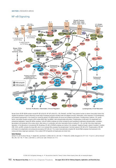
Nuclear factor-κB (NF-κB)/Rel proteins include NF-κB2 p52/p100, NF-κB1 p50/p105, c-Rel, RelA/p65, and RelB. These proteins function as dimeric transcription factors that<br />
regulate the expression of genes influencing a broad range of biological processes including innate and adaptive immunity, inflammation, stress responses, B-cell development,<br />
and lymphoid organogenesis. In the classical (or canonical) pathway, NF-κB/Rel proteins are bound and inhibited by IκB proteins. Proinflammatory cytokines, LPS, growth<br />
factors, and antigen receptors activate an IKK complex (IKKβ, IKKα, and NEMO), which phosphorylates IκB proteins. Phosphorylation of IκB leads to its ubiquitination and<br />
proteasomal degradation, freeing NF-κB/Rel complexes. Active NF-κB/Rel complexes are further activated by post-translational modifications (phosphorylation, acetylation,<br />
glycosylation) and translocate to the nucleus where, either alone or in combination with other transcription factors including AP-1, Ets, and Stat, they induce target gene<br />
expression. In the alternative (or noncanonical) NF-κB pathway, NF-κB2 p100/RelB complexes are inactive in the cytoplasm. Signaling through a subset of receptors, including<br />
LTβR, CD40, and BR3, activates the kinase NIK, which in turn activates IKKα complexes that phosphorylate C-terminal residues in NF-κB2 p100. Phosphorylation of NF-κB2<br />
p100 leads to its ubiquitination and proteasomal processing to NF-κB2 p52. This creates transcriptionally competent NF-κB p52/RelB complexes that translocate to the<br />
nucleus and induce target gene expression. Only a subset of NF-κB agonists and target genes are shown here.<br />
Select Reviews:<br />
Gilmore T.D. (2014) www.nf-kb.org • Hayden M.S. and Ghosh S. (2008) Cell 132, 344–362. • Perkins N.D. (2006) Oncogene 25, 6717–30. • Sun S-C. (2012) Immunol<br />
Rev. 246, 125–140. • Chen J. and Chen Z.J. (2013) Curr. Opin. Immunol. 25, 4–12.<br />
TNF<br />
CYLD<br />
Tumor Immunology<br />
MMPs<br />
VEGF<br />
T cell apoptosis<br />
IL-10<br />
IDO<br />
DC<br />
T Reg<br />
FoxP3<br />
CD4 +<br />
IL-10<br />
TGF-β<br />
IL-35<br />
Th1<br />
T-bet<br />
TNF-α<br />
IL-2<br />
CCL22<br />
MMPs<br />
VEGF<br />
Angiogenesis<br />
IFNγ<br />
T cell<br />
Immune<br />
Checkpoint<br />
MSC<br />
Arginase<br />
IL-10<br />
TGF-β<br />
IFNγ-R<br />
CTL<br />
T-bet<br />
T cell<br />
priming<br />
IL-1β<br />
TNF-α<br />
CD4 +<br />
T cell<br />
Tumor-draining Lymph Node<br />
Proliferation and production<br />
of anti-tumor antibodies<br />
Jak3<br />
Stat3<br />
cytotoxic<br />
granules<br />
Stat1<br />
B cell<br />
IL-2, 4, 5<br />
Tumor-specific<br />
CD8 + T cell<br />
TIM-3<br />
Galectin-9<br />
<br />
B7-H3<br />
Class I antigen<br />
presentation;<br />
IDO; PD-L1<br />
Activation/Response Change<br />
<br />
B7-H4<br />
Genes of cell growth and survival;<br />
PD-L1, VEGF, IL-6, IL-10<br />
PD-1<br />
PD-L1<br />
Nucleus<br />
NF-κB<br />
TCR<br />
MHC<br />
Stat3<br />
Akt<br />
MAPK<br />
DC<br />
M2<br />
Macrophage<br />
IFNγ<br />
cytotoxic<br />
granules<br />
TNF-R<br />
oncogenic<br />
signaling<br />
IL-6<br />
NK<br />
T-bet<br />
Jak<br />
TNF-α<br />
IL-1β<br />
Tumor-promoting<br />
Macrophage<br />
IL-10<br />
TGF-β<br />
M1<br />
Macrophage<br />
IL-6R<br />
Tumor Cell<br />
MMPs<br />
IL-4<br />
CCL22<br />
Inflammation<br />
IL-1β<br />
TNF-α<br />
ALK<br />
ROS1<br />
RTK: EGFR<br />
HER2<br />
etc.<br />
Mast cell<br />
Tumor cells employ multiple defense strategies to evade detection by the immune system. One common strategy, upregulation of immune checkpoint proteins and ligands,<br />
takes advantage of a natural immune mechanism for self-tolerance and prevention of collateral tissue damage. Immune checkpoint receptors, such as PD-1, CTLA-4, and<br />
many others, are located on T cells and engage with their corresponding ligand on tumor cells and dendritic cells, sending inhibitory signals that repress T cell activation<br />
or response. One of the first discovered checkpoint proteins, CTLA-4, plays a role at the stage of T cell priming by binding to the CD28 ligands CD80 or CD86 to prevent<br />
co-stimulatory signals necessary for T cell activation. In contrast, the PD-1/PD-L1 checkpoint acts later in the process, inhibiting anti-tumor immune responses by effector<br />
T cells such as CD4 + T helper 1 (Th1) cells and CD8 + cytotoxic T lymphocytes (CTLs), leading to decreases in IFNγ production and cytolytic activity. Upregulation of PD-L1<br />
expression on the tumor cell surface is mediated by IFNγR signaling to Stat1, as well as oncogenic signaling through several receptor tyrosine kinases (EGFR, ALK, ROS,<br />
HER2, and others) to activate the MAPK, Akt, and Stat3 pathways.<br />
Cells in the tumor microenvironment can also influence tumor progression. FoxP3 + /CD4 + T regulatory cells (Tregs) and myeloid-derived suppressor cells (MDSCs) secrete immunosuppressive<br />
cytokines IL-10 and TGF-β to inhibit the activity of Th1 cells and CTLs. Natural killer (NK) cells release cytotoxic granules against the tumor cell and secrete<br />
IFNγ, which stimulates surrounding pro-inflammatory M1 macrophages. Pro-tumorigenic M2 macrophages suppress anti-tumor immune responses via production of IL-10<br />
and TGF-β and promote metastasis through release of MMPs. MMPs and TGF-β are also released by surrounding mast cells.<br />
Select Reviews:<br />
Pardoll, D.M. (2012) Nat. Rev. Cancer 12, 252–264. • Vanneman, M. and Dranoff, G. (2012) Nat. Rev. Cancer 12, 237–251. • Kawakami, Y., Yaguchi, T., and Park, J.H.,<br />
et al. (2013) Front. Oncol. 3, 136. • Elinav, E., Nowarski, R., Thaiss, C.A., et al. (2013) Nat. Rev. Cancer 13, 759–771. • Mentlik, J.A., Cohen, A.D., and Campbell, K.S.<br />
(2013) Front. Immunol. 4, 481. • Gajewski, T.F., Schreiber, H., and Fu, Y.X. (2013) Nat. Immunol. 14, 1014–1022. • Krstic, J. and Santibanez, J.F. (2014) ScientificWorld-<br />
Journal, 521754.<br />
MMPs<br />
TGF-β<br />
Th2<br />
IL-13<br />
© 2009–2015 Cell Signaling Technology, Inc. • We would like to thank Prof. Thomas D. Gilmore, Boston University, Boston, MA, for reviewing this diagram.<br />
182 For Research Use Only. Not For Use in Diagnostic Procedures. See pages 302 & 303 for Pathway Diagrams, Application, and Reactivity keys.<br />
© 2014–2015 Cell Signaling Technology, Inc. • We would like to thank Glenn Dranoff, M.D., Susanne H.C. Baumeister, M.D.,<br />
Karrie Wong, Ph.D., and Girija Goyal, Dana Farber Cancer Institute and Harvard Medical School, Boston, MA, for reviewing this diagram.<br />
www.cellsignal.com/cstpathways 183