

CST Guide:

Create successful ePaper yourself
Turn your PDF publications into a flip-book with our unique Google optimized e-Paper software.
Section I: Research Areas<br />
Intermediate filament<br />
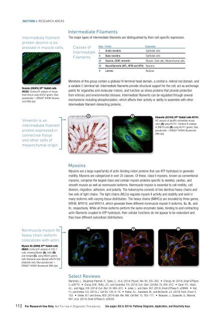
protein desmin is expressed<br />
in muscle cells.<br />
Desmin (D93F5) XP ® Rabbit mAb<br />
#5332: Confocal IF analysis of mouse<br />
heart tissue using #5332 (green). Blue<br />
pseudocolor = DRAQ5 ® #4084 (fluorescent<br />
DNA dye).<br />
Vimentin is an<br />
intermediate filament<br />
protein expressed in<br />
connective tissue<br />
and other cells of<br />
mesenchymal origin.<br />
Nonmuscle myosin IIb<br />
heavy chain isoform<br />
colocalizes with actin.<br />
Myosin IIb (D8H8) XP ® Rabbit mAb<br />
#8824: Confocal IF analysis of COS-7<br />
cells, showing Myosin (A), Actin (B),<br />
and merged (C), using #8824 (green).<br />
Actin filaments were labeled with DY-554<br />
phalloidin (red). Blue pseudocolor =<br />
DRAQ5 ® #4084 (fluorescent DNA dye).<br />
Intermediate Filaments<br />
The major types of intermediate filaments are distinguished by their cell-specific expression.<br />
Classes of<br />
Intermediate<br />
Filaments<br />
Class Protein<br />
Expression<br />
I Acidic keratins Epithelial cells<br />
II Basic keratins Epithelial cells<br />
III Desmin, GFAP, vimentin Muscle, Glial cells, Mesenchymal cells<br />
IV Neurofilaments (NFL, NFM and NFH) Neurons<br />
V Lamins Nucleus<br />
Members of this group contain a globular N-terminal head domain, a central α−helical rod domain, and<br />
a variable C-terminal tail. Intermediate filaments provide structural support for the cell, act as anchorage<br />
points for organelles and molecular motors, and function as stress proteins that provide protection<br />
from intrinsic and environmental stresses. Intermediate filaments can be regulated through several<br />
mechanisms including phosphorylation, which affects their activity or ability to assemble with other<br />
intermediate filament-interacting proteins.<br />
A<br />
A<br />
112 For Research Use Only. Not For Use in Diagnostic Procedures. See pages 302 & 303 for Pathway Diagrams, Application, and Reactivity keys.<br />
B<br />
B<br />
Vimentin (D21H3) XP ® Rabbit mAb #5741:<br />
IHC analysis of paraffin-embedded mouse<br />
colon (A) using #5741. Confocal IF analysis<br />
of SNB19 cells (B) using #5741 (green). Blue<br />
pseudocolor = DRAQ5 ® #4084 (fluorescent<br />
DNA dye).<br />
Myosins<br />
Myosins are a large superfamily of actin-binding motor proteins that use ATP hydrolysis to generate<br />
motility. Myosins are categorized in over 25 classes. Of these, class II myosins, known as conventional<br />
myosins, comprise the largest class and contain myosin proteins specific to skeletal, cardiac, and<br />
smooth muscle as well as nonmuscle isoforms. Nonmuscle myosin is essential to cell motility, cell<br />
division, migration, adhesion, and polarity. The holoenzyme consists of two identical heavy chains and<br />
two sets of light chains. The light chains (MLCs) regulate myosin II activity and stability and exist in<br />
many isoforms with varying tissue distribution. The heavy chains (NMHCs) are encoded by three genes,<br />
MYH9, MYH10, and MYH14, which generate three different nonmuscle myosin II isoforms, IIa, IIb, and<br />
IIc, respectively. While all three isoforms perform the same enzymatic tasks, binding to and contracting<br />
actin filaments coupled to ATP hydrolysis, their cellular functions do not appear to be redundant and<br />
they have different subcellular distributions.<br />
Select Reviews<br />
Blanchoin, L., Boujemaa-Paterski, R., Sykes, C., et al. (2014) Physiol. Rev. 94, 235−263. • Chircop, M. (2014) Small GTPases<br />
5, e29770. • Chung, B.M., Rotty, J.D., and Coulombe, P.A. (2013) Curr. Opin. Cell Biol. 25, 600−612. • Gurel, P.S., Hatch,<br />
A.L., and Higgs, H.N. (2014) Curr. Biol. 24, 660−672. • Julian, L. and Olson, M.F. (2014) Small GTPases 5, e29846. • Kull,<br />
F.J. and Endow, S.A. (2013) J. Cell Sci. 126, 9−19. • Parker, A.L., Kavallaris, M., and McCarroll, J.A. (2014) Front. Oncol. 4,<br />
153. • Snider, N.T. and Omary, M.B. (2014) Nat. Rev. Mol. Cell Biol. 15, 163−177. • Wojnacki, J., Quassollo, G., Marzolo,<br />
M.P., et al. (2014) Small GTPases 5, e28430.<br />
C<br />
Commonly Studied Cytoskeletal Regulation Targets<br />
Target M P Target M P Target M P<br />
14-3-3 Family • Diap1<br />
• Keratin 8/18 •<br />
14-3-3 β/α • Diap2<br />
• Keratin 17 •<br />
14-3-3 γ • DOCK180 • Phospho-Keratin 17 (Ser44) •<br />
14-3-3 ε • DRP1<br />
• Keratin 17/19 •<br />
14-3-3 ζ/δ • Phospho-DRP1 (Ser616) • • Keratin 18 •<br />
14-3-3 η • • Phospho-DRP1 (Ser637) • • Keratin 19 •<br />
14-3-3 τ • DSG2<br />
• Keratin 20 •<br />
Phospho-Ack1 (Tyr284) • EB-1<br />
• Phospho-KIF1B (Ser1487) •<br />
Pan-Actin • • Emerin<br />
• • KIFC1<br />
•<br />
α-Actinin • • EML4<br />
• • KIF3A<br />
•<br />
Afadin<br />
• • EPAC1<br />
• KIF3B<br />
•<br />
Annexin V<br />
• EPAC2<br />
• Kinectin 1 • •<br />
Phospho-AP2M1 (Thr156) • • EpCAM<br />
• • Lamin A/C • •<br />
Phospho-ARHGAP42 • EPLIN<br />
• Phospho-Lamin A/C (Ser22) • •<br />
(Tyr376)<br />
Erlin-1<br />
Cool2/αPix •<br />
• Lamin B1 •<br />
Erlin-2<br />
Cool1/βPix<br />
•<br />
• Lamin B2 •<br />
EVL<br />
ARP2<br />
• •<br />
• LAMP1<br />
•<br />
Ezrin<br />
ARP3<br />
•<br />
• LASP1<br />
•<br />
Phospho-Ezrin (Tyr353)<br />
Atlastin-1 •<br />
• • LCP1<br />
• •<br />
Ezrin/Moesin/Radixin<br />
β-Actin<br />
• •<br />
• Phospho-LCP1 (Tyr28) •<br />
Phospho-Ezrin (Thr567)/<br />
Phospho-Catenin δ-1 •<br />
• • LIMK1<br />
•<br />
Radixin (Thr564)/Moesin<br />
(Ser320)<br />
(Thr558)<br />
Phospho-LIMK1 (Thr508)/ •<br />
β2-Chimerin<br />
LIMK2 (Thr505)<br />
• Fascin<br />
• LIMK2<br />
Caldesmon-1 • •<br />
•<br />
Fer<br />
• LLGL1<br />
Caveolin-1 • •<br />
•<br />
Fes<br />
• LMAN1<br />
Phospho-Caveolin-1 (Tyr14) •<br />
•<br />
Fibrillarin • Phospho-MARK Family<br />
Caveolin-2 •<br />
•<br />
Filamin A<br />
• (Activation Loop)<br />
CD2AP<br />
• Phospho-Filamin A<br />
MARK1<br />
•<br />
•<br />
CDC37<br />
(Ser2152)<br />
• •<br />
MARK2<br />
•<br />
Filamin B<br />
Phospho-CDC37 (Ser13) •<br />
• • MARK3<br />
•<br />
Filamin C<br />
Cdc42<br />
• •<br />
• MARK4<br />
•<br />
Flotillin-1<br />
CD71<br />
•<br />
• MCF2/Dbl<br />
•<br />
Flotillin-2<br />
CdGAP<br />
•<br />
• • Moesin<br />
•<br />
FYVE-CENT<br />
Centrin-2<br />
•<br />
• M-RIP<br />
•<br />
GEF-H1<br />
Chronophin/PDXP •<br />
• • MTSS1<br />
•<br />
Gelsolin<br />
CIN85<br />
•<br />
• • Myosin IIa<br />
•<br />
Phospho-GIT2 (Tyr392)<br />
Claudin-1 • •<br />
• Phospho-Myosin IIa •<br />
GM130<br />
CLIP1/CLIP170 •<br />
• • (Ser1943)<br />
Golgin-97<br />
Myosin IIb<br />
Cofilin<br />
• •<br />
•<br />
• •<br />
HEF1/NEDD9<br />
Myosin IIc<br />
Phospho-Cofilin (Ser3) • •<br />
•<br />
• •<br />
Importin β1<br />
Myosin Va<br />
Cortactin<br />
•<br />
•<br />
•<br />
IQGAP1<br />
Myosin VI<br />
Phospho-Cortactin (Tyr421) •<br />
•<br />
•<br />
IQGAP2<br />
Myosin Light Chain 2<br />
CrkII<br />
•<br />
•<br />
• •<br />
Integrin α2b<br />
Phospho-Myosin Light Chain<br />
Phospho-CrkII (Tyr221) •<br />
•<br />
• •<br />
2 (Ser19)<br />
Pan-Keratin<br />
Dab2<br />
• •<br />
• Phospho-Myosin Light Chain •<br />
Keratin 7<br />
2 (Thr18/Ser19)<br />
Desmin<br />
• •<br />
•<br />
chapter 04: Cell Biology<br />
These protein targets represent key nodes<br />
within cytoskeletal regulation signaling<br />
pathways and are commonly studied in<br />
cytoskeletal regulation research. Primary<br />
antibodies, antibody conjugates, and<br />
antibody sampler kits containing these<br />
targets are available from <strong>CST</strong>.<br />
Listing as of September 2014. See our<br />
website for current product information.<br />
M Monoclonal Antibody<br />
P Polyclonal Antibody<br />
www.cellsignal.com/cstcytoskeletal 113